Embed Size (px)
Citation preview

Prof. Dr. med. Michael Bajka, MDDept OB/GYN, University Hospital of Zurich, USZ
Swiss Society of Ultrasound – Section Obstetrics & Gynecology (www.SGUMGG.ch)
Congenital Uterine AnomaliesCUA
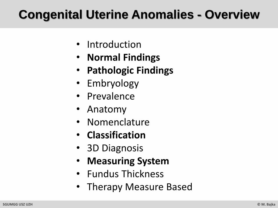
Congenital Uterine Anomalies - Overview
• Introduction• Normal Findings • Pathologic Findings• Embryology• Prevalence• Anatomy • Nomenclature• Classification• 3D Diagnosis• Measuring System• Fundus Thickness• Therapy Measure Based
SGUMGG USZ UZH © M. Bajka

Congenital Uterine Anomalies
Introduction
SGUMGG USZ UZH © M. Bajka
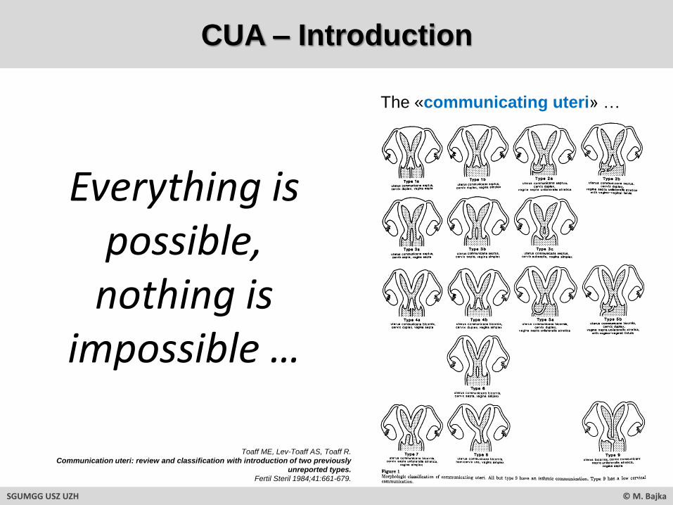
CUA – Introduction
Everything is possible,
nothing is impossible …
SGUMGG USZ UZH © M. Bajka
The «communicating uteri» …
Toaff ME, Lev-Toaff AS, Toaff R.
Communication uteri: review and classification with introduction of two previously
unreported types.
Fertil Steril 1984;41:661-679.

CUA – Introduction
Functional endometrium without outside communication:• Symptoms occur with menarche, mostly pain• Typically pediatric patient• Often misinterpreted as endometriosis• Often surgery needed
SGUMGG USZ UZH © M. Bajka
Functional endometrium with outside communication:• Symptoms (if ever) will occur clearly after menarche,
often related to in-/fertility and bleeding disorders• Typically in adult patients• Indication for surgery under hot debate …
CUA?
SGUMGG USZ UZH © M. Bajka
Case

28y Women, Wish for Child, 14th Day
SGUMGG USZ UZH © M. Bajka
sagittal transversal

SGUMGG USZ UZH © M. Bajka
ESGE/ESHRE: U0, normal uterusAFS/ASRM: VI, arcuate uterus
28y Women, Wish for Child, 14th Day
Normal Findings
Congenital Uterine Anomalies
SGUMGG USZ UZH © M. Bajka
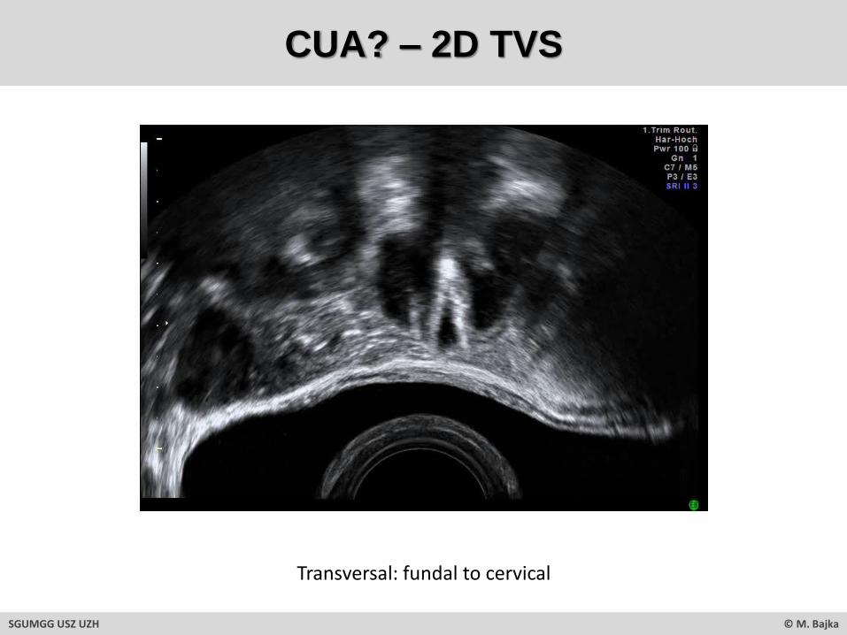
CUA? – 2D TVS
Transversal: fundal to cervical
SGUMGG USZ UZH © M. Bajka

Sagittal: right to left
CUA? – 2D TVS
SGUMGG USZ UZH © M. Bajka
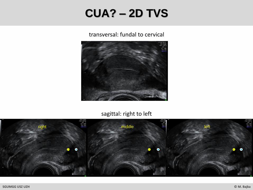
transversal: fundal to cervical
sagittal: right to left
right middle left
CUA? – 2D TVS
SGUMGG USZ UZH © M. Bajka
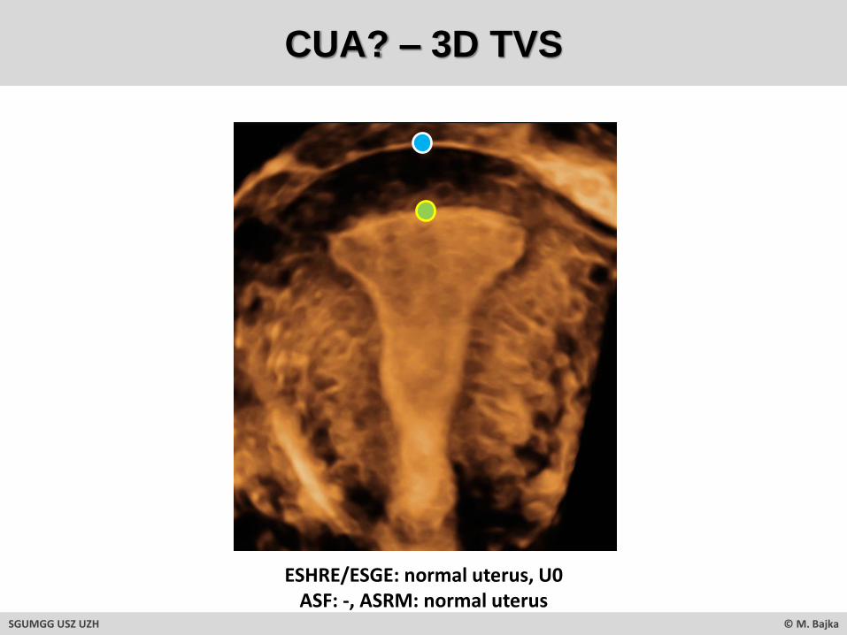
CUA? – 3D TVS
ESHRE/ESGE: normal uterus, U0ASF: -, ASRM: normal uterus
SGUMGG USZ UZH © M. Bajka

Congenital Uterine Anomalies
SGUMGG USZ UZH © M. Bajka
Pathologic Findings
Pathologic Findings
SGUMGG USZ UZH © M. Bajka
Case 1

Transversal: up > down
CUA? – 2D TVS
SGUMGG USZ UZH © M. Bajka

Sagittal: right > left
CUA? – 2D TVS
SGUMGG USZ UZH © M. Bajka

Transversal: up > down
Sagittal: left > right
CUA? – 2D TVS
SGUMGG USZ UZH © M. Bajka

CUA – 2D TVS
SGUMGG USZ UZH © M. Bajka
ESGE / ESHRE Partial septate uterus U2aAFS / ASRM Partial septate uterus Vb
2D sagittal median 3D transversal virtual

CUA - 3D TVS
virtual 3D volumevirtual 3D layers & 3D volume
ESGE / ESHRE Partial septate uterus U2aAFS / ASRM Partial septate uterus Vb
SGUMGG USZ UZH © M. Bajka
3D transversal virtual

Uterus Septum Treatment - Planning
12mm
12mm
13mm
13mm
SGUMGG USZ UZH © M. Bajka
25mm Individual total fundus thickness13mm Part of the septum to be resected12mm Remaining anatomical fundus thickness = goal of anatomical septum resection
Bajka M, Badir S.
Fundus thickness assessment by 3D transvaginal ultrasound allows metrics-based diagnosis and treatment of congenital uterine anomalies.
Ultraschall in Med, 2017;38:183-189.

Uterus Septum Treatment - Before and After
Bevor …
After …
SGUMGG USZ UZH © M. Bajka
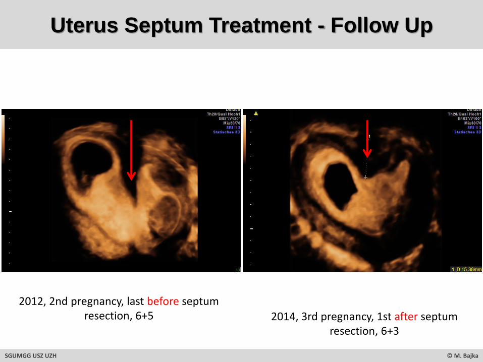
2012, 2nd pregnancy, last before septum resection, 6+5 2014, 3rd pregnancy, 1st after septum
resection, 6+3
SGUMGG USZ UZH © M. Bajka
Uterus Septum Treatment - Follow Up

Pre-treatment… Post-treatment, at 3m…
After 2y… After 5y…
SGUMGG USZ UZH © M. Bajka
Uterus Septum Treatment - Follow Up

SGUMGG USZ UZH © M. Bajka
Case 2
Pathologic Findings

Case 2
SGUMGG USZ UZH © M. Bajka
38y pat, 1 child born at 33w, reg MPA injections, amenorrhea

ESGE/ESHRE: U4b, hemi uterus with no rudimentary cavityASF/ASRM: IId, unicornuate, no horn
SGUMGG USZ UZH © M. Bajka
Case 2

SGUMGG USZ UZH © M. Bajka
Case 3
Pathologic Findings

SGUMGG USZ UZH © M. Bajka
Case 3
35y pat, after prim C-section at 38+1, CUA?
Transversal fundal Transversal cervical

ESGE/ESHRE: U2b C2 V1AFS/ASRM: Va
SGUMGG USZ UZH © M. Bajka
Case 3
35y pat, CUA, after prim C-section at 38+1 , 2nd pregnancy 6+1

ESGE/ESHRE: U2b C2 V1AFS/ASRM: Va
SGUMGG USZ UZH © M. Bajka
Case 3
6+1 9+3
35y pat, CUA, 2nd pregnancy 9+3 gemini mono-di, after prim C-section at 38+1

Quiz
SGUMGG USZ UZH © M. Bajka
Congenital Uterine Anomalies

Case 1
SGUMGG USZ UZH © M. Bajka
Transverse view
ESGE/ESHRE:AFS: -, ASRM:
Sagittal view Coronal view
cranial
caudal
right
left
Grigoris F. Grimbizis, Rudi Campo, Basil C. Tarlatzis, Stephan Gordts, Editors.
Female Genital Tract Congenital Malformations. Classification, Diagnosis and Management.
Springer, 2015.

Case 2
Transverse view Sagittal view Coronal view
SGUMGG USZ UZH © M. Bajka
cranial
caudal
right
left
Grigoris F. Grimbizis, Rudi Campo, Basil C. Tarlatzis, Stephan Gordts, Editors.
Female Genital Tract Congenital Malformations. Classification, Diagnosis and Management.
Springer, 2015.
ESGE/ESHRE:AFS: -, ASRM:
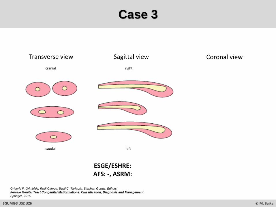
Case 3
Transverse view Sagittal view Coronal view
SGUMGG USZ UZH © M. Bajka
cranial
caudal
right
left
Grigoris F. Grimbizis, Rudi Campo, Basil C. Tarlatzis, Stephan Gordts, Editors.
Female Genital Tract Congenital Malformations. Classification, Diagnosis and Management.
Springer, 2015.
ESGE/ESHRE:AFS: -, ASRM:

Grigoris F. Grimbizis, Rudi Campo, Basil C. Tarlatzis, Stephan Gordts, Editors.
Female Genital Tract Congenital Malformations. Classification, Diagnosis and Management.
Springer, 2015.
Case 4
Transverse view Coronal view
SGUMGG USZ UZH © M. Bajka
cranial
caudal
ESGE/ESHRE:AFS: -, ASRM:
Congenital Uterine Anomalies
Embryology
SGUMGG USZ UZH © M. Bajka
CUA – Embryology (Crosby & Hill)Paramesonephric (Müller) Ducts
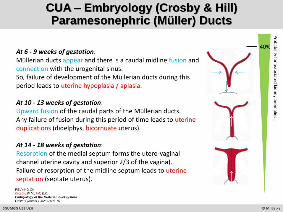
At 6 - 9 weeks of gestation: Müllerian ducts appear and there is a caudal midline fusion and connection with the urogenital sinus. So, failure of development of the Müllerian ducts during this period leads to uterine hypoplasia / aplasia.
At 10 - 13 weeks of gestation: Upward fusion of the caudal parts of the Müllerian ducts. Any failure of fusion during this period of time leads to uterine duplications (didelphys, bicornuate uterus).
At 14 - 18 weeks of gestation: Resorption of the medial septum forms the utero-vaginal channel uterine cavity and superior 2/3 of the vagina).Failure of resorption of the midline septum leads to uterine septation (septate uterus).
Pro
bab
ilityfo
rasso
ciatedkid
ney
ano
malies
…
40%
SGUMGG USZ UZH © M. Bajka
RELYING ON
Crosby, W.M., Hill, E.C.
Embryology of the Müllerian duct system.
Obstet Gynecol 1962;20:507-15.

“There are intermediate and incomplete forms of bicornuate and septate uteri, due to simultaneous lack of fusion and reabsorption of paramensonephric(Müllerian) ducts …”
Bermejo C, Martinez Ten P, Cantarero R, Diaz D, Perez Pedregosa J, Barro E, Labrador E, Ruiz Lopez L.
Three-dimensional ultrasound in the diagnosis of Müllerian duct anomalies and concordance with magnetic resonance imaging.
Ultrasound Obstet Gynecol 2010; 35: 593–601.
CUA - Heterogenity
SGUMGG USZ UZH © M. Bajka
Congenital Uterine Anomalies
SGUMGG USZ UZH © M. Bajka
Prevalence
CUA – Prevalence – Review
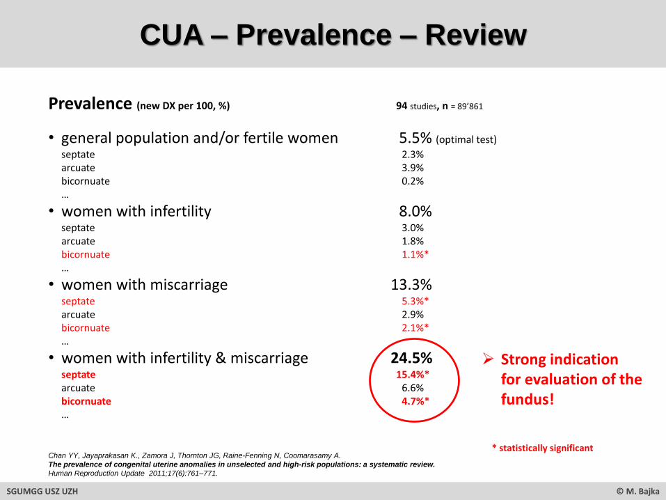
Prevalence (new DX per 100, %) 94 studies, n = 89’861
• general population and/or fertile women 5.5% (optimal test)
septate 2.3%arcuate 3.9%bicornuate 0.2%…
• women with infertility 8.0% septate 3.0%arcuate 1.8%bicornuate 1.1%*…
• women with miscarriage 13.3% septate 5.3%*arcuate 2.9%bicornuate 2.1%*…
• women with infertility & miscarriage 24.5% septate 15.4%*arcuate 6.6%bicornuate 4.7%*…
Chan YY, Jayaprakasan K., Zamora J, Thornton JG, Raine-Fenning N, Coomarasamy A.
The prevalence of congenital uterine anomalies in unselected and high-risk populations: a systematic review.
Human Reproduction Update 2011;17(6):761–771.
➢ Strong indication for evaluation of the fundus!
* statistically significant
SGUMGG USZ UZH © M. Bajka

CUA – Prevalence of Classes
Classification of uterine malformations according to the American Fertility Society 1988relying on embryological, clinical factors, prognosis & treatment options
The American Fertility Society classifications of adnexal adhesions, distal tubal obstruction, tubal occlusion secondary to tubal ligation, tubal
pregnancies, Mullerian anomalies and intrauterine adhesions.
Fertil Steril 1988; 49: 944–955.
1%44% 33%
15%3%
2%
0.3%
Bermejo C, Martinez Ten P, Cantarero R, Diaz D, Perez Pedregosa J, Barro E, Labrador E, Ruiz Lopez L.
Three-dimensional ultrasound in the diagnosis of Müllerian duct anomalies and concordance with magnetic resonance imaging.
Ultrasound Obstet Gynecol 2010; 35: 593–601.
SGUMGG USZ UZH © M. Bajka
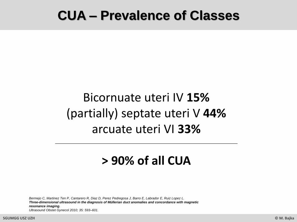
CUA – Prevalence of Classes
Bicornuate uteri IV 15%(partially) septate uteri V 44%
arcuate uteri VI 33%
> 90% of all CUA
SGUMGG USZ UZH © M. Bajka
Bermejo C, Martinez Ten P, Cantarero R, Diaz D, Perez Pedregosa J, Barro E, Labrador E, Ruiz Lopez L.
Three-dimensional ultrasound in the diagnosis of Müllerian duct anomalies and concordance with magnetic
resonance imaging.
Ultrasound Obstet Gynecol 2010; 35: 593–601.
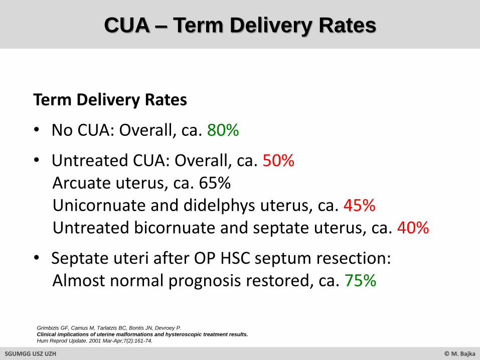
Grimbizis GF, Camus M, Tarlatzis BC, Bontis JN, Devroey P.
Clinical implications of uterine malformations and hysteroscopic treatment results.
Hum Reprod Update. 2001 Mar-Apr;7(2):161-74.
Term Delivery Rates
• No CUA: Overall, ca. 80%
• Untreated CUA: Overall, ca. 50%Arcuate uterus, ca. 65%Unicornuate and didelphys uterus, ca. 45%Untreated bicornuate and septate uterus, ca. 40%
• Septate uteri after OP HSC septum resection:Almost normal prognosis restored, ca. 75%
SGUMGG USZ UZH © M. Bajka
CUA – Term Delivery Rates

Congenital Uterine Anomalies
Anatomy
SGUMGG USZ UZH © M. Bajka

CUA - Anatomy
OUC Outer uterine contourV indentation
Inner uterine contourV protrusion/septation
IUC
Clinicalexamination
FTH
FTH Fundus thickness
2D / 3D US
OUC
IUC
FTH
SGUMGG USZ UZH © M. Bajka
www.netterimages.com
Congenital Uterine Anomalies
SGUMGG USZ UZH © M. Bajka
Nomenclature
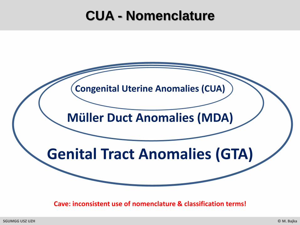
CUA - Nomenclature
Genital Tract Anomalies (GTA)
Cave: inconsistent use of nomenclature & classification terms!
SGUMGG USZ UZH © M. Bajka
Müller Duct Anomalies (MDA)
Congenital Uterine Anomalies (CUA)

CUA - Nomenclature
Uterus duplex unicollis Uterus duplex with double vagina
Uterus didelphys Uterus
septus with
single vagina
Uterus subseptus Uterus arcuatus
Uterus unicornis with
rudimentary contralateral
hemiuterus
+/- slightly different terms & imprecise definitions = inconsistent nomenclature!
glowm.com The Global Library of Women's Medicine
???
www.netterimages.com
SGUMGG USZ UZH © M. Bajka

Congenital Uterine Anomalies
SGUMGG USZ UZH © M. Bajka
Classification 1

SGUMGG USZ UZH © M. Bajka
CUA Classification – AFS

CUA Classification – AFS
Classification of uterine malformations according to the American Fertility Society 1988relying on embryological, clinical factors, prognosis & treatment options
Th
e A
meri
can
Fe
rtilit
y S
oc
iety
cla
ssif
icati
on
s o
f ad
ne
xal
ad
he
sio
ns
, d
ista
l tu
ba
l o
bs
tru
cti
on
, tu
ba
l
oc
clu
sio
n s
eco
nd
ary
to
tu
ba
l lig
ati
on
, tu
ba
l p
reg
na
ncie
s,
Mu
lleri
an
an
om
alies a
nd
in
trau
teri
ne
ad
he
sio
ns
.
Fe
rtil
Ste
ril1988;
49:
944–955.
Arc
uat
eu
teru
sC
om
ple
te C
UA
co
mm
en
tary
!
SGUMGG USZ UZH © M. Bajka
!Bu
ttra
m&
Gib
bo
ns:
Mü
lleria
na
no
ma
lies:
A p
rop
ose
d c
lassific
atio
n (
an
an
aly
sis
of 1
44
ca
se
s),
Fe
rtil
Ste
ril1
97
9;3
2:4
0-4
6.
CUA Classification – AFS
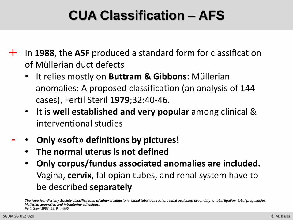
In 1988, the ASF produced a standard form for classification of Müllerian duct defects• It relies mostly on Buttram & Gibbons: Müllerian
anomalies: A proposed classification (an analysis of 144 cases), Fertil Steril 1979;32:40-46.
• It is well established and very popular among clinical & interventional studies
• Only «soft» definitions by pictures!• The normal uterus is not defined• Only corpus/fundus associated anomalies are included.
Vagina, cervix, fallopian tubes, and renal system have to be described separately
SGUMGG USZ UZH © M. Bajka
+
-
The American Fertility Society classifications of adnexal adhesions, distal tubal obstruction, tubal occlusion secondary to tubal ligation, tubal pregnancies,
Mullerian anomalies and intrauterine adhesions.
Fertil Steril 1988; 49: 944–955.

CUA – AFS Based Basic Therapy
SGUMGG USZ UZH © M. Bajka
• Recommendations for surgical interventions base on CUA classifications, mostly on the 1988 ASF Classification
• TherapyVery simplified …- (partially) septate uteri qualify for OP HSC septum
resection (metroplasty)- bicornuate eventually for OPEN metroplasty- arcuate for no surgery.
The American Fertility Society classifications of adnexal adhesions, distal tubal obstruction, tubal occlusion secondary to tubal ligation, tubal pregnancies,
Mullerian anomalies and intrauterine adhesions.
Fertil Steril 1988; 49: 944–955.

SGUMGG USZ UZH © M. Bajka
CUA AFS V/VI – GUBBINI 3D-TVS

CUA AFS V/VI – GUBBINI 3D-TVSG
ubbin
i G
, D
i S
pie
zio
Sard
o A
, N
ascett
i D
, M
arr
a E
, S
pin
elli
M, G
reco E
, C
asadio
P,
Nappi C
.
New
ou
tpati
en
t su
bc
lassif
icati
on
syste
m f
or
Am
eri
can
Fe
rtilit
y S
oc
iety
Cla
sses V
an
d V
I
ute
rin
e a
no
malies.
J M
inim
Invasiv
e G
ynecol 2009;1
6(5
):554
-61.
SGUMGG USZ UZH © M. Bajka

Gubbini G, Di Spiezio Sardo A, Nascetti D, Marra E, Spinelli M, Greco E, Casadio P, Nappi C.
New outpatient subclassification system for American Fertility Society Classes V and VI uterine anomalies.
J Minim Invasive Gynecol 2009;16(5):554-61.
? ?
? ?
Definitions of «X» given by Gubbini et al.:• intercornual distance …?• the line joining the interstital portions of the
fallopian tubes …?• outer interostial distance/line??
X ?Z
Y
Z = «Septum»
SGUMGG USZ UZH © M. Bajka
Y = «Fundus»
CUA AFS V/VI – GUBBINI 3D-TVS

CUA Diagnosis – Tubal Part Intramural
SGUMGG USZ UZH © M. Bajka
Sporadically, they arevisible as thin tubes
Infrequently, they arevisible as broad tubes
Typically, they are notvisible
Bajka M, Badir S.
Fundus thickness assessment by 3D transvaginal ultrasound allows metrics-based diagnosis and treatment of congenital uterine anomalies.
Ultraschall in Med, 2017;38:183-189.
Is the intramural part of the tubes visible in 3D TVS?

Gubbin
iG
, D
i S
pie
zio
Sard
oA
, N
ascett
iD
, M
arr
aE
, S
pin
elli
M, G
reco E
, C
asadio
P,
NappiC
.
New
ou
tpati
en
t su
bc
lassif
icati
on
syste
m f
or
Am
eri
can
Fe
rtilit
y S
oc
iety
Cla
sses V
an
d V
I
ute
rin
e a
no
malies.
J M
inim
Invasiv
e G
ynecol2009;1
6(5
):554
-61.
“… This is crucial to preserve adequate fundus thickness Y (1.5 cm(!?)) to avert intraoperative uterine perforation or uterine rupture during pregnancy or labor…”
A:
Y>
1.5
cm
OHSC OHSC NHSC NHSC
no surg OHSC NHSC -
no surg - - -
SGUMGG USZ UZH © M. Bajka
CUA Classification – GUBBINI 3D-TVS
B:
1.5
cm
>Y
>0
C:
0>
Y>
1c
m1: Z >5mm 2: Z 1/3 cav 3: Z 2/3 cav 4: Z 3/3 cav
XZ
Y
OHSC = office HSC
NHSC = narcosis HSC

Gubbin
i G
, D
i S
pie
zio
Sard
o A
, N
ascett
i D
, M
arr
a E
, S
pin
elli
M, G
reco E
, C
asadio
P,
Nappi C
.
New
ou
tpati
en
t su
bc
lassif
icati
on
syste
m f
or
Am
eri
can
Fe
rtilit
y S
oc
iety
Cla
sses V
an
d V
I
ute
rin
e a
no
malies.
J M
inim
Invasiv
e G
ynecol 2009;1
6(5
):554
-61.
• …According to some authors, clinicians dealing with such uterine anomalies should attempt to describe them according to their component parts rather than categorize them into the class that most approximates the dominant feature.
• Our new classification system directly focuses on the architecture of the uterine cavity, describing the uterine fundus thickness (not in absolute
numbers!) and the endocavitary development of the septum…
• Our subclassification system has a number of advantages in terms of diagnosis of uterine anomalies because it enables differentiationbetween malformations previously classified to the same class in the AFS system (i.e, A1–A3 or B1–B3 or A2, B2, and C2). Furthermore, it enables diagnosis of malformations that are not suitable for surgical correction….
CUA Classification – GUBBINI 3D-TVS
SGUMGG USZ UZH © M. Bajka

Congenital Uterine Anomalies
SGUMGG USZ UZH © M. Bajka
3D Diagnosis

CUA
SGUMGG USZ UZH © M. Bajka

CUA - 3D TVS Illustration of the AFS Classification - BERMEJO
Berm
ejo
C, M
art
inez
Te
n P
, C
anta
rero
R,
Dia
z D
, P
ere
z P
edre
gosa
J, B
arr
oE
, Labra
dor
E, R
uiz
Lopez
L.
Th
ree-d
imen
sio
na
l u
ltra
so
un
d in
th
e d
iag
no
sis
of
Mü
lleri
an
du
ct
an
om
alies a
nd
co
nc
ord
an
ce w
ith
mag
ne
tic
reso
na
nce i
mag
ing
.
Ultra
sound O
bste
tG
ynecol2010;
35:
593–601.
SGUMGG USZ UZH © M. Bajka

CUA - 3D TVS Practical Aspects - BERMEJO
• Transvaginal acquisition (except for three patients with intact hymen)
• Initially 2D ultrasound in strict mid-sagittal view, adjusting the capture window to obtain the optimal 3D volume
• Sweep angle of 90◦ bisecting the capture plane• Anomalies with large transverse diameter (didelphic uterus, wide
septate, bicornuate uterus, communicating unicornuate) volume obtained from a transverse plane so that both uterine horns could be visualized
• Volumes manipulated until a satisfactory surface rendered image was of the fundus ,uterine cavity and cervical canal
• Luminosity and contrast curves adjusted for multiplanar and rendered images, and for threshold and transparency.
• Rendering modes mixture of surface/gradient of light of 10/60 to 60/10.
Berm
ejo
C, M
art
inez
Te
n P
, C
anta
rero
R,
Dia
z D
, P
ere
z P
edre
gosa
J, B
arr
oE
, Labra
dor
E, R
uiz
Lopez
L.
Th
ree-d
imen
sio
na
l u
ltra
so
un
d in
th
e d
iag
no
sis
of
Mü
lleri
an
du
ct
an
om
alies a
nd
co
nc
ord
an
ce w
ith
mag
ne
tic
reso
na
nce i
mag
ing
.
Ultra
sound O
bste
tG
ynecol2010;
35:
593–601.
SGUMGG USZ UZH © M. Bajka

CUA - Diagnosis 3D-TVS vs MRI - BERMEJO
SGUMGG USZ UZH © M. Bajka
Berm
ejo
C, M
art
inez
Te
n P
, C
anta
rero
R,
Dia
z D
, P
ere
z P
edre
gosa
J, B
arr
oE
, Labra
dor
E, R
uiz
Lopez
L.
Th
ree-d
imen
sio
na
l u
ltra
so
un
d in
th
e d
iag
no
sis
of
Mü
lleri
an
du
ct
an
om
alies a
nd
co
nc
ord
an
ce w
ith
mag
ne
tic
reso
na
nce i
mag
ing
.
Ultra
sound O
bste
tG
ynecol2010;
35:
593–601.

CUA - 3D-TVS vs MRI – Conclusions - BERMEJO
• High degree of concordance between 3D US (n=286) and MRI (n=65) in the diagnosis of uterine malformations (based on AFS) (2 differences: 1x 3D US/1x MRI correct)
• Relationship between cavity and fundus is visualized equally well with 3D ultrasound and MRI
• 3D ultrasound was of most use when distinguishing between bicornuate (IV), septate (V), and arcuate (VI)
• Few differences observed only when the lower part of the uterus was studied (cervix, vagina)
• 3D US should be complemented always by careful gynecological exploration in order to identify any alterations in the cervix & vagina (MRI detects cervical and vaginal septa!)
• 3D US is cheaper and better tolerated by patients.
Bermejo C, Martinez Ten P, Cantarero R, Diaz D, Perez Pedregosa J, Barro E, Labrador E, Ruiz Lopez L.
Three-dimensional ultrasound in the diagnosis of Müllerian duct anomalies and concordance with magnetic resonance imaging.
Ultrasound Obstet Gynecol 2010; 35: 593–601.
SGUMGG USZ UZH © M. Bajka
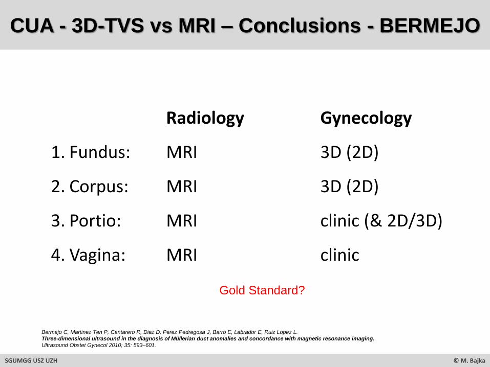
Radiology Gynecology
1. Fundus: MRI 3D (2D)
2. Corpus: MRI 3D (2D)
3. Portio: MRI clinic (& 2D/3D)
4. Vagina: MRI clinic
Gold Standard?
CUA - 3D-TVS vs MRI – Conclusions - BERMEJO
Bermejo C, Martinez Ten P, Cantarero R, Diaz D, Perez Pedregosa J, Barro E, Labrador E, Ruiz Lopez L.
Three-dimensional ultrasound in the diagnosis of Müllerian duct anomalies and concordance with magnetic resonance imaging.
Ultrasound Obstet Gynecol 2010; 35: 593–601.
SGUMGG USZ UZH © M. Bajka

Congenital Uterine Anomalies
SGUMGG USZ UZH © M. Bajka
Classification 2
SGUMGG USZ UZH © M. Bajka
CUA Classification – ESHRE/ESGE

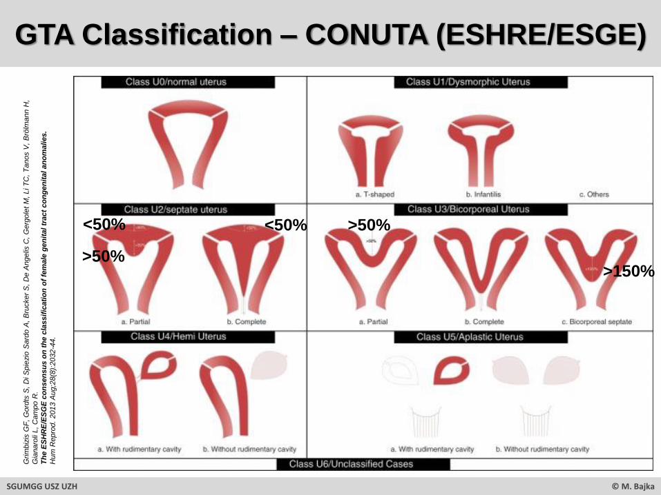
GTA Classification – CONUTA (ESHRE/ESGE)
SGUMGG USZ UZH © M. Bajka
Grim
biz
isG
F, G
ord
tsS
, D
i S
pie
zio
Sard
oA
, B
rucker
S, D
e A
ngelis
C,
Gerg
ole
tM
, Li T
C,
Ta
nos
V,
Brö
lmann
H,
Gia
naro
liL,
Cam
po R
.
Th
e E
SH
RE
/ES
GE
co
ns
en
su
s o
n t
he
cla
ssif
icati
on
of
fem
ale
ge
nit
al
tract
co
ng
en
ital an
om
alies.
Hum
Repro
d.
2013 A
ug;2
8(8
):2032
-44.
GTA Classification – CONUTA (ESHRE/ESGE) G
rim
biz
isG
F, G
ord
tsS
, D
i S
pie
zio
Sard
oA
, B
rucker
S, D
e A
ngelis
C,
Gerg
ole
tM
, Li T
C,
Ta
nos
V,
Brö
lmann
H,
Gia
naro
liL,
Cam
po R
.
Th
e E
SH
RE
/ES
GE
co
ns
en
su
s o
n t
he
cla
ssif
icati
on
of
fem
ale
ge
nit
al
tract
co
ng
en
ital an
om
alies.
Hum
Repro
d.
2013 A
ug;2
8(8
):2032
-44.
<50%
>50%
>50%<50%
>150%
SGUMGG USZ UZH © M. Bajka

GTA Classification – CONUTA (ESHRE/ESGE) G
rig
oris F
. G
rim
biz
is, R
udi C
am
po, B
asil
C.
Ta
rla
tzis
, S
tephan G
ord
tsE
ditors
Fem
ale
Ge
nit
al T
ract
Co
nge
nit
al M
alfo
rmat
ion
s -
Cla
ssif
icat
ion
, D
iagn
osi
s an
d M
anag
em
en
t.Sp
rin
ger
Ver
lag,
20
15
SGUMGG USZ UZH © M. Bajka
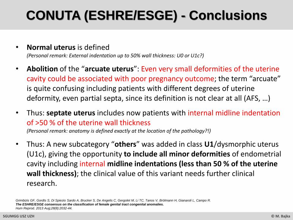
• Normal uterus is defined(Personal remark: External indentation up to 50% wall thickness: U0 or U1c?)
• Abolition of the “arcuate uterus”: Even very small deformities of the uterine cavity could be associated with poor pregnancy outcome; the term “arcuate” is quite confusing including patients with different degrees of uterine deformity, even partial septa, since its definition is not clear at all (AFS, …)
• Thus: septate uterus includes now patients with internal midline indentation of >50 % of the uterine wall thickness (Personal remark: anatomy is defined exactly at the location of the pathology?!)
• Thus: A new subcategory “others” was added in class U1/dysmorphic uterus (U1c), giving the opportunity to include all minor deformities of endometrial cavity including internal midline indentations (less than 50 % of the uterine wall thickness); the clinical value of this variant needs further clinical research.
CONUTA (ESHRE/ESGE) - Conclusions
Grimbizis GF, Gordts S, Di Spiezio Sardo A, Brucker S, De Angelis C, Gergolet M, Li TC, Tanos V, Brölmann H, Gianaroli L, Campo R.
The ESHRE/ESGE consensus on the classification of female genital tract congenital anomalies.
Hum Reprod. 2013 Aug;28(8):2032-44.
SGUMGG USZ UZH © M. Bajka
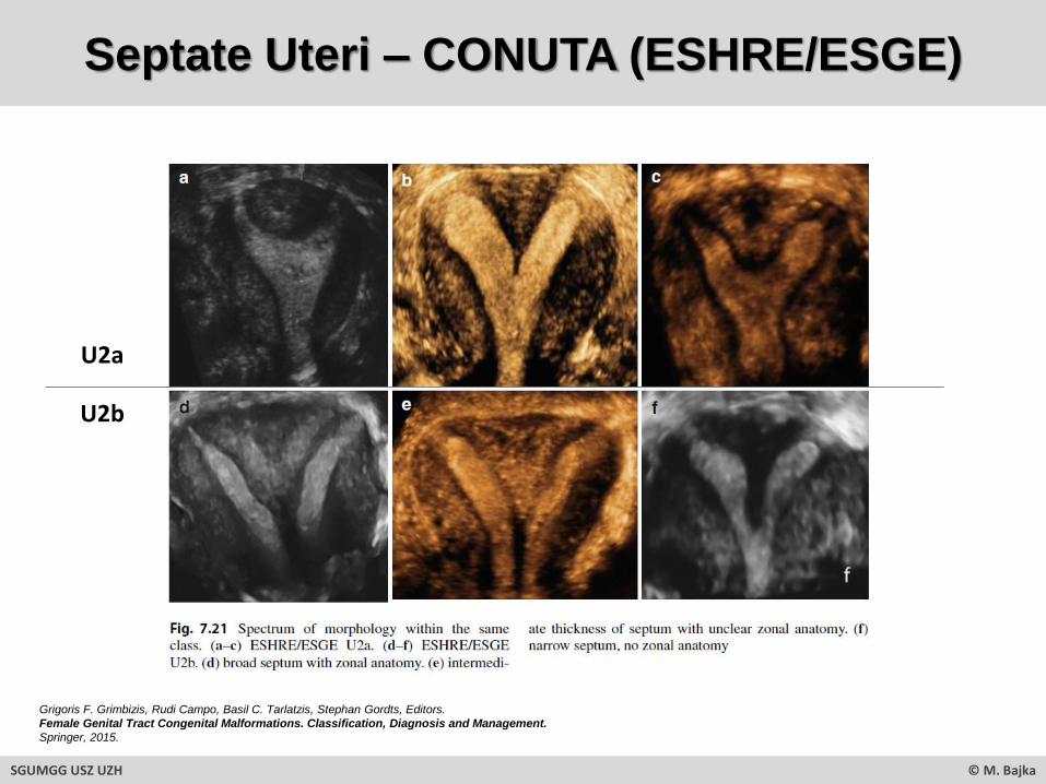
U2a
Septate Uteri – CONUTA (ESHRE/ESGE)
SGUMGG USZ UZH © M. Bajka
Grigoris F. Grimbizis, Rudi Campo, Basil C. Tarlatzis, Stephan Gordts, Editors.
Female Genital Tract Congenital Malformations. Classification, Diagnosis and Management.
Springer, 2015.
U2b
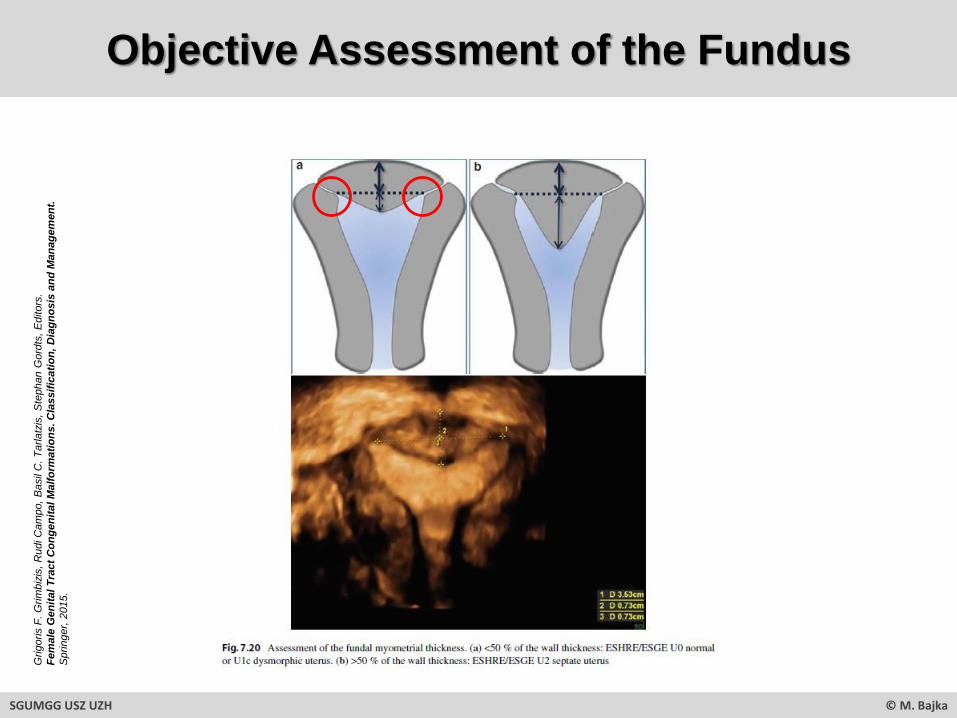
Objective Assessment of the Fundus
SGUMGG USZ UZH © M. Bajka
Grig
oris F
. G
rim
biz
is,
Rudi C
am
po,
Basil
C.
Ta
rla
tzis
, S
tephan G
ord
ts, E
ditors
.
Fe
male
Gen
ital
Tra
ct
Co
ng
en
ital
Malf
orm
ati
on
s. C
lassif
icati
on
, D
iag
no
sis
an
d M
an
ag
em
en
t.
Sprin
ger,
2015.

CUA – Detailed Description of a Case
CUA
• ESGE/ESHRE: Complete bicorporeal uterus (U3b, C0, V0)
• AFS/ASRM: Complete bicornuate uterus (IVa)
• Deutlich in zwei fast gleichgrossen Anteilen vorliegendes Corpus-Fundus uteri (rechts etwas grösser als links, uterine Cavum-Länge re 34mm, li 33mm) mit jeweils gut ausgebildeten Endometriumstreifen, jeweils deutlich nach lateral gekippt, die Separation beginnt direkt kranial der Zervix (kein klassischer Uterus duplex!)
• Zervix: normal ausgebildet (totale Länge 43mm)
• Vagina: normal ausgebildet, keine Septen darstellbar
• Ovar links: normal ausgebildet (unter OH-Suppression)
• Ovar rechts: heute nicht gesehen, zuvor dargestellt
• Tuben: nicht abgeklärt (ein HyFoSy könnte versucht werden)
• Nierenanatomie: kursorisch unauffällig
• Kohabitationsbeschwerden: keine
• Hypermenorrhoe: ausschliesslich zu Beginn der Menstruation zwei Tage (vor OH)
• Dysmenorrhoe: keine
• Schwangerschaften: bisher keine
• Konzeptionswunsch: derzeit nicht vorhanden, OH (Valette) seit diesem Zyklus im Einsatz
SGUMGG USZ UZH © M. Bajka

Congenital Uterine Anomalies
Therapy
SGUMGG USZ UZH © M. Bajka

The Lasting Dilemma: Suboptimal Treatment
Infertility before 2010: After 2x “blind” HSC septum resection, finally no children ...
SGUMGG USZ UZH © M. Bajka

Congenital Uterine Anomalies
SGUMGG USZ UZH © M. Bajka
Measuring System

CUA
SGUMGG USZ UZH © M. Bajka
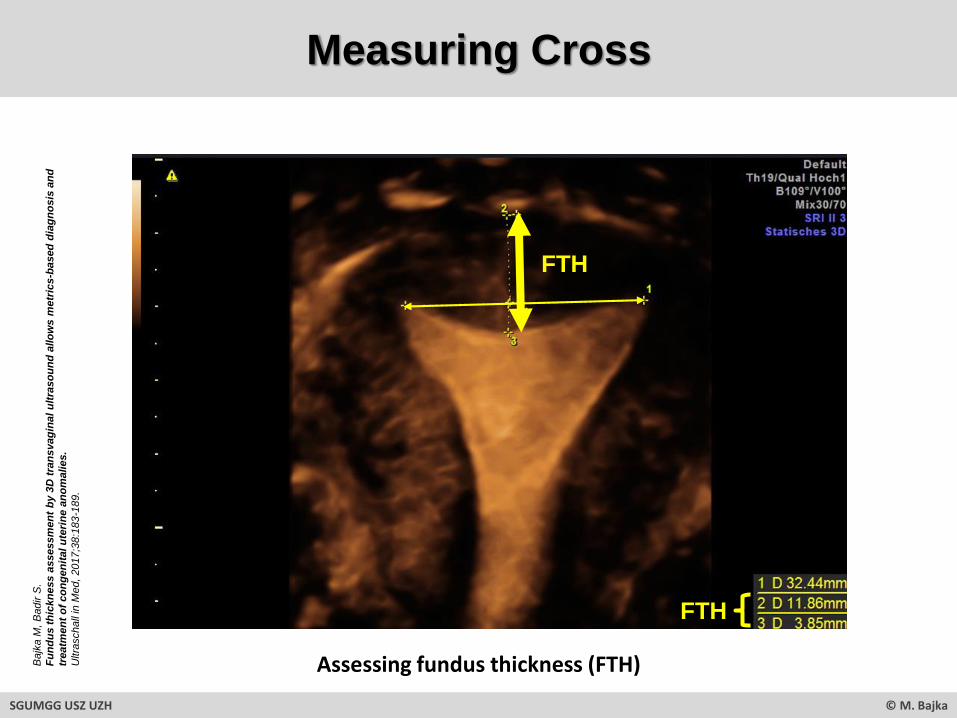
Measuring Cross
FTH
FTH
Assessing fundus thickness (FTH)Bajk
a M
, B
adir S
.
Fu
nd
us
th
ickn
ess a
ssessm
en
t b
y 3
D t
ran
svag
ina
lu
ltra
so
un
d a
llo
ws
metr
ics-b
ased
dia
gn
os
is a
nd
treatm
en
t o
f co
ng
en
ital u
teri
ne
an
om
alies.
Ultra
schall
in M
ed,
2017;3
8:1
83
-189.
SGUMGG USZ UZH © M. Bajka

CUA – Principal of Fundus Therapy
Surgical goal:Anatomical reconstruction, but …
What is normal FTH?
Bajka M, Badir S.
Fundus thickness assessment by 3D transvaginal ultrasound allows metrics-based diagnosis and treatment of congenital uterine anomalies.
Ultraschall in Med, 2017;38:183-189.
SGUMGG USZ UZH © M. Bajka

Normal Fundus in Premenopausal Women
nFTH μ +/- SD
p-valueFTH minima FTH maxima
[mm] [mm] [mm]
All 100 12.02 +/-2.03 6.16 19.43
Groups
Parous 55 12.95 +/- 1.90 8.27 19.43
Nulliparous 45 10.92 +/- 1.86 < 0.0001* 6.16 18.72
Sub-groups according to parity
P No-LNG-IUS 28 12.4 +/- 1.90 8.27 17.48
P LNG-IUS 27 13.38 +/- 1.89 1.08 10.48 19.43
0P No-LNG-IUS 35 10.90 +/- 2.07 6.16 18.72
0P LNG-IUS 10 11.02 +/- 1.34 5.4 8.04 13.74
Sub-groups according to LNG IUS status
P No-LNG-IUS 28 12.4 +/- 1.90 8.27 17.48
0P No-LNG-IUS 35 10.90 +/- 2.07 < 0.0001* 6.16 18.72
P LNG-IUS 27 13.38 +/- 1.89 10.48 19.43
0P LNG-IUS 10 11.02 +/- 1.34 0.02* 8.04 13.74
Ø FTH 12mm(range 6.16 – 19.43mm)
SGUMGG USZ UZH © M. Bajka
Bajk
a M
, B
adir S
.
Fu
nd
us
th
ickn
ess a
ssessm
en
t b
y 3
D t
ran
svag
ina
lu
ltra
so
un
d a
llo
ws
metr
ics-b
ased
dia
gn
os
is a
nd
treatm
en
t o
f co
ng
en
ital u
teri
ne
an
om
alies.
Ultra
schall
in M
ed,
2017;3
8:1
83
-189.

CUA – Possible Fundus Therapy
Bajka M, Badir S.
Fundus thickness assessment by 3D transvaginal ultrasound allows metrics-based diagnosis and treatment of congenital uterine anomalies.
Ultraschall in Med, 2017;38:183-189.
SGUMGG USZ UZH © M. Bajka

CUA - Classifications and FTH - Comparison
FTH: 23.28mm
AFS: partial septate Vb
ESHRE/ESGE: partial septate U2a
FTH: 14.61mm
AFS: arcuate VI
ESHRE/ESGE: normal U0
FTH: 18.17mm
AFS: arcuate VI
ESHRE/ESGE: normal U0
FTH: 19.16mm
AFS: arcuate VI
ESHRE/ESGE: partial septate U2a
FTH: 21.56mm
AFS: arcuate VI
ESHRE/ESGE: partial septate U2a
FTH: 26.99mm
AFS: partial bicornuate IVb
ESHRE/ESGE: bicorporeal septate
U3c
FTH: 35.03mm
AFS: complete septate Va
ESHRE/ESGE: complete septate U2b
2
1
3
D1 30.70mm
D2 11.20mm
D3 7.96mm
D1 31.26mm
D2 14.12mm
D3 4.05mm
D1 29.31mm
D2 9.91mm
D3 4.70mm
D1 45.62mm
D2 8.67mm
D3 12.89mm
D1 29.45mm
D2 4.77mm
D3 18.51mm
D1 44.54mm
D2 - 0.71mm
D3 27.70mm
D1 29.13mm
D2 14.76mm
D3 20.27mm
FTH: 17.10mm
ASF: arcuate VI
ESHRE/ESG: normal U0
D1 36.41mm
D2 14.42mm
D3 2.68mm
SGUMGG USZ UZH © M. Bajka
Bajk
a M
, B
adir S
.
Fu
nd
us
th
ickn
ess a
ssessm
en
t b
y 3
D t
ran
svag
ina
lu
ltra
so
un
d a
llo
ws m
etr
ics
-ba
sed
dia
gn
os
is a
nd
treatm
en
t o
f co
ng
en
ital u
teri
ne
an
om
alies.
Ultra
schall
in M
ed,
2017;3
8:1
83
-189.
Bajk
a M
, B
adir S
.
Fu
nd
us
th
ickn
ess a
ssessm
en
t b
y 3
D t
ran
svag
ina
lu
ltra
so
un
d a
llo
ws m
etr
ics
-ba
sed
dia
gn
os
is a
nd
treatm
en
t o
f co
ng
en
ital u
teri
ne
an
om
alies.
Ultra
schall
in M
ed,
2017;3
8:1
83
-189.

CUA
SGUMGG USZ UZH © M. Bajka
CUA Classification – ASRM Specification
SGUMGG USZ UZH © M. Bajka
CUA Classification – ASRM Specification
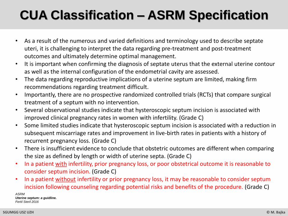
• As a result of the numerous and varied definitions and terminology used to describe septate uteri, it is challenging to interpret the data regarding pre-treatment and post-treatment outcomes and ultimately determine optimal management.
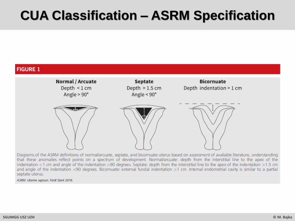
• It is important when confirming the diagnosis of septate uterus that the external uterine contour as well as the internal configuration of the endometrial cavity are assessed.
• The data regarding reproductive implications of a uterine septum are limited, making firm recommendations regarding treatment difficult.
• Importantly, there are no prospective randomized controlled trials (RCTs) that compare surgical treatment of a septum with no intervention.
• Several observational studies indicate that hysteroscopic septum incision is associated with improved clinical pregnancy rates in women with infertility. (Grade C)
• Some limited studies indicate that hysteroscopic septum incision is associated with a reduction in subsequent miscarriage rates and improvement in live-birth rates in patients with a history of recurrent pregnancy loss. (Grade C)
• There is insufficient evidence to conclude that obstetric outcomes are different when comparing the size as defined by length or width of uterine septa. (Grade C)
• In a patient with infertility, prior pregnancy loss, or poor obstetrical outcome it is reasonable to consider septum incision. (Grade C)
• In a patient without infertility or prior pregnancy loss, it may be reasonable to consider septum incision following counseling regarding potential risks and benefits of the procedure. (Grade C)
SGUMGG USZ UZH © M. Bajka
ASRM
Uterine septum: a guidline.
Fertil Steril 2016

CUA – ASRM Specification
According to ASRM 2016
TML
V/VIseptate / arcuate
bicornuate
IV
VInormal/arcuate
Vseptate
Case 1
ASRM
Uterine septum: a guidline.
Fertil Steril 2016
SGUMGG USZ UZH © M. Bajka
OP

The “CUA-Classification-Fight” …
According to ESHRE/ESGE 2013According to ASRM 2016
TMLTML
50%
V/VIseptate / arcuate
bicornuate
IV
VInormal/arcuate
Vseptate
WTHU2
septate
U0normal
U3bicorporeal
U0normal
50%
50%
U2septate
U3bicorporeal
Case 1
ASRM
Uterine Septum: a guidline.
Fertil Steril 2016.
Grimbizis GF, Gordts S, Di Spiezio Sardo A, Brucker S, De Angelis C, Gergolet
M, Li TC, Tanos V, Brölmann H, Gianaroli L, Campo R.
The ESHRE/ESGE consensus on the classification of female genital tract
congenital anomalies.
Hum Reprod. 2013 Aug;28(8):2032-44.
SGUMGG USZ UZH © M. Bajka
OP OP
1. Classification means great simplification
2. We need a detailed description of the individual malformation(s) including reliable metrics
3. 3D US seem to be GYN screening method of choice
4. The rules for individual treatment should base on RCT multicenter studies relying on pre- and postoperative metric assessment with prospective evaluation of the
outcome (fertility, pain reduction, IUC placement)
CUA - Classification Summary
SGUMGG USZ UZH © M. Bajka

1. Consent on measuring system in 3D TVS
CUA – Proposal: Pipeline of Classification System Development
SGUMGG USZ UZH © M. Bajka
2. Perform measurements before and after surgery
3. Measure the outcome (fertility, pain/bleeding, effectiveness of IUC)
4. Define measure and outcome dependent classification system

CUA - Summary
• Screen the genital tract for anomalies by clinical examination & 2D/3D TVUS (transversal plane!) MRI only if necessary (complex malformation, unclear findings, …)
• Describe the anomaly found in detail (& metrics!)
• Assign any abnormality to the most appropriate class and state the classification system used
• Clear inner & outer contour of the fundus, apply the measuring cross and measure the fundus thickness
• The 2013 ESHRE/ESGE GTA Classification is under evaluation
SGUMGG USZ UZH © M. Bajka

Michael Bajka
Medical Director VirtaMed AG
Phone: +41 44 500 96 90
Mobile: +41 79 629 51 81
Email: [email protected]
VirtaMed AG
Badenerstrasse 141
CH-8004 Zurich, Switzerland
www.virtamed.com
Vielen Dank für Ihre Aufmerksamkeit!