Embed Size (px)
Citation preview
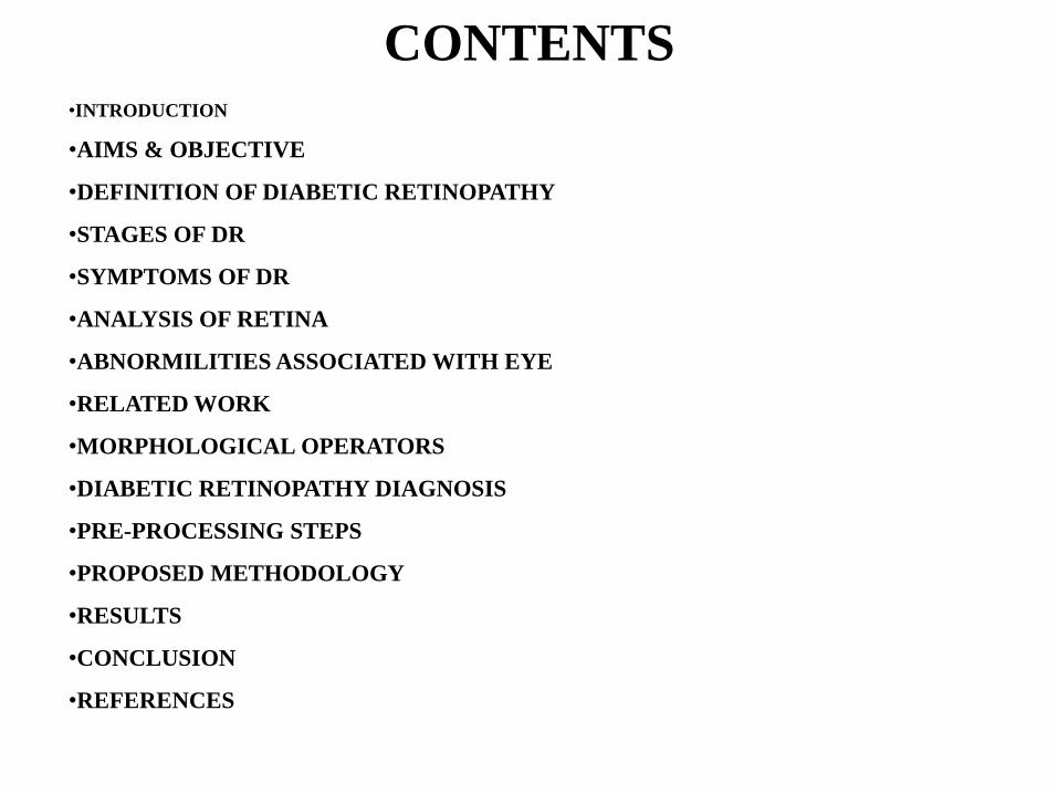
CONTENTS•INTRODUCTION
•AIMS & OBJECTIVE
•DEFINITION OF DIABETIC RETINOPATHY
•STAGES OF DR
•SYMPTOMS OF DR
•ANALYSIS OF RETINA
•ABNORMILITIES ASSOCIATED WITH EYE
•RELATED WORK
•MORPHOLOGICAL OPERATORS
•DIABETIC RETINOPATHY DIAGNOSIS
•PRE-PROCESSING STEPS
•PROPOSED METHODOLOGY
•RESULTS
•CONCLUSION
•REFERENCES
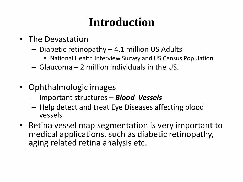
Introduction
• The Devastation– Diabetic retinopathy – 4.1 million US Adults
• National Health Interview Survey and US Census Population
– Glaucoma – 2 million individuals in the US.
• Ophthalmologic images– Important structures – Blood Vessels– Help detect and treat Eye Diseases affecting blood
vessels
• Retina vessel map segmentation is very important to medical applications, such as diabetic retinopathy, aging related retina analysis etc.
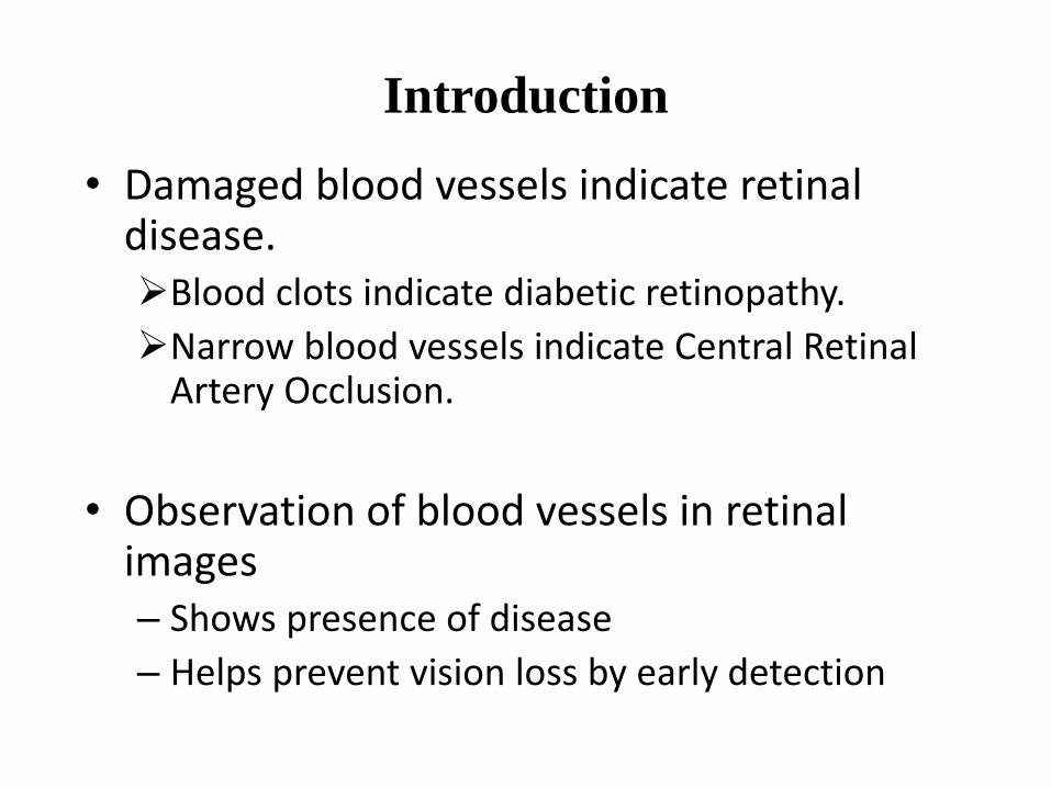
Introduction
• Damaged blood vessels indicate retinal disease.Blood clots indicate diabetic retinopathy.
Narrow blood vessels indicate Central Retinal Artery Occlusion.
• Observation of blood vessels in retinal images– Shows presence of disease
– Helps prevent vision loss by early detection
Aims and Objective
• The primary aim of this project is to develop a system
that will be able to identify patients with BDR and
PDR from either colour image or grey level image
obtained from the retina of the patient.
• The secondary aim includes developing a MATLAB
based Graphic User Interface (GUI) tool to be used
by the ophthalmologist in marking fundus images.
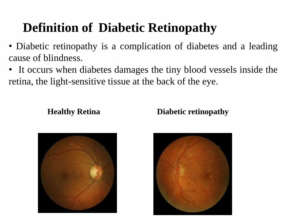
Definition of Diabetic Retinopathy
• Diabetic retinopathy is a complication of diabetes and a leading
cause of blindness.
• It occurs when diabetes damages the tiny blood vessels inside the
retina, the light-sensitive tissue at the back of the eye.
Healthy Retina Diabetic retinopathy
Stages of Diabetic Retinopathy
1. Mild Nonproliferative Retinopathy
2. Moderate Nonproliferative Retinopathy
3. Severe Nonproliferative Retinopathy
4. Proliferative Retinopathy
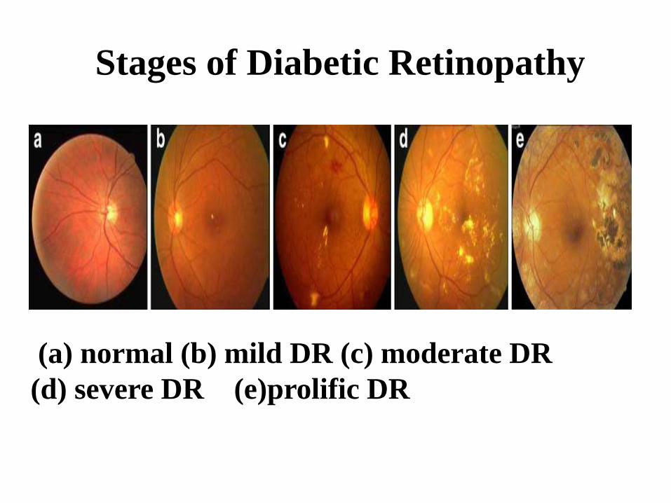
Stages of Diabetic Retinopathy
(a) normal (b) mild DR (c) moderate DR
(d) severe DR (e)prolific DR
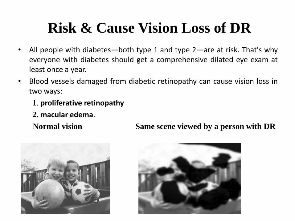
Risk & Cause Vision Loss of DR
• All people with diabetes—both type 1 and type 2—are at risk. That's whyeveryone with diabetes should get a comprehensive dilated eye exam atleast once a year.
• Blood vessels damaged from diabetic retinopathy can cause vision loss intwo ways:
1. proliferative retinopathy
2. macular edema.
Normal vision Same scene viewed by a person with DR
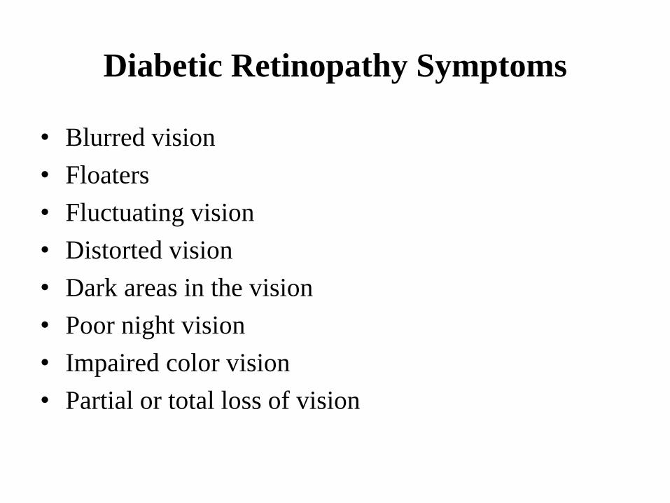
Diabetic Retinopathy Symptoms
• Blurred vision
• Floaters
• Fluctuating vision
• Distorted vision
• Dark areas in the vision
• Poor night vision
• Impaired color vision
• Partial or total loss of vision
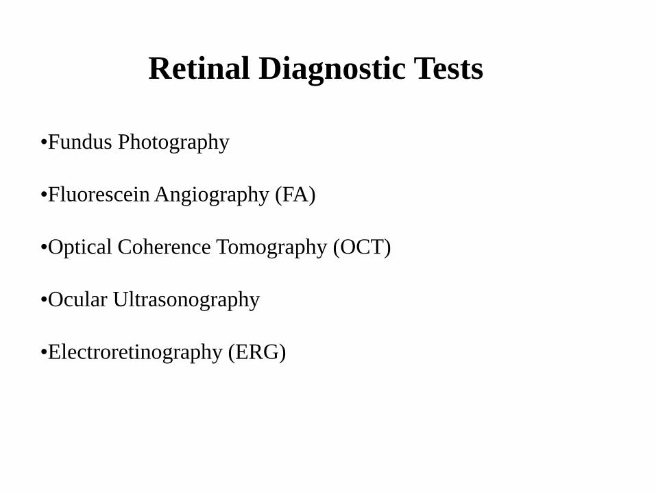
Retinal Diagnostic Tests
•Fundus Photography
•Fluorescein Angiography (FA)
•Optical Coherence Tomography (OCT)
•Ocular Ultrasonography
•Electroretinography (ERG)
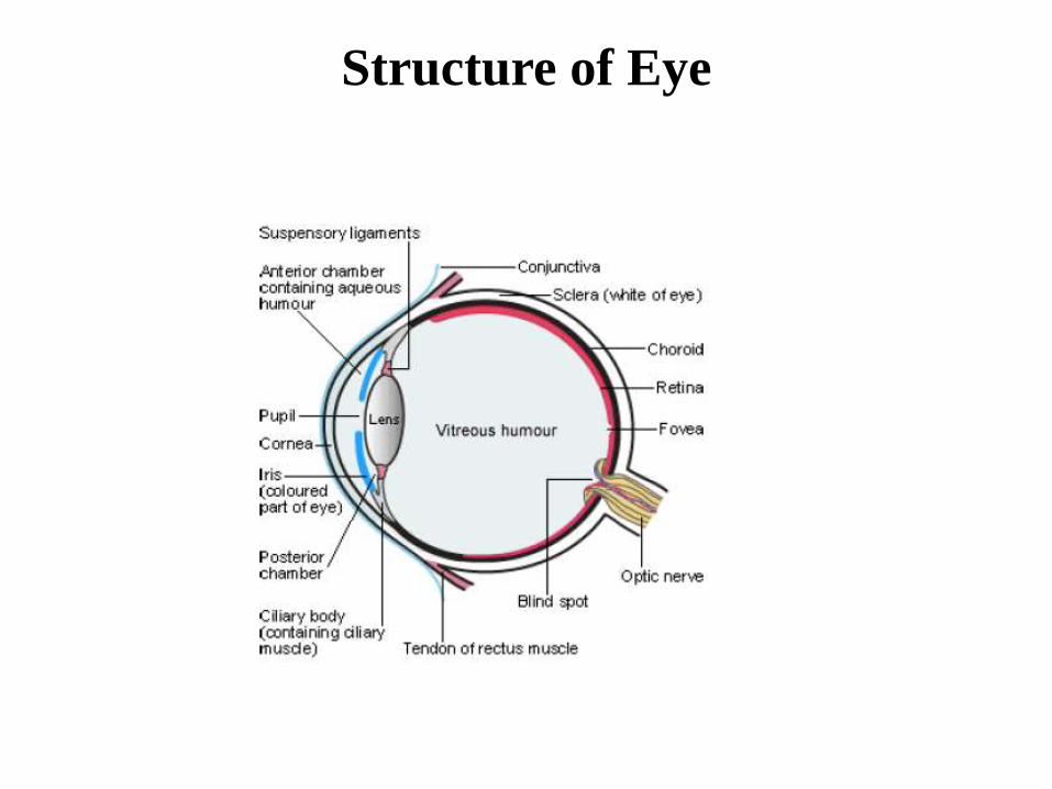
Structure of Eye
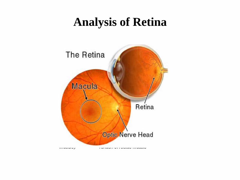
Analysis of Retina
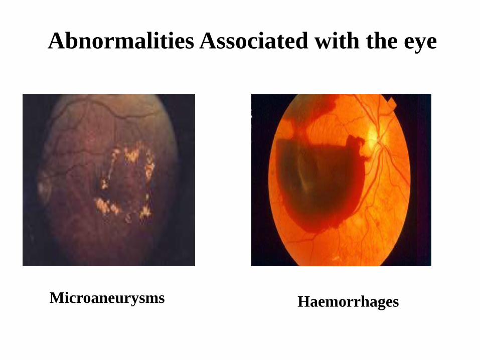
Abnormalities Associated with the eye
Microaneurysms Haemorrhages
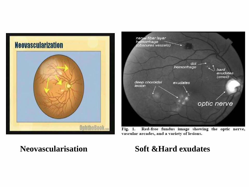
Neovascularisation Soft &Hard exudates
Related Work
• Akara et. al discussed about the set of optimally
adjusted morphological operators used for exudates
detection for thai patients.
• Selvathi et. al uses fundus images are segmented &
they are classified as normal or DR images by
extracting features from these structures & GLCM
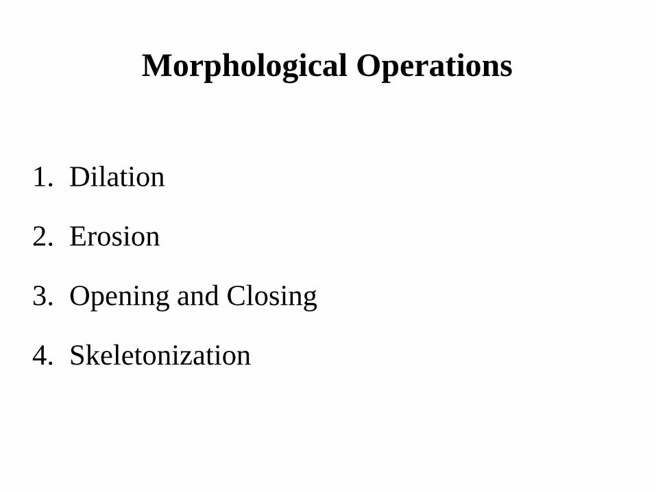
Morphological Operations
1. Dilation
2. Erosion
3. Opening and Closing
4. Skeletonization
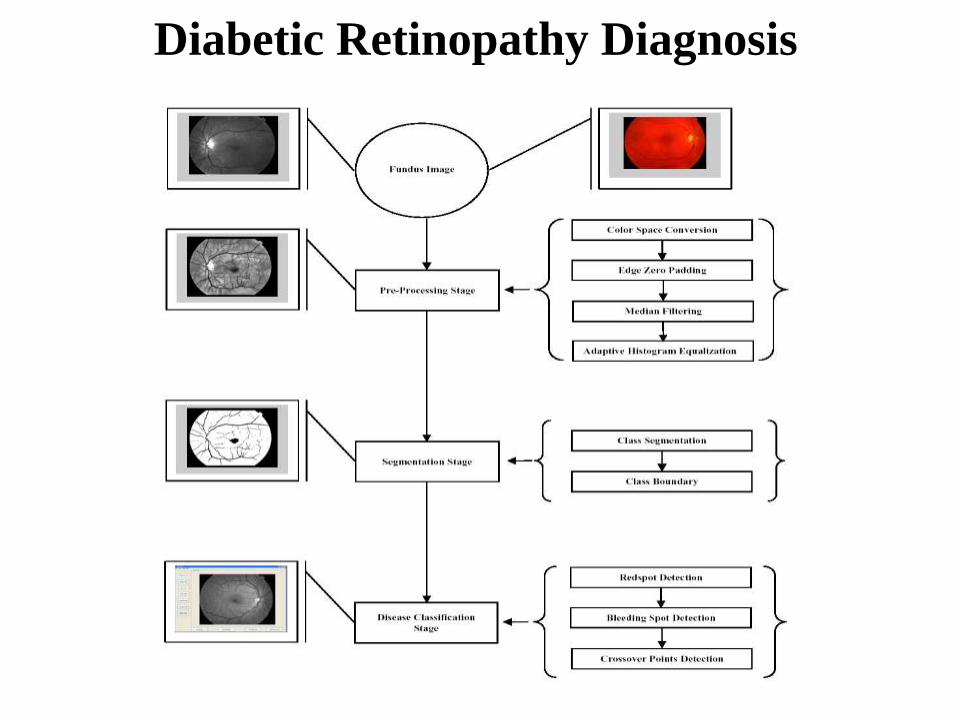
Diabetic Retinopathy Diagnosis
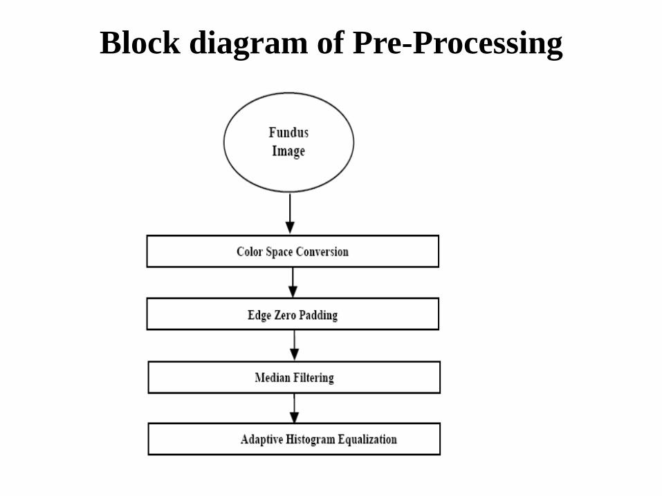
Block diagram of Pre-Processing
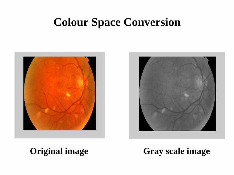
Colour Space Conversion
Original image Gray scale image
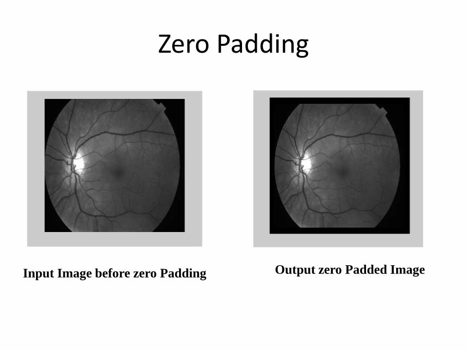
Zero Padding
Input Image before zero Padding Output zero Padded Image
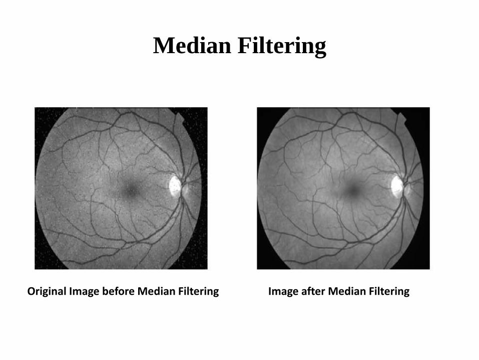
Median Filtering
Original Image before Median Filtering Image after Median Filtering
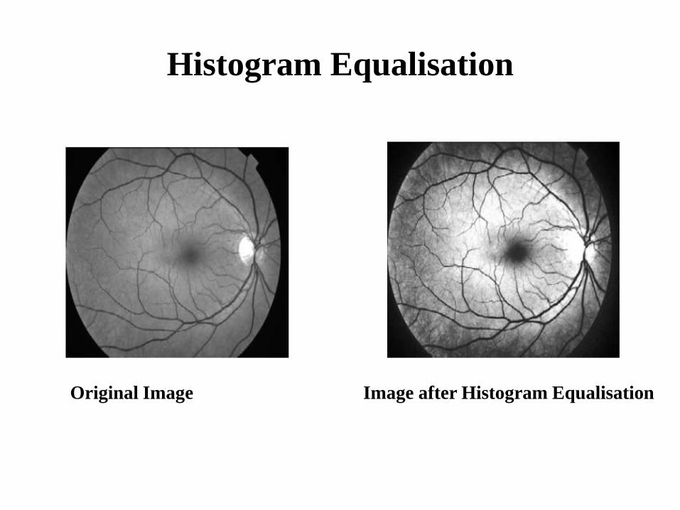
Histogram Equalisation
Original Image Image after Histogram Equalisation
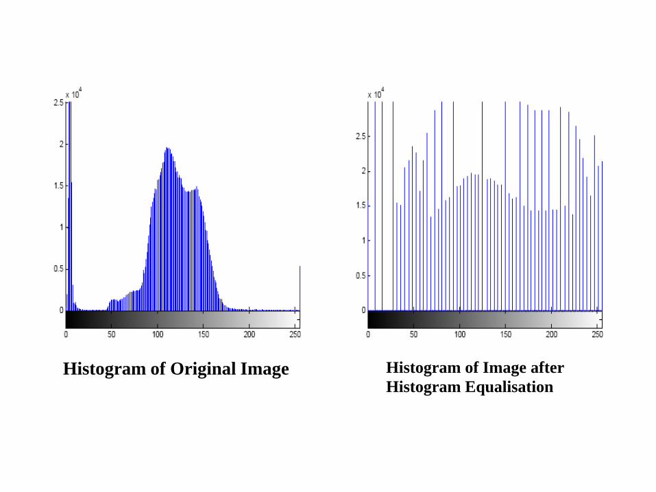
Histogram of Original Image Histogram of Image after
Histogram Equalisation
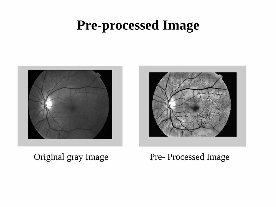
Pre-processed Image
Original gray Image Pre- Processed Image
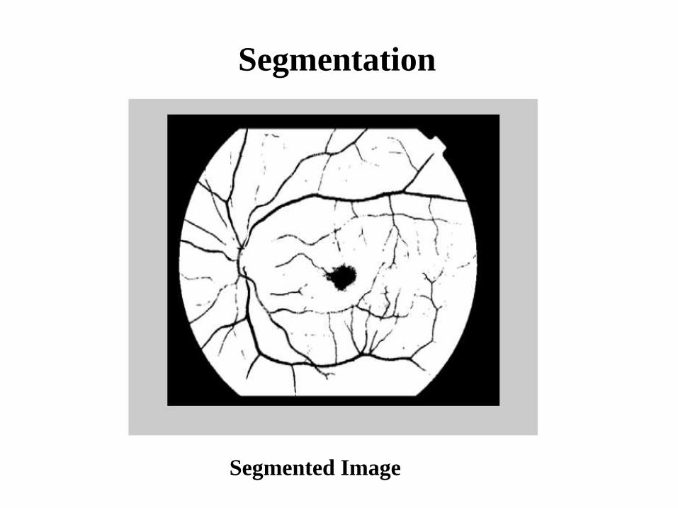
Segmentation
Segmented Image
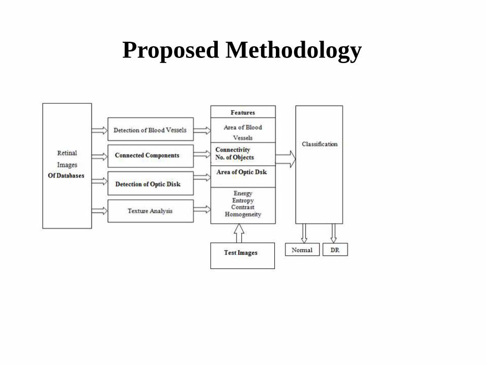
Proposed Methodology
5 methods for Proposed Methodology
• DATABASES: Publicly avalilable databases such as DRIVE, DIARETDB1and MESSIDOR are used in this work.
• SEGMENTATION OF RETINAL STRUCTURE:
Detection of Blood Vessels: It is used for enhancing the retinal image.
Detection of Exudates and Microaneurysms: Morphological operatorsare used for the detection of exudates and MA in this work.
• TEXTURE ANALYSIS:It gives information about the arrangement ofsurface pixels and their relationship with the surrounding pixels
• FEATURE EXTRACTION:The feature vector used for classificationconsists of seven features obtained from segmentation of retinal structures andtexture analysis.
• CLASSIFICATION

RESULTS AND DISCUSSION
OPTIC DISK DETECTION
Optic Disc Location (a) RGB Image, (b) I band after Pre-processing, (c) Image after Closing, (d) Thresholded Image, (e)Marker Image, (f) Reconstructed Image, (g) Threshold on Difference Image, (h) Optic Disc Area.
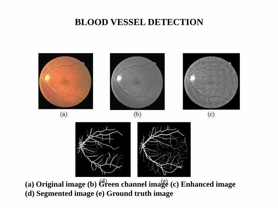
BLOOD VESSEL DETECTION
(a) Original image (b) Green channel image (c) Enhanced image
(d) Segmented image (e) Ground truth image
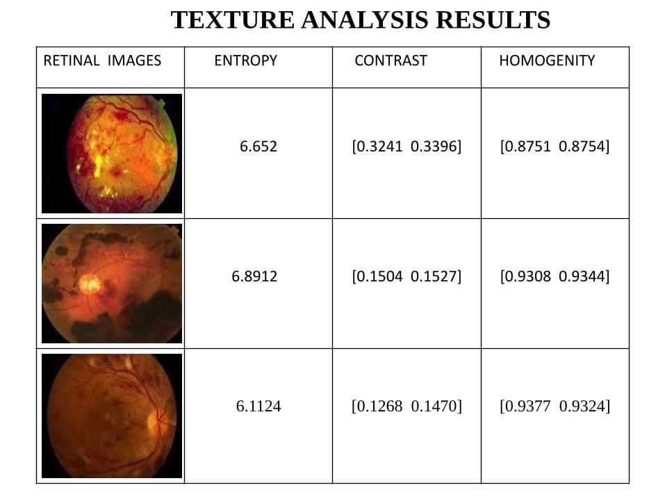
RETINAL IMAGES ENTROPY CONTRAST HOMOGENITY
6.652 [0.3241 0.3396] [0.8751 0.8754]
6.8912 [0.1504 0.1527] [0.9308 0.9344]
6.1124 [0.1268 0.1470] [0.9377 0.9324]
TEXTURE ANALYSIS RESULTS
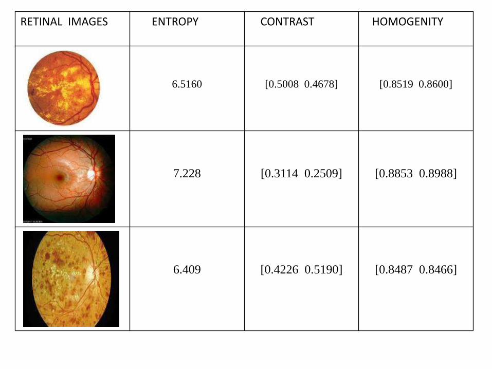
RETINAL IMAGES ENTROPY CONTRAST HOMOGENITY
6.5160 [0.5008 0.4678] [0.8519 0.8600]
7.228 [0.3114 0.2509] [0.8853 0.8988]
6.409 [0.4226 0.5190] [0.8487 0.8466]
Conclusion and Future work
• Development of a system that will be able to identify patients with BDR and PDR from either color image or grey level fundus image
• Develop techniques for not only better detection of vessel edges,
• More anatomically exacting regarding medical image standards.
• Extraction of Minute blood vessels.
REFERENCES
• DRIVE Database: http://www.isi.uu.nl/Research/Database/DRIVE
• Selvathi D, Neethi Balagopal (2012) Detection of Retinal Blood Vessels using Curvelet Transform. IEEE International Conference on devices, Circuits and Systems 325-329. doi: 10.1109/ICDCSyst.2012.6188730
• Pierre Soille (2002) Morphological Image Analysis, Principles and Applications. 2nd Edition. Springer
• Bevilacqua,V., Cambò,S., Cariello,L., Mastronardi, G., ‘A combined method to detect Retinal Fundus Features’, Conference on EACDA, Italy, September, 2005.
• Martin M. and Tosunoglu S., 2000, “Image Processing Techniques for Machine Vision”,Conference on Recent Advances in Robotics, Florida, pp. 1-9.

THANK YOU

ANY QUESTIONS


![Grading Fundus Images for Diabetic Retinopathy · 2017-10-17 · Grading Fundus Images for Diabetic Retinopathy 3885 Apart from OD, the exudates[10] and the cotton wool spots also](https://img.pdfslide.us/doc/110x75/5e3ecf3000efdb1dd03b8d22/grading-fundus-images-for-diabetic-retinopathy-2017-10-17-grading-fundus-images.jpg)

![The Guide - Diabetic Retinopathy - Vision Lossvisionloss.org.au/wp-content/uploads/2016/05/The... · the guide [diabetic retinopathy] What is Diabetic Retinopathy? Diabetic Retinopathy](https://img.pdfslide.us/doc/110x75/5e3ed00bf9c32e41ea6578a8/the-guide-diabetic-retinopathy-vision-the-guide-diabetic-retinopathy-what.jpg)