Embed Size (px)
Citation preview
3/23/2016
1
Fundus AutofluorescenceBrittany Bateman, BS
Fundus Autofluorescence
2
“Fundus autofluorescence imaging is used to record fluorescence that may occur naturally in ocular structures or as a byproduct of a disease process. This technique allows the
topographic mapping of lipofuscin distribution in the RPE. The intensity portrayed by FAF corresponds to the accumulation of lipofuscin, which increases with aging, RPE cell
dysfunction or an abnormal metabolic load on the RPE.”
-Saudi Journal of Ophthalmology
Visible Spectrum
3
The RPE
4
The RPE
5
• Retinal Pigment Epithelium –single layer of cells which separates the choroid from the neurosensory retina
• Five main roles, one of which is phagocytosis of the outer segment
• Allows renewal process necessary to maintain photoreceptors (rods and cones) functioning normally
The RPE
6
• Aging can result in incomplete or partial breakdown of these segments which causes an accumulation of lipofuscin.
• Excessive lipofuscin is associated with visual loss.• This accumulation is evident in ocular disease even before the visual
cycle begins to degrade.
3/23/2016
2
Fluorescence and Phosphorescence
7
• Fluorescence – emission of light by a substance that has absorbed light. – Immediate release of light.– emitted light has longer wavelength– electron excited to a higher energy state and relaxes to ground state
• Phosphorescence – emission of light lasts after excitation has stopped– electrons cannot relax into ground state therefore emission lasts after excitation
Fundus Autofluorescence
8
• Autofluorescence – fluorescence occurs naturally, without a dye
• Fluorescence occurs when molecules get excited by a specific light and then emit light at a longer wavelength which makes them glow
• Fundus autofluorescence works by shining a specific color/wavelength of light into the eye and capturing how it naturally fluoresces without dye
• The major source of fundus autofluorescene is this lipofuscin.
Healthy Eyes
9
Lipofuscin and FAF
10
• The RPE ages or becomes more damaged which means there is more lipofuscin
• Lipofuscin properties fluoresces at about 500nm to 750nm
• Intensity of signal emitted relates to the health level of the RPE based upon what is normal.
• Hyperfluorescence (white/really bright areas) means that the RPE cells are failing
• Hypofluroescence (black/really dark areas) means that the RPE is dead/ atrophy
Types – Fundus Camera
11
• Fundus camera uses white light• Wideband exciter filter and wideband barrier filter• Only a single image• Limiting factors such as ocular media opacities• The first fundus camera technique used filters around 580nm for the exciter and a
barrier filter centered at 695nm.
Fundus Camera Light
12
3/23/2016
3
Types – Fundus Camera
13
Types - cSLO
14
• Confocal Scanning Laser Ophthalmoscope• Laser beam of specific wavelength on the retina to excite it• Images “build” to reduce background noise
• Acquire images at nearly a video rate and add the frames together• Provides higher-quality if there is minimal movement by patient.
• Two main types Heidelberg Retina Angiograph and the Optos
Types - cSLO
15
http://www.heidelbergengineering.com/us/products/spectralis-models/technology/confocal-scanning-laser-ophthalmoscopy/
cSLO Light
16
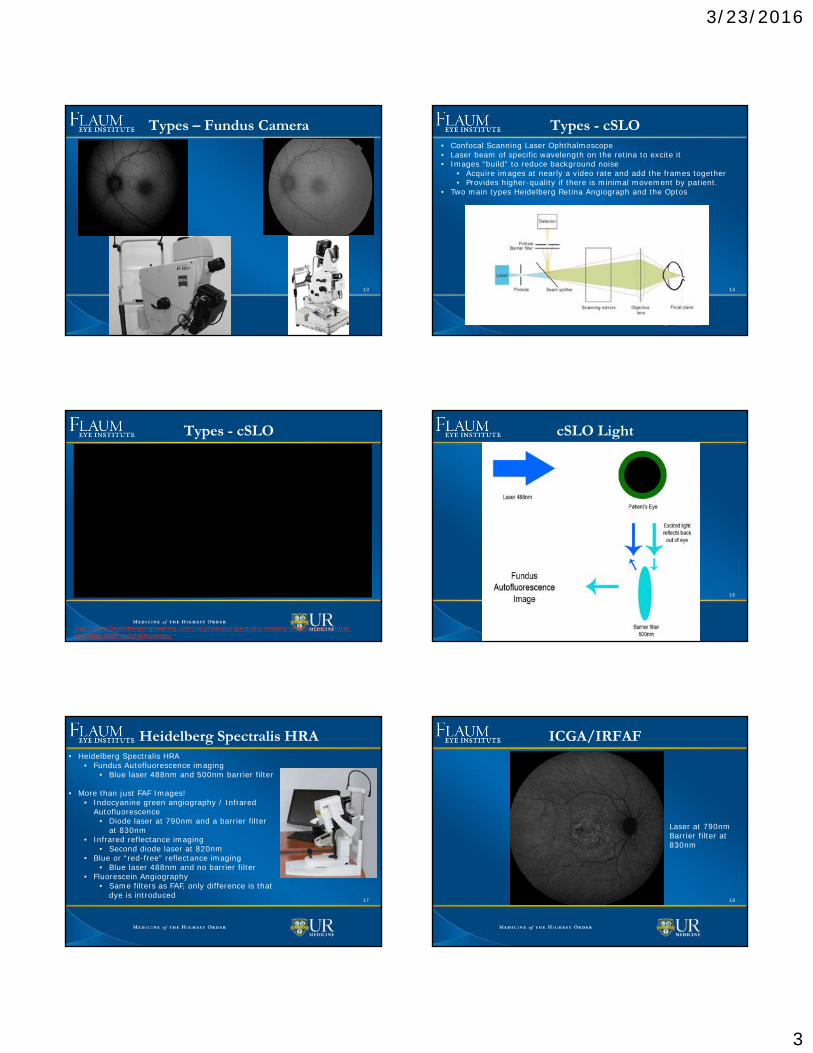
Heidelberg Spectralis HRA
17
• Heidelberg Spectralis HRA• Fundus Autofluorescence imaging
• Blue laser 488nm and 500nm barrier filter
• More than just FAF Images!• Indocyanine green angiography / Infrared
Autofluorescence• Diode laser at 790nm and a barrier filter
at 830nm• Infrared reflectance imaging
• Second diode laser at 820nm• Blue or “red-free” reflectance imaging
• Blue laser 488nm and no barrier filter• Fluorescein Angiography
• Same filters as FAF, only difference is that dye is introduced
ICGA/IRFAF
18
Laser at 790nm Barrier filter at 830nm
3/23/2016
4
Infrared and Blue Reflectance
19
Infrared Reflectance Red-Free Blue Reflectance Laser at 820nm Laser at 488nm
Options
20
Infrared
Red-Free
ICGA/IRAF
FAF
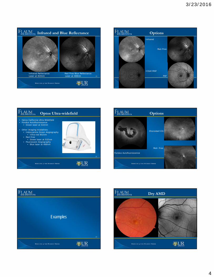
Optos Ultra-widefield
21
• Optos California Ultra-Widefield• Fundus Autofluorescence
• Green laser at 532nm
• Other imaging modalities:• Indocyanine Green Angiography
• Infra-red 802nm• Red-Free
• Green laser at 532nm• Fluorescein Angiography
• Blue laser at 488nm
Options
22
Choroidal/ICG
Red- Free
Fundus Autofluorescence
23
Examples
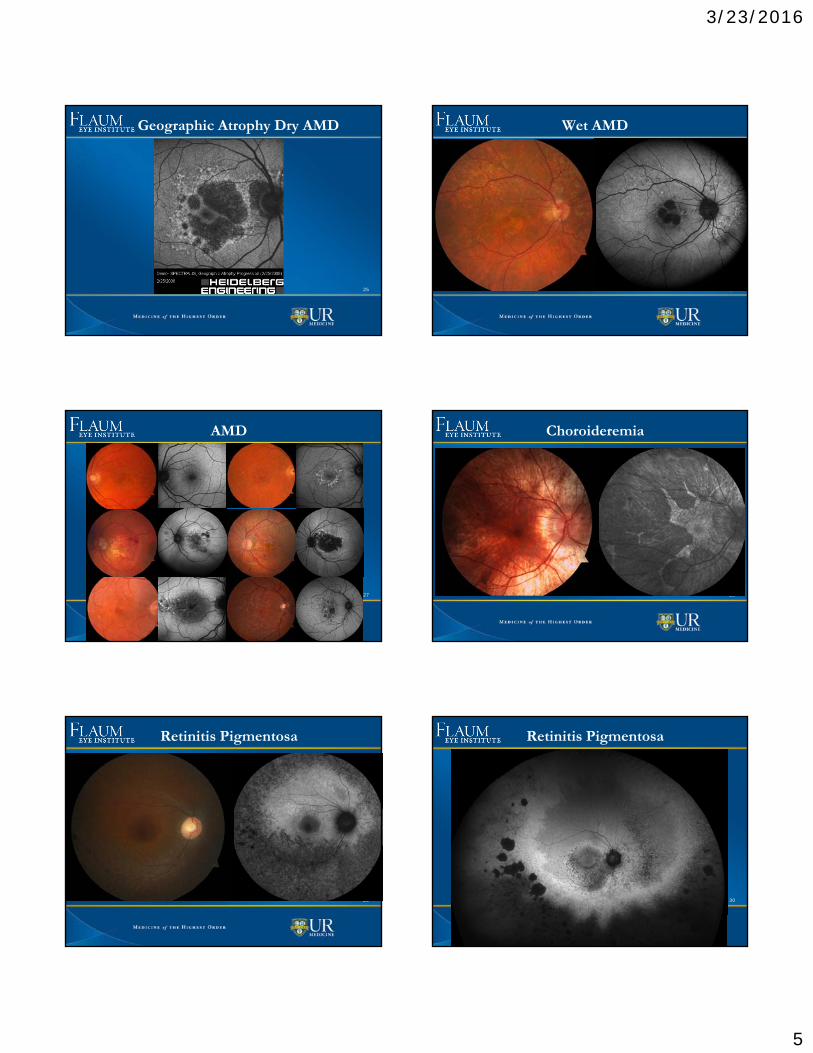
Dry AMD
24
3/23/2016
5
Geographic Atrophy Dry AMD
25
Wet AMD
26
AMD
27
Choroideremia
28
Retinitis Pigmentosa
29
Retinitis Pigmentosa
30

3/23/2016
6
Chorioretinitis
31
Chorioretinitis
32
33
Non - Atrophy Examples
Pigment
34
Optic Nerve Head Drusen
35
Summary
36
• Fundus Autofluorescence shows break down of RPE by accumulation of lipofuscin.
• White areas means that the RPE cells are failing• Black areas means that there is RPE atrophy
• Fundus cameras or cSLO imaging can be used• cSLO Heidelberg Spectralis HRA 488nm and Optos 532nm• Fundus cameras 500-600nm

3/23/2016
7
37
Credit• Sources:
• http://www.aao.org/eyenet/article/nuts-bolts-of-fundus-autofluorescence-imaging• http://www.heidelbergengineering.com/us/• http://www.optos.com/en-US/Products/Retinal-imaging-products/Retinal-imaging-
products/California/• http://www.opsweb.org/?page=Autofluorescence• https://nei.nih.gov/• http://www.amd.org/• http://clinicalgate.com/autofluorescence-imaging/• http://www.blindness.org/eye-conditions/• http://www.sciencedirect.com/science/article/pii/S131945341400037X• http://www.reviewofoptometry.com/continuing_education/tabviewtest/lessonid/108843/• http://www.hindawi.com/journals/joph/2013/309192/• Saudi Journal of Ophthalmology• Atlas of Fundus Autofluorescence Imaging F.G. Holz, S. Schmitz-Valckenberg, R.F.
Spaide, A.C. Bird• https://www.researchgate.net/publication/233722317_Near_Infrared_Autofluorescence_
Imaging_of_Retinal_Diseases• http://www.comtecmed.com/cophy/2011/Uploads/assets/von%20strachwitz.pdf
38
Credit• Diagrams
• Clinical Atlas of Ophthalmology, Spalton• http://www.closerlookatstemcells.org/stem-cells-and-medicine/macular-
degeneration• https://en.wikipedia.org/wiki/Light• http://www.neurology.org/content/80/1/47/F1.large.jpg
• Images courtesy of Flaum Eye Institute Diagnostic Team and Imagers• Brittany Bateman• Brittany Richardson, COA OCT-C CRA• Kassandra Mundt• Patricia Artman, COA• Rachel Hollar, OCT-C CRA• Taylor Pannell, OCT-C CRA
39
Thank you


![SPECTRALIS - INNOVA · Fundus Autofluorescence in the Abca4[-]/[-] Mouse Model of Stargardt Disease - Correlation With Accumulation of A2E, Retinal Function, and Histology doi: 10.1167/iovs.13-11688](https://img.pdfslide.us/doc/110x75/5ec1d3ad12d1a659545b86a4/spectralis-innova-fundus-autofluorescence-in-the-abca4-mouse-model-of-stargardt.jpg)