Embed Size (px)
Citation preview
Congenital Dyserythropoietic Anemia type III (CDA III) - diagnostics, genetics and morbidity
Maria Liljeholm
Department of Radiation Sciences, Hematology Umeå 2016

Responsible publisher under Swedish law: the Dean of the Medical Faculty This work is protected by the Swedish Copyright Legislation (Act 1960:729) New series no: 1784 ISBN: 978-91-7601-424-0 ISSN: 0346-6612 Cover image: Bone marrow smear from a patient with CDA III Electronic version available at http://umu.diva-portal.org/ Printed by: Print & Media, Umeå university Umeå, Sweden 2016

”What makes the desert beautiful,” said the little prince, ”is that somewhere it hides a well...”
Antoine de Saint-Exupéry

i
Table of Contents
Abstract iii Populärvetenskaplig sammanfattning på svenska vi Original papers xi Introduction 1
CDA I 1 CDA II 2 CDA III 3 CDA variants 4 Cell cycle, mitosis and cytokinesis 6 Red cell membrane 8 Hemolytic disorders 9
DAT-negative hemolysis 9 Diagnostic approach in DAT-negative hemolysis 11
Estimation of erythrocyte fragility 11 Flow cytometry 11 Sodium dodecyl sulphate-polyacrylamide gel electrophoresis (SDS-PAGE) 12 Diagnostic considerations 12
Iron 13 Iron homeostasis 13 Iron overload 14
AIMS 16 Study population and methods 17
Study population 17 Methods 19
Paper I 19 Paper II 21 Paper III 22
Results 23 Paper I 23
CDA III and KIF23 23 KIF23 expression 26 KIF23 functional analysis 27
Paper II 29 Flow cytometry in CDA III 29
Paper III 30 HFE status 30 Clinical appearance 30

ii
Discussion 32 Paper I 32 Paper II 34 Paper III 35
Conclusions 37 Future perspectives 38 Acknowledgements 40 References 43 Paper I-III
iii
Abstract
The Congenital Dyserythropoietic Anemias (CDA) are rare hereditary hemolytic disorders with large bi- to multi-nucleated erythroblasts in the bone marrow. Hemolysis is negative in a direct antiglobulin test (DAT). Based on morphology and clinical picture, three major forms of CDAs, type I, II, and III have been defined. CDA III, dominantly inherited, constitutes the rarest type with a majority of cases belonging to a family in Västerbotten, Sweden. The genetic background of CDA I and CDA II has been linked to mutations in CDAN1 and SEC23B respectively. The mutation of CDA III has been linked to 15q22 in earlier studies.
In this project we have defined the causative genetic lesion in two families with CDA III. The novel mutation KIF23 c.2747C>G (p.P916R) was shown to segregate with CDA III in the Swedish and American CDA III families and was absent in 356 healthy controls. KIF23 encodes mitotic kinesin-like protein 1 (MKLP1), which plays a central role in the last step of cytokinesis. RNAi-based knock-down and rescue experiments demonstrated that the p.P916R mutation causes cytokinesis failure in HeLa cells, resulting in increasing number of bi-nuclear cells, consistent with appearance of large multinucleated erythroblasts in CDA III patients. We conclude that CDA III is caused by a mutation in KIF23, encoding MKLP1, a conserved mitotic kinesin crucial for cytokinesis.
Flow cytometry with eosin-5´-maleimide (EMA), anti-CD55 and anti-CD59 is commonly used when investigating non-autoimmune hemolytic anemias. Reduced fluorescence of EMA, typically detected in hereditary spherocytosis, is also seen in CDA II, while reduction of CD55 and CD59 characterizes paroxysmal nocturnal hemoglobinuria (PNH). We studied the flow cytometric profile of EMA, CD55, and CD59 on erythrocytes in CDA III. We found no abnormality of the erythrocyte membrane in CDA III and concluded that standard flow cytometry cannot be used to discriminate between CDA III and normal controls.
In CDA I and CDA II a majority of patients, including those who are not transfusion dependent, suffer from iron overload, which, according to earlier studies, is not the case in CDA III. We found that individuals of the Västerbotten CDA III family carry mutations in the hemochromatosis (HFE) gene. Three CDA III patients with heterozygous or compound HFE mutations need treatment with phlebotomy due to iron overload. One of them carries heterozygous H63D mutation, which is not reported to lead to iron overload by itself in otherwise healthy individuals. We propose that molecular genetic testing of the HFE gene is indicated in all patients with CDA, including CDA III.
iv
Abbreviations
AGLT Acid glycerol lysis time test
ASF1 Anti-silencing function protein 1
CDA Congenital dyserythropoietic anemia
CLL Chronic lymphatic leukemia
DAT Direct antiglobulin test
EMA Eosin-5´-maleimide
ER Endoplasmic reticulum
ERFE Erythroferrone
GAPDH Glyceraldehyde-3-phosphate dehydrogenase
GDF15 Growth differentiation factor 15
GPA Glycophorin A
Hb Hemoglobin
HEMPAS Hereditary erythroblastic multinuclearity associated with a positive acidified-serum test
HE Hereditary elliptocytosis
HH Hereditary hemochromatosis
HPP Hereditary pyropoikilocytosis
HS Hereditary spherocytosis
IL-6 Interleukin-6
LD Lactate dehydrogenase
MCF Mean channel fluorescence
MCV Mean cellular volume
MGUS Monoclonal gammopathy of unknown significance
MKLP1 Mitotic kinesin-like protein 1
MRI Magnetic resonance imaging
MVK Mevalonate kinase gene
NGS Next generation sequencing
OF Osmotic fragility test

v
PCR Polymerase chain reaction
PNH Paroxysmal nocturnal hemoglobinuria
RHAG Rh-associated glycoprotein
RT- PCR Reverse transcription PCR
SDS-PAGE Sodium dodecyl sulphate polyacrylamide gel electrophoresis
Tf Transferrin
TfR Transferrin receptor
TTP Thrombotic thrombocytopenic purpura
TSAT Transferrin iron saturation
TWSG1 Twisted gastrulation BMP signaling modulator 1
Wt Wild type
vi
Populärvetenskaplig sammanfattning på svenska
Kongenitala dyserytropoetiska anemier (CDA) är en grupp ovanliga ärftliga anemier med ineffektiv bildning av röda blodkroppar (erytropoes) och varierande grad av anemi. Baserat på benmärgsmorfologi, klinisk bild och laboratoriefynd indelas CDA i flera undergrupper, varav I-III varit kända sedan länge. De senaste decennierna har nya former identifierats, vilka klassificerats som typ IV-VII, framförallt baserat på enstaka fallrapporter. CDA I-III är de mest studerade. Gemensamma karakteristika utgörs av icke autoimmun hemolys samt dyserytropoes med stora, dubbel- till fler-kärniga, erytroblaster i benmärgen.
CDA III utgör den ovanligaste formen av de dyserytropoetiska anemierna. Den globala prevalensen av CDA III är svårbedömd men majoriteten av rapporterade fall tillhör en släkt med ursprung i Västerbotten. I denna familj har individer med CDA III kartlagts 6 generationer bakåt, till mitten av 1800-talet. Totalt har 47 individer fått diagnosen CDA III. Denna anemiform har ett dominant nedärvningsmönster och en benmärgsbild som präglas av aktiv erytropoes med stora mångkärniga erytroblaster. Den kliniska bilden varierar från mild till måttlig hemolytisk anemi. I tidigare arbeten av Sandström och medarbetare påvisades ett antal diagnostiska kriterier för CDA III, baserat på benmärgsmorfologi och perifera blodprover med analys av hemolysparametrar samt tymidinkinas. Den sjukdomsorsakande genen lokaliserades till 15q22. Individer med CDA III visades ha ökad förekomst av ögonbottenförändringar (angioid streaks) samt ökad risk att drabbas av blodcancern multipelt myelom och dess förstadium monoklonal gammopati utan signifikans (MGUS).
Vid utredning av patient med icke autoimmun hemolytisk anemi bör flödescytometri utföras med frågeställningarna hereditär sfärocytos (HS) samt paroxysmal nocturn hemoglobinuri (PNH). HS, med en prevalens i norra Europa på 1 % och i norra Sverige upp mot 5 %, är en ärftlig hemolytisk anemi med varierande grad av anemi och hemolysorsakade komplikationer i form av gulsot (ikterus) och gallstensproblematik. Svårighetsgraden varierar även på individnivå, med försämrad symtombild vid såväl bakteriella som virusorsakade infektioner. PNH bör alltid uteslutas vid icke autoimmun hemolys på grund av de allvarliga komplikationer, framförallt tromboser och organpåverkan, som denna sjukdom kan medföra. Flödescytometri med eosin-5´-maleimid (EMA) visar minskat upptag hos röda blodkroppar vid HS, medan minskat uttryck av CD59 på ytan av såväl röda som vita blodceller utgör ett karakteristikum för PNH. Vid EMA-flöde ses liknande mönster vid CDA II som vid HS.

vii
Vid CDA I och II utgör järninlagring ett stort kliniskt problem. Att järnnivåerna är ökade vid hemolytiska tillstånd är vanligt, beroende på den ökade destruktionen av röda blodkroppar samt påverkan på hepcidinregleringen. Hepcidin, ett hormon med central roll i järnomsättningen, ser till att absorptionen av järn från tarmen och frisättningen av järn från kroppens järnlager justeras efter aktuellt behov. Vid hemolys och ineffektiv erytropoes fungerar inte denna regleringsmekanism optimalt. Trots detta har järndepåerna vid CDA III i tidigare studier ej visat sig förhöjda, sannolikt beroende på att patienterna, på grund av intravasal hemolys, kontinuerligt förlorar järn i urinen.
Hos i övrigt friska personer utgör, i norra Europa, hereditär hemokromatos den vanligaste orsaken till förhöjda järnnivåer. Sjukdomen nedärvs autosomalt recessivt och orsakas av mutationer i HFE-genen, vilket leder till ökat upptag av järn från tarmen. Muterat anlag på båda kromosomerna (homozygot mutation) krävs för att utveckla ökade järnnivåer, men även hos homozygoter är det bara ca 20 % som utvecklar sjukdomen. I Sverige är prevalensen av hereditär hemokromatos 0,5 %, men i Norrland är sjukdomen vanligare. Omkring 7 % av befolkningen är anlagsbärare. Obehandlad kan sjukdomen leda till inlagring av järn i inre organ med organsvikt som följd. De vanligaste symtomen är ledvärk, lever och hjärtpåverkan samt diabetes. Om behandling i form av regelbundna blodtappningar påbörjas i tid kan denna utveckling helt förhindras. Diagnostiken består av genetisk analys avseende HFE-mutationer tillsammans med provtagning avseende järn, transferrinmättnad (TSAT) och ferritin i serum. I samband med vårt projekt uppmärksammades att mutationer i HFE-genen förekommer hos individer i Västerbottenssläkten.
Syfte
- att identifiera den mutation som ger upphov till CDA III.
- att undersöka ytan på de röda blodkropparna vid CDA III, med de flödescytometriska analyser som, i klinisk rutin, utförs vid utredning av icke-autoimmun hemolytisk anemi.
- att kartlägga förekomsten av hereditär hemokromatos i Västerbottenssläkten samt undersöka den kliniska bilden vid hereditär hemokromatos hos individer med och utan CDA III.
viii
Patienter och metoder Kartläggning av Västerbottenssläkten gjordes av Bergström och Jacobsson i början av 1960-talet och uppdaterades av Sandström och medarbetare i mitten av 90-talet. Släktträdet sträcker sig över sex generationer och tar sin början i mitten av 1800-talet. Sedan 1990-talet har ytterligare barn tillkommit i släkten och en uppdatering av släktträdet har genomförts i detta projekt. DNA har samlats in och nytillkomna individer har utretts med avseende på CDA III. I tidigare arbete av Sandström och medarbetare insamlades DNA på alla då levande individer i släkten. Totalt finns idag DNA preparerat från 60 individer i denna släkt.
För att få fram kandidatgener har i arbete I DNA analyserats med molekylärgenetisk metod i form av next generation sequencing (NSG). För ytterligare konfirmerande studier av mutation i KIF23 har PCR-baserade metoder använts.
I arbete II har flödescytometri med EMA samt anti CD55 och anti CD59 använts i enlighet med klinisk rutin. Flödescytometri utfördes på perifert blod från 16 CDA III positiva individer och 14 CDA III negativa syskon.
I arbete III har HFE-mutationsstatus analyserats med PCR-baserad metod. HFE-mutationsstatus bedömdes på totalt 58 individer i släkten, 37 CDA III positiva och 21 CDA III negativa syskon. För vidare analys med koppling till järnstatus och hemolysprover inkluderades 32 CDA III positiva och 18 CDA III negativa i studien. TSAT, ferritinnivåer och hemolysparametrar analyserades enligt klinisk rutin på laboratoriet för klinisk kemi.
Resultat I arbete I har vi identifierat den genetiska mutation i KIF23 som ger upphov till CDA III. Mutationen leder till dysfunktionellt KIF23, även kallat Mitosis Kinesin-Like Protein 1 (MKLP1). Detta protein har en central roll i bildningen och funktionen av den så kallade ”midbody”, vilken reglerar avslutningen av celldelningen. Mutationen leder till att cellen inte delar sig trots bildning av två eller flera cellkärnor. Störning i midbody-funktionen har experimentellt visats ge upphov till flerkärniga celler.
I arbete II har vi visat att flödescytometri med EMA ger ett obetydligt ökat upptag hos erytrocyter vid CDA III, sannolikt beroende på ökad cellvolym hos de röda blodkropparna. Flödescytometri med anti CD55 och anti CD59 ger helt normalt utfall. Flödescytometri kan alltså inte användas för diagnostik av CDA III. CDA III riskerar inte heller att misstas för HS eller PNH vid flödescytometrisk analys av röda blodkroppar.
I arbete III har förekomsten av HFE-mutationer i Västerbottenssläkten kartlagts och klinisk presentation avseende järnbalansen har undersökts hos
ix
CDA III positiva individer och deras syskon. Bland CDA III negativa syskon har en individ med homozygot mutation och ökade järnnivåer påbörjat blodtappningar. Av de CDA III positiva patienterna hade ingen homozygot mutation men tre befanns bära på heterozygot HFE-mutation tillsammans med laboratoriemässiga fynd förenliga med ökade järndepåer. Alla tre har påbörjat blodtappningar med sjunkande ferritin och TSAT som följd. Tappningarna har tolererats väl trots den kontinuerliga hemolys som föreligger vid CDA III. Ingen av de CDA III negativa syskonen befanns ha störd järnomsättning trots förekomst av heterozygot HFE-mutation, vilket är förväntat, då heterozygot HFE-mutation ej ger upphov till ökade järndepåer hos i övrigt friska personer.
Diskussion I arbete I har vi fastslagit den molekylärgenetiska bakgrunden till CDA III, vilket bidragit till ökad förståelse för celldelningens slutstadium. Kunskap om KIF23 och dess protein KIF23 (MKLP1) kan utgöra en viktig pusselbit avseende utveckling av flerkärniga celler vid exempelvis olika cancerformer. Den höga proliferationen hos många cancerceller ställer också krav på ökad tillgång till proteiner, såsom KIF23, vilka möjliggör celldelning. Att KIF23 är uppreglerat vid flera tumörtyper är känt. Överuttryck finns beskrivet vid såväl hjärntumören gliom som lung-, bröst-, lever- och magsäcks- cancer. Nedreglering av KIF23 har också visat att celldelningen i dessa cancerformer avstannat.
Individer med CDA III har ökad risk at utveckla blodcancern myelom. Vi genomför nu insamling av data från Cancerregistret och Dödsorsaksregistret samt planerar morfologiska studier av obduktionspreparat från individer med CDA III. Detta för att undersöka om även annan cancersjukdom kan kopplas till KIF23 mutation, samt om flerkärniga celler även förekommer i andra cellinjer än den röda blodkroppsbildningen. Vi avser också att studera effekterna av den specifika genetiska mutationen i KIF23 efter att ha infört denna i försöksdjur.
I arbete II har vi undersökt ytan på de röda blodkropparna vid CDA III med de flödescytometriska metoder som utgör basen vid utredning av icke-autoimmun hemolys. Den flödescytometriska profilen vid CDA II kan förväxlas med HS, vilket kan medföra risk för feldiagnostik tidigt i utredningsförloppet av icke-autoimmun hemolytisk anemi. I detta projekt visar vi att så inte är fallet beträffande CDA III. Vid negativt svar på flödescytometri med EMA samt CD59 antikropp bör benmärgsundersökning utföras, vilken i förekommande fall kommer att inge misstanke om CDA. Molekylärgenetisk diagnostik är därefter att föredra för ytterligare subklassificering, vilket numera är möjligt för alla tre huvudtyperna av CDA.
I arbete III visar vi att hereditär hemokromatos förekommer i Västerbottenssläkten. Vid homozygot mutation av HFE-genen kan detta, hos
x
friska CDA III negativa syskon, leda till ökad risk för järninlagring. Utveckling av hereditär hemokromatos förebyggs med blodtappning. Hos CDA III positiva individer kan även heterozygot mutation leda till patologiska järnnivåer. Kombinationen av CDA II och hereditär hemokromatos finns beskriven hos en patient från Italien. Blodtappningar var i detta fall svåra att genomföra på grund av patientens anemi, men våra patienter med CDA III har tolererat behandlingen väl. Även hos individer med blodcancern myelodysplastiskt syndrom eller hemoglobinopatin thalassemi har heterozygot HFE-mutation visats tillräcklig för att leda till patologisk järninlagring. Vårt arbete bekräftar att heterozygot HFE-mutation är tillräckligt för att orsaka höga järnnivåer, när mutationen förekommer tillsammans med annan sjukdom med ineffektiv erytropoes, såsom CDA III. Eftersom mutationer i HFE-genen är vanligt förekommande i befolkningen, bör screening avseende dessa utföras hos patienter med annan hematologisk sjukdom med påverkad järnomsättning, såsom vid hemolytiska sjukdomar och tillstånd med ineffektiv erytropoes.

xi
Original papers
This thesis is based on the following studies, and referred to by their roman numerals in the text.
I. Maria Liljeholm, Andrew F. Irvine, Ann-Louise Vikberg, Anna Norberg, Stacy Month, Herbert Sandström, Anders Wahlin, Masanori Mishima, Irina Golovleva. Congenital Dyserythropoietic Anemia type III (CDA III) is caused by a mutation in kinesin family member, KIF23.
Blood 2013;121(23):4791-9
II. Maria Liljeholm, Elisabeth Grönlund, Irina Golovleva, Herbert Sandström, Anders Wahlin. Erythrocyte Flow Cytometric Analysis in Congenital Dyserythropoietic Anemia Type III - Evaluation of Eosin-5´-Maleimide, CD55, and CD59.
J Blood Disord Transfus 2013;4:172. doi:10.4172/2155-9864.1000172
III. Maria Liljeholm, Ann-Louise Vikberg, Irina Golovleva, Herbert Sandström, Anders Wahlin. Congenital Dyserythropoietic Anemia type III and primary hemochromatosis; coexistence of mutations in KIF23 and HFE.
J Hematol Blood Disord 2016;1(2):203
Reprints for this thesis were made with permission from the journals.

xii
1
Introduction
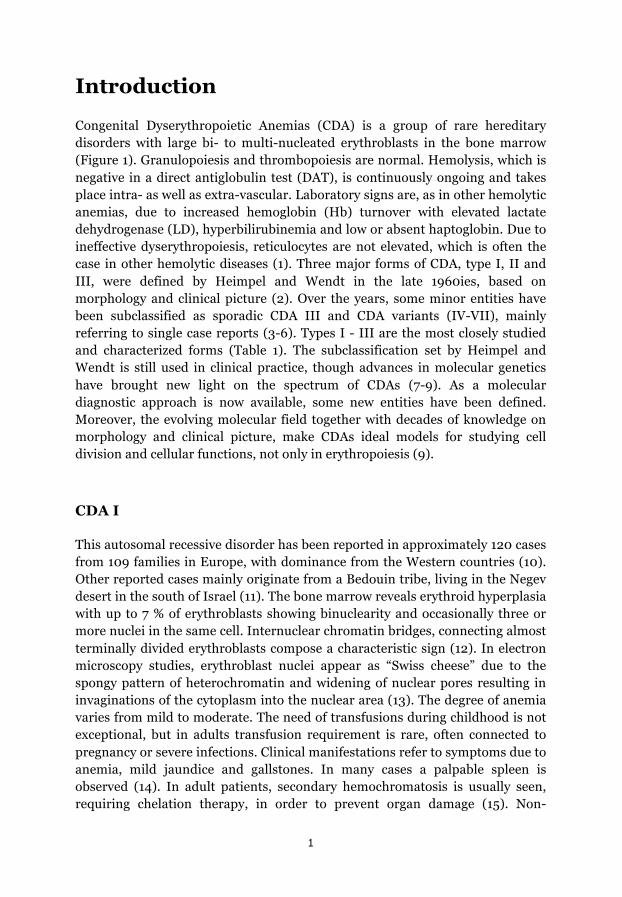
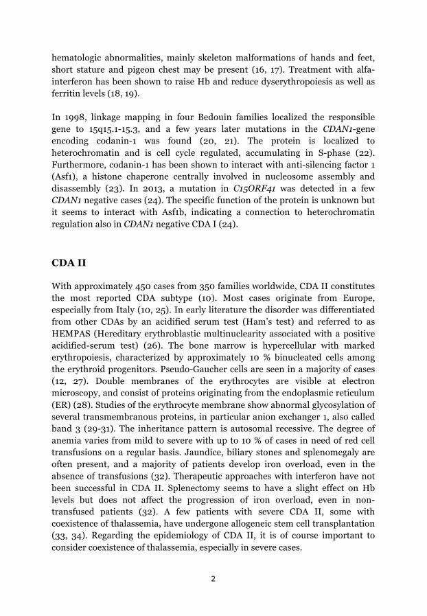
Congenital Dyserythropoietic Anemias (CDA) is a group of rare hereditary disorders with large bi- to multi-nucleated erythroblasts in the bone marrow (Figure 1). Granulopoiesis and thrombopoiesis are normal. Hemolysis, which is negative in a direct antiglobulin test (DAT), is continuously ongoing and takes place intra- as well as extra-vascular. Laboratory signs are, as in other hemolytic anemias, due to increased hemoglobin (Hb) turnover with elevated lactate dehydrogenase (LD), hyperbilirubinemia and low or absent haptoglobin. Due to ineffective dyserythropoiesis, reticulocytes are not elevated, which is often the case in other hemolytic diseases (1). Three major forms of CDA, type I, II and III, were defined by Heimpel and Wendt in the late 1960ies, based on morphology and clinical picture (2). Over the years, some minor entities have been subclassified as sporadic CDA III and CDA variants (IV-VII), mainly referring to single case reports (3-6). Types I - III are the most closely studied and characterized forms (Table 1). The subclassification set by Heimpel and Wendt is still used in clinical practice, though advances in molecular genetics have brought new light on the spectrum of CDAs (7-9). As a molecular diagnostic approach is now available, some new entities have been defined. Moreover, the evolving molecular field together with decades of knowledge on morphology and clinical picture, make CDAs ideal models for studying cell division and cellular functions, not only in erythropoiesis (9).
CDA I
This autosomal recessive disorder has been reported in approximately 120 cases from 109 families in Europe, with dominance from the Western countries (10). Other reported cases mainly originate from a Bedouin tribe, living in the Negev desert in the south of Israel (11). The bone marrow reveals erythroid hyperplasia with up to 7 % of erythroblasts showing binuclearity and occasionally three or more nuclei in the same cell. Internuclear chromatin bridges, connecting almost terminally divided erythroblasts compose a characteristic sign (12). In electron microscopy studies, erythroblast nuclei appear as “Swiss cheese” due to the spongy pattern of heterochromatin and widening of nuclear pores resulting in invaginations of the cytoplasm into the nuclear area (13). The degree of anemia varies from mild to moderate. The need of transfusions during childhood is not exceptional, but in adults transfusion requirement is rare, often connected to pregnancy or severe infections. Clinical manifestations refer to symptoms due to anemia, mild jaundice and gallstones. In many cases a palpable spleen is observed (14). In adult patients, secondary hemochromatosis is usually seen, requiring chelation therapy, in order to prevent organ damage (15). Non-
2
hematologic abnormalities, mainly skeleton malformations of hands and feet, short stature and pigeon chest may be present (16, 17). Treatment with alfa-interferon has been shown to raise Hb and reduce dyserythropoiesis as well as ferritin levels (18, 19).
In 1998, linkage mapping in four Bedouin families localized the responsible gene to 15q15.1-15.3, and a few years later mutations in the CDAN1-gene encoding codanin-1 was found (20, 21). The protein is localized to heterochromatin and is cell cycle regulated, accumulating in S-phase (22). Furthermore, codanin-1 has been shown to interact with anti-silencing factor 1 (Asf1), a histone chaperone centrally involved in nucleosome assembly and disassembly (23). In 2013, a mutation in C15ORF41 was detected in a few CDAN1 negative cases (24). The specific function of the protein is unknown but it seems to interact with Asf1b, indicating a connection to heterochromatin regulation also in CDAN1 negative CDA I (24).
CDA II
With approximately 450 cases from 350 families worldwide, CDA II constitutes the most reported CDA subtype (10). Most cases originate from Europe, especially from Italy (10, 25). In early literature the disorder was differentiated from other CDAs by an acidified serum test (Ham’s test) and referred to as HEMPAS (Hereditary erythroblastic multinuclearity associated with a positive acidified-serum test) (26). The bone marrow is hypercellular with marked erythropoiesis, characterized by approximately 10 % binucleated cells among the erythroid progenitors. Pseudo-Gaucher cells are seen in a majority of cases (12, 27). Double membranes of the erythrocytes are visible at electron microscopy, and consist of proteins originating from the endoplasmic reticulum (ER) (28). Studies of the erythrocyte membrane show abnormal glycosylation of several transmembranous proteins, in particular anion exchanger 1, also called band 3 (29-31). The inheritance pattern is autosomal recessive. The degree of anemia varies from mild to severe with up to 10 % of cases in need of red cell transfusions on a regular basis. Jaundice, biliary stones and splenomegaly are often present, and a majority of patients develop iron overload, even in the absence of transfusions (32). Therapeutic approaches with interferon have not been successful in CDA II. Splenectomy seems to have a slight effect on Hb levels but does not affect the progression of iron overload, even in non-transfused patients (32). A few patients with severe CDA II, some with coexistence of thalassemia, have undergone allogeneic stem cell transplantation (33, 34). Regarding the epidemiology of CDA II, it is of course important to consider coexistence of thalassemia, especially in severe cases.
3
In 2009 two research groups detected the causative gene SEC23B, earlier localized to chromosome 20 (35, 36). The encoded protein SEC23B, is a component of cytoplasmic coat protein II (COP II), which is centrally involved in transportation of membrane and protein components from the ER towards the Golgi apparatus (37). Approximately 80 mutations have been found throughout the gene, giving rise to slightly different phenotypes (38). Two founder mutations have been described in the Italian population (39).
CDA III
This is the rarest form of the three major subtypes of CDA. Globally, a familial form has been detected in three families. The majority originates from a family in Västerbotten, Sweden, and the others include a few cases reported from one family in the United States and one family in Argentina (40, 41). In addition, some sporadic cases have been diagnosed and reported as CDA III. These are single case reports with diverse phenotypes, including hepatosplenomegaly, iron overload and mental retardation, which are not present in familial CDA III (4, 42-45). The mode of inheritance is dominant in the familial cases but appears to be recessive in the sporadic forms (4). The earliest reported familial CDA III cases, a mother and her three children from the American family, were described in 1951 by Wolff and von Hofe and referred to as “familial erythroid multinuclearity” (40). In 1962, the first cases of the Swedish family were described by Bergström et al (46). The pedigree composed 15 affected individuals, reaching back to a couple living in the middle of the 19th century. Today the pedigree of the Swedish Västerbotten CDA III family covers six generations with 47 diagnosed CDA III cases. Throughout the years the family has been closely studied, defining morphology features, as well as laboratory and clinical signs (47). Bone marrow microscopy reveals erythroid hyperplasia with bi- and multinucleated erythroblasts, some with up to 12 nuclei in the same cell, so called gigantoblasts (46, 48). Electron microscopy studies show non-specific signs such as intranuclear clefts and ironladen mitochondria (48). Observations of the erythrocyte membrane, performed in two patients, revealed a slight reduction of glycosylation of band 3 while CD44, CD47, CD59 and Rh-related proteins appeared to be normal (48).
Laboratory signs are consistent with hemolysis with absent haptoglobin and elevated LD and bilirubinemia. Anemia is usually mild and the need of transfusions is very rare. In contrast to CDA I and II, a major part of hemolysis takes place intravascular, leading to hemosiderinuria with loss of iron in the urine. This has been believed to explain why iron overload has not been detected in patients with CDA III (47). Serum thymidine kinase is highly

4
elevated in CDA III and has been used as a diagnostic tool in the CDA III family when bone marrow examination has not been feasible (49). Splenomegaly is uncommon in these patients but about 20 % of cases occasionally experience jaundice and biliary symptoms. Weakness, fatigue and headache have been reported in approximately one third of the patients, symptoms that are aggravated in pregnancy or infection (50).
Although not statistically significant, Bergström et al noted a high frequency of cancer in the medical records of the CDA III cases observed in their study (46). Sandström et al confirmed a connection between CDA III and the precancerous plasma cell disorder monoclonal gammopathy of undetermined significance (MGUS) and multiple myeloma (47). Angioid streaks, rarely occurring retinal changes in patients with other hemolytic disorders such as thalassemia and sickle cell anemia, were found in some CDA III patients (51). The causative gene was localized to 15q21-q25 (52).
In the present study we focus on the Västerbotten CDA III family, defining the genetic cause of the disease, analysing the erythrocyte membrane with flow cytometry, and report that iron overload is present in this family, attributable to coexistence of hereditary mutations of the HFE gene. We are presently investigating the incidence of cancer in this family.
CDA variants
All reported CDAs do not fulfil the criteria of the CDA subtypes described above. According to bone marrow findings and clinical phenotype they have been diagnosed as CDA IV-VII, including only a few patients in each group. In addition, some cases do not fit into these groups either and are regarded as CDA variants (3-6). Hence, CDA subtypes represent a very heterogeneous group, probably originating from different genetic defects connected to erythroid regulation. Some genes, as KLF1 and GATA-1, encoding erythroid transcription factors and mutations in genes where CDA is a part of a clinical syndrome have been described. CDA type IV is caused by mutation in KLF1, while GATA-1 mutation has been detected in X-linked thrombocytopenia with CDA (53-55).
CDA may constitute one of several clinical presentations in syndromes such as Majeed syndrome (mutation in LPIN2), CDA with exocrine pancreatic insufficiency and calvarial hyperostosis (mutation in COX4I2), and the inborn error of metabolism, mevalonate kinase deficiency (mutation in MVK) (56-58).
5
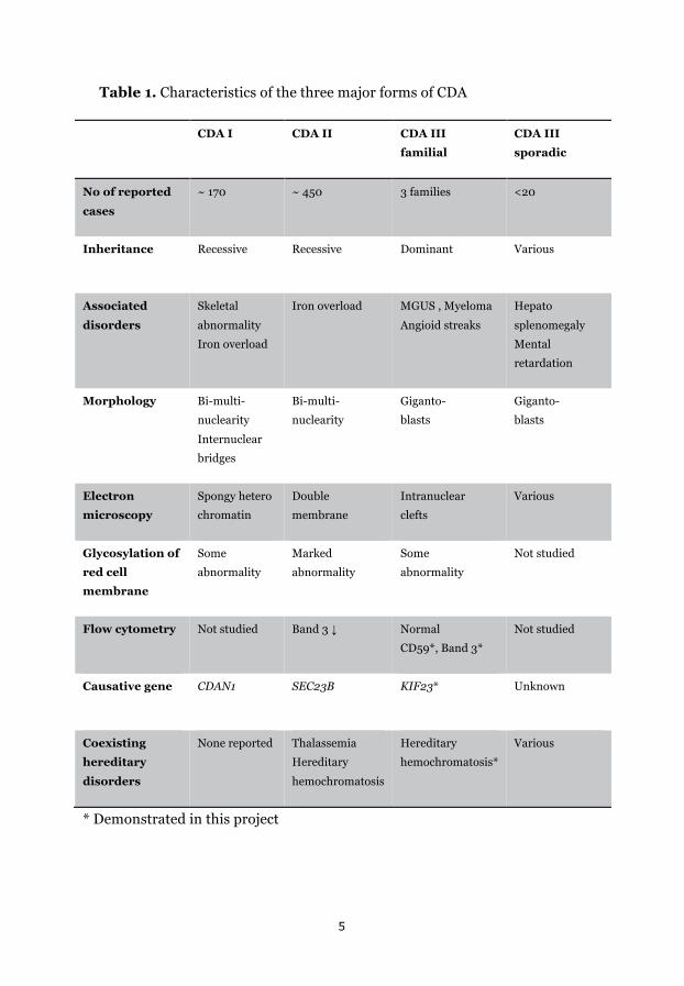
Table 1. Characteristics of the three major forms of CDA
CDA I CDA II CDA III
familial
CDA III
sporadic
No of reported
cases
~ 170 ~ 450 3 families <20
Inheritance
Recessive Recessive Dominant Various
Associated
disorders
Skeletal
abnormality
Iron overload
Iron overload MGUS , Myeloma
Angioid streaks
Hepato
splenomegaly
Mental
retardation
Morphology Bi-multi-
nuclearity
Internuclear
bridges
Bi-multi-
nuclearity
Giganto-
blasts
Giganto-
blasts
Electron
microscopy
Spongy hetero
chromatin
Double
membrane
Intranuclear
clefts
Various
Glycosylation of
red cell
membrane
Some
abnormality
Marked
abnormality
Some
abnormality
Not studied
Flow cytometry
Not studied Band 3 ↓ Normal
CD59*, Band 3*
Not studied
Causative gene
CDAN1 SEC23B KIF23* Unknown
Coexisting
hereditary
disorders
None reported Thalassemia
Hereditary
hemochromatosis
Hereditary
hemochromatosis*
Various
* Demonstrated in this project

6
Figure 1. Bone marrow smears in the major forms of CDA. Erythroblasts with internuclear bridge in CDA I, a binuclear erythroblast in CDA II, and a gigantoblast in CDA III. CDA I and CDA II photos reprinted with permission from Dr Tamary and Prof Iolascon, respectively.
Cell cycle, mitosis and cytokinesis
The cell cycle represents the events that enable a cell to duplicate. The process is strictly regulated, assuring genetic material to transfer properly through generations of cells. Correct segregation of chromosomes and the ability to fulfil cell division is highly dependent on microtubules and kinesins, which are the most central proteins of these processes (59, 60). The microtubules are filamentous structures running through the cell, building up the cytoskeleton and enabling transportation through the cell. The movement of the tubules and the ability to move cargo along the cytoskeleton is carried out by kinesins, so called motor proteins, with the ability to walk along the microtubules. More than 30 kinesins have been defined in the different stages of the cell cycle (59).
The cell cycle is either divided into five phases, GAP0 (G0), GAP1 (G1), Synthesis (S), GAP2 (G2), and Mitosis (M) or the three major stages of interphase, mitosis, and cytokinesis (61, 62). In G0, the cell is per definition not in cell cycle, since it is not dividing or preparing for cell division but fulfilling the tasks it was destined for. Going into G1 the cell grows and gets ready for DNA synthesis. When it is fully prepared for this task it enters the next phase, the S phase, where DNA is replicated. Thereafter it enters the phase of mitosis, which ends in cell division (61).
CDA I CDA II CDA III

7
The definitions of interphase, mitosis and cytokinesis enable a more detailed description of the cell cycle since interphase and mitosis are subdivided into several steps and cytokinesis composes an entity of its own (61, 62). G1, S and G2 correspond to interphase while M includes mitosis and cytokinesis. Here I will focus on the steps of mitosis and cytokinesis.
Mitosis is divided into four major steps, prophase, metaphase, anaphase and telophase. In prophase, the chromatin condensates into the 92 chromosomes present after DNA duplication. The cell poles form as centrosomes are pulled apart by microtubules to opposite sides of the cell, forming the “mitotic spindle”. In metaphase the chromosomes line up at the equator of the cell, each chromosome attached to one of the cell poles by microtubules. Sister chromatids line up tightly together, enabling segregation to opposite poles during anaphase. At the end of anaphase the mitotic spindle elongates as parallel microtubules form the “central spindle”, further forming the cleavage furrow. At the end of telophase the furrow is contracted into the formation of the midbody, a dense bundle of microtubules that tightly connects to the plasma membrane (63).
The cell now enters the last step of the cell cycle, cytokinesis, which involves the processes resulting in the final division of the cells. As the daughter cells are defined, the midbody elongates forming an intercellular bridge. This structure is essential for final cleavage of the cytoplasm, as it enables membrane trafficking and membrane fusion (64).
Mutations of genes encoding for microtubules or kinesins can affect the cell cycle at all stages, depending on the gene involved. Malfunction of proteins, essential in cell division can cause damaged cells due to failure of correct segregation of chromosomes. In addition, impaired regulation mechanisms of the cell cycle can allow cells to divide out of control, being one of the fundamental conditions in cancer development.
In CDA III, erythropoiesis seems to be affected by failure of cytokinesis, since multiple nuclei gather in the same cell. Hence, mitosis seems to proceed properly until the very final step of cell separation. In this project we identify the causative gene in CDA III and show that the encoded protein is centrally involved in the final steps of cytokinesis.
8
Red cell membrane
The red cell is unique among human cells. In its mature form all cell organelles are lost, including the nucleus. Consequently, the properties of the cell are fully dependent on the membrane and the cytoskeleton. Apart from the skeletal network, the cytosol mainly consists of the oxygen transport protein hemoglobin (Hb). During the 120 days lifespan of an erythrocyte, the ability to undergo reversible deformation is essential for the capacity to pass through small capillaries, carrying oxygen to all parts of the body. The cell membrane is very strong and elastic, closely interacting with the cytoskeleton. Even as the red cell is highly extended passing through capillary vessels, this structure enables it to retain the surface area. This property is very important to protect the cell from undergoing hemolysis. The membrane consists of a bilayer of cholesterol and phospholipids, penetrated by transmembranous proteins anchoring the two-dimensional spectrin-based network of the cytoskeleton. More than 50 types of proteins penetrate the membrane in various quantities. About half of them carry different blood group antigens such as the Rhesus protein (Rh) and the oligosaccharides A and B (65).
The transmembrane proteins enable transportation through the membrane, adherence to other cells and activation of signalling pathways (65). The most abundant membrane proteins are band 3, also called anion exchanger 1 (AE1), and glycophorin A (GPA). Interacting with other proteins, they form vertical chains, linking the membrane to the cytoskeleton. Band 3 is a large protein whose integral part serves as a transporter of bicarbonate (HCO3-) and chloride (Cl-) through the membrane. The cytosolic part links to spectrin through the anchoring proteins protein 4.2 and ankyrin. The function of band 3 can be influenced by horizontal interaction with GPA and the Rh-complex, which includes Rh-associated glycoprotein (RhAg), glycophorin B (GPB), intracellular adhesion molecule-4 (ICAM-4) and integrin associated glycoprotein (IAP) (66, 67). Dysfunction of the vertical linkage system leads to failure of red cell integrity and results in hemolysis.
Attached to the external surface of the membrane are glycolipids and glycoproteins, forming a glycocalyx surrounding the cell. Many of these molecules belong to the blood group system or are part of the complement system. The latter plays an important role in the defence against pathogens, and in the removal of damaged cells. Activation of the cascade system causes a cascade of protein activation resulting in hemolysis due to the formation of a membrane attack complex (MAC), which forms pores through the membrane. Regulation of complement activation is essential to protect normal red cells from undergoing hemolysis. Two of the most relevant proteins, in regard to hemolytic disorders, are the decay accelerating factor (DAF) and MAC-
9
inhibitory protein (MAC-IP) (68). Dysfunction of these proteins causes hemolysis due to failure of complement inhibition.
Hemolytic disorders
The first thing that must be defined in a patient with hemolysis is whether it is caused by autoantibodies or not. This is done by the direct antiglobulin test (DAT), also referred to as Coomb’s test. The test reveals autoantibodies to the patient´s own red cells and is an important tool to distinguish between autoimmune and non-autoimmune mediated hemolysis. The most common form is caused by IgG-autoantibodies and often responds promptly to corticosteroids, while complement activating autoimmunity, through IgM-autoantibodies, is more difficult to treat. Autoimmune hemolysis is often connected to other diseases such as lymphoproliferative disorders, other malignancies, or autoimmune diseases (69).
DAT-negative hemolysis
Non-autoimmune hemolytic disorders or DAT-negative hemolysis is a diverse group of dysfunctions of red cells, leading to increased red cell turnover. Lysis is caused by alterations of the membrane, complement regulation, Hb-chains, or enzymes involved in red cell metabolism.
The clinical appearance varies widely from asymptomatic cases to requirement of regular transfusions and chelation therapy. A majority of these disorders are constitutional but the symptoms of hemolysis often vary throughout life, with more active episodes during, for example, infection or pregnancy. However, some disorders, such as paroxysmal nocturnal hemoglobinuria (PNH), are acquired and can be connected with other disorders or malignancies of the bone marrow.
Enzymopathies such as deficiency of glucose-6-phosphate dehydrogenase and pyruvate kinase, and hemoglobinopathies such as thalassemia and sickle cell anemia represent common causes of DAT-negative hemolysis in a global perspective. These diseases compose entities of its own, and pathogenesis and diagnostic approach in these disorders will not be further discussed here. Rather I will focus on the rarer forms of DAT-negative hemolytic disorders, the membranopathies and complement-induced hemolysis, where CDA is an important differential diagnosis to bear in mind.
10
Membranopathies
Hereditary spherocytosis (HS) is the most common hereditary membrane disorder in Caucasians with an incidence of about 1:2000. The disease is caused by deficiency or dysfunction of one of the proteins connected to the band 3 vertical linkage system: that is band 3, protein 4.2, ankyrin or spectrin. Pattern of inheritance is mostly dominant even if recessive cases have been described (70).
Hereditary elliptocytosis (HE) has a similar prevalence as HS but is more common in malaria endemic regions. Hereditary pyropoikilocytosis (HPP) and South Asian ovalocytosis (SAO) compose variants of the disease. HE and SAO are dominantly inherited while HPP has a recessive trait. The elliptic shape of the red cells in HE and HPP is due to dysfunction of the interaction between the cytoskeleton proteins actin, spectrin and protein 4.1. In SAO the interaction of band 3 and ankyrin is disturbed (70).
In the dominantly inherited disorder hereditary stomatocytosis, widening of the membrane pores causes defects in ion and water transportation (71). As in HS, the red cells fail to undergo proper deformation under stress, leading to increased phagocytosis of erythrocytes, mainly in the spleen.
Complement induced hemolysis
PNH is an uncommon acquired hemolytic disorder, sometimes connected to disorders such as aplastic anemia (AA) and bone marrow malignancies such as myelodysplastic syndrome (MDS). Intravascular hemolysis is due to deficiencies of DAF and MAC-IP, which causes inability to inhibit the progress of the complement cascade (72). Except for symptoms of anemia, thrombotic events and organ failure, such as pulmonary hypertension and kidney failure, are common complications.
Congenital dyserythropoietic anemia
Among the CDAs, the red cells in CDA II carry a double membrane and the glycosylation of band 3 is altered (12). In CDA I and CDA III minor aberrations have been found in membrane proteins in studies of single patients, including band 3 in CDA III (48, 73).
11
Diagnostic approach in DAT-negative hemolysis
Estimation of erythrocyte fragility
Malfunction of proteins involved in the vertical linkage system or cytoskeleton can result in red cells with spherical appearance, so called spherocytes. These cells fail to retain their surface area under deformation, which causes fragility and hemolysis. This vulnerability can be tested by different resistance tests. In the osmotic fragility (OF) test, the proportion of hemolysis is determined when red cells are suspended in a hypotonic solution. In the acid glycerol lysis time test (AGLT) and the Pink test, lysis is measured after suspension in buffered glycerol solutions (74).
Irrespective of the cause, these tests determine inability to resist stress but cannot differ between different membrane and cytoskeleton defects (71, 74). Both HS and CDA II will appear with raised fragility in the OF-test and shortened lysis time in the AGLT-test (74). In CDA III osmotic fragility has appeared normal, when studied in ten patients (46).
Flow cytometry
Surface antigens, identified by immunophenotyping, are named according to the Clusters of Differentiation (CD) protocol. They are present in various combinations in different cell types and stages of maturation and serve mainly as receptors or ligands. More than 350 CDs have been identified in human cells so far (75). By binding monoclonal antibodies to CDs, single cells can be identified by immunophenotyping or flow cytometry. In flow cytometry, a monoclonal antibody attached to a fluorochrome adheres to a specific CD or membrane protein. As each single cell passes through a laser beam, the fluorescence is detected and spotted in a scatter plot, enabling characterization of single cells.
In erythrocytes, the surface antigens MAC-IP and DAF are named CD59 and CD55, respectively. The transmembrane protein IAP is referred to as CD47. Consequently, deficiencies or abnormalities of these antigens can be detected by flow cytometry using antibodies against these CDs (76). Thus, the diagnosis of PNH mainly relies on flow cytometry with anti-CD55 and anti-CD59 (77).
The dye eosin-5´-maleimide (EMA) interacts with the first extracellular loop of band 3 and some Rh-related integral proteins, including CD47, and has become an important tool in the diagnostics of membranopathies of red cells (78, 79). HS, HPP and CDA II show reduced fluorescence with this method (80, 81).
12
Sodium dodecyl sulphate-polyacrylamide gel electrophoresis (SDS-PAGE)
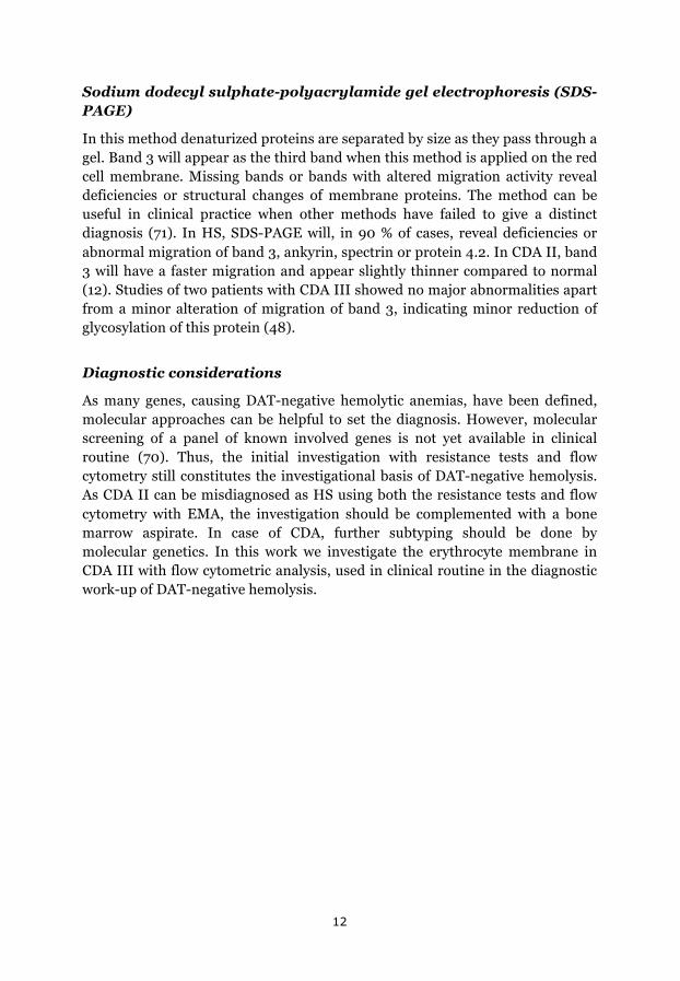
In this method denaturized proteins are separated by size as they pass through a gel. Band 3 will appear as the third band when this method is applied on the red cell membrane. Missing bands or bands with altered migration activity reveal deficiencies or structural changes of membrane proteins. The method can be useful in clinical practice when other methods have failed to give a distinct diagnosis (71). In HS, SDS-PAGE will, in 90 % of cases, reveal deficiencies or abnormal migration of band 3, ankyrin, spectrin or protein 4.2. In CDA II, band 3 will have a faster migration and appear slightly thinner compared to normal (12). Studies of two patients with CDA III showed no major abnormalities apart from a minor alteration of migration of band 3, indicating minor reduction of glycosylation of this protein (48).
Diagnostic considerations
As many genes, causing DAT-negative hemolytic anemias, have been defined, molecular approaches can be helpful to set the diagnosis. However, molecular screening of a panel of known involved genes is not yet available in clinical routine (70). Thus, the initial investigation with resistance tests and flow cytometry still constitutes the investigational basis of DAT-negative hemolysis. As CDA II can be misdiagnosed as HS using both the resistance tests and flow cytometry with EMA, the investigation should be complemented with a bone marrow aspirate. In case of CDA, further subtyping should be done by molecular genetics. In this work we investigate the erythrocyte membrane in CDA III with flow cytometric analysis, used in clinical routine in the diagnostic work-up of DAT-negative hemolysis.

13
Iron
Iron homeostasis
Being an important part of the Hb-molecule, with a major capacity to link oxygen, iron (Fe) plays a crucial role in the transportation of oxygen around the body. On the other hand, excess and accumulation of iron is highly toxic, causing organ failure due to toxic oxidative processes. To keep a favourable balance of this two-edged metal, iron homeostasis needs to be strictly regulated.
The body contains about four to five grams of iron, whereof about two thirds bound in the red cells. Iron can neither be produced nor degraded by the body. Thus, intake through absorption in the duodenum needs to be in equilibrium with losses, mainly through the stools. Daily intake of iron is about 10-20 mg per day, of which about 10 % is absorbed. After absorption by iron transporters on the surface of enterocyte, iron is further transported to the blood by the iron exchange channel, ferroportin. When released into the blood, iron binds to transferrin (Tf) and is further transported around in the body as a Tf-Fe-complex. A majority (approx. 80 %) is directed to the bone marrow to take part in erythropoiesis. The remaining 20 % is transported to other organs to participate in metabolic processes. The Fe-Tf-complex is internalized into cells by the transferrin receptors (TfRs). Excess of cellular iron is stored mainly as ferritin and to a minor extent as hemosiderin. The pool of reserve iron is mainly kept in hepatocytes and macrophages (82).
Erythrocytes are degraded mainly in the spleen and iron is then redirected to the bone marrow to be recycled in the build-up of red cells. A healthy individual loses about 1-2 mg of iron every day through epithelial shedding, mainly from the intestine, but also from the skin and urinary tract. In addition, fertile women lose iron every month due to menstruation (82).
Despite periods of larger losses as in delivery or in minor trauma, iron plasma concentration and iron reserves are kept relatively constant due to the feedback mechanism of hepcidin. Hepcidin is a small peptide, produced by the liver, which binds to and inhibits the iron transporter ferroportin. In a healthy individual, a situation of iron deficiency with diminished reserves and low plasma iron concentration causes reduced production of hepcidin. This results in a reduction of ferroportin inhibition and consequently enhanced ability to absorb iron from the food. In parallel, iron release from the storages in hepatocytes and macrophages is enhanced. On the other hand, in a situation with filled iron stores, hepcidin is increased, resulting in inhibition of ferroportin. This leads to reduction of iron uptake by enterocytes and decreased release of iron from macrophages and hepatocytes (83).
14
Hepcidin is also known to be suppressed by ineffective erythropoiesis, through an erythroid regulation pathway. A pathway that is not clearly understood, but begins to clarify. Overexpression of Growth Differentiation Factor 15 (GDF15) in CDA I and CDA II has, as in thalassemia, been shown to limit hepcidin expression (84-86). However, in more recent studies GDF15 has not been found to contribute to hepcidin suppression in anemias with effective erythropoiesis such as iron deficiency, anemia of chronic disease and myeloproliferative malignancies (87). Its role in ineffective erythropoiesis remains to be elucidated. In 2014, Krautz et al identified the new hormone erythroferrone (ERFE), produced by erythroblasts in response to erythropoietin. Further they showed that ERFE is needed for the mediation of hepcidin suppression in situations with expanded erythropoiesis in mice after phlebotomy or EPO-injections. In mice with thalassemia, thus with ineffective erythropoiesis, ERFE was shown to be highly expressed, leading to suppression of hepcidin (88). Levels of ERFE in CDAs have not been reported.
Iron overload
Iron overload is a big clinical problem in patients with chronic anemias, in need of regular transfusions. In hemolytic disorders, such as thalassemia and CDA I and CDA II, iron overload is most often present even in non-transfused patients. As described above, this is due to disturbed regulation of hepcidin, caused by ineffective erythropoiesis (84-86, 88). In addition, many patients with thalassemia, CDA I and CDA II suffer from secondary hemochromatosis due to transfusions. In clinical practice, iron overload can be estimated by ferritin, transferrin iron saturation (TSAT) and magnetic resonance imaging (MRI) of the liver or heart, or eventually liver biopsy. Treatment is based on chelation therapy or, in cases without marked anemia, regular phlebotomies.
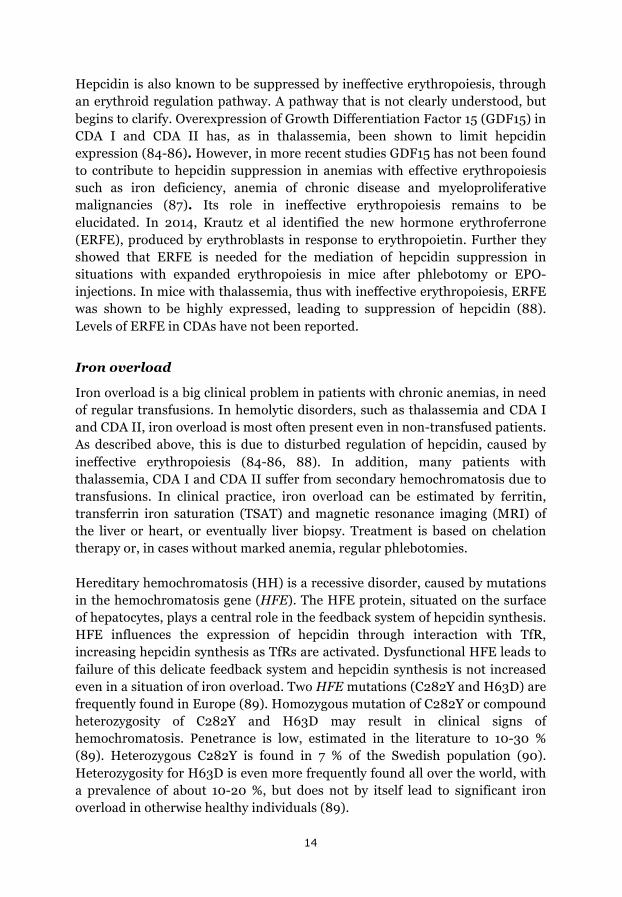
Hereditary hemochromatosis (HH) is a recessive disorder, caused by mutations in the hemochromatosis gene (HFE). The HFE protein, situated on the surface of hepatocytes, plays a central role in the feedback system of hepcidin synthesis. HFE influences the expression of hepcidin through interaction with TfR, increasing hepcidin synthesis as TfRs are activated. Dysfunctional HFE leads to failure of this delicate feedback system and hepcidin synthesis is not increased even in a situation of iron overload. Two HFE mutations (C282Y and H63D) are frequently found in Europe (89). Homozygous mutation of C282Y or compound heterozygosity of C282Y and H63D may result in clinical signs of hemochromatosis. Penetrance is low, estimated in the literature to 10-30 % (89). Heterozygous C282Y is found in 7 % of the Swedish population (90). Heterozygosity for H63D is even more frequently found all over the world, with a prevalence of about 10-20 %, but does not by itself lead to significant iron overload in otherwise healthy individuals (89).

15
In earlier studies, patients with CDA III were not found to have higher ferritin levels compared to their healthy siblings (47). This has been explained by the continuous loss of iron in the urine, due to intravascular hemolysis present in CDA III.
However, in this project we found that some individuals in the Västerbotten CDA III family did have increased levels of ferritin. As mutations of the HFE gene were found in these individuals we decided to screen the entire family for C282Y and H63D. In this project we study the prevalence of HFE mutations in the Västerbotten CDA III family and investigate the clinical appearance concerning iron balance in patients with coexistence of CDA III and mutation of the HFE gene.
16
AIMS
The overall aim of this project was to investigate the genetic etiology of CDA III.
Furthermore, we wanted to study CDA III in a clinical perspective, concerning diagnostic approach and morbidity connected to iron metabolism.
Specific aims were
- to identify the causative gene in CDA III.
- to examine the erythrocyte membrane in CDA III with flow cytometry.
- to study the presence and clinical effect of HFE mutations in individuals of the Västerbotten CDA III family.
17
Study population and methods
Study population
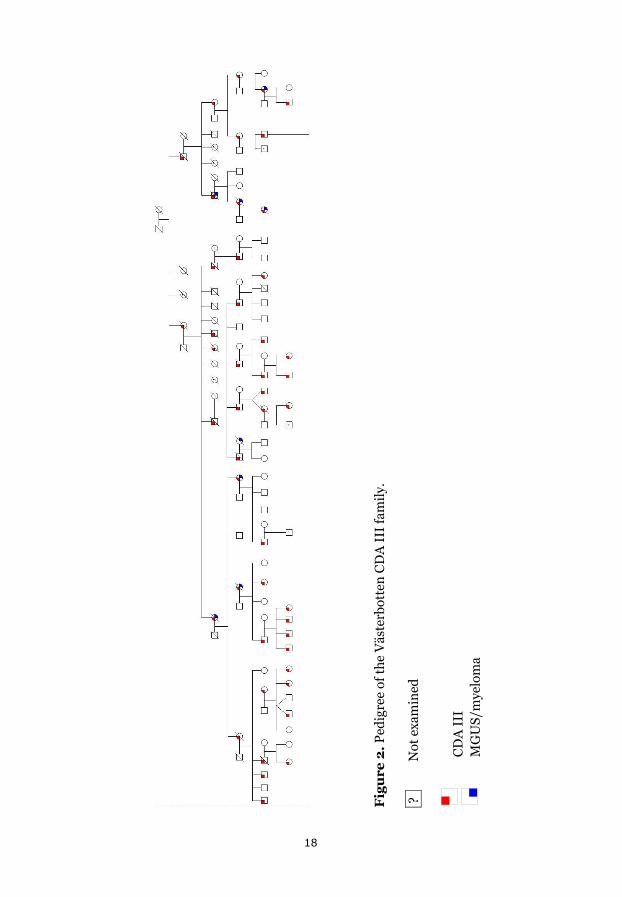
Individuals from the Västerbotten CDA III family form the basis of these studies. Furthermore, six individuals from the American CDA III family are included in paper I. The Västerbotten CDA III family consists of six generations reaching back to the middle of the 19th century. In this project, the pedigree has been updated and complemented with a few children in the 6th generation and some adults. Today the family consists of 85 individuals with confirmed risk of having CDA III. CDA III has been established in 47 of them, while 28 are non-affected siblings. Except for the two probands, eight individuals have not been examined, five are deceased, one lives abroad, and two did not want to participate (Figure 2).
In paper I, analyses were performed on DNA from 60 individuals from the Västerbotten CDA III family (39 CDA III positive and 21 CDA III negative siblings) and six individuals from the American CDA III family (four affected and two unaffected). Furthermore, DNA from 356 control individuals from a geographically matched Swedish population was analysed. CDA III status was confirmed by Hb, haptoglobin, LD, thymidine kinase or bone marrow morphology.
In paper II, 16 individuals with CDA III and 14 non-affected siblings were included in the flow cytometry analyses with EMA. Three normal controls were used per assay. Flow cytometry with anti-CD55 and anti-CD59 was performed on erythrocytes from 12 CDA III positive and 7 CDA III negative relatives with one normal control per assay.
In paper III, HFE genotyping was performed in 37 CDA III positive and 21 CDA III negative siblings. Further evaluation, concerning hematological parameters and iron status, was performed in 32 CDA III patients and 18 CDA III negative siblings.
18
Fig
ure
2. P
edig
ree
of th
e V
äste
rbot
ten
CD
A I
II fa
mily
.
?
Not
exa
min
ed
C
DA
III
M
GU
S/m
yelo
ma
19
Methods
Paper I
Targeted sequencing and data analysis
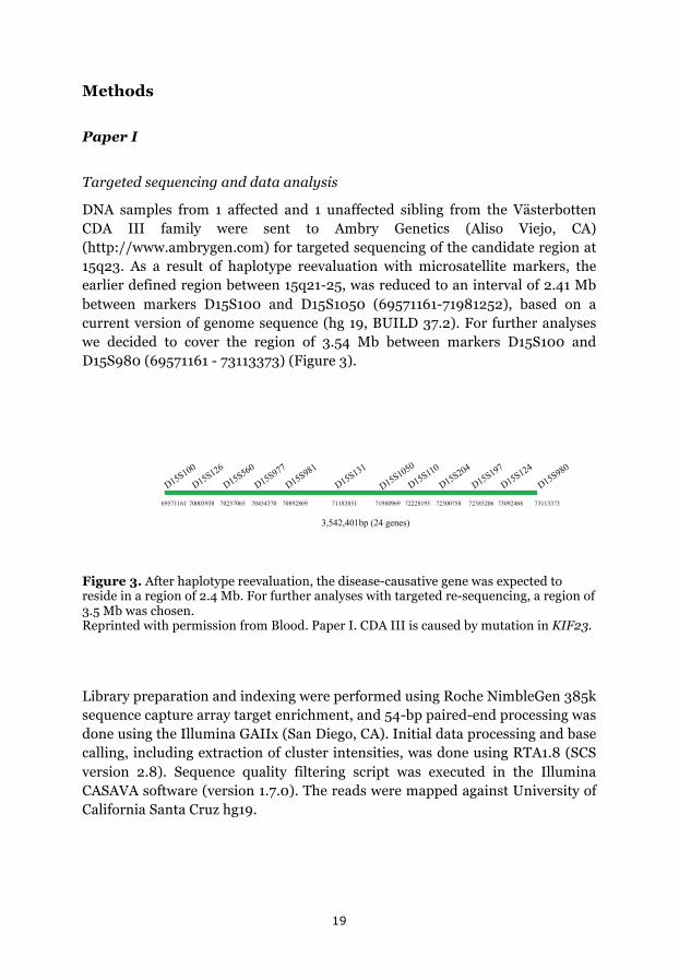
DNA samples from 1 affected and 1 unaffected sibling from the Västerbotten CDA III family were sent to Ambry Genetics (Aliso Viejo, CA) (http://www.ambrygen.com) for targeted sequencing of the candidate region at 15q23. As a result of haplotype reevaluation with microsatellite markers, the earlier defined region between 15q21-25, was reduced to an interval of 2.41 Mb between markers D15S100 and D15S1050 (69571161-71981252), based on a current version of genome sequence (hg 19, BUILD 37.2). For further analyses we decided to cover the region of 3.54 Mb between markers D15S100 and D15S980 (69571161 - 73113373) (Figure 3).
Figure 3. After haplotype reevaluation, the disease-causative gene was expected to reside in a region of 2.4 Mb. For further analyses with targeted re-sequencing, a region of 3.5 Mb was chosen. Reprinted with permission from Blood. Paper I. CDA III is caused by mutation in KIF23.
Library preparation and indexing were performed using Roche NimbleGen 385k sequence capture array target enrichment, and 54-bp paired-end processing was done using the Illumina GAIIx (San Diego, CA). Initial data processing and base calling, including extraction of cluster intensities, was done using RTA1.8 (SCS version 2.8). Sequence quality filtering script was executed in the Illumina CASAVA software (version 1.7.0). The reads were mapped against University of California Santa Cruz hg19.
!"""#$%& !"'((!&# !"'#&"() !'$$''!'!$$#'#&$)%&!$$)$ !(#%"#)%!(*&*'!(!("&!()&!((('%'# !$%#(%)% !'(%"*)#
'+&*"+*($,-./"*.012134
20
Bioinformatics
All bioinformatics tools were available via the Alamut software version 2.0 (Interactive Biosoftware, Rouen, France).
Sequence variants found in the two sisters analysed with targeted sequencing were investigated by bioinformatics to evaluate impact on protein function. Missense mutations were analysed by Sorting Intolerant from Tolerant (http://sift.jcvi.org) and Polymorphism Phenotyping (http://genetics.bwh. harvard.edu/pph). Variants detected in intronic sequences were analysed with the splice site prediction programs GeneSplicer (http://www.cbcb. umd.edu/software/GeneSplicer) and Splice Site Finder (www.genet.sickkids. on.ca/ali/splicesitefinder).
Sequencing of KIF23, MYO9A and TLE3
For bidirectional sequencing of KIF23 (MIM605064, ENSG00000137807), intronic sequences adjacent to exon 21 were amplified from genomic DNA. Sequences for KIF23 primers were 21F:5´gctcattttggaggaacagaa and 21R:5´gggagttcctgatgaagtgg, designed with Primer3 software. Amplification and sequencing were performed by polymerase chain reaction (PCR). The products of sequencing reactions were analysed on ABI 3500xL Dx Genetic Analyser (Applied Biosystems, Foster City, CA).
Sequencing of MYO9A and two TLE3 variants was similarly done by PCR. Primers are reported in Paper I.
Segregation analysis
To investigate whether mutations of KIF23, MYO9A or TLE3 segregated in the CDA III family, restriction fragment length polymorphism analyses was done. For digestion of KIF23 exon 21, PCR-products were digested by endonuclease, HpyCH4IV, separated by electrophoresis on an agarose gel and visualized after staining with ethidium bromide.
KIF23 expression
Two known isoforms of KIF23, generated by alternative splicing of exon 18, are described in the literature (91). One full-length transcript containing exon 18, ENST00000260363, encoding a 960 AA protein and one form lacking exon 18 (∆18), ENST00000559279, encoding a 856 AA protein. RNA from normal tissues was obtained from First Choice Human Total RNA Survey Panel (Ambion Life Technologies, Carlsbad, CA). We used RNA from brain, colon, heart, kidney, liver, lung, small intestine, spleen, thymus, placenta, ovary,
21
skeletal muscles, prostate, testis, and thyroid. In addition, RNA was extracted from lymphocytes of whole blood of a CDA III patient and one control case. Reverse transcription (RT)-PCR was performed with KIF23-specific primers to detect the two known transcripts, along with control primers for glyceraldehyde-3-phosphate dehydrogenase (GAPDH). RT-PCR products were separated and visualised as in the segregation analyses. Primers are reported in Paper I.
KIF23 functional analysis
Knock-down and rescue analyses were done by the research group of Masanori Mishima, Associate Professor, Division of Biomedical Cell Biology at University of Warwick in the United Kingdom. Analyses were done in HeLa cells. Interfering RNA (siRNA), targeted to the 3´-untranslated region of KIF23 was used to block translation. DNA construct of the KIF23 ∆18 (ENST00000559279) with or without P916R mutation was introduced into the cells, both tagged with a green fluorescent protein (GFP). Time lapse observation by differential interference contrast and GFP fluorescence microscopy was performed to visualize the process of cell division and cytokinesis.
Statistical analysis
No statistical analyses were done to confirm the relationship between CDA III and KIF23 c.2747G>C since 100 % of CDA III patients expressed the mutation and none of the CDA III negative siblings nor the control population carried the mutation. Data on cytokinesis failure were analysed by the research group of M Mishima, with generalized linear model for binomial data using R (http://www.r-project.org).
Paper II
Peripheral blood was obtained for analyses of Hb, erythrocyte mean cellular volume (MCV), and flow cytometric assays.
Flow cytometry
Flow cytometric assays, measuring fluorescence intensity of intact red cells after incubation with EMA were performed on a BD Biosciences FACS Calibur flow cytometer and Mean channel fluorescence (MCF) levels were determined by Cellquest software. Mean channel fluorescence (MCF) was determined for 30000 events. Each assay contained one study sample from a patient of the Västerbotten CDA III family together with three normal controls, randomly

22
selected from the Department of Laboratory Chemistry, University Hospital of Umeå.
In flow cytometry regarding CD55 and CD59, each study sample was analysed together with one normal control. 15000 events were recorded. A threshold for populations of erythrocytes expressing CD55 and CD59 in the normal control was identified and set at the same level in the study sample.
Statistical analyses
Ratios of MCF in study samples and controls in each assay were calculated. Study sample MCF and control MCF values were normally distributed according to Kolmogorov-Smirnov test. Results were presented as MCF ratios and range. The Sign test was used to compare MCF of study samples and their normal controls. P < 0.05 was regarded as significant. Pearson’s correlation test was used for analysis of correlation between EMA fluorescence and MCV.
Paper III
Detection of HFE mutations
HFE genotyping was performed as in clinical routine, at the Department of Clinical Genetics, University Hospital of Umeå. Applied Biosystems® Assay-by-design TaqMan probes were used to establish presence of C282Y and H63D.
Clinical appearance
Peripheral blood was obtained for analyses of iron status (ferritin and TSAT) and hematological parameters (Hb and LD). Ferritin and TSAT were analysed according to standard procedures at the Department of Laboratory Chemistry, University Hospital of Umeå. Samples were collected between 1997 and 2015. All individuals with HFE mutation and elevated ferritin or TSAT were offered clinical and laboratory investigation to confirm or reject iron overload. Phlebotomy was initiated in individuals with iron overload.
Statistical analyses
Ferritin, TSAT, Hb and LD are presented as median values and range. Two-sided Mann-Whitney U-test was used for comparison between groups. P < 0.05 was regarded as significant.
23
Results
Paper I
CDA III and KIF23
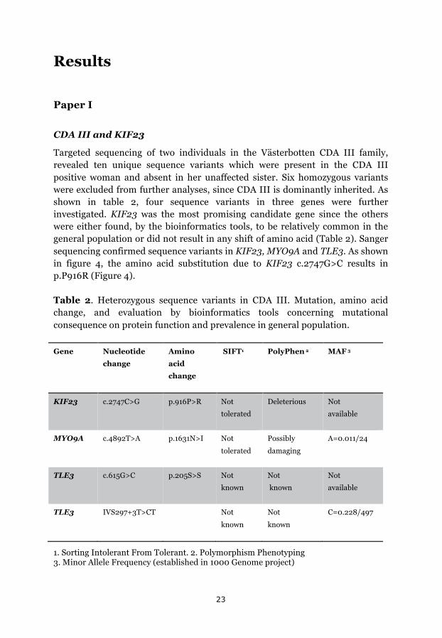
Targeted sequencing of two individuals in the Västerbotten CDA III family, revealed ten unique sequence variants which were present in the CDA III positive woman and absent in her unaffected sister. Six homozygous variants were excluded from further analyses, since CDA III is dominantly inherited. As shown in table 2, four sequence variants in three genes were further investigated. KIF23 was the most promising candidate gene since the others were either found, by the bioinformatics tools, to be relatively common in the general population or did not result in any shift of amino acid (Table 2). Sanger sequencing confirmed sequence variants in KIF23, MYO9A and TLE3. As shown in figure 4, the amino acid substitution due to KIF23 c.2747G>C results in p.P916R (Figure 4).
Table 2. Heterozygous sequence variants in CDA III. Mutation, amino acid change, and evaluation by bioinformatics tools concerning mutational consequence on protein function and prevalence in general population.
Gene Nucleotide
change
Amino
acid
change
SIFT1 PolyPhen 2 MAF 3
KIF23 c.2747C>G p.916P>R Not
tolerated
Deleterious Not
available
MYO9A c.4892T>A p.1631N>I Not
tolerated
Possibly
damaging
A=0.011/24
TLE3 c.615G>C p.205S>S Not
known
Not
known
Not
available
TLE3 IVS297+3T>CT Not
known
Not
known
C=0.228/497
1. Sorting Intolerant From Tolerant. 2. Polymorphism Phenotyping 3. Minor Allele Frequency (established in 1000 Genome project)
24
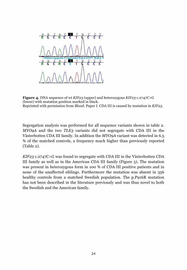
Figure 4. DNA sequence of wt KIF23 (upper) and heterozygous KIF23 c.2747C>G (lower) with mutation position marked in black. Reprinted with permission from Blood. Paper I. CDA III is caused by mutation in KIF23.
Segregation analysis was performed for all sequence variants shown in table 2. MYO9A and the two TLE3 variants did not segregate with CDA III in the Västerbotten CDA III family. In addition the MYO9A variant was detected in 6.5 % of the matched controls, a frequency much higher than previously reported (Table 2).
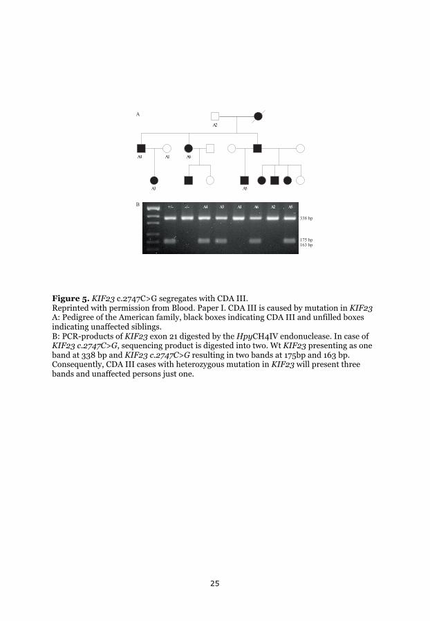
KIF23 c.2747C>G was found to segregate with CDA III in the Västerbotten CDA III family as well as in the American CDA III family (Figure 5). The mutation was present in heterozygous form in 100 % of CDA III positive patients and in none of the unaffected siblings. Furthermore the mutation was absent in 356 healthy controls from a matched Swedish population. The p.P916R mutation has not been described in the literature previously and was thus novel to both the Swedish and the American family.
25
Figure 5. KIF23 c.2747C>G segregates with CDA III. Reprinted with permission from Blood. Paper I. CDA III is caused by mutation in KIF23 A: Pedigree of the American family, black boxes indicating CDA III and unfilled boxes indicating unaffected siblings. B: PCR-products of KIF23 exon 21 digested by the HpyCH4IV endonuclease. In case of KIF23 c.2747C>G, sequencing product is digested into two. Wt KIF23 presenting as one band at 338 bp and KIF23 c.2747C>G resulting in two bands at 175bp and 163 bp. Consequently, CDA III cases with heterozygous mutation in KIF23 will present three bands and unaffected persons just one.
!"
!#
!$
!%
!& !'!" !#$%& &%&
!
(
!"
!$ !&
!'
!' !( !) !*
%%)*+,
$-'*+,$&%*+,
26
KIF23 expression
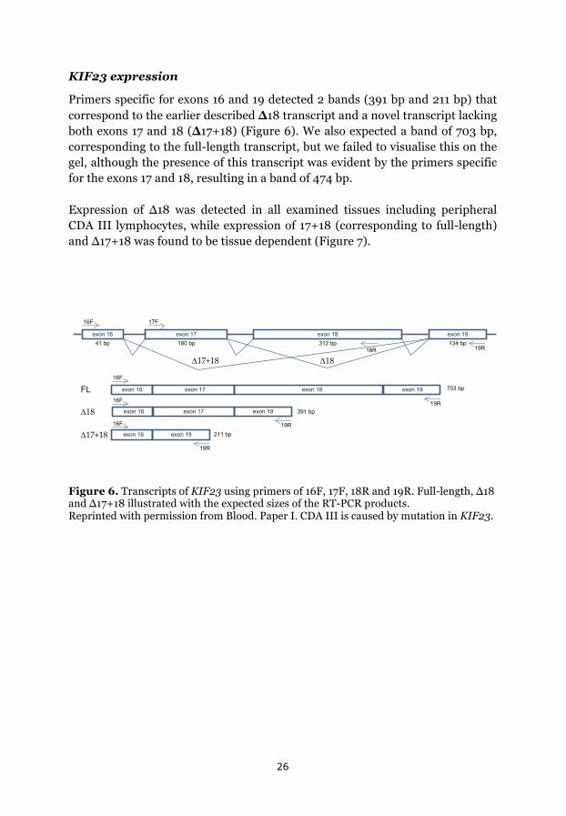
Primers specific for exons 16 and 19 detected 2 bands (391 bp and 211 bp) that correspond to the earlier described ∆18 transcript and a novel transcript lacking both exons 17 and 18 (∆17+18) (Figure 6). We also expected a band of 703 bp, corresponding to the full-length transcript, but we failed to visualise this on the gel, although the presence of this transcript was evident by the primers specific for the exons 17 and 18, resulting in a band of 474 bp.
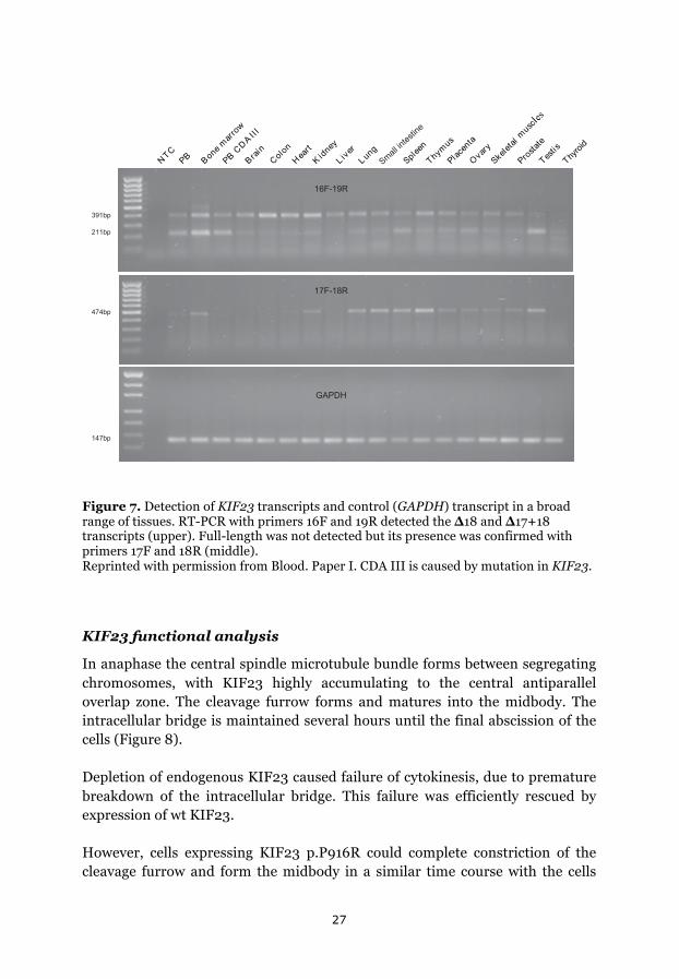
Expression of ∆18 was detected in all examined tissues including peripheral CDA III lymphocytes, while expression of 17+18 (corresponding to full-length) and ∆17+18 was found to be tissue dependent (Figure 7).
Figure 6. Transcripts of KIF23 using primers of 16F, 17F, 18R and 19R. Full-length, ∆18 and ∆17+18 illustrated with the expected sizes of the RT-PCR products. Reprinted with permission from Blood. Paper I. CDA III is caused by mutation in KIF23.
!"#
$!%&'
!"
#(
!)#
!*+!,+!,-%&' .!/%&' !.$%&'
0123%!" 0123%!) 0123%!, 0123%!*
0123%!) 0123%!*0123%!" 0123%!,
0123%!" 0123%!) 0123%!*
!#$!" 0123%!" 0123%!*
!"!#$!"
!"#
!*+
!*+
!*+
!"#
!"#
)-.%&'
%.*!%&'
/!!%&'
27
Figure 7. Detection of KIF23 transcripts and control (GAPDH) transcript in a broad range of tissues. RT-PCR with primers 16F and 19R detected the ∆18 and ∆17+18 transcripts (upper). Full-length was not detected but its presence was confirmed with primers 17F and 18R (middle). Reprinted with permission from Blood. Paper I. CDA III is caused by mutation in KIF23.
KIF23 functional analysis
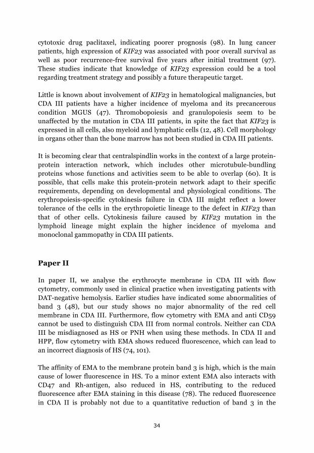
In anaphase the central spindle microtubule bundle forms between segregating chromosomes, with KIF23 highly accumulating to the central antiparallel overlap zone. The cleavage furrow forms and matures into the midbody. The intracellular bridge is maintained several hours until the final abscission of the cells (Figure 8).
Depletion of endogenous KIF23 caused failure of cytokinesis, due to premature breakdown of the intracellular bridge. This failure was efficiently rescued by expression of wt KIF23.
However, cells expressing KIF23 p.P916R could complete constriction of the cleavage furrow and form the midbody in a similar time course with the cells
!"#$% %&
'()*+,,&-
$%)#./00 0
% ,+1'
#&2&'
3(+,45 16'(7
8 19(,8 :';
<=2(('
">7*:?
$2+@('4+
A9+,7
<B(2(4+2*:?@2!"
$,&?4+4(
"(?41?
">7,&16
CDCE=
<*+22)1'4(?41'(
FGGE=
HIGE=
GJKLGIM
GDKLGNM
GCDE=
O/$.3

28
expressing wt KIF23, but regressed to form binucleate cells about 2 hours after midbody formation (Figure 8). These data indicate that the p.P916R mutation impairs the function of KIF23, essential for the completion of cytokinesis.
Figure 8. Upper images showing complete cell division with wt KIF23. KIF23 p.P916R mutant (lower) was localized to the spindle midzone (white arrow) and condensed to form the midbody (arrowheads) in a similar manner to the wt KIF23 (upper white arrow and arrowheads). However, the cell membrane of the intercellular bridge was detached from the midbody approx. 3,5 hour after its formation, creating a binucleate cell (lower). The remnant of the midbody (arrowhead) was incorporated to the cell on the right after proper cell division (upper), and incorporated into the binucleate cell after cytokinesis failure (lower). Reprinted with permission from Blood, paper I; CDA III is caused by mutation in KIF23.
29
Paper II
Flow cytometry in CDA III
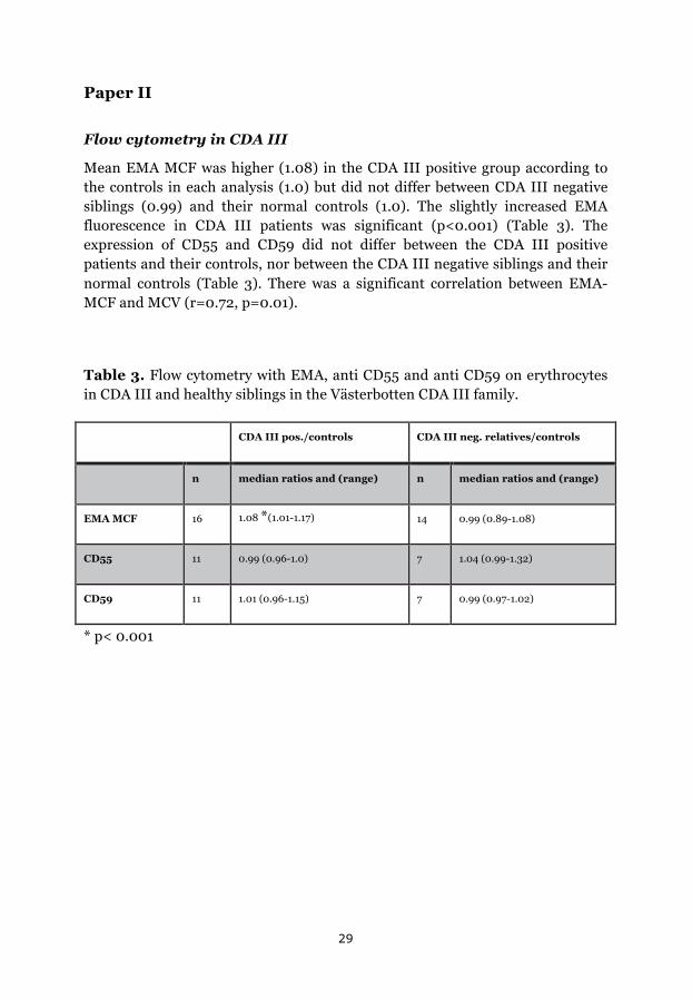
Mean EMA MCF was higher (1.08) in the CDA III positive group according to the controls in each analysis (1.0) but did not differ between CDA III negative siblings (0.99) and their normal controls (1.0). The slightly increased EMA fluorescence in CDA III patients was significant (p<0.001) (Table 3). The expression of CD55 and CD59 did not differ between the CDA III positive patients and their controls, nor between the CDA III negative siblings and their normal controls (Table 3). There was a significant correlation between EMA-MCF and MCV (r=0.72, p=0.01).
Table 3. Flow cytometry with EMA, anti CD55 and anti CD59 on erythrocytes in CDA III and healthy siblings in the Västerbotten CDA III family.
CDA III pos./controls CDA III neg. relatives/controls
n median ratios and (range) n median ratios and (range)
EMA MCF 16 1.08 *(1.01-1.17) 14 0.99 (0.89-1.08)
CD55 11 0.99 (0.96-1.0) 7 1.04 (0.99-1.32)
CD59 11 1.01 (0.96-1.15) 7 0.99 (0.97-1.02)
* p< 0.001

30
Paper III
HFE status
HFE genotyping was successfully performed in all 58 cases. HFE mutation was found in 24 out of 37 CDA III patients (65 %) and 13 out of 21 CDA III negative siblings (62 %). Among the 50 cases with available iron status, 21 out of 32 CDA III positive (66 %) and 11 out of 18 CDA III negative siblings (61 %) were found to have HFE mutations. 16 of the CDA III patients with HFE-mutation were heterozygous either for C282Y or H63D, and five were compound heterozygotes. Seven of the 18 CDA III negative siblings were heterozygous carriers of HFE mutations, two were compound heterozygotes, and two had homozygous HFE mutations. In individuals with heterozygous C282Y (9 CDA III positive and 6 CDA III negative cases), ferritin levels were significantly higher in CDA III patients, p=0.036.
Clinical appearance
The group of patients with CDA III and available iron status and hematological parameters (Hb and/or LD), consisted of 16 males and 16 females with a median age of 53 years, and the CDA III negative group consisted of 10 males and 8 females, with a median age of 44 years. Median Hb among CDA III patients was 116 g/L (94-157) and LD 6.2 ukat/L (4.2-10). In the CDA III negative group median Hb was 141 g/L and LD 2.7 ukat/L. Ferritin was higher (108 umol/L) in the CDA III positive group than in the CDA III negative group (76 umol/L), and TSAT was also higher among CDA III patients, 46 % vs. 34 %, but the differences were not significant (p=0.055 for both comparisons).
Laboratory evidence of iron overload was noted in four patients, one CDA III negative case with homozygous C282Y, and three CDA III positive individuals with heterozygous HFE mutations (Table 4). Magnetic resonance imaging (MRI) of the liver and S-hepcidin analysis was performed in the CDA III positive patient with heterozygous H63D mutation. Liver iron was 6700 ug/g dry tissue (normal range 300-1500) and hepcidin was 46 ug/L (normal range 8-76). To normalize ferritin and TSAT, treatment with phlebotomy was started in four patients; the CDA III negative sibling who was homozygous for C282Y and three CDA III patients, one compound heterozygote and two with heterozygous mutations, one C282Y, and one H63D.
A total of 18 individuals, 11 CDA III cases and 7 CDA III negative siblings, had wt HFE. Ferritin levels did not differ between these groups (p=0.86) One of these CDA III positive males, suffering from hypertension and hyperlipidemia,
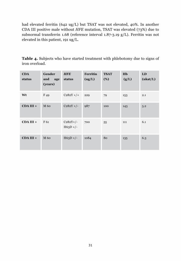
31
had elevated ferritin (642 ug/L) but TSAT was not elevated, 40%. In another CDA III positive male without HFE mutation, TSAT was elevated (73%) due to subnormal transferrin 1.68 (reference interval 1.87-3.19 g/L). Ferritin was not elevated in this patient, 191 ug/L.
Table 4. Subjects who have started treatment with phlebotomy due to signs of iron overload.
CDA
status
Gender
and age
(years)
HFE
status
Ferritin
(ug/L)
TSAT
(%)
Hb
(g/L)
LD
(ukat/L)
Wt F 49 C282Y +/+ 229 79 153 2.1
CDA III + M 60 C282Y +/- 987 100 143 5.2
CDA III + F 61 C282Y+/-
H63D +/-
700 55 111 6.1
CDA III + M 60 H63D +/- 1084 80 135 6.3

32
Discussion
In this project we found a genetic cause of familial CDA III. Furthermore we investigated the flow cytometric profile of CDA III erythrocytes from a clinical perspective and conclude that mutations of the HFE gene, even in heterozygous form, can cause iron overload in CDA III patients.
Paper I
In paper I, stepwise laboratory work using targeted sequencing, prioritization of gene sequences with the help of bioinformatics tools, Sanger sequencing and segregation analyses finally enabled us to identify KIF23 as the causative gene in CDA III. The mutation KIF23 c.2747C>G causes cytokinesis failure as the encoded motor protein, kinesin-like protein KIF23, also called mitotic kinesin-like protein 1 (MKLP1), is a crucial player in the formation of the midbody and intercellular bridge. Together with the GTPase regulating protein CYK4, KIF23 forms a heterotetramer, which builds up the protein centralspindlin (92). This protein enables assembly of microtubules in the midbody and through the C-terminus of CYK4, adheres the plasma membrane to the intercellular bridge. This connection makes the intracellular bridge reside several hours before final abscission (63, 93). Several proteins regulate the clustering and adherence capacity of centralspindlin. By binding a 14-3-3 protein, centralspindlin is inactivated, unable to carry out clustering or anchoring activity. At the central spindle, Aurora B disconnects the 14-3-3 protein, switching centralspindlin into an active state. As Aurora B activity peaks between segregating chromosomes in late anaphase and early telophase, centralspindlin is activated as the cell finalizes the last steps of mitosis and enters cytokinesis. This enables centralspindlin to perform the clustering activity that will finally form the midbody. The activity of Aurora B then rapidly declines. Thereafter the activated state is withheld by the GTPase ARF6, which by competitive binding prevents 14-3-3 to bind to, and inactivate, centralspindlin. ARF6 localizes to the midbody and makes sure that centralspindlin remains active until the final abscission of the cells (94).
Thus, as centralspindlin plays a crucial role in cytokinesis, dysfunction of one of its components, KIF23, results in cytokinesis failure and the multinucleated cells, which are the hallmark of CDA III (Figure 9) (95).

33
Figure 9. Failure of cytokinesis, due to mutation in KIF23. This figure was originally published in Blood (95), reprinted with permission from Blood Traxler E. Congenital dyserythropoietic anemias: III's a charm. Blood 2013;121(23):4614-5.
Failure of controlling and regulating the steps of the cell cycle and mitosis are hallmarks of cancer development. The high proliferative ability of cancer cells also requires an excess of proteins, active in the mitotic processes. The last possibility in the cell cycle to slow down or stop proliferation is the step of cytokinesis. As KIF23 is a key regulator of this final process, knowledge about its involvement in cancer is an area of interest.
KIF23 expression is cell cycle dependent, being suppressed in interphase and upregulated during S-phase. Interestingly, the tumor suppressor gene p53 has been shown to reduce expression of KIF23. This repression is due to downregulation of KIF23 promoter activity. Thus, mutations of the promoter region of KIF23 or mutations of p53 can result in uncontrolled proliferation (96).
KIF23 has recently been shown to be overexpressed in lung-, breast-, gastric- and hepatocellular cancer as well as in glioma (97-100). In addition, down regulation of KIF23 suppresses cell division in glioma, breast cancer, and lung cancer cell lines as well as in a glioma mouse model (97, 99, 100). In gastric cancer KIF23 was found to be upregulated in cell lines with resistance to the
34
cytotoxic drug paclitaxel, indicating poorer prognosis (98). In lung cancer patients, high expression of KIF23 was associated with poor overall survival as well as poor recurrence-free survival five years after initial treatment (97). These studies indicate that knowledge of KIF23 expression could be a tool regarding treatment strategy and possibly a future therapeutic target.
Little is known about involvement of KIF23 in hematological malignancies, but CDA III patients have a higher incidence of myeloma and its precancerous condition MGUS (47). Thromobopoiesis and granulopoiesis seem to be unaffected by the mutation in CDA III patients, in spite the fact that KIF23 is expressed in all cells, also myeloid and lymphatic cells (12, 48). Cell morphology in organs other than the bone marrow has not been studied in CDA III patients.
It is becoming clear that centralspindlin works in the context of a large protein-protein interaction network, which includes other microtubule-bundling proteins whose functions and activities seem to be able to overlap (60). It is possible, that cells make this protein-protein network adapt to their specific requirements, depending on developmental and physiological conditions. The erythropoiesis-specific cytokinesis failure in CDA III might reflect a lower tolerance of the cells in the erythropoietic lineage to the defect in KIF23 than that of other cells. Cytokinesis failure caused by KIF23 mutation in the lymphoid lineage might explain the higher incidence of myeloma and monoclonal gammopathy in CDA III patients.
Paper II
In paper II, we analyse the erythrocyte membrane in CDA III with flow cytometry, commonly used in clinical practice when investigating patients with DAT-negative hemolysis. Earlier studies have indicated some abnormalities of band 3 (48), but our study shows no major abnormality of the red cell membrane in CDA III. Furthermore, flow cytometry with EMA and anti CD59 cannot be used to distinguish CDA III from normal controls. Neither can CDA III be misdiagnosed as HS or PNH when using these methods. In CDA II and HPP, flow cytometry with EMA shows reduced fluorescence, which can lead to an incorrect diagnosis of HS (74, 101).
The affinity of EMA to the membrane protein band 3 is high, which is the main cause of lower fluorescence in HS. To a minor extent EMA also interacts with CD47 and Rh-antigen, also reduced in HS, contributing to the reduced fluorescence after EMA staining in this disease (78). The reduced fluorescence in CDA II is probably not due to a quantitative reduction of band 3 in the

35
erythrocyte membrane but rather an affected configuration, leading to a decreased affinity to EMA (79).
The subtle increase in EMA fluorescence in CDA III is probably due to the increase in erythrocyte volume (MCV) exposing a larger surface per cell to binding of EMA to band 3, Rhesus-antigen and CD47. Earlier data on the correlation of MCV and MCF in flow cytometry with EMA is a bit contradictory. Correlation has been confirmed in individuals with high MCV due to overconsumption of alcohol, but in DAT-positive hemolytic anemia no correlation between MCV and MCF has been observed (80). Low MCV due to iron deficiency does not seem to affect EMA MCF (79-80).
Since methods analysing erythrocyte fragility can also be misleading for differentiating between HS and other DAT-negative hemolytic anemias, caution should be taken in the diagnostic work up of these diseases. If there is no family history of CDA, the diagnosis is easy to overlook in the absence of a bone marrow investigation. When a bone marrow smear raises the suspicion of CDA, further subclassification should be carried out by molecular genetics, searching for causative genes, which are now defined in the three major forms of CDA.
Paper III
In paper III, we show that mutations of the HFE gene are present in the Västerbotten CDA III family. 65 % of CDA III positive cases and 62 % of their CDA III negative siblings carried C282Y and/or H63, the two major HFE mutations present in a European population. Two CDA III patients with heterozygous HFE mutations, one CDA III patient who was compound heterozygous, and one CDA III negative case with homozygous C282Y, needed treatment with phlebotomy due to iron overload.
One of the heterozygous cases carried H63D, which is not reported to lead to iron overload by itself in otherwise healthy individuals. MRI of the liver confirmed iron overload in this patient. Hepcidin was in the normal range, indicating dysfunction of the feedback system, which in a healthy individual would stimulate hepcidin expression due to iron overload. We suggest that ineffective erythropoiesis suppresses hepcidin through the ERFE-pathway and that HFE mutation contributes to further reduction of hepcidin, leading to iron overload.
All CDA III patients with HFE mutations do not develop iron overload. It is also known, even in healthy individuals, that the penetrance of HFE mutations is
36
relatively low, with about only 10-30 % of homozygous C282Y carriers developing hemochromatosis (89). Thus, there are other factors protecting the individual from accumulating iron. In addition it is also a question about time and gender. In the case of CDA III, as earlier stated, the continuous loss of iron in the urine may contribute to keep the balance in iron homeostasis (47).
None of the family members with wt HFE, nor healthy siblings carrying heterozygous or compound heterozygous HFE mutations, had laboratory signs of iron overload. One CDA III positive individual with wt HFE had elevated ferritin and one had elevated TSAT. Further investigation showed that these patients did not fulfil the criteria of iron overload.
This study indicates that heterozygous HFE mutation, even H63D, can cause iron overload when coexisting with a hemolytic disorder such as CDA III. Earlier studies on HFE mutations in patients with hemolysis where iron overload can be a problem, such as thalassemia and myelodysplastic syndrome (MDS), are contradictory. Some studies show iron overload in thalassemia patients with co-existing HFE mutations, even in heterozygous forms (102-104). Recently, non-transfused MDS patients with heterozygous HFE mutations were found to have higher ferritin level and inferior overall survival compared to MDS patients with wt HFE (105). However, other studies have not verified that heterozygous mutation affects iron overload in thalassemia (106, 107).
So far, phlebotomies have been performed without problems in spite of the continuous hemolysis in our CDA III patients, and phlebotomy alone is sufficient to control ferritin and TSAT levels.
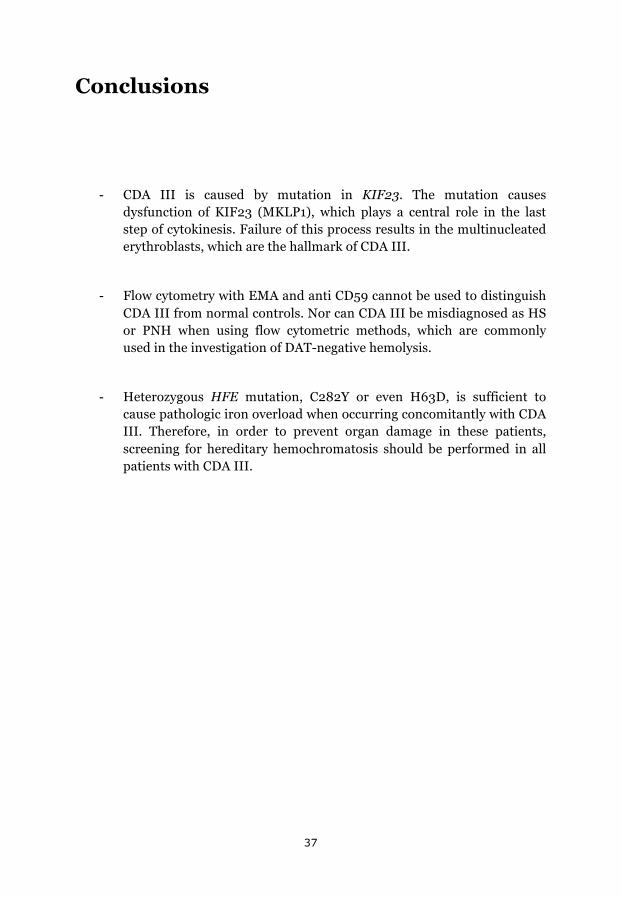
37
Conclusions
- CDA III is caused by mutation in KIF23. The mutation causes dysfunction of KIF23 (MKLP1), which plays a central role in the last step of cytokinesis. Failure of this process results in the multinucleated erythroblasts, which are the hallmark of CDA III.
- Flow cytometry with EMA and anti CD59 cannot be used to distinguish CDA III from normal controls. Nor can CDA III be misdiagnosed as HS or PNH when using flow cytometric methods, which are commonly used in the investigation of DAT-negative hemolysis.
- Heterozygous HFE mutation, C282Y or even H63D, is sufficient to cause pathologic iron overload when occurring concomitantly with CDA III. Therefore, in order to prevent organ damage in these patients, screening for hereditary hemochromatosis should be performed in all patients with CDA III.
38
Future perspectives
Rare anemias constitute a small health problem from a global perspective. Nevertheless, patients with these disorders have the right to find help and knowledge when they turn to health care. Furthermore, taken together as a group, patients suffering from either of these rare anemias are quite numerous. Thus, to gather and spread knowledge on rare diseases and to enable good research, collaboration and networking between centres involved in this field is essential.
The European Network for Rare and Congenital Anaemias (ENERCA) is such a collaboration, attempting to offer improved health service to professional medical practitioners and patients in every aspect of rare anemias. The project is cofounded by the European Commission through its Executive Agency for Health and Consumers (EAHC), being a part of the EU Health Programme, which focuses on reducing health inequalities across the European Union. The project is now working on a registry with the attempt to cover every patient who gets a diagnosis of a rare anemia. A good registry would enable good research and hopefully lead to new insights in how these patients should be followed and treated. As KIF23 now has been found to be mutated in CDA III in one family in Västerbotten as well as in one American family, it would be of great interest to investigate sporadic CDA III to search for mutations in KIF23. Hopefully, the collaboration of ENERCA could facilitate such projects.
Moreover, it is important to look at the results of this project from a broader perspective, where CDA III can serve as a model for both cytokinesis failure and DAT-negative hemolysis.
KIF23 and its involvement in cancer development and the opportunity to use KIF23 expression for risk stratification or treatment options in different cancers, should be further looked into. Since we know that plasma cell neoplasia, myeloma and MGUS, is overrepresented in CDA III, it would be of interest to look at KIF23 expression and its correlation with clinical parameters in myeloma patients.
Furthermore it would be interesting to look at the connection between KIF23 and p53 from a clinical point of view, since we know that many hematological malignancies with mutation in or loss of p53 have very bad outcome. For example, KIF23 expression in chronic lymphatic leukemia (CLL) with deletion of 17p or mutations in p53 could be further looked into.

39
Iron overload is a great problem in many hemolytic diseases, such as thalassemia and other hemoglobinopathies. In addition, secondary iron overload is a frequent problem in hematologic disorders and malignancies requiring regular transfusions. Knowledge on iron metabolism and its regulation has been expanding in the last decade. It is of further interest to look at these regulation mechanisms and how they work together. Since HFE mutations are frequent in Europe, knowledge on how these mutations can influence development of iron overload in patients already at risk of iron accumulation could be valuable. If mutations lead to a faster iron accumulation this could influence how these patients should be followed and when treatment should be considered.
40
Acknowledgements
Most things in life you can’t do by yourself, a thesis is not an exception. Many people have helped me to accomplish this. To all of you, I am most grateful. In particular I want to thank.
Anders Wahlin, my supervisor, for all your patience and support, encouraging me even when I felt like giving up. For your humble way of sharing knowledge, always able to give a wise advice, in research, as well as in the most troublesome situations of clinical practice. It is a great pleasure working with you.
Irina Golovleva, my co-supervisor, for generously sharing your great knowledge in the field of genetics.
Herbert Sandström, my co-supervisor, for guiding me through all the technicalities of the doctoral studies.
Ann-Louise Vikberg, for guiding me at the lab and for your indispensable laboratory work.
Beatrice Melin, my examinator, for excellent guidance.
Maria Wing, secretary at the Department of Radiation Sciences, for always answering all my questions with a smile.
All staff at the hematopathology department and especially Elisabeth Grönlund and Göran Roos for sharing your knowledge on flow cytometry and Elisabeth for performing all the flow cytometry analyses in paper II.
Lena Lundkvist, at Regionalt Cancercentrum Norr, for your excellent help with the pedigrees.
All my hard working colleagues at the Hematology department. I could never have done this without your support.
Karin Forsberg, former head of the Hematology department and my wise colleague for many years, thanks for all support and advices and for helping us at work instead of being a full time retiree.

41
Gunnel Sundström, my mentor during speciality training, for being such a good role model.
All staff at the Hematology department for excellent teamwork. A special thank to Ulla Lindkvist, Britt-Marie Gezelius, Lars Skagerlind and Fredrik Åström for daily helpful support, and to Carita Johansson for keeping track on all patients and blood sampling in these studies.
Current head of Cancercentrum, Elisabeth O Karlsson and deputy head of Cancercentrum, Martin Erlanson, for encouraging me to prioritize this work.
Katrin Sundh, secretary of Cancercentrum, for always being so helpful.
Eva Hellström Lindberg for inviting me to Center for Hematology and Regenerative Medicine (HERM) and for giving me a Swedish and European perspective on hematology.
Jan Samuelsson, for giving me valuable knowledge and insights throughout the work for the Swedish Society of Hematology and Maria Samuelsson for all your invaluable help.
Mum and dad for all your love, and for giving me and my sister the courage to face new challenges and to believe in ourselves.
Malin, my sister and dearest friend, on whom I can always rely. Martin, Alexander and David for all good moments that we share.
Håkan and Kristina, my parents in law, for all your help through the years and for accepting me the way I am.
My lovely family, you give me purpose. Per, for firmly standing by my side since the 8th of March 1996, in good times and in bad. Life is good together with you. Erik and Emil, always on my mind and in my heart.
The new (four-footed) friend of the family, Malte, together we explore new paths.

42
Financial support was provided by Cancerforskningsfonden Norrland, Blodforskningsfonden i Umeå, Umeå University through regional agreement between Umeå University and Västerbotten County Council on cooperation in the field of Medicine, Odontology and Health (ALF).
Last but not least I want to thank all members of the Västerbotten CDA III family for their consent to participate in these studies. Without their generosity this project would not have been possible.

43
References
1. Lewis SM, Pitcher CS, Nelson D. Congenital dyserythropoietic anaemia. Br J Haematol 1972;22(5):638-42.
2. Heimpel H, Wendt F. Congenital dyserythropoietic anemia with karyorrhexis and multinuclearity of erythroblasts Helv Med Acta 1968;34(2):103-15.
3. Wickramasinghe SN, Vora AJ, Will A, Winfield DA, Hughes RG, et al. Transfusion-dependent congenital dyserythropoietic anaemia with non-specific dysplastic changes in erythroblasts. Eur J Haematol 1998; 60(2):140-2.
4. Wickramasinghe SN, Wood WG. Advances in the understanding of the congenital dyserythropoietic anaemias. Br J Haematol 2005;131(4):431-46.
5. Carter C, Darbyshire PJ, Wickramasinghe SN. A congenital dyserythropoietic anaemia variant presenting as hydrops fetalis. Br J Haematol 1989;72(2):289-90.
6. Wickramasinghe SN, Andrews VE, O'Hea AM. Congenital dyserythropoiesis characterized by marked macrocytosis, vitamin B12- and folate-independent megaloblastic change and absence of the defining features of congenital dyserythropoietic anaemia types I or III. Br J Haematol 1996;95(1):73-6.
7. Iolascon A, Esposito MR, Russo R. Clinical aspects and pathogenesis of CDAs: from morphology to molecular approach. Haematologica 2012;97(12):1786–1794.
8. Iolascon A, Heimpel H, Wahlin A, Tamary H. Congenital dyserythropoietic anemias: molecular insights and diagnostic approach. Blood 2013;122(13):2162-6.
9. Gambale A, Iolascon A, Andolfo I, Russo R. Diagnosis and management of congenital dyserythropoietic anemias. Expert Rev Hematol 2016 Jan 6:1-14. [Epub ahead of print]
44
10. Heimpel H, Matuschek A, Ahmed M, Bader-Meunier B, Colita A, Delaunay J, et al. Frequency of congenital dyserythropoietic anemias in Europe. Eur J Haematol 2010;85(1):20-5.
11. Tamary H, Shalev H, Luria D, Shaft D, Zoldan M, Shalmon L, et al. Clinical features and studies of erythropoiesis in Israeli Bedouins with congenital dyserythropoietic anemia type I. Blood 1996;87(5):1763-70.
12. Wickramasinghe SN. Congenital dyserythropoietic anaemias: clinical features, haematological morphology and new biochemical data. Blood Rev 1998;12(3):178-200.
13. Lewis SM, Nelson DA, Pitcher CS. Clinical and ultrastructural aspects of congenital dyserythropoietic anaemia type I. Br J Haematol 1972;23(1):113-9.
14. Heimpel H, Schwarz K, Ebnother M, Goede JS, Heydrich D, Kamp T, et al. Congenital dyserythropoietic anemia type I (CDA I): molecular genetics, clinical appearance, and prognosis based on long-term observation. Blood 2006;107(1):334-40.
15. Shalev H, Kapleushnik Y, Haeskelzon L, Degani O, Kransnov T, Sphilber O, et al. Clinical and laboratory manifestations of congenital dyserythropoietic anemia type I in young adults. Eur J Haematol 2002;68(3):170-4.
16. Goede JS, Benz R, Fehr J, Schwarz K, Heimpel H. Congenital dyserythropoietic anemia type I with bone abnormalities, mutations of the CDAN I gene, and significant responsiveness to alpha-interferon therapy. Ann Hematol 2006;85(9):591-5.
17. El-Sheikh AA, Hashem H, Holman C, Vyas YM. Congenital dyserythropoietic anemia type I presenting as persistent pulmonary hypertension with pigeon chest deformity. Pediatr Blood Cancer 2014;61(8):1460-2.
18. Lavabre-Bertrand T, Blanc P, Navarro R, Saghroun M, Vannereau H, Braun M, et al. Alpha-Interferon therapy for congenital dyserythropoiesis type I. Br J Haematol 1995;89(4):929-32.
45
19. Lavabre-Bertrand T, Ramos J, Delfour C, Henry L, Guiraud I, Carillo S, et al. Long-term alpha interferon treatment is effective on anaemia and significantly reduces iron overload in congenital dyserythropoiesis type I. Eur J Haematol 2004;73(5):380-3.
20. Tamary H, Shalmon L, Shalev H, Halil A, Dobrushin D, Ashkenazi N, et al. Localization of the gene for congenital dyserythropoietic anemia type I to a <1-cM interval on chromosome 15q15.1-15.3. Am J Hum Genet 1998;62(5):1062-9.
21. Dgany O, Avidan N, Delaunay J, Krasnov T, Shalmon L, Shalev H, et al. Congenital dyserythropoietic anemia type I is caused by mutations in codanin-1. Am J Hum Genet 2002;71(6):1467-74.
22. Noy-Lotan S, Dgany O, Lahmi R, Marcoux N, Krasnov T, Yissachar N et al. Codanin-1, the protein encoded by the gene mutated in congenital dyserythropoietic anemia type I (CDAN1), is cell cycle-regulated. Haematologica 2009;94(5):629-37.
23. Ask K, Jasencakova Z, Menard P, Feng Y, Almouzni G, Groth A. Codanin-1, mutated in the anaemic disease CDAI, regulates Asf1 function in S-phase histone supply. Embo J 2012;31(8):2013-23.
24. Babbs C, Roberts NA, Sanchez-Pulido L, McGowan SJ, Ahmed MR, Brown JM, et al. Homozygous mutations in a predicted endonuclease are a novel cause of congenital dyserythropoietic anemia type I. Haematologica 2013;98(9):1383-7.
25. Iolascon A, Servedio V, Carbone R, Totaro A, M Carella, S Perrotta et al. Geographic distribution of CDA-II: did a founder effect operate in Southern Italy? Haematologica 2000;85(5):470-4.
26. Crookston JH, Crookston MC, Burnie KL, Francombe WH, Dacie JV, Davis JA, et al. Hereditary erythroblastic multinuclearity associated with a positive acidified-serum test: a type of congenital dyserythropoietic anaemia. Br J Haematol 1969;17(1):11-26.
27. Heimpel H, Kellermann K, Neuschwander N, Hogel J, Schwarz K. The morphological diagnosis of congenital dyserythropoietic anemia: results of a quantitative analysis of peripheral blood and bone marrow cells. Haematologica 2010;95(6):1034-6.
46
28. Alloisio N, Texier P, Denoroy L, Berger C, Miraglia del Giudice E, Perrotta S, et al. The cisternae decorating the red blood cell membrane in congenital dyserythropoietic anemia (type II) originate from the endoplasmic reticulum. Blood 1996;87(10):4433-9.
29. Iolascon A, D'Agostaro G, Perrotta S, Izzo P, Tavano R, Miraglia del Giudice E. Congenital dyserythropoietic anemia type II: molecular basis and clinical aspects. Haematologica 1996;81(6):543-59.
30. Fukuda MN, Papayannopoulou T, Gordon-Smith EC, Rochant H, Testa U. Defect in glycosylation of erythrocyte membrane proteins in congenital dyserythropoietic anaemia type II (HEMPAS). Br J Haematol 1984;56(1):55-68.
31. Fukuda MN. HEMPAS. Hereditary erythroblastic multinuclearity with positive acidified serum lysis test. Biochim Biophys Acta 1999;1455(2-3):231-9.
32. Heimpel H, Anselstetter V, Chrobak L, Denecke J, Einsiedler B, Gallmeier K, et al. Congenital dyserythropoietic anemia type II: epidemiology, clinical appearance, and prognosis based on long-term observation. Blood 2003;102(13):4576-81.
33. Modi G, Shah S, Madabhavi I, Panchal H, Patel A, Uparkar U, et al. Successful Allogeneic Hematopoietic Stem Cell Transplantation of a Patient Suffering from Type II Congenital Dyserythropoietic Anemia A Rare Case Report from Western India. Case Rep Hematol 2015;1:792485: doi: 10.1155/2015/792485.
34. Iolascon A, Sabato V, de Mattia D, Locatelli F. Bone marrow transplantation in a case of severe, type II congenital dyserythropoietic anaemia (CDA II). Bone Marrow Transplant 2001;27(2):213-5.
35. Bianchi P, Fermo E, Vercellati C, Boschetti C, Barcellini W, Iurlo A, et al. Congenital dyserythropoietic anemia type II (CDAII) is caused by mutations in the SEC23B gene. Hum Mutat 2009;30(9):1292-8.
36. Schwarz K, Iolascon A, Verissimo F, Trede NS, Horsley W, Chen W, et al. Mutations affecting the secretory COPII coat component SEC23B cause congenital dyserythropoietic anemia type II. Nat Genet 2009;41(8):936-40.

47
37. Watson P, Stephens DJ. ER-to-Golgi transport: form and formation of vesicular and tubular carriers. Biochim Biophys Acta 2005;1744(3):304-15.
38. Russo R, Gambale A, Langella C, Andolfo I, Unal S, Iolascon A. Retrospective cohort study of 205 cases with congenital dyserythropoietic anemia type II: definition of clinical and molecular spectrum and identification of new diagnostic scores. Am J Hematol 2014;89(10):e169-75.
39. Russo R, Gambale A, Esposito MR, Serra ML, Troiano A, De Maggio I, et al. Two founder mutations in the SEC23B gene account for the relatively high frequency of CDA II in the Italian population. Am J Hematol 2011 Sep;86(9):727-32.
40. Wolff JA, Von Hofe FH. Familial erythroid multinuclearity. Blood 1951;6(12):1274-83.
41. Accame EA, de Tezanos Pinto M. Congenital dyserythropoiesis with erythroblastic polyploidy. Report of a variety found in Argentinian Mesopotami. Sangre 1981;26(5):545-55.
42. Goudsmit R, Beckers D, De Bruijne JI, Engelfriet CP, James J, Morselt AF, et al. Congenital dyserythropoietic anaemia, type 3. Br J Haematol 1972;23(1):97-105.
43. Jijina F, Ghosh K, Yavagal D, Pathare AV, Mohanty D. A patient with congenital dyserythropoietic anaemia type III presenting with stillbirths. Acta Haematol 1998;99(1):31-3.
44. Rohrig G, Kilter H, Beuckelmann D, Kroner A, Scheid C, Diehl V, et al. Congenital dyserythropoietic anemia type III associated with congenital atrioseptal defect has led to severe cardiac problems in a 32-year-old patient. Am J Hematol 2000;64(4):314-6.
45. Sigler E, Shaft D, Shtalrid M, Shvidel L, Berrebi A, Resnitzky P. New sporadic case of congenital dyserythropoietic anemia type III in an aged woman: detailed description of ultrastructural findings. Am J Hematol 2002;70(1):72-6.
46. Bergstrom I, Jacobsson L. Hereditary benign erythroreticulosis. Blood 1962;19:296-303.
48
47. Sandstrom H, Wahlin A, Eriksson M, Bergstrom I, Wickramasinghe SN. Intravascular haemolysis and increased prevalence of myeloma and monoclonal gammopathy in congenital dyserythropoietic anaemia, type III. Eur J Haematol 1994;52(1):42-6.
48. Wickramasinghe SN, Wahlin A, Anstee D, Parsons SF, Stopps G, Berstrom I, et al. Observations on two members of the Swedish family with congenital dyserythropoietic anaemia, type III. Eur J Haematol 1993;50(4):213-21.
49. Sandstrom H, Wahlin A, Eriksson M, Bergstrom I. Serum thymidine kinase in congenital dyserythropoietic anaemia type III. Br J Haematol 1994;87(3):653-4.
50. Sandstrom H, Wahlin A. Congenital dyserythropoietic anemia type III. Haematologica 2000;85(7):753-7.
51. Sandstrom H, Wahlin A, Eriksson M, Holmgren G, Lind L, Sandgren O. Angioid streaks are part of a familial syndrome of dyserythropoietic anaemia (CDA III). Br J Haematol 1997 ;98(4):845-9.
52. Lind L, Sandstrom H, Wahlin A, Eriksson M, Nilsson-Sojka B, Sikström C, et al. Localization of the gene for congenital dyserythropoietic anemia type III, CDAN3, to chromosome 15q21-q25. Hum Mol Genet 1995;4(1):109-12.
53. Arnaud L, Saison C, Helias V, Lucien N, Steschenko D, Giarratana MC, et al. A dominant mutation in the gene encoding the erythroid transcription factor KLF1 causes a congenital dyserythropoietic anemia. Am J Hum Genet 2010;87(5):721-7.
54. Jaffray JA, Mitchell WB, Gnanapragasam MN, Seshan SV, Guo X, Westhoff CM, et al. Erythroid transcription factor EKLF/KLF1 mutation causing congenital dyserythropoietic anemia type IV in a patient of Taiwanese origin: review of all reported cases and development of a clinical diagnostic paradigm. Blood Cells Mol Dis 2013;51(2):71-5.
55. Nichols KE, Crispino JD, Poncz M, White JG, Orkin SH, Maris JM, et al. Familial dyserythropoietic anaemia and thrombocytopenia due to an inherited mutation in GATA1. Nat Genet 2000;24(3):266-70.
49
56. Samkari A, Borzutzky A, Fermo E, Treaba DO, Dedeoglu F, Altura RA. A novel missense mutation in MVK associated with MK deficiency and dyserythropoietic anemia. Pediatrics 2010;125(4):e964-8.
57. Ferguson PJ, Chen S, Tayeh MK, Ochoa L, Leal SM, Pelet A, et al. Homozygous mutations in LPIN2 are responsible for the syndrome of chronic recurrent multifocal osteomyelitis and congenital dyserythropoietic anaemia (Majeed syndrome). J Med Genet 2005;42(7):551-7.
58. Shteyer E, Saada A, Shaag A, Al-Hijawi FA, Kidess R, Revel-Vilk S, et al. Exocrine pancreatic insufficiency, dyserythropoeitic anemia, and calvarial hyperostosis are caused by a mutation in the COX4I2 gene. Am J Hum Genet 2009;84(3):412-7.
59. Vicente JJ, Wordeman L. Mitosis, microtubule dynamics and the evolution of kinesins. Exp Cell Res 2015;334(1):61-9.
60. Lee KY, Davies T, Mishima M. Cytokinesis microtubule organisers at a glance. J Cell Sci 2012;125(15):3495-500.
61. Hartwell LH, Hood L, Goldberg ML, Reynolds AE, Silver LM. Genetics, from genes to genomes. 4th ed. New York: McGraw-Hill; 2011.
62. Leblond CP, El-Alfy M. The eleven stages of the cell cycle, with emphasis on the changes in chromosomes and nucleoli during interphase and mitosis. Anat Rec 1998;252(3):426-43.
63. Lekomtsev S, Su KC, Pye VE, Blight K, Sundaramoorthy S, Takaki T, et al. Centralspindlin links the mitotic spindle to the plasma membrane during cytokinesis. Nature 2012;492(7428):276-9.
64. D'Avino PP, Capalbo L. Regulation of midbody formation and function by mitotic kinases. Semin Cell Dev Biol 2016 Jan 21: doi: 10.1016/j.semcdb.2016.01.018. [Epub ahead of print].
65. Mohandas N, Gallagher PG. Red cell membrane: past, present, and future. Blood 2008;112(10):3939-48.
66. Bruce LJ, Beckmann R, Ribeiro ML, Peters LL, Chasis JA, Delaunay J, et al. A band 3-based macrocomplex of integral and peripheral proteins in the RBC membrane. Blood 2003;101(10):4180-8.
50
67. Van den Akker E, Satchwell TJ, Williamson RC, Toye AM. Band 3 multiprotein complexes in the red cell membrane; of mice and men. Blood Cells Mol Dis 2010;45(1):1-8.
68. Brodsky RA. Complement in hemolytic anemia. Blood 2015;126(22):2459-65.
69. Zanella A, Barcellini W. Treatment of autoimmune hemolytic anemias. Haematologica 2014;99(10):1547-54.
70. Da Costa L, Galimand J, Fenneteau O, Mohandas N. Hereditary spherocytosis, elliptocytosis, and other red cell membrane disorders. Blood Rev 2013;27(4):167-78.
71. Barcellini W, Bianchi P, Fermo E, Imperiali FG, Marcello AP, Vercellati C, et al. Hereditary red cell membrane defects: diagnostic and clinical aspects. Blood Transfus 2011;9(3):274-7.
72. Brodsky RA. Paroxysmal nocturnal hemoglobinuria. Blood 2014 Oct 30;124(18):2804-11.
73. Alloisio N, Jaccoud P, Dorleac E, Morle L, et al. Alterations of globin chain synthesis and of red cell membrane proteins in congenital dyserythropoietic anemia I and II. Pediatr Res 1982;16(12):1016-21.
74. King MJ, Zanella A. Hereditary red cell membrane disorders and laboratory diagnostic testing. Int J Lab Hematol 2013;35(3):237-43.
75. Human Cell Differentiation Molecules.com [homepage on the internet]. Barcelona: Department of Cellular Biology, Immunology & Neurosciences, Medical School, University of Barcelona;2013. Available from: http://www.hcdm.org
76. Bene MC, Le Bris Y, Robillard N, Wuilleme S, Fouassier M, Eveillard M. Flow cytometry in hematological nonmalignant disorders. Int J Lab Hematol 2016;38(1):5-16.

51
77. Borowitz MJ, Craig FE, Digiuseppe JA, Illingworth AJ, Rosse W, Sutherland DR, et al. Guidelines for the diagnosis and monitoring of paroxysmal nocturnal hemoglobinuria and related disorders by flow cytometry. Cytometry B Clin Cytom 2010;78(4):211-30.
78. King MJ, Smythe JS, Mushens R. Eosin-5-maleimide binding to band 3 and Rh-related proteins forms the basis of a screening test for hereditary spherocytosis. Br J Haematol 2004 Jan;124(1):106-13.
79. Kedar PS, Colah RB, Kulkarni S, Ghosh K, Mohanty D. Experience with eosin-5'-maleimide as a diagnostic tool for red cell membrane cytoskeleton disorders. Clin Lab Haematol 2003;25(6):373-6.
80. King MJ, Behrens J, Rogers C, Flynn C, Greenwood D, Chambers K. Rapid flow cytometric test for the diagnosis of membrane cytoskeleton-associated haemolytic anaemia. Br J Haematol 2000;111(3):924-33.
81. King MJ, Jepson MA, Guest A, Mushens R. Detection of hereditary pyropoikilocytosis by the eosin-5-maleimide (EMA)-binding test is attributable to a marked reduction in EMA-reactive transmembrane proteins. Int J Lab Hematol 2011;33(2):205-11.
82. Ganz T. Systemic iron homeostasis. Physiol Rev 2013;93(4):1721-41.
83. Brissot P, Loreal O. Iron metabolism and related genetic diseases: A cleared land, keeping mysteries. J Hepatol 2016;64(2):505-15.
84. Casanovas G, Swinkels DW, Altamura S, Schwarz K, Laarakkers CM, Gross HJ, et al. Growth differentiation factor 15 in patients with congenital dyserythropoietic anaemia (CDA) type II. J Mol Med 2011;89(8):811-6.
85. Tamary H, Shalev H, Perez-Avraham G, Zoldan M, Levi I, Swinkels DW, et al. Elevated growth differentiation factor 15 expression in patients with congenital dyserythropoietic anemia type I. Blood 2008;112(13):5241-4.
52
86. Tanno T, Bhanu NV, Oneal PA, Goh SH, Staker P, Lee YT, et al. High levels of GDF15 in thalassemia suppress expression of the iron regulatory protein hepcidin. Nat Med 2007;13(9):1096-101.
87. Kim A, Nemeth E. New insights into iron regulation and erythropoiesis. Curr Opin Hematol 2015;22(3):199-205.
88. Kautz L, Jung G, Valore EV, Rivella S, Nemeth E, Ganz T. Identification of erythroferrone as an erythroid regulator of iron metabolism. Nat Genet 2014;46(7):678-84.
89. Ekanayake D, Roddick C, Powell LW. Recent advances in hemochromatosis: a 2015 update : A summary of proceedings of the 2014 conference held under the auspices of Hemochromatosis Australia. Hepatol Int 2015;9(2):174-82.
90. Stal P, Hultcrantz R. Hereditary hemochromatosis--a common genetic disease. Lakartidningen 2012;109(46):2097-9.
91. Kuriyama R, Gustus C, Terada Y, Uetake Y, Matuliene J. CHO1, a mammalian kinesin-like protein, interacts with F-actin and is involved in the terminal phase of cytokinesis. J Cell Biol 2002;156(5):783-90.
92. Mishima M, Kaitna S, Glotzer M. Central spindle assembly and cytokinesis require a kinesin-like protein/RhoGAP complex with microtubule bundling activity. Dev Cell 2002;2(1):41-54.
93. White EA, Glotzer M. Centralspindlin: at the heart of cytokinesis. Cytoskeleton (Hoboken) 2012;69(11):882-92.
94. Joseph N, Hutterer A, Poser I, Mishima M. ARF6 GTPase protects the post-mitotic midbody from 14-3-3-mediated disintegration. Embo J 2012;31(11):2604-14.
95. Traxler E, Weiss MJ. Congenital dyserythropoietic anemias: III's a charm. Blood 2013;121(23):4614-5.
96. Fischer M, Grundke I, Sohr S, Quaas M, Hoffmann S, Knorck A, et al. p53 and cell cycle dependent transcription of kinesin family member 23 (KIF23) is controlled via a CHR promoter element bound by DREAM and MMB complexes. PLoS One 2013;8(5):e63187.

53
97. Kato T, Wada H, Patel P, Hu HP, Lee D, Ujiie H, et al. Overexpression of KIF23 predicts clinical outcome in primary lung cancer patients. Lung Cancer 2016;92:53-61.
98. Murakami H, Ito S, Tanaka H, Kondo E, Kodera Y, Nakanishi H. Establishment of new intraperitoneal paclitaxel-resistant gastric cancer cell lines and comprehensive gene expression analysis. Anticancer Res 2013;33(10):4299-307.
99. Zou JX, Duan Z, Wang J, Sokolov A, Xu J, Chen CZ, et al. Kinesin family deregulation coordinated by bromodomain protein ANCCA and histone methyltransferase MLL for breast cancer cell growth, survival, and tamoxifen resistance. Mol Cancer Res 2014;12(4):539-49.
100. Takahashi S, Fusaki N, Ohta S, Iwahori Y, Iizuka Y, Inagawa K, et al. Downregulation of KIF23 suppresses glioma proliferation. J Neurooncol 2012;106(3):519-29.
101. King MJ, Telfer P, MacKinnon H, Langabeer L, McMahon C, Darbyshire P et al. Using the eosin-5-maleimide binding test in the differential diagnosis of hereditary spherocytosis and hereditary pyropoikilocytosis. Cytometry B Clin Cytom 2008;74(4):244-50.
102. Martins R, Picanco I, Fonseca A, Ferreira L, Rodrigues O, Coelho M, et al. The role of HFE mutations on iron metabolism in beta-thalassemia carriers. J Hum Genet 2004;49(12):651-5.
103. Ruiz-Arguelles GJ, Garces-Eisele J, Reyes-Nunez V, Sanchez-Anzaldo J, Ruiz-Delgado GJ, Jiménez-González C, et al. Heterozygosity for the H63D mutation in the hereditary hemochromatosis (HFE) gene may lead into severe iron overload in beta-thalassemia minor: observations in a thalassemic kindred. Rev Invest Clin 2001;53(2):117-20.
104. Sharma V, Panigrahi I, Dutta P, Tyagi S, Choudhry VP, Saxena R. HFE mutation H63D predicts risk of iron over load in thalassemia intermedia irrespective of blood transfusions. Indian J Pathol Microbiol 2007;50(1):82-5.

54
105. Lucijanic M, Pejsa V, Mitrovic Z, Stoos-Veic T, Livun A, Jakšić O, et al. Hemochromatosis gene mutations may affect the survival of patients with myelodysplastic syndrome. Hematology 05. doi: 10.1179/1607845415Y.0000000015.
106. Lopez-Escribano H, Ferragut JF, Parera MM, Guix P, Castro JA, Ramon MM, et al. Effect of co-inheritance of beta-thalassemia and hemochromatosis mutations on iron overload. Hemoglobin 2012;36(1):85-92.
107. Jazayeri M, Bakayev V, Adibi P, Haghighi Rad F, Zakeri H, Kalantar E, et al. Frequency of HFE gene mutations in Iranian beta-thalassaemia minor patients. Eur J Haematol 2003;71(6):408-11.

I


Regular Article
RED CELLS, IRON, AND ERYTHROPOIESIS
Congenital dyserythropoietic anemia type III (CDA III) is caused bya mutation in kinesin family member, KIF23Maria Liljeholm,1 Andrew F. Irvine,2 Ann-Louise Vikberg,3 Anna Norberg,3 Stacy Month,4 Herbert Sandstrom,5
Anders Wahlin,1 Masanori Mishima,2 and Irina Golovleva3
1Department of Radiation Sciences, Umea University, Umea, Sweden; 2Mechanochemical Cell Biology, Division of Biomedical Cell Biology, Warwick
Medical School, University of Warwick, Coventry, United Kingdom; 3Department of Medical Biosciences/Medical and Clinical Genetics, Umea University, Umea,
Sweden; 4Department of Pediatric Hematology-Oncology, Kaiser-Permanente, Oakland, CA; and 5Department of Public Health and Clinical Medicine,
Family Medicine, Umea University, Umea, Sweden
Key Points
• KIF23/MKLP1 mutation foundin the CDA III patients causescytokinesis failure.
Haplotype analysis and targeted next-generation resequencing allowed us to identify
a mutation in the KIF23 gene and to show its association with an autosomal dominant
form of congenital dyserythropoietic anemia type III (CDA III). The region at 15q23 where
CDA III was mapped in a large Swedish family was targeted by array-based sequence
capture in a female diagnosed with CDA III and her healthy sister. Prioritization of all
detected sequence changes revealed 10 variants unique for the CDA III patient. Among
those variants, a novel mutation c.2747C>G (p.P916R) was found in KIF23, which encodes mitotic kinesin-like protein 1 (MKLP1).
This variant segregates with CDA III in the Swedish and American families but was not found in 356 control individuals. RNA
expression of the 2 known splice isoforms of KIF23 as well as a novel one lacking the exons 17 and 18 was detected in a broad range
of human tissues. RNA interference–based knock-down and rescue experiments demonstrated that the p.P916R mutation causes
cytokinesis failure in HeLa cells, consistent with appearance of largemultinucleated erythroblasts in CDA III patients. We conclude that
CDA III is caused by a mutation in KIF23/MKLP1, a conserved mitotic kinesin crucial for cytokinesis. (Blood. 2013;121(23):4791-4799)
Introduction
Congenital dyserythropoietic anemia (CDA) is a group of rarehereditary disorders with ineffective erythropoiesis and distinctdyserythropoietic changes in the bone marrow. Three major types(I, II, and III) and several subtypes have been described.1-3 CDA IIIis the rarest form of the 3 classical CDAs, with about 60 casesdescribed globally, the majority belonging to a family in Sweden.The disease is characterized by intravascular hemolysis in com-bination with dyserythropoiesis with large multinucleated erythro-blasts (gigantoblasts) in the bone marrow. The anemia is mild tomoderate and red blood cell transfusions are rarely needed. In theSwedish family, retinal angioid streaks, monoclonal gammopathy ofundetermined significance, and myeloma have also developed ina substantial number of patients.4 The first family with CDA IIIidentified and described by Wolff and von Hofe in 1951 was anAmerican family consisting of a mother and her 3 affected children.The anemia was named “familial erythroid multinuclearity.”5 Later,a family, with church records dating back to the 18th century wasdescribed in the Swedish County Vasterbotten and the condition wasnamed benign hereditary erythroreticulosis.6 A dominant pattern ofinheritance was noted in both the American and the Swedish fam-ilies. A small Argentinean family with autosomal dominant CDA IIIhas also been described.7
The few reported sporadic cases of CDA III have shown con-siderable differences in clinical presentation, with severe erythroid
hyperplasia associated with skeletal disorders, mental retardation,and hepatosplenomegaly. The pattern of inheritance seems to beautosomal recessive in these cases, suggesting a different geneticalteration.8,9
CDA III, as it appears in the Vasterbotten family, has the char-acteristic morphology in bone marrow smears with large multinu-cleated erythroblasts. When a bone marrow smear is not available,elevated thymidine kinase or laboratory data indicating intravascularhemolysis, with elevated lactate dehydrogenase (LDH) and un-detectable haptoglobin, confirm the CDA III diagnosis, provided that1 of the parents is affected.4,10 By linkage analysis the genetic lo-cation of CDA III (MIM 105600) was mapped to an 11-cM intervalon chromosome 15q21-q25, but the disease gene was not identified.11
The genes responsible for recessive forms of CDA, CDA I(MIM 224120), and CDA II (MIM 224100) have been reported.CDA I is caused by mutations in codanin I (CDAN1; MIM607465), also located on chromosome 15, whereas CDA II dependson mutations in a component of coat protein complex II–coatedvesicles (SEC23B, MIM 610512) situated on chromosome 20.12-14
Recently, a fourth type of CDA with an autosomal-dominantinheritance pattern (CDA IV, MIM 613673) and genetic defect ina transcriptional activator on chromosome 19p13 (KLF, 600599)was identified.15 In the present study, we aimed at identification ofthe gene responsible for CDA III.
Submitted October 12, 2012; accepted March 29, 2013. Prepublished online
as Blood First Edition paper, April 9, 2013; DOI 10.1182/blood-2012-10-
461392.
The online version of this article contains a data supplement.
There is an Inside Blood commentary in this issue.
The publication costs of this article were defrayed in part by page charge
payment. Therefore, and solely to indicate this fact, this article is hereby
marked “advertisement” in accordance with 18 USC section 1734.
© 2013 by The American Society of Hematology
BLOOD, 6 JUNE 2013 x VOLUME 121, NUMBER 23 4791
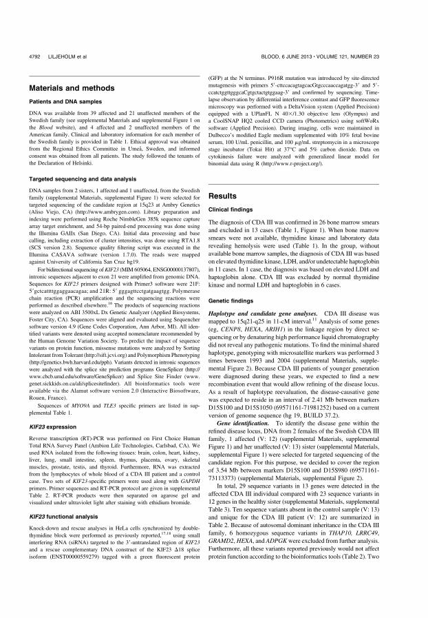
Materials and methods
Patients and DNA samples
DNA was available from 39 affected and 21 unaffected members of theSwedish family (see supplemental Materials and supplemental Figure 1 onthe Blood website), and 4 affected and 2 unaffected members of theAmerican family. Clinical and laboratory information for each member ofthe Swedish family is provided in Table 1. Ethical approval was obtainedfrom the Regional Ethics Committee in Umea, Sweden, and informedconsent was obtained from all patients. The study followed the tenants ofthe Declaration of Helsinki.
Targeted sequencing and data analysis
DNA samples from 2 sisters, 1 affected and 1 unaffected, from the Swedishfamily (supplemental Materials, supplemental Figure 1) were selected fortargeted sequencing of the candidate region at 15q23 at Ambry Genetics(Aliso Viejo, CA) (http://www.ambrygen.com). Library preparation andindexing were performed using Roche NimbleGen 385k sequence capturearray target enrichment, and 54-bp paired-end processing was done usingthe Illumina GAIIx (San Diego, CA). Initial data processing and basecalling, including extraction of cluster intensities, was done using RTA1.8(SCS version 2.8). Sequence quality filtering script was executed in theIllumina CASAVA software (version 1.7.0). The reads were mappedagainst University of California San Cruz hg19.
For bidirectional sequencing ofKIF23 (MIM 605064, ENSG00000137807),intronic sequences adjacent to exon 21 were amplified from genomic DNA.Sequences for KIF23 primers designed with Primer3 software were 21F:59gctcattttggaggaacagaa; and 21R: 59 gggagttcctgatgaagtgg. Polymerasechain reaction (PCR) amplification and the sequencing reactions wereperformed as described elsewhere.16 The products of sequencing reactionswere analyzed on ABI 3500xL Dx Genetic Analyzer (Applied Biosystems,Foster City, CA). Sequences were aligned and evaluated using Sequenchersoftware version 4.9 (Gene Codes Corporation, Ann Arbor, MI). All iden-tified variants were denoted using accepted nomenclature recommended bythe Human Genome Variation Society. To predict the impact of sequencevariants on protein function, missense mutations were analyzed by SortingIntolerant from Tolerant (http://sift.jcvi.org) and Polymorphism Phenotyping(http://genetics.bwh.harvard.edu/pph). Variants detected in intronic sequenceswere analyzed with the splice site prediction programs GeneSplicer (http://www.cbcb.umd.edu/software/GeneSplicer) and Splice Site Finder (www.genet.sickkids.on.ca/ali/splicesitefinder). All bioinformatics tools wereavailable via the Alamut software version 2.0 (Interactive Biosoftware,Rouen, France).
Sequences of MYO9A and TLE3 specific primers are listed in sup-plemental Table 1.
KIF23 expression
Reverse transcription (RT)-PCR was performed on First Choice HumanTotal RNA Survey Panel (Ambion Life Technologies, Carlsbad, CA). Weused RNA isolated from the following tissues: brain, colon, heart, kidney,liver, lung, small intestine, spleen, thymus, placenta, ovary, skeletalmuscles, prostate, testis, and thyroid. Furthermore, RNA was extractedfrom the lymphocytes of whole blood of a CDA III patient and a controlcase. Two sets of KIF23-specific primers were used along with GAPDHprimers. Primer sequences and RT-PCR protocol are given in supplementalTable 2. RT-PCR products were then separated on agarose gel andvisualized under ultraviolet light after staining with ethidium bromide.
KIF23 functional analysis
Knock-down and rescue analyses in HeLa cells synchronized by double-thymidine block were performed as previously reported,17,18 using smallinterfering RNA (siRNA) targeted to the 39-untranslated region of KIF23and a rescue complementary DNA construct of the KIF23 D18 spliceisoform (ENST00000559279) tagged with a green fluorescent protein
(GFP) at the N terminus. P916R mutation was introduced by site-directedmutagenesis with primers 59-cttccacagtagcacGtgcccaaccagatgg-39 and 59-ccatctggttgggcaCgtgctactgtggaag-39 and confirmed by sequencing. Time-lapse observation by differential interference contrast and GFP fluorescencemicroscopy was performed with a DeltaVision system (Applied Precision)equipped with a UPlanFL N 403/1.30 objective lens (Olympus) anda CoolSNAP HQ2 cooled CCD camera (Photometrics) using softWoRxsoftware (Applied Precision). During imaging, cells were maintained inDulbecco’s modified Eagle medium supplemented with 10% fetal bovineserum, 100 U/mL penicillin, and 100 mg/mL streptomycin in a microscopestage incubator (Tokai Hit) at 37°C and 5% carbon dioxide. Data oncytokinesis failure were analyzed with generalized linear model forbinomial data using R (http://www.r-project.org/).
Results
Clinical findings
The diagnosis of CDA III was confirmed in 26 bone marrow smearsand excluded in 13 cases (Table 1, Figure 1). When bone marrowsmears were not available, thymidine kinase and laboratory datarevealing hemolysis were used (Table 1). In the group, withoutavailable bone marrow samples, the diagnosis of CDA III was basedon elevated thymidine kinase, LDH, and/or undetectable haptoglobinin 11 cases. In 1 case, the diagnosis was based on elevated LDH andhaptoglobin alone. CDA III was excluded by normal thymidinekinase and normal LDH and haptoglobin in 6 cases.
Genetic findings
Haplotype and candidate gene analyses. CDA III disease wasmapped to 15q21-q25 in 11-cM interval.11 Analysis of some genes(eg, CENP8, HEXA, ARIH1) in the linkage region by direct se-quencing or by denaturing high performance liquid chromatographydid not reveal any pathogenic mutations. To find the minimal sharedhaplotype, genotyping with microsatellite markers was performed 3times between 1993 and 2004 (supplemental Materials, supple-mental Figure 2). Because CDA III patients of younger generationwere diagnosed during these years, we expected to find a newrecombination event that would allow refining of the disease locus.As a result of haplotype reevaluation, the disease-causative genewas expected to reside in an interval of 2.41 Mb between markersD15S100 and D15S1050 (69571161-71981252) based on a currentversion of genome sequence (hg 19, BUILD 37.2).
Gene identification. To identify the disease gene within therefined disease locus, DNA from 2 females of the Swedish CDA IIIfamily, 1 affected (V: 12) (supplemental Materials, supplementalFigure 1) and her unaffected (V: 13) sister (supplemental Materials,supplemental Figure 1) were selected for targeted sequencing of thecandidate region. For this purpose, we decided to cover the regionof 3.54 Mb between markers D15S100 and D15S980 (69571161-73113373) (supplemental Materials, supplemental Figure 2).
In total, 29 sequence variants in 13 genes were detected in theaffected CDA III individual compared with 23 sequence variants in12 genes in the healthy sister (supplemental Materials, supplementalTable 3). Ten sequence variants absent in the control sample (V: 13)and unique for the CDA III patient (V: 12) are summarized inTable 2. Because of autosomal dominant inheritance in the CDA IIIfamily, 6 homozygous sequence variants in THAP10, LRRC49,GRAMD2,HEXA, and ADPGKwere excluded from further analysis.Furthermore, all these variants reported previously would not affectprotein function according to the bioinformatics tools (Table 2). Two
4792 LILJEHOLM et al BLOOD, 6 JUNE 2013 x VOLUME 121, NUMBER 23

Table 1. Diagnostic characteristics in the CDA III Swedish family
Sex Bone marrow morphology* Hb, g/L† Haptoglobin, g/L‡ LDH, mkat/L Thymidine kinase§ CDA III||
III:6 M 1 143 ,0.1 5.2 52.5 1
III:9 M 2 ND ND ND 1.5 2
III:10 M 1 107 ,0.1 5.8 36.5 1
III:12 F 2 ND ND ND 1.4 2
III:14 M 1 116 0.1 4.3 18.1 1
III:18 M 2 162 0.6 2.7 2.6 2
III:20 F 1 128 ,0.1 6.3 36.2 1
IV:2 F 1 80 ,0.1 5.3 27.9 1
IV:4 F 1 126 ,0.1 8.5 250.0 1
IV:5 M 2 149 1.6 3.1 1.9 2
IV:7 F 1 105 ,0.1 8.0 39.1 1
IV:8 M 1 157 ,0.1 6.8 37.7 1
IV:10 M 1 107 ,0.1 6.2 46.5 1
IV:12IV:12 M 1 ND ND ND 56.8 1
IV:14 M 2 153 1.8 2.0 2.2 2
IV:15 M 1 146 ,0.1 6.2 28.9 1
IV:17 M 1 129 ,0.1 6.2 59.1 1
IV:20 F 1 120 ,0.1 4.8 53.3 1
IV:21 F 2 135 1.1 1.9 2.7 2
IV:22 M 2 154 0.6 2 4.5 2
IV:24 F ND 105 ,0.1 ND 65.0 1
IV:26 F 1 114 ,0,1 6.1 62.3 1
V:1 M 1 118 ,0.1 5.7 43.6 1
V:3 M 1 127 ,0.1 7.1 75.0 1
V:4 M 2 145 0.5 2.8 ND 2
V:5 M 1 106 ,0.1 6.8 64.3 1
V:7 F 1 94 ,0.1 7.7 149.0 1
V:8 F 2 107 0.5 2.4 1.7 2
V:9 M 1 135 ,0.1 6.3 65,6 1
V:12 F ND 114 ,0.1 6.7 184.0 1
V:13 F 2 145 1.9 2.0 2.6 2
V:14 M 1 113 ,0.1 5.7 41.9 1
V:17 M 2 130 0.6 2.9 1.0 2
V:18 F 2 136 1.0 2.0 3.4 2
V:19 F ND 153 0.7 2.1 4.0 2
V:20 M ND 158 1.0 ND 3.3 2
V:22 F 1 121 ,0.1 6.1 89.0 1
V:23 M 1 135 ,0.1 6.4 139.0 1
V:24 M ND 105 ,0.1 ND 52.3 1
V:26 M ND 120 ,0.1 ND 108.0 1
V:27 M ND 147 0.6 2.7 5.0 2
V:28 M ND 145 0.3 3.1 8.0 2
V:30 F ND 109 ,0.1 4.1 120.0 1
V:31 M ND 118 0.8 ND 3.8 2
V:32 M ND 127 ND 3.4 7.0 2
V:33 F 1 119 0.1 4.3 18.5 1
V:35 M ND ND ,0.1 ND 69.0 1
V:36 F 1 124 ,0.1 8.3 167.0 1
V:38 F 2 138 2,6 2.7 6.4 2
VI:1 F ND 102 ,0.1 7.5 279.0 1
VI:2 F ND 133 1.22 2.2 30.0 2
VI:3 F ND 114 1.87 2.6 ND 2
VI:4 M 1 123 ,0.1 10.0 ND 1
VI:5 M ND 149 0.53 3.3 ND 2
VI:6 F ND 116 ,0.1 5.9 116 1
VI:7 F ND 109 ,0.1 6.6 120 1
VI:8 M ND 125 ND 4.6 134 1
VI:10 M 1 108 ,0.1 5.8 286 1
VI:11 F ND 115 ND 5.3 102 1
VI:14 F ND 107 ,0.1 8.8 ND 1
F, female; CDA III Hb, hemoglobin; LDH, lactate dehydrogenase; M, male; ND, no data.
*Consistent with CDA III.
†Normal range for males is 134-170 and for females 117-153.
‡Normal range 0.24-1.9.
§Normal range ,3.4, adjusted according to Nordic Reference Interval Project.
||CDA III final diagnosis.
BLOOD, 6 JUNE 2013 x VOLUME 121, NUMBER 23 KIF23 IS A GENETIC CAUSE OF CDA III 4793
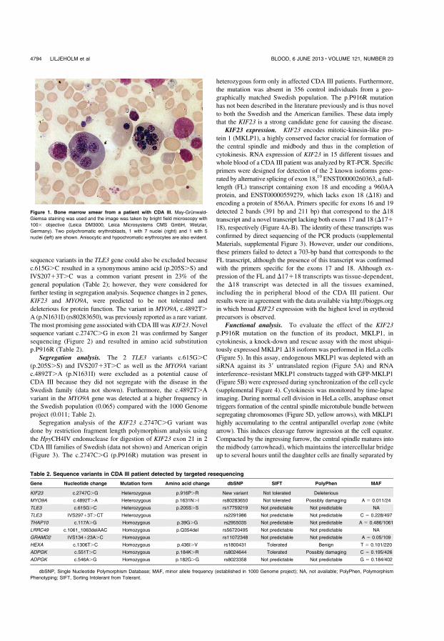
sequence variants in the TLE3 gene could also be excluded becausec.615G.C resulted in a synonymous amino acid (p.205S.S) andIVS20713T.C was a common variant present in 23% of thegeneral population (Table 2); however, they were considered forfurther testing in segregation analysis. Sequence changes in 2 genes,KIF23 and MYO9A, were predicted to be not tolerated anddeleterious for protein function. The variant in MYO9A, c.4892T.A (p.N1631I) (rs80283650), was previously reported as a rare variant.The most promising gene associated with CDA III was KIF23. Novelsequence variant c.2747C.G in exon 21 was confirmed by Sangersequencing (Figure 2) and resulted in amino acid substitutionp.P916R (Table 2).
Segregation analysis. The 2 TLE3 variants c.615G.C(p.205S.S) and IVS20713T.C as well as the MYO9A variantc.4892T.A (p.N1631I) were excluded as a potential cause ofCDA III because they did not segregate with the disease in theSwedish family (data not shown). Furthermore, the c.4892T.Avariant in the MYO9A gene was detected at a higher frequency inthe Swedish population (0.065) compared with the 1000 Genomeproject (0.011; Table 2).
Segregation analysis of the KIF23 c.2747C.G variant wasdone by restriction fragment length polymorphism analysis usingthe HpyCH4IV endonuclease for digestion of KIF23 exon 21 in 2CDA III families of Swedish (data not shown) and American origin(Figure 3). The c.2747C.G (p.P916R) mutation was present in
heterozygous form only in affected CDA III patients. Furthermore,the mutation was absent in 356 control individuals from a geo-graphically matched Swedish population. The p.P916R mutationhas not been described in the literature previously and is thus novelto both the Swedish and the American families. These data implythat the KIF23 is a strong candidate gene for causing the disease.
KIF23 expression. KIF23 encodes mitotic-kinesin-like pro-tein 1 (MKLP1), a highly conserved factor crucial for formation ofthe central spindle and midbody and thus in the completion ofcytokinesis. RNA expression of KIF23 in 15 different tissues andwhole blood of a CDA III patient was analyzed by RT-PCR. Specificprimers were designed for detection of the 2 known isoforms gene-rated by alternative splicing of exon 18,19 ENST00000260363, a full-length (FL) transcript containing exon 18 and encoding a 960AAprotein, and ENST00000559279, which lacks exon 18 (D18) andencoding a protein of 856AA. Primers specific for exons 16 and 19detected 2 bands (391 bp and 211 bp) that correspond to the D18transcript and a novel transcript lacking both exons 17 and 18 (D17118), respectively (Figure 4A-B). The identity of these transcripts wasconfirmed by direct sequencing of the PCR products (supplementalMaterials, supplemental Figure 3). However, under our conditions,these primers failed to detect a 703-bp band that corresponds to theFL transcript, although the presence of this transcript was confirmedwith the primers specific for the exons 17 and 18. Although ex-pression of the FL and D17118 transcripts was tissue-dependent,the D18 transcript was detected in all the tissues examined,including the in peripheral blood of the CDA III patient. Ourresults were in agreement with the data available via http://biogps.orgin which broad KIF23 expression with the highest level in erythroidprecursors is observed.
Functional analysis. To evaluate the effect of the KIF23p.P916R mutation on the function of its product, MKLP1, incytokinesis, a knock-down and rescue assay with the most ubiqui-tously expressed MKLP1 D18 isoform was performed in HeLa cells(Figure 5). In this assay, endogenous MKLP1 was depleted with ansiRNA against its 39 untranslated region (Figure 5A) and RNAinterference–resistant MKLP1 constructs tagged with GFP-MKLP1(Figure 5B) were expressed during synchronization of the cell cycle(supplemental Figure 4). Cytokinesis was monitored by time-lapseimaging. During normal cell division in HeLa cells, anaphase onsettriggers formation of the central spindle microtubule bundle betweensegregating chromosomes (Figure 5D, yellow arrows), with MKLP1highly accumulating to the central antiparallel overlap zone (whitearrow). This induces cleavage furrow ingression at the cell equator.Compacted by the ingressing furrow, the central spindle matures intothe midbody (arrowhead), which maintains the intercellular bridgeup to several hours until the daughter cells are finally separated by
Figure 1. Bone marrow smear from a patient with CDA III. May-Grunwald-
Giemsa staining was used and the image was taken by bright field microscopy with
1003 objective (Leica DM3000, Leica Microsystems CMS GmbH, Wetzlar,
Germany). Two polychromatic erythroblasts, 1 with 7 nuclei (right) and 1 with 5
nuclei (left) are shown. Anisocytic and hypochromatic erythrocytes are also evident.
Table 2. Sequence variants in CDA III patient detected by targeted resequencing
Gene Nucleotide change Mutation form Amino acid change dbSNP SIFT PolyPhen MAF
KIF23 c.2747C.G Heterozygous p.916P.R New variant Not tolerated Deleterious
MYO9A c.4892T.A Heterozygous p.1631N.I rs80283650 Not tolerated Possibly damaging A 5 0.011/24
TLE3 c.615G.C Heterozygous p.205S.S rs17759219 Not predictable Not predictable NA
TLE3 IVS29713T.CT Heterozygous rs2291986 Not predictable Not predictable C 5 0.228/497
THAP10 c.117A.G Homozygous p.39G.G rs2955035 Not predictable Not predictable A 5 0.486/1061
LRRC49 c.1061_1063delAAC Homozygous p.Q354del rs56720495 Not predictable Not predictable NA
GRAMD2 IVS134123A.C Homozygous rs11072348 Not predictable Not predictable A 5 0.05/109
HEXA c.1306T.C Homozygous p.436I.V rs1800431 Tolerated Benign T 5 0.101/220
ADPGK c.551T.C Homozygous p.184K.R rs8024644 Tolerated Possibly damaging C 5 0.195/426
ADPGK c.546A.G Homozygous p.182G.G rs8023358 Not predictable Not predictable G 5 0.184/402
dbSNP, Single Nucleotide Polymorphism Database; MAF, minor allele frequency (established in 1000 Genome project); NA, not available; PolyPhen, Polymorphism
Phenotyping; SIFT, Sorting Intolerant from Tolerant.
4794 LILJEHOLM et al BLOOD, 6 JUNE 2013 x VOLUME 121, NUMBER 23

abscission (double-headed arrow). MKLP1 plays essential roles inthe formation of the central spindle and the stable maintenance ofthe midbody. Failure of cytokinesis by depletion of the endogenousMKLP1 was efficiently rescued by expression of wild-type GFP-MKLP1 (Figure 5C). In contrast, GFP-MKLP1 with the p.P916Rmutation (P812R mutation in this isoform) showed little or no rescueactivity above the level of the control empty vector (Figure 5C,P916R vs GFP). Cells expressing GFP-MKLP1-P916R could
complete constriction of the cleavage furrow and form the midbodyin a similar time course with the cells expressing the wild-typeGFP-MKLP1, but regressed to form binucleate cells about 2 hoursafter midbody formation (180 6 148 nub [mean 6 standard devia-tion.], mode at 105 min, n 5 225) (Figure 5D-E and supplementalVideos 1 and 2). These data indicate that the p.P916R mutationimpairs the function of MKLP1 essential for the completion ofcytokinesis.
Figure 2. A novel c.2747C>G (p.P916R) mutation in the KIF23 gene. (A) Exon structure of the KIF23 gene with DNA and protein sequence of the exon 21 where the
mutation resides. (B) DNA sequence showing c.2747C.G mutation. (Upper) Wild type and (lower) c.2747C.G heterozygous mutations, with mutation position marked in
black. (C) Phylogenetic alignment of partial KIF23 protein sequence.
Figure 3. c.2747C>G segregates CDA III in the
American family. (A) Pedigree of the family with
affected individuals shown in black symbols and healthy
individuals shown as unfilled symbols. DNA was avail-
able only for cases marked with A1–A6. (B) Restriction
length fragment polymorphism assay. Amplification of
exon 21 of KIF23 was done as described in “Materials
and Methods.” PCR products were digested using the
HpyCH4IVendonuclease.c.2747C.Gcreatesarestriction
site; therefore, all mutation carriers show 3 bands—338,
175, and 163 bp (small fragments are poorly resolved
on agarose gel)—whereas all unaffected individuals show
only 1 band (338 bp).
BLOOD, 6 JUNE 2013 x VOLUME 121, NUMBER 23 KIF23 IS A GENETIC CAUSE OF CDA III 4795
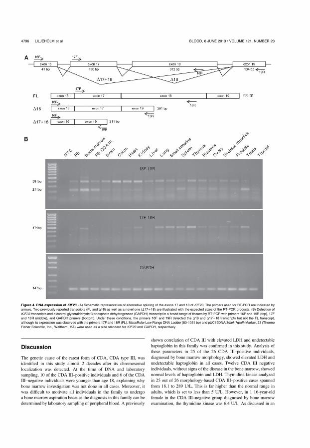
Discussion
The genetic cause of the rarest form of CDA, CDA type III, wasidentified in this study almost 2 decades after its chromosomallocalization was detected. At the time of DNA and laboratorysampling, 10 of the CDA III–positive individuals and 6 of the CDAIII–negative individuals were younger than age 18, explaining whybone marrow investigation was not done in all cases. Moreover, itwas difficult to motivate all individuals in the family to undergoa bone marrow aspiration because the diagnosis in this family can bedetermined by laboratory sampling of peripheral blood. A previously
shown correlation of CDA III with elevated LDH and undetectablehaptoglobin in this family was confirmed in this study. Analysis ofthese parameters in 25 of the 26 CDA III–positive individuals,diagnosed by bone marrow morphology, showed elevated LDH andundetectable haptoglobin in all cases. Twelve CDA III negativeindividuals, without signs of the disease in the bone marrow, showednormal levels of haptoglobin and LDH. Thymidine kinase analyzedin 25 out of 26 morphology-based CDA III–positive cases spannedfrom 18.1 to 289 U/L. This is far higher than the normal range inadults, which is set to less than 5 U/L. However, in 1 16-year-oldfemale in the CDA III–negative group diagnosed by bone marrowexamination, the thymidine kinase was 6.4 U/L. As discussed in an
Figure 4. RNA expression of KIF23. (A) Schematic representation of alternative splicing of the exons 17 and 18 of KIF23. The primers used for RT-PCR are indicated by
arrows. Two previously reported transcripts (FL and D18) as well as a novel one (D17118) are illustrated with the expected sizes of the RT-PCR products. (B) Detection of
KIF23 transcripts and a control glyceraldehyde-3-phosphate dehydrogenase (GAPDH) transcript in a broad range of tissues by RT-PCR with primers 16F and 19R (top), 17F
and 18R (middle), and GAPDH primers (bottom). Under these conditions, the primers 16F and 19R detected the D18 and D17118 transcripts but not the FL transcript,
although its expression was observed with the primers 17F and 18R (FL). MassRuler Low Range DNA Ladder (80-1031 bp) and pUC19DNA/MspI (HpaII) Marker, 23 (Thermo
Fisher Scientific, Inc., Waltham, MA) were used as a size standard for KIF23 and GAPDH, respectively.
4796 LILJEHOLM et al BLOOD, 6 JUNE 2013 x VOLUME 121, NUMBER 23
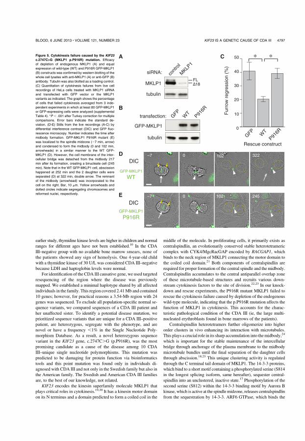
earlier study, thymidine kinase levels are higher in children and normalranges for different ages have not been established.10 In the CDAIII–negative group with no available bone marrow smears, none ofthe patients showed any sign of hemolysis. One 4-year-old childwith a thymidine kinase of 30 U/L was considered CDA III–negativebecause LDH and haptoglobin levels were normal.
For identification of the CDA III causative gene, we used targetedresequencing of the region where the disease was previouslymapped. We established a minimal haplotype shared by all affectedindividuals in the family. This region covered 2.41Mb and contained10 genes; however, for practical reasons a 3.54-Mb region with 24genes was sequenced. To exclude all population-specific normal se-quence variants, we compared sequences of a CDA III patient andher unaffected sister. To identify a potential disease mutation, weprioritized sequence variants that are unique for a CDA III–positivepatient, are heterozygous, segregate with the phenotype, and arenovel or have a frequency ,1% in the Single Nucleotide Poly-morphism Database. As a result, a novel heterozygous sequencevariant in the KIF23 gene, c.2747C.G (p.P916R), was the mostpromising candidate as a cause of the disease among 10 CDAIII–unique single nucleotide polymorphisms. This mutation waspredicted to be damaging for protein function via bioinformaticstools and this point mutation was found only in individuals di-agnosed with CDA III and not only in the Swedish family but also inthe American family. The Swedish and American CDA III familiesare, to the best of our knowledge, not related.
KIF23 encodes the kinesin superfamily molecule MKLP1 thatplays critical roles in cytokinesis.19,20 It has a kinesin motor domainon its N terminus and a domain predicted to form a coiled coil in the
middle of the molecule. In proliferating cells, it primarily exists ascentralspindlin, an evolutionarily conserved stable heterotetramericcomplex with CYK4/MgcRacGAP encoded by RACGAP1, whichbinds to the neck region of MKLP1 connecting the motor domain tothe coiled coil domain.21 Both components of centralspindlin arerequired for proper formation of the central spindle and the midbody.Centralspindlin accumulates to the central antiparallel overlap zoneof these microtubule-based structures and recruits various down-stream cytokinesis factors to the site of division.22,23 In our knock-down and rescue experiments, the P916R mutant MKLP1 failed torescue the cytokinesis failure caused by depletion of the endogenouswild-type molecule, indicating that the p.P916R mutation affects thefunction of MKLP1 in cytokinesis. This accounts for the charac-teristic pathological condition of the CDA III (ie, the large multi-nucleated erythroblasts found in bone marrows of the patients).
Centralspindlin heterotetramers further oligomerize into higherorder clusters in vivo enhancing its interaction with microtubules.This plays a crucial role in its sharp accumulation into themidbody,24
which is important for the stable maintenance of the intercellularbridge through anchorage of the plasma membrane to the midbodymicrotubule bundles until the final separation of the daughter cellsthrough abscission.18,25 This unique clustering activity is regulatedthrough the C terminal tail domain of MKLP1. The 14-3-3 proteins,which bind to a short motif containing a phosphorylated serine (S814in the longest splicing isoform, same hereafter), sequester central-spindlin into an unclustered, inactive state.17 Phosphorylation of thesecond serine (S812) within the 14-3-3 binding motif by Aurora Bkinase, which is active at the spindlemidzone, releases centralspindlinfrom the sequestration by 14-3-3. ARF6 GTPase, which binds the
Figure 5. Cytokinesis failure caused by the KIF23
c.2747C>G (MKLP1 p.P916R) mutation. Efficacy
of depletion of endogenous MKLP1 (A) and equal
expression of wild-type (WT) and P916R GFP-MKLP1
(B) constructs was confirmed by western blotting of the
whole cell lysates with anti-MKLP1 (A) or anti-GFP (B)
antibody. Tubulin was also blotted as a loading control.
(C) Quantitation of cytokinesis failures from live cell
recordings of HeLa cells treated with MKLP1 siRNA
and transfected with GFP vector or the MKLP1
variants as indicated. The graph shows the percentage
of cells that failed cytokinesis averaged from 3 inde-
pendent experiments in which at least 80 GFP-MKLP1
or GFP-expressing cells were analyzed (supplemental
Table 4). *P , .001 after Turkey correction for multiple
comparisons. Error bars indicate the standard de-
viation. (D-E) Stills from the live recordings (A-C) by
differential interference contrast (DIC) and GFP fluo-
rescence microscopy. Number indicates the time after
midbody formation. GFP-MKLP1 P916R mutant (E)
was localized to the spindle midzone (27 min, arrow)
and condensed to form the midbody (0 and 102 min,
arrowheads) in a similar manner to the WT GFP-
MKLP1 (D). However, the cell membrane of the inter-
cellular bridge was detached from the midbody 217
min after its formation, creating a binucleate cell (245
min). Note that in the WT GFP-MKLP1 cell, abscission
happened at 252 min and the 2 daughter cells were
separated (D) at 322 min, double arrow. The remnant
of the midbody (arrowhead) was incorporated to the
cell on the right. Bar, 10 mm. Yellow arrowheads and
dotted circles indicate segregating chromosomes and
reformed nuclei, respectively.
BLOOD, 6 JUNE 2013 x VOLUME 121, NUMBER 23 KIF23 IS A GENETIC CAUSE OF CDA III 4797
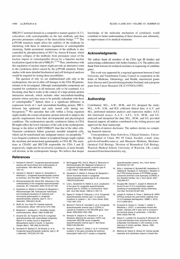
MKLP1 C terminal domain in a competitive manner against 14-3-3,colocalizes with centralspindlin on the late midbody and thusprevents premature collapse of the intercellular bridge.18,26 Thep.P916R mutation might affect the stability of the midbody byinterfering with these or unknown regulations of centralspindlinclustering. Stable postmitotic maintenance of the midbody is alsocontrolled by phosphorylation of S911 by Aurora B kinase, whichprevents collapse of the midbody from premature onset of thenuclear import of centralspindlin driven by a bipartite nuclearlocalization signal in the tail of MKLP1.27,28 Thus, interference withthis regulation of nuclear import might provide an alternative routeto the cytokinesis defect caused by the p.P916R mutation. Futurestudies with more detailed biochemical and cell biological analyseswould be required for testing these possibilities.
The question of why we see multinucleated cells only in theerythropoiesis, but not in other cell lineages in the CDA III patients,remains to be investigated. Although centralspindlin components areessential for cytokinesis in all metazoan cells so far examined, it isbecoming clear that it works in the context of a large protein–proteininteraction network, which includes other microtubule-bundlingproteins whose activities seem to be partially redundant with thoseof centralspindlin.29 Indeed, there is a significant difference inexpression levels of 1 such microtubule-bundling protein, PRC1,between late epidermal and early neural cells during em-bryogenesis.30 We speculate that cells within a living organismmight modify the conserved protein–protein network to adapt to thespecific requirements from their developmental and physiologicalconditions. The erythropoiesis-specific cytokinesis failure in CDAIII might reflect the lower tolerance of the cells in the erythropoieticlineage to the defect in KIF23/MKLP1 than that of the other cells.Transient cytokinesis failure generates unstable tetraploid cells,which can be transformed into malignant tumors via aneuploidy.31
Less frequent cytokinesis failure in lymphoid lineage might explainthe myeloma and monoclonal gammopathy in CDA III. The muta-tions in CDAN1 and SEC23B responsible for CDA I and II,respectively, might also be involved in cytokinesis, or more broadlycell division, in the erythropoietic lineage. We believe that deeper
knowledge of the molecular mechanism of cytokinesis wouldcontribute to better understanding of these diseases and, ultimately,to improvement of its medical treatments.
Acknowledgments
The authors thank all members of the CDA type III families andacknowledge collaborationwithAmbryGenetics, CA. The authors alsothank Frida Jonsson for technical assistance in sequencing of candidategenes.
This work was supported by a regional agreement between UmeaUniversity and Vasterbotten County Council in cooperation in thefields of Medicine, Odontology, and Health, unrestricted grantsfromAlexion and Cancerforskningsfonden Norrland, and a programgrant from Cancer Research UK (C19769/A11985).
Authorship
Contribution: M.L., A.W., M.M., and I.G. designed the study;M.L., A.W., S.M., and H.S. collected clinical data; A.-L.V. andM.L. performed mutation analyses and expression studies; A.F.I.did functional assays; A.-L.V., A.F.I., A.N., M.M., and I.G.analyzed and interpreted the data; M.L., M.M., and I.G. providedfinancial support; all authors contributed to the writing process andapproved the final manuscript.
Conflict-of-interest disclosure: The authors declare no compet-ing financial interests.
Correspondence: Irina Golovleva, Clinical Genetics, Univer-sity Hospital of Umea, 901 85 Umea, Sweden; e-mail: [email protected]; and Masanori Mishima, Mechano-chemical Cell Biology, Division of Biomedical Cell Biology,Warwick Medical School, University of Warwick, UK; e-mail:[email protected].
References
1. Heimpel H, Wendt F. Congenital dyserythropoieticanemia with karyorrhexis and multinuclearity oferythroblasts. Helv Med Acta. 1968;34(2):103-115.
2. Heimpel H, Wendt F, Klemm D, Schubothe H,Heilmeyer L. Congenital dyserythropoietic anemia[in German]. Arch Klin Med. 1968;215(2):174-194.
3. Wickramasinghe SN, Wood WG. Advances in theunderstanding of the congenital dyserythropoieticanaemias. Br J Haematol. 2005;131(4):431-446.
4. Sandstrom H, Wahlin A, Eriksson M, Bergstrom I,Wickramasinghe SN. Intravascular haemolysisand increased prevalence of myeloma andmonoclonal gammopathy in congenitaldyserythropoietic anaemia, type III. Eur JHaematol. 1994;52(1):42-46.
5. Wolff JA, Von Hofe FH. Familial erythroidmultinuclearity. Blood. 1951;6(12):1274-1283.
6. Bergstrom I, Jacobsson L. Hereditary benignerythroreticulosis. Blood. 1962;19:296-303.
7. Accame EA, de Tezanos Pinto M. Congenitaldyserythropoiesis with erythroblastic polyploidy.Report of a variety found in ArgentinianMesopotamia (author’s transl). In Spanish].Sangre (Barc). 1981;26(5-A):545-555.
8. Goudsmit R, Beckers D, De Bruijne JI, et al.Congenital dyserythropoietic anaemia, type 3. BrJ Haematol. 1972;23(1):97-105.
9. McCluggage WG, Hull D, Mayne E, Bharucha H,Wickramasinghe SN. Malignant lymphoma incongenital dyserythropoietic anaemia type III.J Clin Pathol. 1996;49(7):599-602.
10. Sandstrom H, Wahlin A, Eriksson M, Bergstrom I.Serum thymidine kinase in congenitaldyserythropoietic anaemia type III. Br J Haematol.1994;87(3):653-654.
11. Lind L, Sandstrom H, Wahlin A, et al. Localizationof the gene for congenital dyserythropoieticanemia type III, CDAN3, to chromosome 15q21-q25. Hum Mol Genet. 1995;4(1):109-112.
12. Dgany O, Avidan N, Delaunay J, et al. Congenitaldyserythropoietic anemia type I is caused bymutations in codanin-1. Am J Hum Genet. 2002;71(6):1467-1474.
13. Bianchi P, Fermo E, Vercellati C, et al. Congenitaldyserythropoietic anemia type II (CDAII) iscaused by mutations in the SEC23B gene. HumMutat. 2009;30(9):1292-1298.
14. Schwarz K, Iolascon A, Verissimo F, et al.Mutations affecting the secretory COPII coatcomponent SEC23B cause congenitaldyserythropoietic anemia type II. Nat Genet.2009;41(8):936-940.
15. Arnaud L, Saison C, Helias V, et al. A dominantmutation in the gene encoding the erythroidtranscription factor KLF1 causes a congenital
dyserythropoietic anemia. Am J Hum Genet.2010;87(5):721-727.
16. Kohn L, Kadzhaev K, Burstedt MS, Haraldsson S,Hallberg B, Sandgren O, Golovleva I. Mutation inthe PYK2-binding domain of PITPNM3 causesautosomal dominant cone dystrophy (CORD5) intwo Swedish families. Eur J Hum Genet. 2007;15(6):664-671.
17. Douglas ME, Davies T, Joseph N, Mishima M.Aurora B and 14-3-3 coordinately regulateclustering of centralspindlin during cytokinesis.Curr Biol. 2010;20(10):927-933.
18. Joseph N, Hutterer A, Poser I, Mishima M. ARF6GTPase protects the post-mitotic midbody from14-3-3-mediated disintegration. EMBO J. 2012;31(11):2604-2614.
19. Kuriyama R, Gustus C, Terada Y, Uetake Y,Matuliene J. CHO1, a mammalian kinesin-likeprotein, interacts with F-actin and is involved inthe terminal phase of cytokinesis. J Cell Biol.2002;156(5):783-790.
20. Nislow C, Lombillo VA, Kuriyama R, McIntosh JR.A plus-end-directed motor enzyme that movesantiparallel microtubules in vitro localizes to theinterzone of mitotic spindles. Nature. 1992;359(6395):543-547.
21. Mishima M, Kaitna S, Glotzer M. Central spindleassembly and cytokinesis require a kinesin-like
4798 LILJEHOLM et al BLOOD, 6 JUNE 2013 x VOLUME 121, NUMBER 23

protein/RhoGAP complex with microtubulebundling activity. Dev Cell. 2002;2(1):41-54.
22. Green RA, Paluch E, Oegema K. Cytokinesis inanimal cells. Annu Rev Cell Dev Biol. 2012;28:29-58.
23. White EA, Glotzer M. Centralspindlin: at the heartof cytokinesis. Cytoskeleton (Hoboken). 2012;69(11):882-892.
24. Hutterer A, Glotzer M, Mishima M. Clustering ofcentralspindlin is essential for its accumulation tothe central spindle and the midbody. Curr Biol.2009;19(23):2043-2049.
25. Lekomtsev S, Su KC, Pye VE, et al.Centralspindlin links the mitotic spindle to the
plasma membrane during cytokinesis. Nature.2012;492(7428):276-279.
26. Makyio H, Ohgi M, Takei T, et al. Structural basisfor Arf6-MKLP1 complex formation on theFlemming body responsible for cytokinesis.EMBO J. 2012;31(11):2590-2603.
27. Neef R, Klein UR, Kopajtich R, Barr FA. Cooperationbetween mitotic kinesins controls the late stagesof cytokinesis. Curr Biol. 2006;16(3):301-307.
28. Liu X, Erikson RL. The nuclear localization signal ofmitotic kinesin-like protein Mklp-1: effect on Mklp-1function during cytokinesis. Biochem Biophys ResCommun. 2007;353(4):960-964.
29. Lee KY, Davies T, Mishima M. Cytokinesismicrotubule organisers at a glance. J Cell Sci.2012;125(Pt 15):3495-3500.
30. Kieserman EK, Glotzer M, Wallingford JB.Developmental regulation of centralspindle assembly and cytokinesis duringvertebrate embryogenesis. Curr Biol. 2008;18(2):116-123.
31. Fujiwara T, Bandi M, Nitta M, Ivanova EV,Bronson RT, Pellman D. Cytokinesis failuregenerating tetraploids promotes tumorigenesisin p53-null cells. Nature. 2005;437(7061):1043-1047.
BLOOD, 6 JUNE 2013 x VOLUME 121, NUMBER 23 KIF23 IS A GENETIC CAUSE OF CDA III 4799

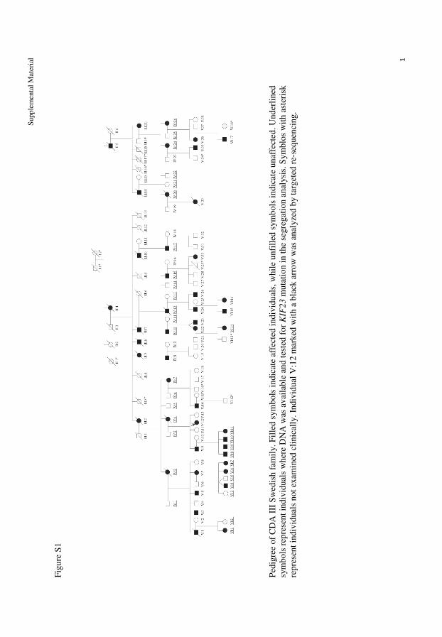
Sup
ple
menta
l M
ater
ial 1
Fig
ure
S1
Ped
igre
e of
CD
A I
II S
wed
ish f
amil
y. F
ille
d s
ym
bols
indic
ate
affe
cted
indiv
idual
s, w
hil
e unfi
lled
sym
bols
indic
ate
unaf
fect
ed. U
nder
lined
sym
bols
rep
rese
nt
indiv
idual
s w
her
e D
NA
was
av
aila
ble
and t
este
d f
or
KIF
23 m
uta
tion i
n t
he
segre
gat
ion a
nal
ysi
s. S
ym
blo
s w
ith a
ster
isk
repre
sent
indiv
idual
s not
exam
ined
cli
nic
ally
. In
div
idual
V:1
2 m
arked
wit
h a
bla
ck a
rrow
was
anal
yze
d b
y t
arget
ed r
e-se
qu
enci
ng.
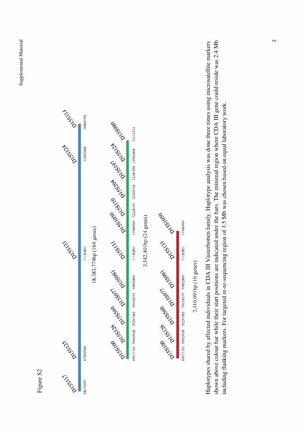
Sup
ple
menta
l M
ater
ial 2
Fig
ure
S2
Hap
loty
pes
shar
ed b
y a
ffec
ted i
ndiv
idual
s in
CD
A I
II V
äste
rbott
en f
amil
y.
Hap
loty
pe
anal
ysi
s w
as d
one
thre
e ti
mes
usi
ng m
icro
sate
llit
e m
ark
ers
show
n a
bove
colo
ur
bar
whil
e th
eir
star
t posi
tions
are
indic
ated
under
th
e b
ars.
The
min
imal
reg
ion w
her
e C
DA
III
gen
e co
uld
res
ide
was
2.4
Mb
incl
udin
g f
lankin
g m
arker
s. F
or
targ
eted
re-
re-s
equen
cin
g r
egio
n o
f 3.5
Mb
was
chose
n b
ased
on e
qual
lab
ora
tory
wo
rk.

Supplemental Material
3
Figure S3
Sequence of a novel KIF23 isoform lacking exons 17 and 18
Arrow shows boundary between end of exon 16 and start of exon 19. Sequence was done on
211 bp PCR fragment amplified with primers 16F and 19R
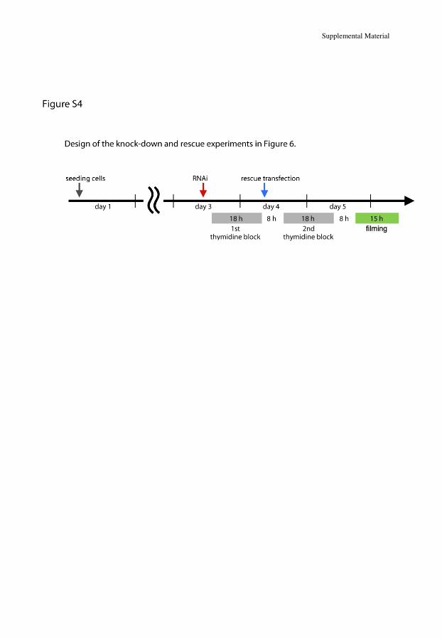
Supplemental Material
4
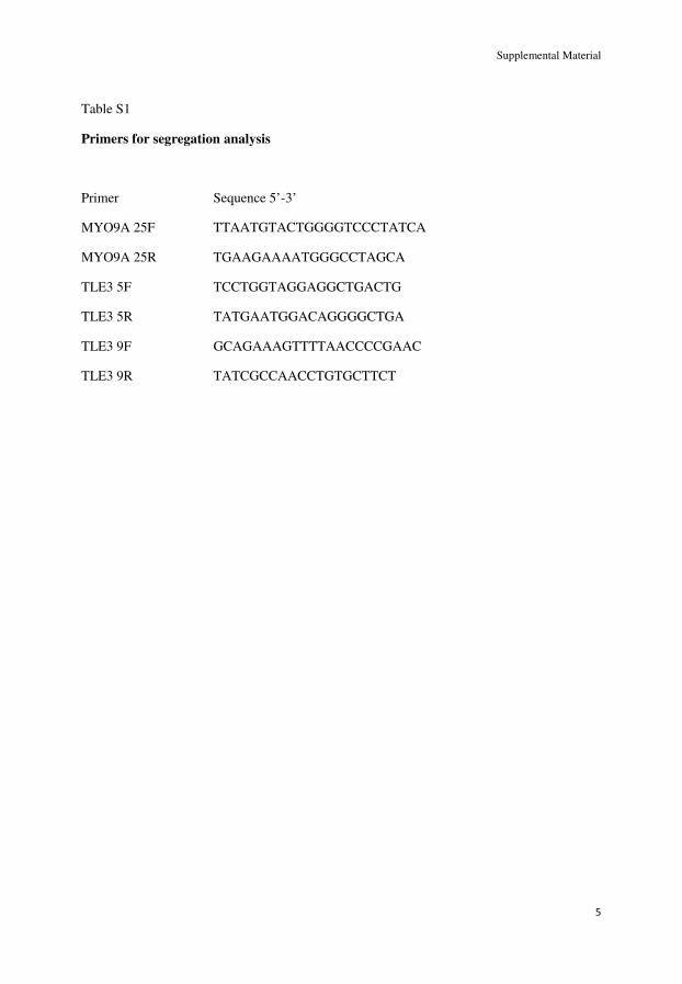
Supplemental Material
5
Table S1
Primers for segregation analysis
Primer Sequence 5’-3’
MYO9A 25F TTAATGTACTGGGGTCCCTATCA
MYO9A 25R TGAAGAAAATGGGCCTAGCA
TLE3 5F TCCTGGTAGGAGGCTGACTG
TLE3 5R TATGAATGGACAGGGGCTGA
TLE3 9F GCAGAAAGTTTTAACCCCGAAC
TLE3 9R TATCGCCAACCTGTGCTTCT

Supplemental Material
6
Table S2
KIF23-specific primers for analysis of RNA expression
Primer Sequence 5’-3’
KIF23 16F GCATGGTGACAGAAACGACA
KIF23 17F GAGCGTAGAGTGGCAGCCAA
KIF23 18R ATTTCTGAATGTAGGAACCAC
KIF23 19R ACATGTGGCTGCATGACTGT
Total RNA was extracted using TRIzol reagent (Invitrogen®Life Technologies, Carlsbad,
CA, USA) according to the manufacturer’instructions. Total RNA was then reverse-
transcribed with SuperScript III RT (Invitrogen®Life Technologies, Carlsbad, CA, USA) and
the resulting cDNA was used as template for PCR analysis. All PCR reactions were
performed with Amplitaq Gold (Applied Biosystems) as recommended with an annealing
temperature of 57°C (16F-19R), 47°C (17F-18R), 54°C (GAPDH) and an extension time of
30 s for a total of 39 cycles.
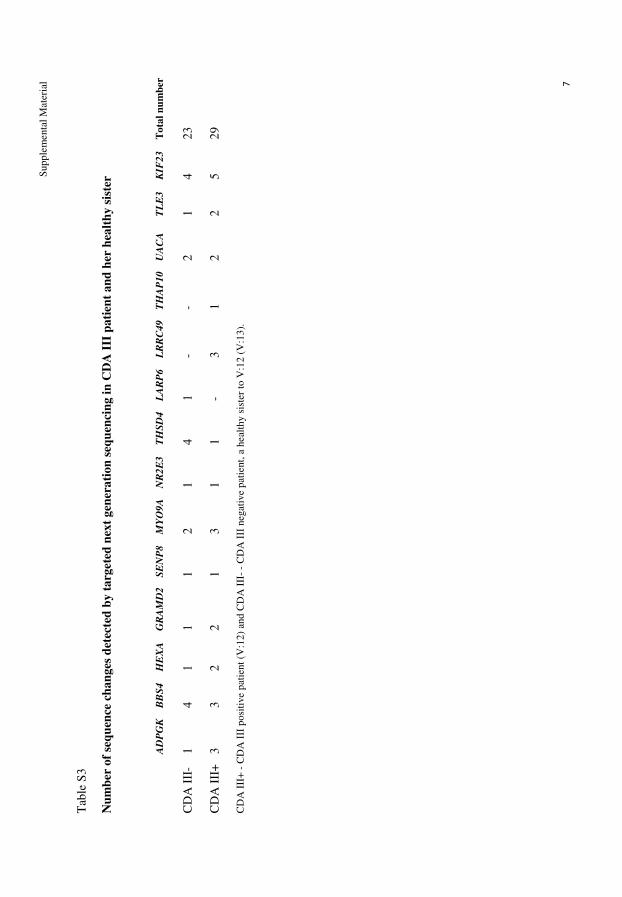
Sup
ple
menta
l M
ater
ial 7
Tab
le S
3
Nu
mb
er o
f se
qu
ence
ch
an
ges
det
ecte
d b
y t
arg
ete
d n
ext
gen
era
tion
seq
uen
cin
g i
n C
DA
III
pati
ent
an
d h
er h
ealt
hy s
iste
r
A
DP
GK
B
BS
4
HE
XA
G
RA
MD
2
SE
NP
8
MY
O9
A
NR
2E
3
TH
SD
4
LA
RP
6
LR
RC
49
T
HA
P1
0
UA
CA
T
LE
3
KIF
23
T
ota
l n
um
ber
CD
A I
II-
1
4
1
1
1
2
1
4
1
- -
2
1
4
23
CD
A I
II+
3
3
2
2
1
3
1
1
- 3
1
2
2
5
29
CD
A I
II+
- C
DA
III
po
siti
ve
pat
ient
(V:1
2)
and
CD
A I
II-
- C
DA
III
neg
ativ
e p
atie
nt,
a h
eal
thy s
iste
r to
V:1
2 (
V:1
3).

Sup
ple
menta
l M
ater
ial 8
Tab
le S
4
Su
mm
ary
of
scori
ng o
f th
e ti
me-
lap
se m
ovie
s in
th
e k
nock
-dow
n a
nd
res
cue
exp
erim
ents
in
Fig
ure
6. s.
d., s
tan
dard
dev
iati
on
resc
ue
con
stru
ct
b
iolo
gic
al
rep
lica
tes
to
tal
cell
s co
un
ted
cy
tok
ines
is f
ail
ure
%
fail
ure
s.
d. (%
)
GF
P
3
334
137
41.0
6.5
GF
P-M
KL
P1 W
T
3
368
74
20.4
1.9
GF
P-M
KL
P1 P
916R
3
574
225
39.3
3.7
L
egen
ds
to S
up
ple
men
tary
Movie
s 1 a
nd
2
Tim
e la
pse
movie
s of
HeL
a ce
lls
expre
ssin
g w
ild-t
ype
GF
P-M
KL
P1 (
Movie
1)
or
P916R
muta
nt
GF
P-M
KL
P1 (
Movie
2)
as r
escu
e co
nst
ruct
in
an R
NA
i-bas
ed k
no
ck-d
ow
n a
nd r
escu
e as
say. F
luore
scen
ce i
mag
e of
GF
P-M
KL
P1 (
gre
en)
was
mer
ged
on D
IC i
mag
e (g
raysc
ale)
. B
ar, 10 µ
m.
Tim
e, h
our:
min
.

II


Research Article Open Access
Liljeholm et al., J Blood Disorders Transf 2013, 4:6http://dx.doi.org/10.4172/2155-9864.1000172
Research article Open Access
BloodDisorders & Transfusion
Volume 4 • Issue 6 • 1000172J Blood Disorders TransfISSN: 2155-9864 JBDT, an open access journal
Erythrocyte Flow Cytometric Analysis in Congenital Dyserythropoietic Anemia Type III-Evaluation of Eosin-5´-Maleimide, CD55, and CD59Maria Liljeholm1*, Elisabeth Gronlund3, Irina Golovleva3, Herbert Sandström2 and Anders Wahlin1
1Department of Radiation Sciences, Umea University, Sweden2Department of Public Health and Clinical Medicine, Umea University, Sweden3Department of Medical Biosciences, Umea University, Sweden
*Corresponding author: Maria Liljeholm, Department of Radiation Sciences, Umea University, SE-90185 Umea, Sweden, E-mail: [email protected]
Received September 24, 2013; Accepted November 06, 2013; Published November 10, 2013
Citation: Liljeholm M, Gronlund E, Golovleva I, Sandström H, Wahlin A (2013) Erythrocyte Flow Cytometric Analysis in Congenital Dyserythropoietic Anemia Type III-Evaluation of Eosin-5´-Maleimide, CD55, and CD59. J Blood Disorders Transf 4: 172. doi: 10.4172/2155-9864.1000172
Copyright: © 2013 Liljeholm M, et al. This is an open-access article distributed under the terms of the Creative Commons Attribution License, which permits unrestricted use, distribution, and reproduction in any medium, provided the original author and source are credited.
Keywords: Congenital dyserythropoietic anemia type III; Flow cytometry; Eosin-5´-maleimide; CD55; CD59
IntroductionFlow cytometry with recording of mean cellular fluorescence
(MCF) after eosin-5’-maleimide (EMA) staining is a valuable tool in the work-up of patients with direct antiglobulin test (DAT) negative anemia [1,2]. EMA binds predominantly to band 3, but also to CD47 and Rhesus (Rh)-related glycoproteins, together leading to the markedly reduced EMA binding in hereditary spherocytosis [3,4]. EMA binding appears to be normal in most common anemias such as DAT positive hemolytic anemia and iron deficiency as well as in hemoglobinopathies such as thalassemia [5]. Normal MCF after EMA staining has also been reported in anemias due to enzymopathies such as glucose-6 phosphate dehydrogenase deficiency and pyruvate kinase deficiency [6]. Reduced binding of monoclonal antibodies against the glycosylphosphatidylinositol anchor proteins CD55 and CD59 to erythrocytes and myeloid cells forms the basis of the flow cytometric diagnosis of paroxysmal nocturnal hemoglobinuria (PNH) [7].
Congenital dyserythropoietic anemias (CDA) constitute a group of rare anemias with DAT-negative hemolysis, dysplastic and ineffective erythropoiesis. Three major types, I, II, and III, and several subgroups have been identified [8]. MCF after EMA staining is reduced in CDA II and pyropoikilocytosis [2,9]. CD59 also seems to be slightly reduced in pyropoikilocytosis, albeit only studied in a few cases [10].
CDA III is the rarest form of CDA. At least two forms of CDA III exist, one familial which is inherited in an autosomal dominant manner, and one sporadic that might be inherited as an autosomal recessive trait or arising as de novo spontaneous mutation [11-13]. The largest known family with CDA III originates from the Swedish county of Vasterbotten [11]. The detection of the mutated gene linked to CDA I, Codanin-1, and the mutated gene SEC23B in CDA II, has facilitated the diagnosis in these two subtypes of CDA [14-16]. We have recently found that a mutation in KIF23 is associated with CDA III in two unrelated families, so a genetic diagnostic approach is now available for the three major types of CDA [17].
We have previously studied the erythrocyte membrane in two patients with CDA III [18]. The study, which was based on monoclonal antibodies and gel electrophoresis, did not reveal any gross alteration of the erythrocyte membrane concerning CD44, CD47, CD59 and Rh-related proteins, but there was a slight reduction of glycosylation of band 3. The osmotic fragility test appeared normal in 10 CDA III patients from the Västerbotten family [11].
The aim of this study was to examine the erythrocyte membrane in CDA III with flow cytometry using EMA-binding test and monoclonal antibodies against CD55 and CD59. These assays are routinely performed in the diagnostic workup of DAT-negative hemolytic anemia. It is unknown if flow cytometry with EMA and monoclonal antibodies against CD55 and CD59 can be used to identify samples from CDA III patients.
The study was approved by the Regional Ethical Review Board in Umea, Sweden, dnr: 2010-8-31.
Materials and MethodsTo our knowledge, there are 59 known persons alive with CDA
III or with one parent diagnosed with CDA III, in the Västerbotten family. Seven affected and one unaffected person, living abroad, was not contacted for the study. Because of mental retardation, two adults
Abstract Introduction: Flow cytometry with eosin-5´-maleimide (EMA), anti-CD55 and anti-CD59 is commonly used
when investigating non-autoimmune hemolytic anemias. Reduced fluorescence of EMA, typically detected in hereditary spherocytosis is also seen in congenital dyserythropoietic anemia type II (CDA II). Reduction of CD55 and CD59 characterizes paroxysmal nocturnal hemoglobinuria (PNH). We studied the flow cytometric profile of EMA, CD55 and CD59 on erythrocytes in congenital dyserythropoietic anemia type III (CDA III).
Methods: Erythrocytes from 16 CDA III positive individuals, 14 CDA III negative relatives and three normal controls per assay were studied with flow cytometry after EMA staining. Flow cytometry after anti-CD55 and anti-CD59 was performed on erythrocytes from 12 CDA III positive and 7 CDA III negative relatives with one normal control per assay.
Results: CDA III - erythrocytes exhibited marginally stronger fluorescence after EMA-staining than normal controls. Correlation between EMA fluorescence and erythrocyte volume was confirmed. CDA III subjects did not differ from normal controls concerning CD55 and CD59.
Conclusion: The results of the present study indicate no abnormality of the erythrocyte membrane in CDA III and show that standard flow cytometry cannot be used to discriminate between CDA III and normal controls.
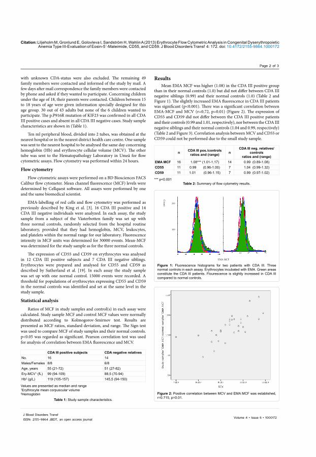
Citation: Liljeholm M, Gronlund E, Golovleva I, Sandström H, Wahlin A (2013) Erythrocyte Flow Cytometric Analysis in Congenital Dyserythropoietic Anemia Type III-Evaluation of Eosin-5´-Maleimide, CD55, and CD59. J Blood Disorders Transf 4: 172. doi: 10.4172/2155-9864.1000172
Page 2 of 3
Volume 4 • Issue 6 • 1000172J Blood Disorders TransfISSN: 2155-9864 JBDT, an open access journal
ResultsMean EMA MCF was higher (1.08) in the CDA III positive group
than in their normal controls (1.0) but did not differ between CDA III negative siblings (0.99) and their normal controls (1.0) (Table 2 and Figure 1). The slightly increased EMA fluorescence in CDA III patients was significant (p<0.001). There was a significant correlation between EMA-MCF and MCV (r=0.72, p=0.01) (Figure 2). The expression of CD55 and CD59 did not differ between the CDA III positive patients and their controls (0.99 and 1.01, respectively), nor between the CDA III negative siblings and their normal controls (1.04 and 0.99, respectively) (Table 2 and Figure 3). Correlation analysis between MCV and CD55 or CD59 could not be performed due to the small study sample.
with unknown CDA-status were also excluded. The remaining 49 family members were contacted and informed of the study by mail. A few days after mail correspondence the family members were contacted by phone and asked if they wanted to participate. Concerning children under the age of 18, their parents were contacted. Children between 15 to 18 years of age were given information specially designed for this age group. 30 out of 43 adults but none of the 6 children wanted to participate. The p.P916R mutation of KIF23 was confirmed in all CDA III positive cases and absent in all CDA III negative cases. Study sample characteristics are shown in (Table 1).
Ten ml peripheral blood, divided into 2 tubes, was obtained at the nearest hospital or in the nearest district health care centre. One sample was sent to the nearest hospital to be analysed the same day concerning hemoglobin (Hb) and erythrocyte cellular volume (MCV). The other tube was sent to the Hematopathology Laboratory in Umeå for flow cytometric assays. Flow cytometry was performed within 24 hours.
Flow cytometry
Flow cytometric assays were performed on a BD Biosciences FACS Calibur flow cytometer. Mean channel fluorescence (MCF) levels were determined by Cellquest software. All assays were performed by one and the same biomedical scientist.
EMA-labelling of red cells and flow cytometry was performed as previously described by King et al. [3]. 16 CDA III positive and 14 CDA III negative individuals were analyzed. In each assay, the study sample from a subject of the Västerbotten family was set up with three normal controls, randomly selected from the hospital routine laboratory, provided that they had hemoglobin, MCV, leukocytes, and platelets within the normal range for our laboratory. Fluorescence intensity in MCF units was determined for 30000 events. Mean-MCF was determined for the study sample as for the three normal controls.
The expression of CD55 and CD59 on erythrocytes was analysed in 12 CDA III positive subjects and 7 CDA III negative siblings. Erythrocytes were prepared and analysed for CD55 and CD59 as described by Sutherland et al. [19]. In each assay the study sample was set up with one normal control. 15000 events were recorded. A threshold for populations of erythrocytes expressing CD55 and CD59 in the normal controls was identified and set at the same level in the study sample.
Statistical analysis
Ratios of MCF in study samples and control(s) in each assay were calculated. Study sample MCF and control MCF values were normally distributed according to Kolmogorov-Smirnov test. Results are presented as MCF ratios, standard deviation, and range. The Sign test was used to compare MCF of study samples and their normal controls. p<0.05 was regarded as significant. Pearson correlation test was used for analysis of correlation between EMA fluorescence and MCV.
200 100
EMA MCF
Count
Figure 1: Fluorescence histograms for two patients with CDA III. Three normal controls in each assay. Erythrocytes incubated with EMA. Green areas constitute the CDA III patients. Flourescence is slightly increased in CDA III compared to normal controls.
Figure 2: Positive correlation between MCV and EMA MCF was established, r=0.715, p=0.01.
Values are presented as median and range1Erythrocyte mean corpuscular volume2Hemoglobin
Table 1: Study sample characteristics.
CDA III positive subjects CDA negative relativesNo. 16 14Males/Females 8/8 6/8Age, years 55 (21-72) 51 (27-82)Ery-MCV1 (fL) 99 (94-109) 88,5 (70-94) Hb2 (g/L) 119 (105-157) 145,5 (94-150)
*** p<0.001Table 2: Summary of flow cytometry results.
n CDA III pos./controlsratios and (range) n
CDA III neg. relatives/controls
ratios and (range)EMA MCF 16 1.08*** (1.01-1.17) 14 0.99 (0.89-1.08)CD55 11 0.99 (0.96-1.00) 7 1.04 (0.99-1.32)CD59 11 1.01 (0.96-1.15) 7 0.99 (0.97-1.02)
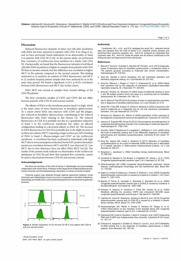
Citation: Liljeholm M, Gronlund E, Golovleva I, Sandström H, Wahlin A (2013) Erythrocyte Flow Cytometric Analysis in Congenital Dyserythropoietic Anemia Type III-Evaluation of Eosin-5´-Maleimide, CD55, and CD59. J Blood Disorders Transf 4: 172. doi: 10.4172/2155-9864.1000172
Page 3 of 3
Volume 4 • Issue 6 • 1000172J Blood Disorders TransfISSN: 2155-9864 JBDT, an open access journal
DiscussionReduced fluorescence intensity of intact red cells after incubation
with EMA has been reported in patients with CDA II by King et al., and we have previously found indications of an abnormality in band 3 in patients with CDA III [3,18]. In the present study, we performed flow cytometry of erythrocytes from members of a family with CDA III. Unexpectedly, we found that the fluorescence intensity of red blood cells after EMA incubation is higher in samples from patients with CDA III than in normal controls, but this difference was correlated to higher MCV in the patients compared to the normal controls. This finding motivated us to perform an analysis of EMA fluorescence and MCV in 22 random hospital patient samples that were analysed by us at the same time period. We found a significant (r=0.5, p=0.02) correlation between EMA fluorescence and MCV also in this cohort.
EMA MCF was normal in samples from normal siblings of the CDA III patients.
The flow cytometric profiles of CD55 and CD59 did not differ between patients with CDA III and normal controls.
The affinity of EMA to the membrane protein band 3 is high, which is the main cause of lower fluorescence in hereditary spherocytosis. To a minor extent EMA also interacts with CD47 and Rh-antigen, also reduced in hereditary spherocytosis, contributing to the reduced fluorescence after EMA staining in this disease [4]. The reduced fluorescence in CDA II is probably not due to a quantitative reduction of band 3 in the erythrocyte membrane but rather an affected configuration leading to a decreased affinity to EMA [6]. The increase in EMA fluorescence in CDA III is probably due to the slight increase of erythrocyte volume (MCV) exposing a larger surface per cell to binding of EMA to band 3, Rhesus-antigen and CD47 on the erythrocyte membrane. A correlation has been confirmed in individuals with high MCV due to overconsumption of alcohol, but in DAT-positive hemolytic anemia no correlation between MCV and MCF was observed [3]. Low MCV due to iron deficiency does not affect EMA MCF [3,6,20]. The results of the present study indicate no abnormality of the erythrocyte membrane in CDA III and show that standard flow cytometry cannot be used to discriminate between CDA III and normal controls.
Acknowledgements
We thank all members of the CDA III family in Västerbotten and acknowledge collaboration with Göran Roos, Professor at the Department of Medical Biosciences, Umeå University and Hematopathology Laboratory at Umeå University hospital.
Financial support was obtained through regional agreement between Umeå University and Västerbotten County Council on cooperation in the field of Medicine, Odontology and Health (ALF), and unrestricted grants from Cancerforskningsfonden Norrland.
Authorship
Contribution: M.L., A.W., and E.G. designed the study; M.L. collected clinical data and samples from the CDA III family; E.G. collected control samples and performed flow cytometry analyses. M.L., and A.W. analyzed an interpreted flow cytometry data. M.L. and A.W. wrote the paper and all authors contributed to the writing process and approved the final manuscript.
References
1. Bianchi P, Fermo E, Vercellati C, Marcello AP, Porretti L, et al. (2012) Diagnostic power of laboratory tests for hereditary spherocytosis: a comparison study in 150 patients grouped according to molecular and clinical characteristics. Haematologica 97: 516-523.
2. King MJ, Zanella A (2013) Hereditary red cell membrane disorders and laboratory diagnostic testing. Int J Lab Hematol 35: 237-243.
3. King MJ, Behrens J, Rogers C, Flynn C, Greenwood D, et al. (2000) Rapid flow cytometric test for the diagnosis of membrane cytoskeleton-associated haemolytic anaemia. Br J Haematol 111: 924-933.
4. King MJ, Smythe JS, Mushens R (2004) Eosin-5-maleimide binding to band 3 and Rh-related proteins forms the basis of a screening test for hereditary spherocytosis. Br J Haematol 124: 106-113.
5. Kar R, Mishra P, Pati HP (2010) Evaluation of eosin-5-maleimide flow cytometric test in diagnosis of hereditary spherocytosis. Int J Lab Hematol 32: 8-16.
6. Kedar PS, Colah RB, Kulkarni S, Ghosh K, Mohanty D (2003) Experience with eosin-5’-maleimide as a diagnostic tool for red cell membrane cytoskeleton disorders. Clin Lab Haematol 25: 373-376.
7. Richards SJ, Rawstron AC, Hillmen P (2000) Application of flow cytometry to the diagnosis of paroxysmal nocturnal hemoglobinuria. Cytometry 42: 223-233.
8. Iolascon A, Esposito MR, Russo R (2012) Clinical aspects and pathogenesis of congenital dyserythropoietic anemias: from morphology to molecular approach. Haematologica 97: 1786-1794.
9. King MJ, Telfer P, MacKinnon H, Langabeer L, McMahon C, et al. (2008) Using the eosin-5-maleimide binding test in the differential diagnosis of hereditary spherocytosis and hereditary pyropoikilocytosis. Cytometry B Clin Cytom 74: 244-250.
10. King MJ, Jepson MA, Guest A, Mushens R (2011) Detection of hereditary pyropoikilocytosis by the eosin-5-maleimide (EMA)-binding test is attributable to a marked reduction in EMA-reactive transmembrane proteins. Int J Lab Hematol 33: 205-211.
11. Bergstrom I, Jacobsson L (1962) Hereditary benign erythroreticulosis. Blood 19: 296-303.
12. Goudsmit R, Beckers D, De Bruijne JI, Engelfriet CP, James J, et al. (1972) Congenital dyserythropoietic anaemia, type 3. Br J Haematol 23: 97-105.
13. Wickramasinghe SN (1998) Congenital dyserythropoietic anaemias: clinical features, haematological morphology and new biochemical data. Blood Rev 12: 178-200.
14. Dgany O, Avidan N, Delaunay J, Krasnov T, Shalmon L, et al. (2002) Congenital dyserythropoietic anemia type I is caused by mutations in codanin-1. Am J Hum Genet 71: 1467-1474.
15. Bianchi P, Fermo E, Vercellati C, Boschetti C, Barcellini W, et al. (2009) Congenital dyserythropoietic anemia type II (CDAII) is caused by mutations in the SEC23B gene. Hum Mutat 30: 1292-1298.
16. Schwarz K, Iolascon A, Verissimo F, Trede NS, Horsley W, et al. (2009) Mutations affecting the secretory COPII coat component SEC23B cause congenital dyserythropoietic anemia type II. Nat Genet 41: 936-940.
17. Liljeholm M, Irvine AF, Vikberg AL, Norberg A, Month S, et al. (2013) Congenital dyserythropoietic anemia type III (CDA III) is caused by a mutation in kinesin family member, KIF23. Blood 121: 4791-4799.
18. Wickramasinghe SN, Wahlin A, Anstee D, Parsons SF, Stopps G, et al. (1993) Observations on two members of the Swedish family with congenital dyserythropoietic anaemia, type III. Eur J Haematol 50: 213-221.
19. Sutherland DR, Kuek N, Davidson J, Barth D, Chang H, et al. (2007) Diagnosing PNH with FLAER and multiparameter flow cytometry. Cytometry B Clin Cytom 72: 167-177.
20. D’Alcamo E, Agrigento V, Sclafani S, Vitrano A, Cuccia L, et al. (2011) Reliability of EMA binding test in the diagnosis of hereditary spherocytosis in Italian patients. Acta Haematol 125: 136-140.
CDA III
Normal control
Count
Figure 3: Similar expression of CD 55 and CD 59 in one patient with CDA III and one normal control.


III

Annex Publishers | www.annexpublishers.com
Research Article Open Access
Volume 1 | Issue 2
Volume 1 | Issue 2
Journal of Hematology and Blood Disorders
Introduction
Congenital Dyserythropoietic Anemia Type III and Primary Hemochromatosis; Coexistence of Mutations in KIF23 and HFELiljeholm M*1, Vikberg AL2, Golovleva I2, Sandström H3 and Wahlin A1
1Department of Radiation Sciences, Umeå University2Department of Medical Biosciences, Umeå University3Department of Public Health and Clinical Medicine, Umeå University*Corresponding author: Liljeholm M, Hematology, University Hospital, SE-90185 Umeå, Sweden, E-mail: [email protected]: Liljeholm M, Vikberg AL, Golovleva I, Sandström H, Wahlin A (2016) Congenital Dyserythropoietic Anemia Type III and Primary Hemochromatosis; Coexistence of Mutations in KIF23 and HFE. J Hematol Blood Disord 1(2): 203
Background: Congenital dyserythropoietic anemia type III (CDA III) can be caused by mutation in KIF23. CDA III differs from CDA I and II in the sense that secondary hemochromatosis has not been reported. However, we have observed elevated serum ferritin in a CDA III family. Since primary hemochromatosis is common in Northern Europe we decided to screen the family for HFE mutations.
Abstract
Keywords: Congenital dyserythropoietic anemia; Hereditary hemochromatosis; Iron overload; HFE gene; KIF23 gene
Congenital dyserythropoietic anemias (CDA) is a group of rare hereditary hemolytic disorders. Three major subtypes (I, II, III) and some minor entities (IV-VII) have been described [1]. The CDAs present with laboratory signs of increased hemoglobin (Hb) turnover, such as low or absent haptoglobin, elevated lactate dehydrogenase (LDH) and bilirubinemia. The bone marrow is hypercellular with a predominance of large bi- to multi-nucleated erythroblasts. Erythroblast morphology differs between CDAs, but the diagnosis of CDA I, II and III is best set by genetic testing [2,3].
Aim: Study clinical appearance and prevalence of HFE gene mutations, C282Y and H63D, in a CDA III family.
Methods: DNA from 37 CDA III patients and 21 non-affected siblings was genotyped. Iron status from EDTA plasma was measured in 32 of the CDA III patients and 18 of the non-affected siblings.Results: Out of 37 CDA III patients, 18 carried heterozygous HFE mutations and six were compound heterozygotes. Out of 21 CDA III negative siblings, nine had heterozygous HFE mutations, two were homozygous (one H63D and one C282Y), and two were compound heterozygous. None of the patients with wt HFE, regardless of CDA III status, suffered from iron overload. Four patients with HFE mutations needed treatment with phlebotomy to normalize ferritin and transferrin iron saturation; one CDA III negative patient with homozygous C282Y, two CDA III patients with heterozygous HFE mutations and one CDA III case with compound heterozygosity.Conclusion: HFE mutations were found in 65 % of CDA III patients and in 62 % of their CDA III negative siblings. Heterozygous HFE mutation, C282Y and even H63D, can cause iron overload when occurring concomitantly with CDA III.
CDA I and II are the most common forms, both with autosomal recessive inheritance [4-6]. Symptoms and signs of CDA I and II are mainly mild to moderate anemia, splenomegaly, and iron overload. The risk of secondary hemochromatosis increases when transfusions are needed [1]. Overexpression of Growth Differentiation Factor 15 (GDF15) in CDA I and II has, as in thalassemia, been shown to limit hepcidin expression thereby increasing iron absorption [7,8].
CDA III is the rarest form of the major CDA subtypes with only about 60 reported cases worldwide. A familial form with dominant inheritance has been described in two families, one American family with four cases and one family in Västerbotten County, Sweden, with the majority of reported cases [9,10]. One family in Argentina with eight cases has been described, although the pattern of inheritance in this family has not been confirmed [10]. A few cases of CDA III with recessive inheritance have been described in the literature [11,12]. One of these patients developed iron overload after erythrocyte transfusions [12].
In the Västerbotten CDA III family, the disease is caused by a mutation in c.2747C>G, p.P916R in KIF23. The mutation results in a dysfunctional KIF23 protein, also known as Mitotic Kinesin Like Protein 1 (MKLP1), leading to failure of cytokinesis, the final stage of mitosis. As a result, the bone marrow in CDA III is characterized by gigantoblasts, large erythroblasts with up to 12 nuclei in the same cell [13].
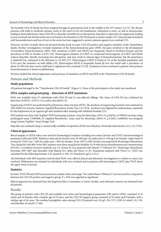
Annex Publishers | www.annexpublishers.com
2
Volume 1 | Issue 2
Journal of Hematology and Blood Disorders
The Swedish CDA III family has been mapped through six generations back to the middle of the 19th century [14,15]. The disease presents with mild to moderate anemia, rarely in the need of red cell transfusions. Hemolysis is extra- as well as intravascular, leading to hemosiderinuria. Since CDA III is a disorder of ineffective erythropoiesis, hepcidin is expected to be suppressed, leading to iron overload, but ferritin levels were not found to be elevated in CDA III patients in earlier studies [15]. Intravascular hemolysis with hemosiderinuria and loss of iron in the urine has been suggested to protect patients against iron overload [15].
However, we have recently observed elevated ferritin levels in some CDA III positive and negative members of the Västerbotten family. Further investigations revealed mutations of the hemochromatosis gene (HFE), the gene involved in the development of hereditary hemochromatosis (HH). HFE mutations (C282Y and H63D) are frequently found in Northern Europe and the prevalence of HH in Sweden is 0.5% [16]. Homozygous mutation of C282Y or compound heterozygosity of C282Y and H63D reduces hepcidin, thereby increasing iron absorption, and may result in clinical signs of hemochromatosis, but the penetrance is relatively low, estimated in the literature to 10-30% [17]. Heterozygous C282Y is found in 7% of the Swedish population and 0.5% carry the mutation on both alleles [16]. Heterozygous H63D is frequently found all over the world with a prevalence of about 10-20% but does not by itself lead to significant iron overload [17,18]. Treatment of iron overload in patients with primary hemochromatosis is based on phlebotomy.
We have studied the clinical appearance and presence of mutations in KIF23 and HFE in the Västerbotten CDA III family.
Statistics Ferritin, TSAT, Hb and LDH are presented as median values and range. Two-sided Mann-Whitney U-test was used for comparison between the CDA III positive and negative group. P < 0.05 was regarded as significant.
The group of patients with CDA III and available iron status and hematological parameters (Hb and/or LDH), consisted of 16 males and 16 females with a median age of 53 years, and the CDA III negative group consisted of 10 males and 8 females, with a median age of 44 years. The median hemoglobin value among CDA III patients was 116 g/L (94-157), LDH 6.2 ukat/L (4.2-10), and bilirubin 18 umol/L (7-86).
Results
Study populationPatients and Methods
All patients belonged to the “Västerbotten CDA III family” (Figure 1). None of the participants in the study was transfused.
DNA samples and genotyping – detection of HFE mutationsDNA was available from 37 individuals with CDA III and 21 non-affected siblings. The status of CDA III was confirmed by detection of KIF23 c.2747C>G as earlier described [13].
Sequencing of KIF23 was performed by polymerase chain reaction (PCR). The products of sequencing reactions were analysed on ABI 3500xL Dx Genetic Analyzer (Applied Biosystems, Foster City, CA). PCR - products were digested by endonuclease, separated by electrophoresis on an Agarose gel and visualized after staining with ethidium bromide [13].
HFE analysis was done with TaqMan® SNP Genotyping Analysis. Assay for detecting c.187C>G, p.H63D, rs1799945 was done using predesigned assay C1085600_10 (Applied Biosystems). Assay used for detecting c.845G>A, p.C282Y, rs1800562 was designed using Custom TaqMan® Assay Design Tool.
Hepcidin was analysed using a commercially available Competetive ELISA test (Bachem, Peninsula Laboratories, LLC, CA, USA).
Clinical appearanceBlood samples in EDTA-tubes were used for hematological analyses including iron status (ferritin and TSAT) and hematological parameters (Hb and LDH). Reference intervals for ferritin were 30-400 ug/L for males and 13-150 ug/L for females. Upper limit of normal for TSAT was < 60% for males and < 50% for females. From 1997 to 2007 ferritin was analysed by Boehringer Mannheims Tina-Quant kit 1661400. From 2007 analyses were done using Roche Modular E170 with electrochemiluminescence immunoassay (ECLIA). Correlation between methods was 1.0. Serum-Fe was analysed with Hitachi 717/Hitachi 911, Boehringer Mannheims Ferrozine 1997-2007 and thereafter with Hitachi 911, delta, till Vitros 5.1 FS. Transferrin analysed with Vitros 5,1. TSAT was calculated by the following formula: S-Fe [µmol/L] x 100 / (S-Transferrin [g/L] x 25,1).
All individuals with HFE mutation and elevated TSAT were offered clinical and laboratory investigation to confirm or reject iron overload. Phlebotomy was initiated in individuals with iron overload and in patients with homozygous C282Y plus TSAT above the upper limit of normal.
Ethical approval was obtained from the Regional Ethics Committee in Umeå, Sweden, and informed consent was obtained from all patients.
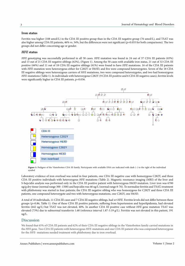
Annex Publishers | www.annexpublishers.com
3 Journal of Hematology and Blood Disorders
Volume 1 | Issue 2
Iron statusFerritin was higher (108 umol/L) in the CDA III positive group than in the CDA III negative group (76 umol/L), and TSAT was also higher among CDA III patients, 46% vs. 34%, but the differences were not significant (p=0.055 for both comparisons). The two groups did not differ concerning age or gender.
Laboratory evidence of iron overload was noted in four patients, one CDA III negative case with homozygous C282Y, and three CDA III positive individuals with heterozygous HFE mutations (Table 2). Magnetic resonance imaging (MRI) of the liver and S-hepcidin analysis was performed only in the CDA III positive patient with heterozygous H63D mutation. Liver iron was 6700 ug/g dry tissue (normal range 300 -1500) and hepcidin was 46 ug/L (normal range 8-76). To normalize ferritin and TSAT, treatment with phlebotomy was started in four patients; the CDA III negative sibling who was homozygous for C282Y and three CDA III patients, one compound heterozygote and two with heterozygous mutations, one C282Y, one H63D.
Figure 1: Pedigree of the Västerbotten CDA III family. Participants with available DNA are indicated with dash (–) to the right of the individual symbol
HFE status
HFE genotyping was successfully performed in all 58 cases. HFE mutation was found in 24 out of 37 CDA III patients (65%) and 13 out of 21 CDA III negative siblings (62%), (Figure 1). Among the 50 cases with available iron status, 21 out of 32 CDA III positive (66%) and 11 out of 18 CDA III negative siblings (61%) were found to have HFE mutations. 16 of the CDA III patients with HFE-mutation were heterozygous either for C282Y or H63D, and five were compound heterozygotes. Seven of the 18 CDA III negative siblings were heterozygous carriers of HFE mutations, two were compound heterozygotes, and two had homozygous HFE-mutations (Table 1). In individuals with heterozygous C282Y (9 CDA III positive and 6 CDA III negative cases), ferritin levels were significantly higher in CDA III patients, p=0.036.
A total of 18 individuals, 11 CDA III cases and 7 CDA III negative siblings, had wt HFE. Ferritin levels did not differ between these groups (p=0.86, Table 1). One of these CDA III positive patients, suffering from hypertension and hyperlipidemia, had elevated ferritin (642 ug/L) but TSAT was not elevated, 40%. In another CDA III positive case without HFE gene mutation TSAT was elevated (73%) due to subnormal transferrin 1.68 (reference interval 1.87-3.19 g/L). Ferritin was not elevated in this patient, 191 ug/L.
DiscussionWe found that 65% of CDA III patients and 62% of their CDA III negative siblings in the Västerbotten family carried mutations in the HFE gene. Two CDA III patients with heterozygous HFE-mutations and one CDA III patient who was compound heterozygous for the HFE- mutations needed treatment with phlebotomy due to iron overload.

Annex Publishers | www.annexpublishers.com
4
Volume 1 | Issue 2
Journal of Hematology and Blood Disorders
One of the heterozygous cases carried H63D, which is not reported to lead to iron overload by itself. MRI of the liver confirmed iron overload in this patient. Hepcidin was in the normal range, but we believe that the level was inappropriate considering the iron status, indicating disruption of the feedback system. We hypothesise that increased erythropoiesis produces larger amounts of erythroferrone suppressing hepcidin that may be further suppressed by HFE mutation.
LDH ukat/LCDA III + CDA III –
Hb g/LCDA III + CDA III –
TSAT %CDA III + CDA III –
Ferritin ug/LCDA III + CDA III –HFE status
2.8(1.9-4.5)
n=7
6.1(4.2-8.0)
n=11
135(118-153)
n=7
119 (105-135)
n=11
32 ***(27-44)
n=7
45 (26-73) **
n=11
90 (28-392)
n=7
116 (28-642)*
n=11HFE wt
2.8(1.9-3.1)
n=6
6.0***(5.2-10.0)
n=9
145(114-154)
n=6
123 (107-157)
n=9
33(21-46)
n=6
45 (37-100)
n=9
73 (13-154)
n=6
89 (71-987)
n=9C282Y +/-
2.1
n=1
153
n=1
79
n=1
229
n=1C282Y +/+
2.4(2.0-2.7)
n=2
6.4***(5.9-8.3)
n=5
154(145-162)
n=2
116(114-124)
n=5
49 (42-56)
n=2
55 (28-80)
n=5
75 (56-94)
n=2
66 (48-700)
n=5
C282Y +/-H63D +/-
2.4
6.7***(5.8-7.7)
n=7
94
105(80-135)
n=7
4
n=1
39 (24-80)
n=7
3
n=1
139(41-1084)
n=7
H63D +/-
2.7
n=1
138
n=1
49
n=1
20
n=1H63D +/+
+/- heterozygous mutation, +/+ homozygous mutation.* elevated ferritin in one patient with hyperlipidemia and normal TSAT.** elevated TSAT in one patient with subnormal Tf and normal ferritin. *** sample missing in one case.Table 1: Iron status, Hb and LDH in individuals of the Västerbotten CDA III family, with and without HFE mutation. Medians and (range)
LDH (ukat/L)Hb (g/L)TSAT (%)Ferritin
(ug/L)HFE statusGender and age (years)
CDA status
2.115379229C282Y +/+F 49wt
5.2143100987C282Y +/-M 60CDA III +
6.111155700C282Y +/-H63D +-F 61CDA III +
6.3135801084H63D +/-M 60CDA III +
Table 2: Individuals in whom phlebotomy has been initiated due to iron overload
None of the healthy siblings carrying HFE-mutations with heterozygous or compound heterozygosity nor CDA III positive cases with wt HFE had laboratory signs of iron overload. One CDA III positive individual with wt HFE had elevated ferritin and one had elevated TSAT. After further investigation none of these patients fulfilled the criteria of iron overload. Ferritin levels were higher among CDA III positive vs CDA III negative individuals with heterozygous C282Y, though study population is limited. Earlier studies have shown that homozygous mutation of HFE C282Y or, eventually, compound heterozygosity of C282Y and H63D is needed to develop clinical signs of hemochromatosis in healthy individuals, though penetrance is relatively low [17]. In this study we found that heterozygous mutation of the HFE- gene, either C282Y or H63D can be sufficient to cause iron overload in patients with CDA III. All patients were screened for diabetes since this is a well-known metabolic factor causing elevated ferritin, although TSAT is not elevated in diabetes [19]. One male CDA III patient, heterozygous for H63D, was diagnosed with type II diabetes mellitus. Except for elevated ferritin (1084 ug/L), TSAT was also elevated (80%) in this patient, indicating iron overload.
Earlier studies on HFE-mutations in patients with hematologic disorders where iron overload can be a problem, such as thalassemia and myelodysplastic syndromes (MDS), are contradictory. Some studies show iron overload in thalassemia patients with co-existing HFE-mutations, even in heterozygous forms [20-22]. Recently, non-transfused MDS-patients, carriers of HFE-mutations, were found to have higher ferritin level and inferior overall survival compared to non-mutated MDS-patients [23]. However, other studies have not verified that heterozygous mutation affects iron overload in thalassemia [24,25].
So far, phlebotomies have been performed without problems in spite of the continuous hemolysis in our CDA III patients and phlebotomy alone is sufficient to control ferritin and TSAT-levels.
5 Journal of Hematology and Blood Disorders
Annex Publishers | www.annexpublishers.com Volume 1 | Issue 2
This study indicates that heterozygous HFE mutation, C282Y or H63D, is sufficient to cause pathologic iron overload when occurring concomitantly with CDA III. Therefore, in order to prevent organ damage in these patients, screening for primary hemochromatosis should be performed in all patients with congenital dyserythropoietic anemia type III.
Conclusion
Financial support was obtained through regional agreement between Umeå University and Västerbotten County Council on cooperation in the field of Medicine, Odontology and Health (ALF), and unrestricted grants from Cancerforskningsfonden Norrland. None of the authors has any conflict of interest to declare.
Conflict of Interest and Sources of Funding
References1. Iolascon A, Esposito MR, Russo R (2012) Clinical aspects and pathogenesis of congenital dyserythropoietic anemias: from morphology to molecular approach. Haematologica 97: 1786-94.
5. Tamary H, Shalev H, Luria D, Shaft D, Zoldan M, et al. (1996) Clinical features and studies of erythropoiesis in Israeli Bedouins with congenital dyserythropoietic anemia type I. Blood 87: 1763-70.
4. Heimpel H (2004) Congenital dyserythropoietic anemias: epidemiology, clinical significance, and progress in understanding their pathogenesis. Ann Hematol 83: 613-21.
3. Iolascon A, Heimpel H, Wahlin A, Tamary H (2013) Congenital dyserythropoietic anemias: molecular insights and diagnostic approach. Blood 122: 2162-6.
2. Wickramasinghe SN, Wood WG (2005) Advances in the understanding of the congenital dyserythropoietic anaemias. Br J Haematol 131: 431-46.
6. Heimpel H, Matuschek A, Ahmed M, Bader-Meunier B, Colia A, et al. (2010) Frequency of congenital dyserythropoietic anemias in Europe. Eur J Haematol 85: 20-5.7. Casanovas G, Swinkels DW, Altamura S, Schwarz K, Laarakkers CM, et al. (2011) Growth differentiation factor 15 in patients with congenital dyserythropoietic anaemia (CDA) type II. J Mol Med 89: 811-6.8. Tamary H, Shalev H, Perez-Avraham G, Zoldan M, Levi I, et al. (2008) Elevated growth differentiation factor 15 expression in patients with congenital dyseryth-ropoietic anemia type I. Blood 112: 5241-4.9. Wolff JA, Von Hofe FH (1951) Familial erythroid multinuclearity. Blood 6: 1274-83.
10. Sandstrom H, Wahlin A (2000) Congenital dyserythropoietic anemia type III. Haematologica 85: 753-7.
11. Goudsmit R, Beckers D, De Bruijne JI, Engelfriet CP, James J, et al. (1972) Congenital Dyserythropoietc anemia type 3. Br J Haematol 23: 97-105.
12. Sigler E, Shaft D, Shtalrid M, Shvidel L, Berrebi A, et al (2002) New sporadic case of congenital dyserythropoietic anemia type III in an aged woman: detailed description of ultrastructural findings. Am J Hematol 70: 72-6.13. Liljeholm M, Irvine AF, Vikberg AL, Norberg A, Month S, et al. (2013) Congenital dyserythropoietic anemia type III (CDA III) is caused by a mutation in kinesin family member, KIF23. Blood 121: 4791-9.14. Bergstrom I, Jacobsson L (1962) Hereditary benign erythroreticulosis. Blood 19: 296-303.
15. Sandstrom H, Wahlin A, Eriksson M, Bergstrom I, Wickramasinghe SN (1994) Intravascular haemolysis and increased prevalence of myeloma and monoclonal gammopathy in congenital dyserythropoietic anaemia, type III. Eur J Haematol 52: 42-6.
16. Stal P, Hultcrantz R (2012) Hereditary hemochromatosis -a common genetic disease. Lakartidningen 109: 2097-9.
17. Ekanayake D, Roddick C, Powell LW (2015) Recent advances in hemochromatosis: a 2015 update : A summary of proceedings of the 2014 conference held under the auspices of Hemochromatosis Australia. Hepatol Int 9: 174-82.18. Hanson EH, Imperatore G, Burke W (2001) HFE gene and hereditary hemochromatosis: a HuGE review. Human Genome Epidemiology. Am J Epidemiol Aug154: 193-206.19. Yeap BB, Divitini ML, Gunton JE, Olynyk JK, Beilby JP, et al. (2015) Higher ferritin levels, but not serum iron or transferrin saturation, are associated with Type 2 diabetes mellitus in adult men and women free of genetic haemochromatosis. Clin Endocrinol (Oxf) 82: 525-32.20. Martins R, Picanco I, Fonseca A, Ferreira L, Rodrigues O, et al. (2004) The role of HFE mutations on iron metabolism in beta-thalassemia carriers. J Hum Genet 49: 651-5.21. Ruiz-Arguelles GJ, Garces-Eisele J, Reyes-Nunez V, Sanchez-Anzaldo J, Ruiz-Delgado GJ, et al. (2001) Heterozygosity for the H63D mutation in the hereditary hemochromatosis (HFE) gene may lead into severe iron overload in beta-thalassemia minor: observations in a thalassemic kindred. Rev Invest Clin 53: 117-20.22. Sharma V, Panigrahi I, Dutta P, Tyagi S, Choudhry VP, et al. (2007) HFE mutation H63D predicts risk of iron over load in thalassemia intermedia irrespective of blood transfusions. Indian J Pathol Microbiol 50: 82-5.23. Lucijanić M, Pejša V, Mitrović Z, Štoos-Veić T, Livun A, et al. (2015) Hemochromatosis gene mutations may affect the survival of patients with myelodysplastic syndrome. Hematology. 24. Lopez-Escribano H, Ferragut JF, Parera MM, Guix P, Castro JA, et al. (2012) Effect of co-inheritance of beta-thalassemia and hemochromatosis mutations on iron overload. Hemoglobin 36: 85-92.25. Jazayeri M, Bakayev V, Adibi P, Haghighi Rad F, Zakeri H, et al. (2003) Frequency of HFE gene mutations in Iranian beta-thalassaemia minor patients. Eur J Haematol 71: 408-11.