Embed Size (px)
Citation preview
Ludwig-Maximilians Universität MünchenFakultät für Biologie
Dissertation
Cloning and characterisation of a Dictyostelium STE20-like protein kinase DST2
Hyunju Son
2002

Cloning and characterisation of a Dictyostelium STE20-like protein kinase DST2
Dissertation der Fakultät für Biologie derLudwig-Maximilians-Universität
München
vorgelegt von
Hyun Ju Sonaus
Süd-Korea
23. September 2002

Ehrenwörtliche Versicherung
Diese Dissertation wurde selbständig und ohne unerlaubte Hilfsmittel angefertigt.
München, September 2002
Hyun-Ju Son
Dissertation eingereicht : September 2002
Erstgutachter : Prof. Dr. C. David
Zweitgutachter: Prof. Dr. H. M. Williams
Sondergutachter: Prof. Dr. M. Schleicher
Parts of this work have already been published or are in the process of being published.
Hyunju Son, Ludwig Eichinger and Michael Schleicher
DST2, a Dictyostelium STE20-like kinase, is phosphorylated by PKA and phosphorylates the
actin binding protein severin (in process).
Hyunju Son, Ludwig Eichinger and Michael Schleicher
DST2, a Dictyostelium STE20-like kinase, is phosphorylated by PKA and phosphorylates the
actin binding protein severin. The American Society for Cell Biology (ASCB) 41st Annual
meeting abstract, December 2001.
Hyunju Son, Ludwig Eichinger and Michael Schleicher
Cloning and characterization of a Dictyostelium STE20-like protein kinase AV09.
European Life Scientist Organisation (ELSO) 1st Annual meeting abstract, September 2000.
The experimental parts of the work presented here were carried out in the laboratory of Prof.Dr. M. Schleicher from May 1999 to December 2001 at the Adolf-Butenandt-Institute of cellbiology, Ludwig-Maximilians-universitaet Muenchen.

Table of Contents 1
ABBREVIATIONS 5
SUMMARY 8
ZUSAMMENFASSUNG 9
1 INTRODUCTION 10
1.1 The cytoskeleton 101.2 Actin and actin binding proteins 101.3 F-actin fragmenting proteins 111.4 Actin-binding proteins as substrates for protein kinases 131.5 STE20-like kinases 141.6 Dictyostelium discoideum 151.7 Goals of the project 17
2 MATERIALS AND METHODS 18
2.1 Materials 182.1.1 Enzymes for molecular biology 182.1.2 Antibodies 182.1.3 Protein inhibitors 192.1.4 Antibiotics 192.1.5 Chemical reagents 192.1.6 Media 202.1.6.1 Media for D. discoideum for culture 202.1.6.2 Media for E. coli culture 212.1.7 Buffers and other solutions 212.1.8 Equipment 222.1.9 Other materials 23
1
2.1.10 Centrifuges and rotors 232.1.11 Computer programmes 242.2 Vectors and Strains 242.2.1 Vectors 242.2.2 Bacterial strains 242.2.3 Cultivation of E. coli 242.2.4 D. discoideum strains 252.2.5 Cultivation of D. discoideum 252.2.5.1 Growth in liquid medium 252.2.5.2 Growth on agar plates 252.2.5.3 Preservation of spores 252.2.5.4 Freezing of Dictyostelium cells 262.2.5.5 Development of D. discoideum 262.3 DNA Methods 262.3.1 Agarose gel electrophoresis 262.3.2 DNA extraction from agarose gels 272.3.3 Determination of DNA concentration 272.3.4 Preparation of plasmid DNA 272.3.4.1 Isolation of plasmid DNA by the method of Homes and Quigley 272.3.4.2 Isolation of plasmid DNA by the method of Qiagen 282.3.4.3 Phenol extraction and precipitation of DNA 282.3.5 DNA cleavage with restriction enzymes 292.3.6 Ligation of DNA into a plasmid vector 292.3.7 Preparation of electroporation competent cells 292.3.8 Electroporation of E. coli 302.3.9 Screening for positive E. coli transformants 302.3.10 E.coli permanent cultures 312.3.11 Transformation of D. discoideum 312.3.12 Polymerase chain reaction (PCR) 322.3.13 Purification of PCR products 322.3.14 Oligonucleotides 332.3.15 Southern blotting 33
2
2.3.16 Northern blotting 342.4 Analysis of DST2 biochemical methods 342.4.1 SDS-Polyacrylamide gel elctrophoresis (SDS-PAGE) 342.4.2 Coomassie blue staining of proteins 362.4.3 Drying of SDS-PAGE gels 362.4.4 Western blotting 362.4.5 Bradford assay 382.4.6 Preparation of actin from rabbit skeletal muscle 382.4.7 Protein purification 392.4.7.1 Purification of Histidine-tagged construct 392.4.7.2 Purification of maltose-binding-protein (MBP) tagged constructs 402.4.8 Low shear viscometry 412.4.9 Severing activity of severin measured by fluorescence spectroscopy 412.4.10 Inhibition of actin depolymerisation analyzed by fluorescence spectrometry 422.4.11 In vitro kinase assay 42
3 RESULTS43
3.1 Molecular characterisation of DST2 433.1.1 Sequence analysis 433.1.2 Sequence comparison with STE20-like kinases 453.1.3 Southern analysis of DST2 493.1.4 Northern analysis of DST2 503.2 Biochemical and cell biological characterisation of DST2 523.2.1 Expression and purification of recombinant DST2 523.2.2 Western analysis of DST2 543.2.3 In vitro kinase assay with recombinant DST2 constructs 553.2.3.1 The influence of Mn2+ and Mg2+ on DST2 kinase activity 583.2.3.2 PKA is a potential upstream kinase of DST2 603.2.4 Expression, purification and biochemical characterisation of recombinant
C-terminally truncated DST2 constructs
61
3.2.5 In vivo kinase assay with C-terminally truncated constructs of DST2 62
3
3.2.6 DST2 is present as a high molecular weight complex in AX2 homogenate 633.2.7 Phosphorylation by DST2 inhibits the severing activity of severin 64
4 DISCUSSION66
4.1 DST2 is a new member of the STE20-like protein kinase family 664.2 DST2 is autophosphorylated and activated in a Mn2+ dependent manner 684.3 Regulation of DST2 activity 684.4 DST2 exists as a high molecular weight complex 704.5 Binding partners of PAK family kinases 704.6 Signalling to the cytoskeleton 714.7 Future prospects 73
5 References 74
CURRICULUM VITAE89
ACKNOWLEDGEMENT 90
4
ABBREVIATIONS
ATP Adenosine 5’-triphosphate
bp Base pair(s)
BSA Bovine serum albumin
C- Carboxy-
cDNA Complementary DNA
CIP Calf intestinal phosphatase
conc Concentrated
D Dalton
DNA Desoxyribonuleic acid
dNTP Desoxiribonucletide triphosphate
DNase Deoxyribonuclease
DMSO Dimethyl sulfoxide
DTT Dithiothreitol
E. coli Escherichia coli
EDTA Ethylene-diamine-tetraacetic acid
EGTA Ethylene glycol-bis-(2-aminoethylether)-N,N’-tetraacetic acid
et al And others
Fig. Figure
F-actin Filamentous (polymerized) actin
FITC Fluorescein isothiocyanate
G-actin Globular actin (monomer)
5
GFP Green fluorescent protein
h Hour(s)
HEPES N-2-hydroxyethylpiperazine-N’-2-ethanesulfonic acid
His-tag Histidine-tag
Hs Homo sapiens
IgG Immunoglobulin G
ITPG Isopropyl-ß-thiogalactopyranoside
kb kilo base(s)
kDa Kilodalton
l Litre(s)
LMW Low molecular weight
µ Micro
M Mol/l
mAb Monoclonal antibody
min Minute(s)
ml Mililitres
µm Micrometer
mM Milimolar
MOPS Morpholinopropanesulfonic acid
nm Nanometer
N- Amino-
NADH Nicotine adenine dinucleotide
OD Optical density
6
PAGE Polyacrylamide gel electrophoresis
PBS Phosphate buffered saline
PCR Polymerase chain reaction
PIP2 Phosphatidylinositol-4,5-diphosphate
PMSF Phenylmethylsulfonyl fluoride
pH Negative decadic logarithm of proton concentration
RNA Ribonucleic acid
RNase Ribonuclease
rpm Revolutions per minute
RT Room temperature
s, sec Seconds(s)
SDS Sodium dodecyl sulfate
TBE Tris/borate/EDTA
TBS Tris buffered saline
TEMED N,N,N’,N’-tetramethylenediamine
Tris Tris-hydroxymethyl-ammonuimethane
Tween 20 Polyoxyethylene-sorbitanmonolaurate
U Units
V Volt
v/v Volume per volume
w/v Weight per volume
7
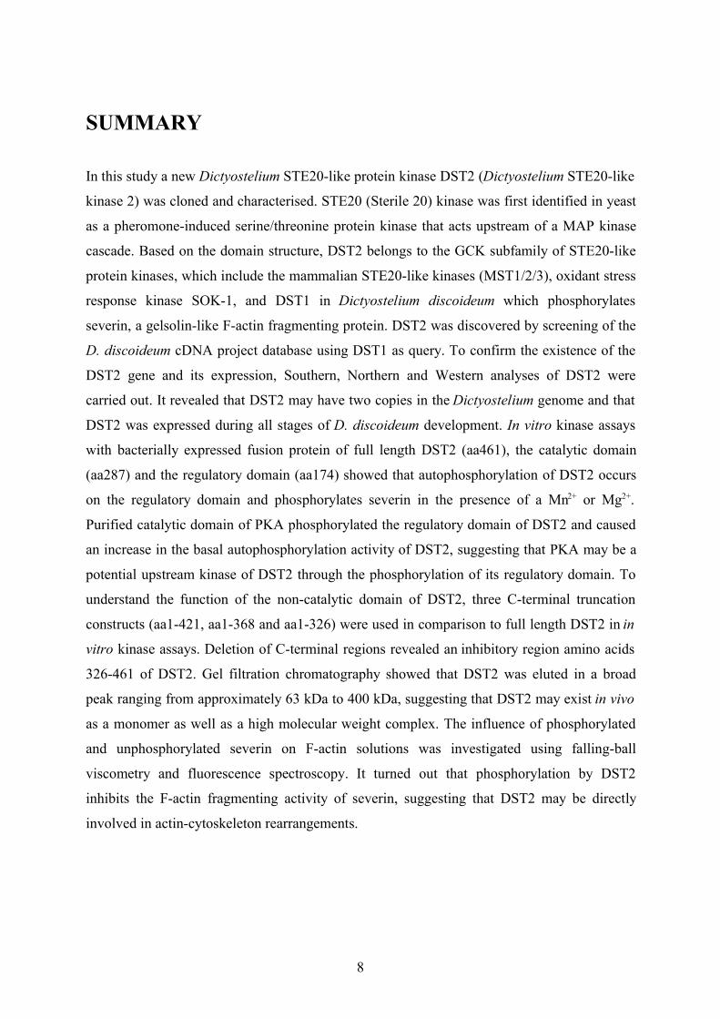
SUMMARY
In this study a new Dictyostelium STE20-like protein kinase DST2 (Dictyostelium STE20-like
kinase 2) was cloned and characterised. STE20 (Sterile 20) kinase was first identified in yeast
as a pheromone-induced serine/threonine protein kinase that acts upstream of a MAP kinase
cascade. Based on the domain structure, DST2 belongs to the GCK subfamily of STE20-like
protein kinases, which include the mammalian STE20-like kinases (MST1/2/3), oxidant stress
response kinase SOK-1, and DST1 in Dictyostelium discoideum which phosphorylates
severin, a gelsolin-like F-actin fragmenting protein. DST2 was discovered by screening of the
D. discoideum cDNA project database using DST1 as query. To confirm the existence of the
DST2 gene and its expression, Southern, Northern and Western analyses of DST2 were
carried out. It revealed that DST2 may have two copies in the Dictyostelium genome and that
DST2 was expressed during all stages of D. discoideum development. In vitro kinase assays
with bacterially expressed fusion protein of full length DST2 (aa461), the catalytic domain
(aa287) and the regulatory domain (aa174) showed that autophosphorylation of DST2 occurs
on the regulatory domain and phosphorylates severin in the presence of a Mn2+ or Mg2+.
Purified catalytic domain of PKA phosphorylated the regulatory domain of DST2 and caused
an increase in the basal autophosphorylation activity of DST2, suggesting that PKA may be a
potential upstream kinase of DST2 through the phosphorylation of its regulatory domain. To
understand the function of the non-catalytic domain of DST2, three C-terminal truncation
constructs (aa1-421, aa1-368 and aa1-326) were used in comparison to full length DST2 in in
vitro kinase assays. Deletion of C-terminal regions revealed an inhibitory region amino acids
326-461 of DST2. Gel filtration chromatography showed that DST2 was eluted in a broad
peak ranging from approximately 63 kDa to 400 kDa, suggesting that DST2 may exist in vivo
as a monomer as well as a high molecular weight complex. The influence of phosphorylated
and unphosphorylated severin on F-actin solutions was investigated using falling-ball
viscometry and fluorescence spectroscopy. It turned out that phosphorylation by DST2
inhibits the F-actin fragmenting activity of severin, suggesting that DST2 may be directly
involved in actin-cytoskeleton rearrangements.
8
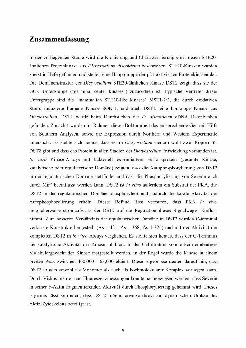
Zusammenfassung
In der vorliegenden Studie wird die Klonierung und Charakterisierung einer neuen STE20-
ähnlichen Proteinkinase aus Dictyostelium discoideum beschrieben. STE20-Kinasen wurden
zuerst in Hefe gefunden und stellen eine Hauptgruppe der p21-aktivierten Proteinkinasen dar.
Die Domänenstruktur der Dictyostelium STE20-ähnlichen Kinase DST2 zeigt, dass sie der
GCK Untergruppe ("germinal center kinases") zuzuordnen ist. Typische Vertreter dieser
Untergruppe sind die "mammalian STE20-like kinases" MST1/2/3, die durch oxidativen
Stress induzierte humane Kinase SOK-1, und auch DST1, eine homologe Kinase aus
Dictyostelium. DST2 wurde beim Durchsuchen der D. discoideum cDNA Datenbanken
gefunden. Zunächst wurden im Rahmen dieser Doktorarbeit das entsprechende Gen mit Hilfe
von Southern Analysen, sowie die Expression durch Northern und Western Experimente
untersucht. Es stellte sich heraus, dass es im Dictyostelium Genom wohl zwei Kopien für
DST2 gibt und dass das Protein in allen Stadien der Dictyostelium Entwicklung vorhanden ist.
In vitro Kinase-Assays mit bakteriell exprimiertem Fusionsprotein (gesamte Kinase,
katalytische oder regulatorische Domäne) zeigten, dass die Autophosphorylierung von DST2
in der regulatorischen Domäne stattfindet und dass die Phosphorylierung von Severin auch
durch Mn2+ beeinflusst werden kann. DST2 ist in vitro außerdem ein Substrat der PKA, die
DST2 in der regulatorischen Domäne phosphoryliert und dadurch die basale Aktivität der
Autophosphorylierung erhöht. Dieser Befund lässt vermuten, dass PKA in vivo
möglicherweise stromaufwärts der DST2 auf die Regulation dieses Signalweges Einfluss
nimmt. Zum besseren Verständnis der regulatorischen Domäne in DST2 wurden C-terminal
verkürzte Konstrukte hergestellt (As 1-421, As 1-368, As 1-326) und mit der Aktivität der
kompletten DST2 in in vitro Assays verglichen. Es stellte sich heraus, dass der C-Terminus
die katalytische Aktivität der Kinase inhibiert. In der Gelfiltration konnte kein eindeutiges
Molekulargewicht der Kinase festgestellt werden, in der Regel wurde die Kinase in einem
breiten Peak zwischen 400,000 - 63,000 eluiert. Diese Ergebnisse deuten darauf hin, dass
DST2 in vivo sowohl als Monomer als auch als hochmolekularer Komplex vorliegen kann.
Durch Viskosimetrie- und Fluoreszenzmessungen konnte nachgewiesen werden, dass Severin
in seiner F-Aktin fragmentierenden Aktivität durch Phosphorylierung gehemmt wird. Dieses
Ergebnis lässt vermuten, dass DST2 möglicherweise direkt am dynamischen Umbau des
Aktin-Zytoskeletts beteiligt ist.
9
CHAPTER 1
Introduction
1.1 The cytoskeleton
The cytoskeleton is a complex network of protein filaments that extend throughout the
cytoplasm. It is responsible for cell shape, cell motility, cell polarity, cytokinesis, intracellular
transport, cytoplasmic streaming and muscle contraction. The diverse activities of the
cytoskeleton depend on three types of protein filaments: actin filaments (about 6-8 nm in
diameter), microtubules (about 25 nm in diameter) and intermediate filaments (about 10 nm in
diameter). It is characteristic of these filamentous systems that they are built by reversible
assembly of monomeric, evolutionarily highly conserved subunits, namely - and -tubulin in
microtubules, the different proteins in intermediate filaments and actin in microfilaments
(Sandoz et al., 1988).
Intermediate filaments stabilize the cell against mechanical stress and structure the cytoplasm
by establishing links to various binding partners (Housewart and Cleveland, 1998). However,
no direct involvement of intermediate filaments in cellular motility has been reported to date.
In contrast, actin filaments and microtubules interact with different proteins to generate
different types of cellular motility. Microtubules are major organizers of the cell interior and
are vitally involved in motility events such as chromosome migration during cell division
(Valiron et al., 2001).
1. 2 Actin and actin binding proteins
The microfilament protein actin is not only the most abundant protein in many eukaryotic
cells, but is also very highly conserved in evolution from human to amoeba. The 42 kDa
globular actin monomer (G-actin) polymerizes into polar, helical filaments (F-actin). Actin
filaments have a polar structure, with two structurally different ends - a fast growing (barbed
or +) end and a slow growing (pointed or -) end (Wegner, 1976).
10
In vivo, actin polymerisation is a highly regulated process controlled both by ATP binding and
hydrolysis, and by the action of a number of actin binding proteins that control the
incorporation of actin monomers into existing filaments, the dynamic equilibrium between G-
and F-actin and the three-dimensional organization of the filamentous network (Schleicher et
al, 1995). For many actin-binding proteins it was shown that their activity is regulated in vitro
by Ca2+, phospholipids, phosphorylation or changes in pH. Based on their interaction with G-
or F-actin, they are placed into different functional groups. One distinguishes between
proteins that bind actin monomers, fragment and/or cap actin filaments, act as molecular
motors, tether actin filaments to the membrane, or crosslink actin filaments (Eichinger et al.,
1999).
The actin-based motility is driven by the assembly of actin filaments. Important regulatory
proteins in the assembly of new actin filament networks are the Arp2/3 complex and the
Wiscott-Aldrich Syndrome Protein (WASP). The Arp2/3 complex is a complex of seven
proteins, including the actin-related proteins Arp2 and Arp3. It localizes to the leading edge of
a variety of cells, binds to the sides of actin filaments and rapidly nucleates branches (Svitkina
and Borisy, 1999; Bear et al., 2002). Members of the WASP and the related Scar (Supressor
of cAMP receptor) family bind directly to the Arp2/3 complex and stimulate its ability to
promote the nucleation of new actin filaments (Machesky and Insall, 1998; Seastone et al.,
2001). F-actin fragmenting and/or capping proteins, that have also been identified as
important regulators of cell motility, are described in the next section.
1.3 F-actin fragmenting proteins
Filament number and length are in part controlled by F-actin fragmenting proteins. At
micromolar Ca2+ concentrations they sever actin filaments, which is usually followed by
capping of the newly created barbed end. This leads to a rapid increase of short capped
filaments together with a dramatic decrease in viscosity. For several members of this family it
has been shown that uncapping is caused by polyphosphoinositides, particularly
phosphatidylinositol 4,5-biphosphate (PIP2) (Janmey et al., 1987; Eichinger and Schleicher,
1992). In vivo this could lead to free barbed ends ready for rapid elongation (Stossel, 1989).
11
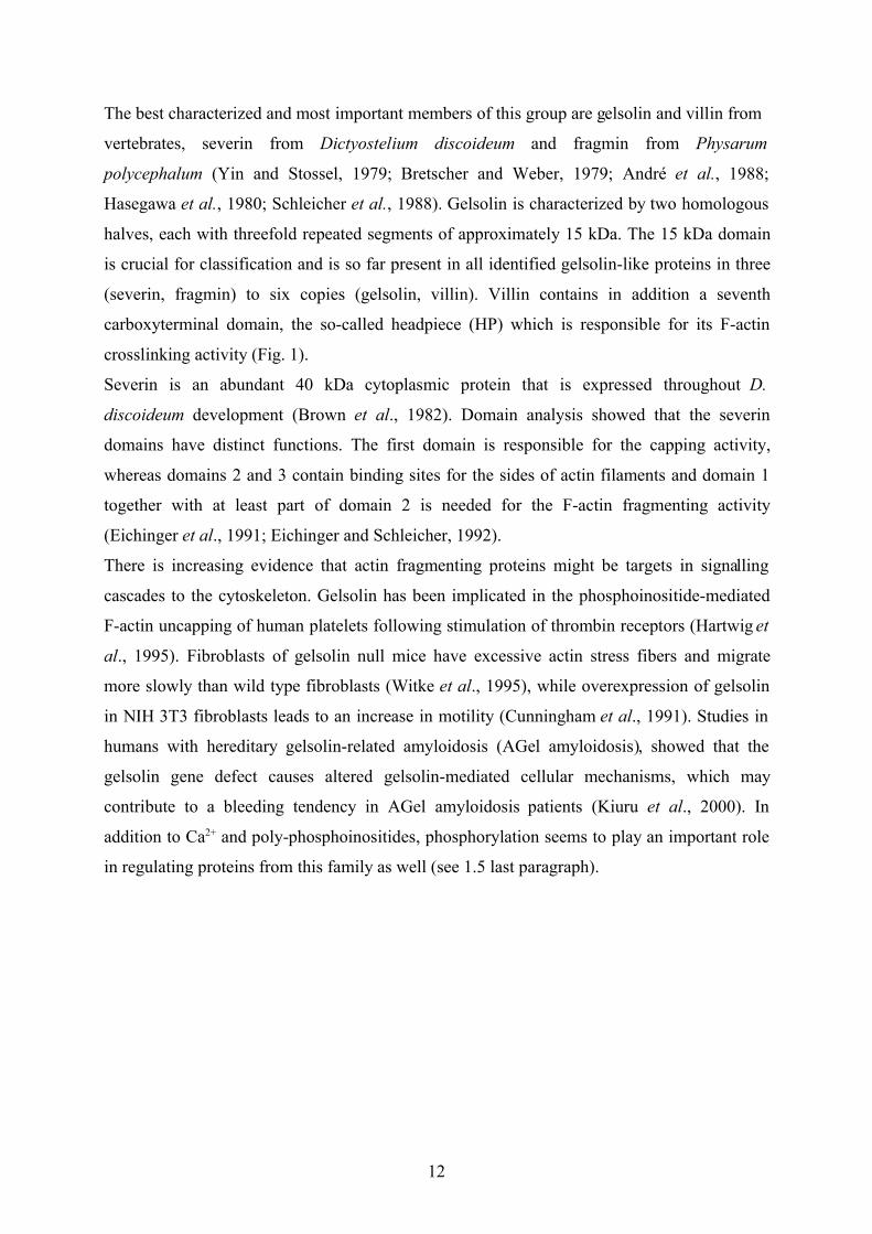
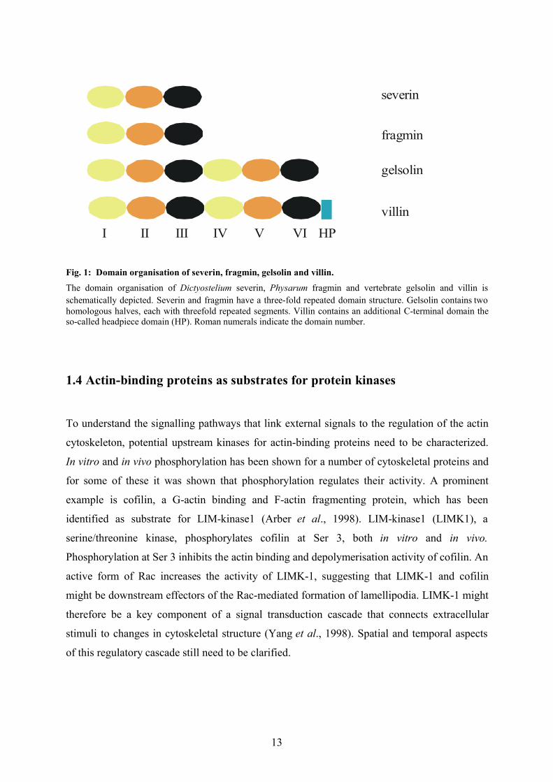
The best characterized and most important members of this group are gelsolin and villin from
vertebrates, severin from Dictyostelium discoideum and fragmin from Physarum
polycephalum (Yin and Stossel, 1979; Bretscher and Weber, 1979; André et al., 1988;
Hasegawa et al., 1980; Schleicher et al., 1988). Gelsolin is characterized by two homologous
halves, each with threefold repeated segments of approximately 15 kDa. The 15 kDa domain
is crucial for classification and is so far present in all identified gelsolin-like proteins in three
(severin, fragmin) to six copies (gelsolin, villin). Villin contains in addition a seventh
carboxyterminal domain, the so-called headpiece (HP) which is responsible for its F-actin
crosslinking activity (Fig. 1).
Severin is an abundant 40 kDa cytoplasmic protein that is expressed throughout D.
discoideum development (Brown et al., 1982). Domain analysis showed that the severin
domains have distinct functions. The first domain is responsible for the capping activity,
whereas domains 2 and 3 contain binding sites for the sides of actin filaments and domain 1
together with at least part of domain 2 is needed for the F-actin fragmenting activity
(Eichinger et al., 1991; Eichinger and Schleicher, 1992).
There is increasing evidence that actin fragmenting proteins might be targets in signalling
cascades to the cytoskeleton. Gelsolin has been implicated in the phosphoinositide-mediated
F-actin uncapping of human platelets following stimulation of thrombin receptors (Hartwig et
al., 1995). Fibroblasts of gelsolin null mice have excessive actin stress fibers and migrate
more slowly than wild type fibroblasts (Witke et al., 1995), while overexpression of gelsolin
in NIH 3T3 fibroblasts leads to an increase in motility (Cunningham et al., 1991). Studies in
humans with hereditary gelsolin-related amyloidosis (AGel amyloidosis), showed that the
gelsolin gene defect causes altered gelsolin-mediated cellular mechanisms, which may
contribute to a bleeding tendency in AGel amyloidosis patients (Kiuru et al., 2000). In
addition to Ca2+ and poly-phosphoinositides, phosphorylation seems to play an important role
in regulating proteins from this family as well (see 1.5 last paragraph).
12
severin
fragmin
gelsolin
villinI II III IV V VI HP
Fig. 1: Domain organisation of severin, fragmin, gelsolin and villin.The domain organisation of Dictyostelium severin, Physarum fragmin and vertebrate gelsolin and villin isschematically depicted. Severin and fragmin have a three-fold repeated domain structure. Gelsolin contains twohomologous halves, each with threefold repeated segments. Villin contains an additional C-terminal domain theso-called headpiece domain (HP). Roman numerals indicate the domain number.
1.4 Actin-binding proteins as substrates for protein kinases
To understand the signalling pathways that link external signals to the regulation of the actin
cytoskeleton, potential upstream kinases for actin-binding proteins need to be characterized.
In vitro and in vivo phosphorylation has been shown for a number of cytoskeletal proteins and
for some of these it was shown that phosphorylation regulates their activity. A prominent
example is cofilin, a G-actin binding and F-actin fragmenting protein, which has been
identified as substrate for LIM-kinase1 (Arber et al., 1998). LIM-kinase1 (LIMK1), a
serine/threonine kinase, phosphorylates cofilin at Ser 3, both in vitro and in vivo.
Phosphorylation at Ser 3 inhibits the actin binding and depolymerisation activity of cofilin. An
active form of Rac increases the activity of LIMK-1, suggesting that LIMK-1 and cofilin
might be downstream effectors of the Rac-mediated formation of lamellipodia. LIMK-1 might
therefore be a key component of a signal transduction cascade that connects extracellular
stimuli to changes in cytoskeletal structure (Yang et al., 1998). Spatial and temporal aspects
of this regulatory cascade still need to be clarified.
13
1.5 STE20-like kinases
The MAPK (Mitogen Activated Protein Kinase) system is an evolutionarily highly conserved
intracellular signalling cascade and plays an essential role in many cellular processes, such as
growth, differentiation and stress-related response. Its core comprises a module of three
kinases consisting of a MAPKKK (MAPK kinase kinase), MAPKK (MAPK kinase) and
MAPK. STE20 (Sterile 20) is a pheromone-induced yeast serine/threonine protein kinase that
acts upstream of a MAP kinase cascade (Wu et al., 1995; Leberer et al., 1992). In response to
activated Cdc42, it activates the MAPK cascade that includes STE11 (MAPKKK), STE7
(MAPKK) and FUS3/KSS1 (MAPK) (Herskowitz et al., 1995). STE20-related protein
kinases have been identified in various eukaryotes, and have a highly conserved catalytic
domain in common with yeast STE20. Based on their structure and regulation, they can be
divided into two subfamilies.
The first group, including yeast STE20 and its mammalian homologues, the PAKs (p21
activated kinases), have a kinase domain at the carboxy terminus and a putative regulatory
domain at the amino terminus, which contains a binding site for Rac1 and Cdc42 (Manser et
al., 1994). PAKs are regulated in vivo and in vitro by the small GTP binding proteins Rac1
and Cdc42 and by phospholipids. PAKs specially regulate the JNK pathway and are involved
in regulating some of the diverse cytoskeletal changes induced by Rac and Cdc42 (Bagrodia
et al., 1995; Kyriakis et al., 1996; Benner et al., 1995; Yu et al., 1998; Bokoch et al., 1998).
They have been shown to be required for processes including neurite formation and axonal
guidance, development of cell polarity and motile responses (Daniels and Bokoch, 1999).
The second group, the so-called GCK (Germinal Center Kinase) subfamily, has a catalytic
domain at the N-terminus and a putative regulatory domain at the C-terminus and lacks a
recognizable GTPase binding site (Sells and Chernoff, 1997). The GCKs subfamily can be
further divided into two groups based on their structure and properties. Group I GCKs are
closely related to GCK. This group consists of GCK, GCKR (GCK-related), GLK (GCK-
like), HPK1 (Hematopoietic Progenitor Kinase-1) and NIK (Nck-Interacting Kinase) (Katz et
al., 1994; Kiefer et al., 1996; Hu et al., 1996; Su et al., 1997; Diener et al., 1997; Shi et al.,
1997; Fu et al., 1999). Mammalian SOK-1 (STE20-like oxidant stress-activated kinase1),
MST1, 2 and 3 (mammalian STE20-like kinase 1, 2 and 3) and LOK (Lymphocyte-Oriented
Kinase), Dictyostelium DST1 and yeast Sps1 belong to the group II GCKs (Pombo et al.,
1996; Creasy and Chernoff, 1995a; Creasy and Chernoff, 1995b; Schinkmann and Blenis,
1997; Friesen et al., 1994; Eichinger et al., 1998).
14
These enzymes are less well understood. Although group II GCKs share homologous
sequence, with the catalytic domain of group I GCKs, their C-terminal regulatory domains
differ significantly from those of the group I kinases (Eichinger et al., 1998; Friesen et al.,
1994; Schweitzer and Philippsen, 1991).
Several members of the GCK subfamily are responsive to cellular stress. Sps1p has been
shown to become activated in response to nutrient deprivation (Friesen et al., 1994). Human
Krs-1 and Krs-2, which are identical with MST1 and MST2, are activated upon treatment of
cells with staurosporine, okadaic acid, high concentrations of sodium arsenite, and extreme
heat shock at 55°C (Taylor et al., 1996; Creasy and Chernoff, 1995a; Creasy and Chernoff,
1995b). Furthermore the activity human SOK-1 was shown to be induced several fold by
oxidant stress. It most likely controls a novel stress response pathway since it is not involved
in already defined MAPK cascades (Pombo et al., 1996). This lead to the assumption that
members from the GCK subfamily are important for the response of eukaryotic cells to
environmental stresses. Recent results suggest that some members of the GCK subfamily
might also be involved in the regulation of the remodelling of the actin cytoskeleton via F-
actin fragmenting proteins. Severin was identified as a substrate for DST1 in vitro.
Phosphorylation of severin was strongly reduced in the presence of Ca2+ (Eichinger et
al.,1998). Though there is no direct proof, several pieces of evidence suggest that severin is
also an in vivo substrate for DST1 (unpublished results). Furthermore, TNIK was shown to
phosphorylate gelsolin. Overexpression of wild type TNIK in NIH3T3 and Hela cells lead to
morphological changes of the cells and resulted in the disruption of F-actin structures and
inhibition of cell spreading (Fu et al., 1999).
1.6 Dictyostelium discoideum
Cellular slime molds were first discovered by O. Brefeld in 1869, but the modern era began
with the discovery of the new species Dictyostelium discoideum (Raper, 1935). D. discoideum
has two alternative life cycles: sexual and asexual. The asexual cycle is easier to produce in
the laboratory and is the one used for almost all experimental studies. D. discoideum feeds on
bacteria as separate amoebae and upon starvation enters a social, multicellular stage which is
mediated by chemotaxis. Chemotactically aggregated cells form a multicellular structure
which undergoes morphogenesis and cell-type differentiation into spore and stalk cells.
15
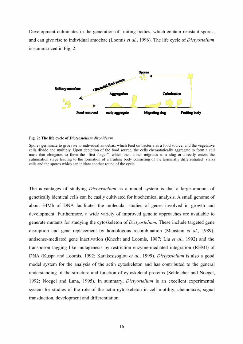
Development culminates in the generation of fruiting bodies, which contain resistant spores,
and can give rise to individual amoebae (Loomis et al., 1996). The life cycle of Dictyostelium
is summarized in Fig. 2.
Fig. 2: The life cycle of Dictyostelium discoideum
Spores germinate to give rise to individual amoebas, which feed on bacteria as a food source, and the vegetativecells divide and multiply. Upon depletion of the food source, the cells chemotatically aggregate to form a cellmass that elongates to form the “first finger”, which then either migrates as a slug or directly enters theculmination stage leading to the formation of a fruiting body consisting of the terminally differentiated stalkscells and the spores which can initiate another round of the cycle.
The advantages of studying Dictyostelium as a model system is that a large amount of
genetically identical cells can be easily cultivated for biochemical analysis. A small genome of
about 34Mb of DNA facilitates the molecular studies of genes involved in growth and
development. Furthermore, a wide variety of improved genetic approaches are available to
generate mutants for studying the cytoskeleton of Dictyostelium. These include targeted gene
disruption and gene replacement by homologous recombination (Manstein et al., 1989),
antisense-mediated gene inactivation (Knecht and Loomis, 1987; Liu et al., 1992) and the
transposon tagging like mutagenesis by restriction enzyme-mediated integration (REMI) of
DNA (Kuspa and Loomis, 1992; Karakesisoglou et al., 1999). Dictyostelium is also a good
model system for the analysis of the actin cytoskeleton and has contributed to the general
understanding of the structure and function of cytoskeletal proteins (Schleicher and Noegel,
1992; Noegel and Luna, 1995). In summary, Dictyostelium is an excellent experimental
system for studies of the role of the actin cytoskeleton in cell motility, chemotaxis, signal
transduction, development and differentiation.
16
1.7 Goals of the projectThe goal of this project was to discover and characterise new protein kinases homologous to
DST1 in D. discoideum. Previous work showed that DST1 phosphorylates the F-actin
fragmenting protein severin (Eichinger et al.,1998). These results suggested that DST1 might
play a regulating role in the rearrangement of the actin cytoskeleton. DST2 (Dictyostelium
STE20-like kinase 2) was discovered by screening the sequences of the D. discoideum data
bases. DST2 is a serine/threonine protein kinase and highly homologous to DST1 (69%
similarity in the kinase domain).
The further aims of this work were to biochemically characterise DST2, to understand its
function in terms of a possible regulatory role in the remodelling of the actin cytoskeleton, and
to investigate in which signalling pathways DST2 might be involved.
To address these questions, the following approaches were taken :
1) DST2 was generated as active recombinant protein kinase and used for biochemical
characterisation and to test various potential substrates.
2) A polyclonal antiserum was raised against recombinant DST2 and used in cell
biological and biochemical studies in D. discoideum.
3) Various C-terminal truncated DST2 constructs were generated and characterised in
biochemical assays to unravel functionally important regions in the C-terminal
domain.
4) Recombinant DST2 was used to study the impact of phosphorylation on severin
activity in viscometry- and fluorescence spectroscopy assays.
17
CHAPTER 2
MATERIALS AND METHODS
2.1 MATERIALS
2.1.1 Enzymes for molecular biology
Calf intestine alkaline phosphatase RocheDNA polymerase 1 RocheLysozyme SigmaRestriction enzymes Amersham, Boehringer, Eurogentec,
Gibco-BRL, New England Biolabs,
PromegaRNase A SigmaT4 DNA ligase Gibco-BRL, PromegaTaq polymerase AmershamPfu Turbo polymerase Stratagene
2.1.2 Antibodies
Anti-Actin (mAb Act1) ( Simpson et al., 1984 )Goat anti-mouse IgG antibody,
coupled with peroxidase
Dianova
Goat anti-mouse IgG antibody,
Conjugated with Cy3 or FITC
Dianova
18
2.1.3 Protease inhibitors
Benzamidine SigmaPEFA-block RothPhenylmethylsulfonylfluoride (PMSF) ServaProtease inhibitor cocktail (p2714) Sigma
2.1.4 Antibiotics
Ampicillin RothBlasticidin S ICN BiomedicalsGeneticin (G418) ICN BiomedicalsHygromycin B CabiochemKanamycin SigmaNalidixic acid SigmaPenicillin/Streptomycin Sigma
2.1.5 Chemical reagents
Unless otherwise stated, chemicals were obtained from Fluka, Merck, Pharmacia, Roth, Serva
or Sigma and have the purity grade of “p.a”.
Agarose (SeaKem Me) FMC BioproductsBacto-agar, -peptone, -tryptone DifcoChloroform p.a Riedel de HaenDE52 (Diethylaminoethyl-cellulose) WhatmanHydroxylapatite Bio-RadIPTG (Isopropyl--D-thiogalactose-pyranosid) GerbuOligonucleotides MWG-biotechPeptone OxoidPhenol AppligenePhosphocellulose (P11) Whatman
19
Proteose peptone OxoidTriton X-100 PierceYeast extract Oxoid
2.1.6 Media
All media and buffers were made with deionized water and sterilized by autoclaving at 120oC
for 20 min. Antibiotics were added to media when cooled to about 50oC.
2.1.6.1 Media for D. discoideum culture
AX medium (pH 6.7) SM agar plates (pH 6.5)14.3 g peptone 9 g agar7.15 g yeast extract 10 g peptone50 mM glucose 50 mM glucose3.5 mM Na2HPO4 1 g yeast extract3.5 mM KH2PO4 4 mM MgSO4
16 mM KH2PO4
5.7 mM K2HPO4
-Both media were filled up to 1 litre with dH2O.
Soerensen Phosphate buffer (pH 6.0) Salt solution14.6 mM KH2PO4 10 mM NaCl2 mM Na2HPO4 10 mM KCl
2.7 mM CaCl2
HL-5 medium Phosphate agar plates (pH 6.0)10 g yeast extract 15 g Bacto agar50 mM glucose - filled up to 1 L with Soerensen buffer8.5 mM KH2PO4
1.25 mM Na2HPO4
-filled up to 2 L with dH2O
20

2.1.6.2 Medium for E. coli culture
LB-medium (pH 7.4)
10 g Bacto-tryptone
5 g yeast extract
86 mM NaCl
-pH adjusted with NaOH and medium filled up to 1 L with dH2O.
For LB-agar plates, 1.5% (W/V) agar added into the medium and selection was provided by
introducing 50 mg/l ampicillin and/or 25 mg/l kanamycin
2.1.7 Buffers and other solutions
Those not shown here will be described in their corresponding sections under Methods.
100x Denhardt’s reagent TE buffer (pH 8.0)2% Ficoll 400 10 mM Tris/HCl2% polyvinylpyrrolidon 1 mM EDTA2% Bovin serum albumin -autoclave
10 x NCP buffer (pH 8.0) 10 x TBE buffer (pH 8.3)100 mM Tris/HCl 890 mM Tris1.5 M NaCl 890 mM Boric acid5 ml Tween 20 20 mM EDTA0.02% NaN3 -autoclave-Filled up to 1 L with dH2O
PBS (pH 7.2) Tris-Phenol (pH 8.0)70 mM Na2HPO4 -1 vol. melted phenol was equilibrated150 mM NaCl with 1 vol. 1 M tris/HCl, pH 8.030 mM KH2PO4
2.7 mM KCl-autoclave
21
2.1.8 Equipment
Axiophot microscope ZeissCCD camera (C5985-10) HamamatsuConductivity meter (LF 537) WTWConfocal laser scanning microcope LeicaDiavert inverse microscope LeicaDigital color video CCD camera (TK-C1380) JVCDounce homogenizer BraunEagle Eye II StratageneElectroporation apparatus BioRadFluorescence spectrophotometer
(Aminco Bowman)
Sopra
FPLC device (BioLogic) BioRadNuclepore filter CostarParr bomb Parr Instrument CompanyPCR thermal cycler BiometraPH meter KnickProtein fraction collector PharmaciaRotary shaker GFLSemi-dry protein transfer Trans-Blot SD BioRadSMART system PharmaciaSpectrophotometer PharmaciaSpeed-Vac concentrator BachhoferStereomicroscope (MZ12) LeicaUltrafiltration centricon AmiconVortex Bender & HobeinWater baths GFLWeighing machines SartoriusX-ray film developing machine (Curix 60) AGFA
22
2.1.9 Other materials
2.2 ml sterile tubes for freezing of cells Nunc3 MM filter paper Whatman4-well borosilicate glass chamber slides Nunc24-well plates CostarDialysis membrane BiomolEppendorf tubes (0.1 ml, 0.5 ml, 1.5 ml) EppendorfFalcon centrifuge tubes (15ml, 30ml) FalconNitrocellulose membranes (BA85) Schleicher & Schuell Petri dishes GreinerPolaroid film (667) PolaroidPolyallomer ultracentrifuge tubes 1.5 ml BeckmanQuartz cuvettes HellmaSterile filters (0.22 m, 0.45 m) MilliporeTissue culture flasks NuncX-ray films (X-Omat) Kodak
2.1.10 Centrifuges and rotors
CentrifugesJ2-21 M/E BeckmanJ6-HC BeckmanG6-SKR BeckmanOptima LE-80K ultracentrifuge BeckmanOptima TL 100 ultracentrifuge BeckmanTable-top centrifuge (5415) Eppendorf
RotorsJA 14, JA 20, JS-4.2, Ti45, Ti 70, TLA 100.3 Beckman
23
2.1.11 Computer programmes
Windows NTBilddatenbank system LeicaSigma Plot 2.01 Jandel scientificWinword 7.0 MicrosoftMacintoshIllustrator 8.0 AdobeNIH Image 1.60 National Institutes of HealthPhotoshop 5.0 AdobeUNIXUWGCG package program (University
of Wisconsin Genetics Computer Group)
2.2 Vectors and Strains
2.2.1 Vectors
The following vectors were used: pMalC2-vector (New England Biolabs), PQE30 (Qiagen).
2.2.2 Bacterial strains
The following Escherichia coli strains were used: XL-1 Blue for cloning (Sambrook et al.,
1989), BL21 and M15 for protein expression.
2.2.3 Cultivation of E. coli
Bacteria were cultivated according to standard methods (Sambrook. et al., 1989) on agar
plates or in liquid culture (240rpm). The cultivation temperature was 37oC. For long-term
storage, 400 l of bacterial culture were mixed with an equal amount of glycerol and stored at
–80oC.
24
2.2.4 D. discoideum strains
The wild type D. discoideum strain AX2, which can be cultivated under axenic
conditions, was used for this study.
2.2.5 Cultivation of D. discoideum
2.2.5.1 Growth in liquid medium (Claviez et al., 1982)
From spores or bacterial lawns on SM-agar plates, the wild type AX strain was inoculated into
AX or HL-5 medium containing the antibiotics streptomycin sulfate (400 g/ml) in order to
K. aerogenes. The generation time at 21oC and 150 rpm is about 10 hours. For large-scale
generation of cells for protein purification, cells cultivated in 4 x 2.5 l cultures up to a density
between 5 x 10⁶ and 1.2 x 10⁷ cells/ml were harvested after centrifugation giving normally a
yield of about 100g wet weight. As for cell biological studies, cells were allowed to grow up
to a maximum density of 5 x 10⁶ cells/ml to avoid the stationary phase.
2.2.5.2 Growth on agar plates
Isolation of transformant clones was carried out on SM agar plates. 100 l of a suspension of
K. aerogenes in salt solution were placed on each agar plate, spread evenly together with the
Dictyostelium cells, and the plates were kept at 21oC for about 2 days. The doubling time is
around 3-4 h and the Dictyostelium colonies appeared as round clearings on the bacteria lawn.
2.2.5.3 Preservation of spores
Cells from the axenic culture were harvested by centrifugation at 300 g for 10 min, washed
once with cold Soerensen buffer and resuspended at a cell density of 2 x 10⁸ cells/ ml. 500 l
of the cell suspension was spread out per phosphate agar plate, and the cells were able to
develop into fruiting bodies within 2-3 days. The spores were then harvested by knocking onto
the lid of the petri dish and taken up in 10 ml of cold Soerensen buffer. 1 ml aliquots were
dispensed into Nunc tubes (2.2 ml), shock-frozen in liquid nitrogen and stored at -70oC.
25
For inoculation of spores, an aliquot was thawed at room temperature and cultured in AX
medium, whereby after 3 days at 21oC and 150 rpm, a cell density of about 5 x 10⁶ cells/ml
was reached.
2.2.5.4 Freezing of Dictyostelium cells
For the preservation of Dictyostelium cells, axenic cultures were harvested and resuspended at
a cell density of about 5 x 10⁷ cells/ml in ice-cold freezing medium (AX or HL-5 medium
+1% (v/v) penicillin–streptomycin solution +5% (v/v) DMSO), and distributed as 1 ml
aliquots into Nunc tubes (2.2 ml) pre-cooled on ice. The aliquots were then placed into the
wells of a pre-cooled (4oC) brass block (10 x 9 x 5 cm) and kept at -70oC for a period of at
least 15h and subsequently stored at -70oC. To recover the cells, a frozen vial was thawed
rapidly under cold running water, the cells washed once with cold AX medium, and then
cultivated in AX medium in petri dishes or 24-wells plates. For the transformant clones,
growth medium was changed after 24h and replaced with medium containing suitable
antibiotics for selection.
2.2.5.5 Development of D. discoideum
Wild-type Dictyostelium (AX2 cells) were cultivated on SM plates at 21°C for 24 hours. Cells
were harvested from different stages of development of Dictyostelium and used for Northern
and Western analysis.
2.3 DNA methods
2.3.1 Agarose gel electrophoresis
The separation of DNA fragments according to length was done in gels with 1% agarose in
TBE buffer. For the detection of DNA fragments, 0.05 g/ml ethidium bromide was added to
the liquid agarose. Gels were run with 50-200 V. Probes were incubated with 1/6 (v/v) sample
dye before loading. After separation, the fragments were detected with a UV illuminator and
documented with the Eagle Eye CCD camera system (Stratagene, Heidelberg).
26

DNA sample buffer40% Sucrose0.5% SDS0.25% Bromophenol blue-taken up in TE buffer
2.3.2 DNA extraction from agarose gels
DNA bands were cut out of the gel with a scalpel and transferred into a sterile micro tube. The
isolation procedure was performed with the DNA extraction kit (Qiagen) according to the
manufacturer’s manual.
2.3.3 Determination of DNA concentration
DNA concentration in agarose gels was estimated by comparing the intensity of the band with
the bands of the molecular weight marker. Alternatively, the OD260 was measured (50 g/ml
of DNA have an OD260 of 1, Sambrook et al., 1989).
2.3.4 Preparation of plasmid DNA
2.3.4.1 Isolation of plasmid DNA by the method of Homes and Quigley (1981)
An inoculum of bacteria was taken from a streak culture and suspended in 200l of
STET/lysozyme buffer. Alternatively, 1 ml of bacteria from a fresh overnight shaking culture
was harvested using a table-top centrifuge (5000 rpm, 1 min, RT), the supernatant discarded
and the pellet resuspended in STET buffer. The suspension was boiled in a water bath for 1
min, centrifuged (14,000 rpm, 10 min, RT), and the insoluble cell debris removed using a
sterile toothpick. The nucleic acids in the supernatant were precipitated with 200 l of
isopropanol for 5 min at RT and sedimented using a table-top centrifuge (14,000 rpm, 30 min,
RT). The DNA pellet was then washed with 70% ethanol, vacuum-dried with a Speed-vac
concentrator, and dissolved in 30 l of TE buffer.
27
STET buffer (pH 8.0)50 mM Tris/HCl50 mM EDTA5% Triton-X-1008% Sucrose1 mg/ml lysozyme (added prior to usage)
2.3.4.2 Isolation of plasmid DNA by the method of Qiagen
Large-scale preparation of plasmid DNA from E. coli was carried out with the Midi-kit
purchased from the Qiagen company. Bacteria were cultivated in 150 ml of LB medium
containing 50 g/ml ampicillin overnight at 37oC with agitation. The cells were harvested by
centrifugation (4,000 g, 5 min, 4o C) and resuspended in 10 ml of buffer P1. For cell lysis, 10
ml of buffer P2 was introduced and the resultant mixture was gently mixed by inverting the
tube several times. After incubation for 5 min at RT, 10 ml of chilled buffer P3 was added, the
suspension mixed gently by inverting tube, and then incubated on ice for 15 min. Following
centrifugation (35,000 g, 30 min, 4o C), the sediment of proteins and cell debris was discarded,
while the supernatant containing the plasmid DNA was centrifuged for a further period of 15
min. The clear supernatant was passed through a Qiagen tip 100 column previously
equilibrated with 5 ml of buffer QBT. After washing twice with 10 ml each of buffer QC, the
plasmid DNA was eluted with 5 ml of buffer QF, precipitated with 0.7 vol. of isopropanol,
spun down (30,000 g, 30 min, 4oC), and the DNA pellet washed with 70% ice-cold ethanol,
air-dried and dissolved in 200 l of TE buffer. The DNA concentration was determined using
a spectrophotometer whereby an OD260 of 1.0 corresponds to 50 g of DNA. The ratio of
OD260/OD280 should be between 1.8 and 2.0.
28

2.3.4.3 Phenol Extraction and Precipitation of DNA
To separate the DNA from contaminating proteins, the DNA solution was mixed with 50%
(v/v) phenol and 50% (v/v) chloroform/isoamylalcohol (24:1). The mixture was heavily
vortexed and centrifuged at 13,000 rpm at room temperature for 5 min. The upper phase was
collected and the DNA was precipitated with ethanol. For ethanol precipitation, 10% (v/v) of
a 3 M Na-acetate solution of pH 5.2 as well as 2.5 volumes of ethanol were added to the DNA
solution. The sample was then incubated at –80oC for 1 hour. After a centrifugation of 30 min
at 13,000 rpm and 4oC, the pellet was washed with 70% ethanol, centrifuged again for 15 min
under the previous conditions and dried in a Speed Vac. The dried pellet was resuspended in a
suitable volume of H2O.
2.3.5 DNA cleavage with restriction enzymes
The cleavage was performed in a volume of 50 l. The buffer suitable for the restriction
enzyme(s) was added and the volume adjusted with H2O. After addition of the enzyme(s), the
tube was incubated at the appropriate temperature for at least 1h. The digest was analysed on
an agarose gel.
2.3.6 Ligation of DNA into a plasmid vector
To avoid re-ligation and concatamer formation of a cut vector, the vector was
dephosphorylated with alkaline phosphatase (CIP, Calf Intestinal Phosphatase). The reaction
was performed according to manufacturer’s instructions (New England Biolabs). The vector
was purified on a gel and incubated with the corresponding insert. The reaction volume was
set to 10 l. 50 ng of vector were ligated with a threefold molar excess of insert. Ligases of
various suppliers were used (Gibco, NEB) together with the supplied buffers. After the
ligation, the DNA was extracted with phenol, precipitated with ethanol and resuspended in 5-
10 l of H20.
29

2.3.7 Preparation of electroporation competent cells
1 liter LB was inoculated with 10 ml of an E. coli overnight culture and grown to an OD600 of
0.6 at 37oC under vigorous shaking. All flasks and solutions subsequently used were sterilized
and cooled to 4oC. Proper cooling was essential for obtaining competent cells of a good
quality. Cells were spun down and resuspended in 1 l of H20. After another centrifugation, the
cells were resuspended in 500 ml of H2O, pelleted again, washed with 20 ml of 10% glycerol
and finally resuspended in 3 ml of 10% glycerol. The cells were stored at -80oC in 50 l
aliquots.
2.3.8 Electroporation of E. coli
For transformation, electroporation competent E. coli cells were thawed on ice. 50 l of cells
were mixed with 1-5 l of DNA or the ligation mixture resolubilized in dH2O, and placed in a
pre-chilled electroporation cuvette (Eurogentec; 2 mm gap between electrodes). After a pulse
of 2.5 kV, 200 and 25 F, 1 ml of SOC medium (Sambrook et al., 1989) was immediately
added and the cells regenerated at 37oC for 45 min with agitation. 1, 10 and 100 l of cells
were plated out on LB agar plates containing 50 g/ml of ampicillin or 25 g/ml of
kanamycin and incubated overnight at 37oC.
SOC medium2% Bacto-tryptone0.5% Yeast extract10 mM NaCl2.5 mM KCl10 mM MgCl2
10 mM MgSO4
20 mM Glucose
30
2.3.9 Screening for positive E.coli transformants
To isolate E. coli colonies carrying the desired DNA fragment, colonies were selected and
mini preparations of plasmid DNA were performed followed by restriction analysis to
determine the correct orientation of the DNA insert. Finally the authenticity of the DNA
sequence was verified by sequencing (Toplab, Martinsried).
2.3.10 E. coli permanent cultures
Important transformants were preserved as permanent cultures. An inoculum of bacteria was
resuspended in 1 ml of LB medium containing 7% of DMSO. The cells were shock frozen in
liquid nitrogen and stored at -70oC.
2.3.11 Transformation of D. discoideum
Recombinant gene expression in D. discoideum was regulated by a plasmid harbouring the
Dictyostelium actin 15 promoter (A15P) and actin 8 terminator (A8T) sequences. The
presence of an appropriate antibiotic resistance cassette on the expression plasmid allowed for
selection of transformants with geneticin or blasticidin.
For transformation, Dictyostelium cells cultivated to a density of 2-3 x 10⁶ cells/ml were
harvested by centrifugation (300 g, 5 min, 4oC), washed once with cold Soerensen buffer,
followed by washing twice with ice-cold electroporation buffer and the cells finally
resuspended in chilled electroporation buffer at a density of 1 x 10⁸ cells/ml. 500 l of the cell
suspension were mixed with 15-30 g of the desired plasmid DNA in a pre-chilled
electroporation cuvette (4 mm gap between electrodes). After a pulse at 1.0 kV and 3 F with
the aid of an electroporation device (Gene Pulser, Biorad), the cells were immediately
transferred to a petri dish and incubated at RT for 10 min, after which CaCl2 and MgCl2 were
added to final concentration of 1 mM each and the cells incubated for an additional 15 min at
RT with gentle agitation. Finally, the cells were diluted with HL-5 medium to a density of 1 x
106 cells/ml and allowed to recover at 21oC for 24 h before selection pressure was added.
31
Electroporation buffer (pH 6.1)
50 mM sucrose
10 mM KH2PO4
-pH was adjusted with KOH solution, and the buffer sterilized by filtration
2.3.12 Polymerase chain reaction (PCR)
The amplification of DNA fragments was carried out by “polymerase chain reaction”. For
PCR (colony screening), Taq polymerase (Boehringer) and Pfu Turbo polymerase
(Boehringer) were used. The reactions contained 2 mM MgCl2, 200 M dNTPs, 0.5 M 5’-
and 3’- primer and 1/250 vol of Taq-polymerase. cDNA, genomic DNA and plasmid DNA
were used as templates. Prior to amplification, the DNA was denaturated for 5 min at 95oC.
The denaturation was followed by 25 amplification cycles: 95oC, 1 min; 50-65oC (depending
on the annealing temperature of the primers used), 1 min; 72oC 1 min-3 min (depending on
the length of the fragment to be amplified). The last elongation step was at 72oC, for 3 min.
Based on the number of guanine and pyrimidine nucleotides, the annealing temperature (Tm)
of a oligonucleotide primer could be calculated according to the formula of Suggs et al.
(1981) : 4(G+C) +2(A+T) – 10 = Tm (oC). Preparative PCR was carried out with the Expand
high fidelity PCR system from Boehringer Mannheim. The reactions were carried out
according to the supplier’s instructions.
2.3.13 Purification of PCR products
For cloning purposes, the PCR products were purified using the QIAquick PCR purification
kit (Qiagen) following the manufacturer’s protocol. Buffers used were provided by the kit and
all centrifugation steps were done at 14,000 rpm at RT using a table-top microcentrifuge. 5
vol. PB buffer were added to 1 vol. of the PCR reaction and mixed. The sample was applied
to a QIAquick spin column and centrifuged for 1 min to bind DNA to the column while the
flow-through collected in 2 ml collection tube was discarded. DNA was washed with 0.75 ml
of PE buffer by centrifuging for 1 min. Residual ethanol was removed by centrifuging for an
extra 1 min. The spin column was then placed in a clean 1.5 ml eppendorf tube and the DNA
eluted by the addition of 50 l of dH2O to the column and centrifuging for 1 min. The purified
DNA was subsequently used for restriction digest for 2-6 h and finally purified from agarose
gel with the aid of the QIAquick gel extraction kit from Qiagen.
32

2.3.14 Oligonucleotides (Primers)
DST2-Nt 5’- CGC GGA TCC ATG TCA ACG CTC AAT GTA CC-3’DST2-Ct 5’-GCG CTG CAG CTA CTA CTT TGA TTT CTT TTC ATC-3’DST2-Ntreg 5’-CGC GGA TCC GAA CAA GAT ATA ATC ATC AAT-3’DST2-CtCat 5’-GCG CTG CAG ATC TAT TAA TGG TAC CAA GAG TG-3’DST2-Ct326 5’-GCG CTG CAG CTA CTA TCC TCT ATT ATC ATC AGA ATC-3’DST2-Ct368 5’-GCG CTG CAG CTA CTA ATT AAA TAC AAC TGT ATC ATA AG-3’DST2-Ct421 5’-GCG CTG CAG CTA CTA CTC TAA GGA ATA ACT AGA G-3’Av09uni1 5’-CGC GGA TCC AGT TAG AAA GAT TCA TAG AG- 3’Av09rev1 5’-CGC GGA TCC AAT TTC TTG AAT GAC TTC-3’Av09uni2 5’-GCA AAA TTC AGG TGG TGA AG-3’Av09rev2 5’-CGC GGA TCC ATA AGA ATT CTT ATT ATC AG-3’
DST2-Nt/DST2-Ct: used for the complete construct of DST2
DST2-Nt/DST2-Ctcat: used for the catalytic domain of DST2
DST2-NtReg/DST2-Ct: used for the regulatory domain of DST2
DST2-Nt/DST2-Ct326: used for the c-terminal truncation construct of DST2 (1-326)
DST2-Nt/DST2-Ct368: used for the c-terminal truncation construct of DST2 (1-368)
DST2-Nt/DST2-Ct421: used for the c-terminal truncation construct of DST2 (1-421)
Av09uni1: used for sequencing
Av09rev1: used for sequencing
Av09uni2: used for sequencing
Av09rev2: used for sequencing
2.3.15 Southern Blotting
Dictyostelium genomic DNA was digested with various restriction enzymes and separated on
0.7% agarose gel (30 V, for 24 h). DNA was denatured in 0.5 M NaOH for 20 min and the gel
was rinsed briefly with H2O. The gel was submerged in neutralization solution ( 1 M Tris, pH
7.5 for 20 min) and equilibrated with 20 x SSC buffer for 20 min. DNA was blotted from the
gel by capillary transfer to the membrane (nitrocellulose filter; Schleicher & Schuell) using 20
x SSC buffer. The membrane was rinsed with 6 x SSC buffer, baked at 80°C for 1.5 h, rinsed
with hybridization buffer and hybridized with 32P-labelled sample overnight.
33
The membrane was rinsed 3 times with 2 x SSC/ 0.1% SDS- buffer at 37°C for 10 min and
rinsed again with wash buffer at 37°C for 1 h.
20 X SSC (pH 7.5)
3 M NaCl
0.4% Na-citrate
2.3.16 Northern blotting
For isolation of RNA, cells were lysed with 1% SDS in the presence of 0.1%
diethylpyrocarbonate. The RNA was further purified by several phenol/chloroform
extractions. RNA was separated on 0.8% agarose gels in the presence of 6% formaldehyde,
transferred to nitrocellulose filters (Schleicher & Schuell) and probed with nick-translated
DNA in 50% formamide, 2 x SSC, 1% sarkosyl, 4 mM EDTA, 0.1% sodium dedocyl sulfate,
4x Denhardt’s solution and 0.12M phosphate buffer, pH 6.8, at 37°C for 16 to 18 h.
2.4 Analysis of DST2 with biochemical methods
2.4.1 SDS-Polyacrylamide gel electrophorosis (SDS-PAGE)
Protein mixtures were separated by discontinuous SDS-PAGE (Laemmli, 1970). For this
purpose, 10-15% resolving gels with 3% stacking gels were used (7.5 x 10 x 0.05 cm). The
stacking gel deposits the polypeptides to the same starting level at the surface of the resolving
gel, and subsequently the SDS-polypeptides complexes are separated in the resolving gel
according to size under uniform voltage and pH. Prior to SDS-PAGE, 1/3 vol. 3 x SDS gel
loading buffer was added to the protein samples to be separated and boiled for 3 min.
Electrophoresis was carried out at a constant voltage of 150 V, after which the gel could be
stained with Coomassie Blue dye and destained for direct observation of the protein bands, or
protein from the gel could be blotted onto nitrocellulose membranes and detected indirectly
via antibodies. As standard, a mixture of proteins of defined molecular masses was
electrophoresed.
34
Stacking gel
125 mM Tris/glycine (pH6.8)0.1% SDS3.3% acrylamide: bisacrylamide (30:0.8)per 16 ml:240 l 20% ammonium persulfate (APS)8 l TEMED
Resolving gel380 mM Tris/glycine (pH 8.8)0.1% SDS10-15% acrylamide :bisacrylamide (30:0.8)per 42 ml :480 l 20% APS12 l TEMED
10x SDS-PAGE running buffers (pH 8.3)250 mM Tris1.9 M Glycine1% SDS
3xSDS gel loading buffer150 mM Tris/HCl (pH 6.8)30% Glycerol6% SDS15% -mercaptoethanol0.3% bromophenol blue
35
2.4.2 Coomassie Blue staining of proteins
Following SDS-PAGE, gels were stained in Coomassie Blue solution for at least 30 min with
agitation, after which the unbound dye was removed by shaking in a destaining solution.
Coomassie Blue solution
0.1% Coomassie Brillant Blue R250
50% methanol
12% glacial acetic acid
-solution filtered via a Whatman filter
Destaining solution
10% ethanol
7% glacial acetic acid
2.4.3 Drying of SDS-PAGE gels
For permanent recording, SDS-polyacrylamide gels after Coomassie Blue staining were
washed in water with agitation with a couple of changes of water to remove the destaining
solution. The gels were then shaken in a drying solution for 20 min, after which each gel was
placed between 2 dialysis membranes pre-wetted in the drying solution and then air-dried
overnight.
Drying solution for polyacrylamide gels
24% ethanol
5% glycerol
2.4.4 Western blotting
Following separation of protein by SDS-PAGE, the proteins were transferred from gels onto
nitrocellulose membranes (Schleicher & Schuell BA85) according to the modified protocol of
Towbin et al. (1979) with the aid of a protein transfer apparatus (Trans-Blot SD, BioRad).
36
In this “semi-dry” method, the gel and its attached nitrocellulose filter were sandwiched
between pieces of Whatman 3MM filter paper which had been soaked in transfer buffer and
protein transfer was carried out at RT at 12 V for 40 min, after which the nitrocellulose filter
was blocked overnight at 4oC in 5% (W/v) milk powder in 1 x NCP buffer (without sodium
azide) and incubated with primary antibody (polyclonal antibody raised against the regulatory
domain of DST2 1:500-1:1000 in NCP buffer) for 1 h. After washing several times with NCP
buffer, the membrane was incubated with a secondary antibody (diluted 1:10,000 in NCP
buffer) for 1h. For the experimental purposes, goat anti-mouse IgG conjugated with
peroxidase (Dianova) was used as the secondary antibody. Finally, the membrane was washed
a few times with NCP buffer and protein bands were detected via chemiluminescence, by
incubating the membrane in enhanced chemiluminescence reagents (ECL, Amersham) for 1
min, and then exposed to X-ray films (X-omat AR5, Kodak).
Transfer buffer
25 mM Tris/HCl (pH 8.5)190 mM Glycine20% Methanol0.02% SDS
20x NCP buffer (pH 7.2)48.4 g Tris348 g NaCl20 ml Tween 20-Filled up to 2 l with dH2O.
Chemiluminescence reagents (light sensitive)200 l Luminol (250 mM in DMSO; Luminol: 3-amino-phthalazinedione)89 l p-Coumaric acid (90 mM in DMSO)2 ml Tris/HCl (1M, pH 8.5)-Filled up to 20 ml with dH2O.
37

2.4.5 Bradford assay
Protein quantification was performed according to the method of Bradford (1976). In
principle, the Coomassie brilliant blue G250 dye interacts primarily with arginine residues of
proteins and causes a shift in the absorbance peak from 465 nm to 595 nm. Bovine serum
albumin was used to make a standard colour response curve from which the concentration of
protein samples could be determined.
2.4.6 Preparation of actin from rabbit skeletal muscle
Actin was prepared from rabbit skeletal muscle according to the methods described by
Spudich and Watt (1971) as well as by Pardee and Spudich (1982). The back and upper thigh
muscles of a freshly bled rabbit were sliced into pieces and extracted with high-salt extraction
buffer for 10 min with agitation. The mixture was centrifuged (4,000 g, 10 min) and re-
extracted to remove myosin, after which the sediment was resuspended in water and the pH
adjusted to between 8.2 and 8.5 with 1 M Na2CO3 solution. Following centrifugation (4,000 g,
10 min), the supernatant was discarded and the process repeated until swelling of the sediment
was observed. The sediment was then washed with cold acetone, dried overnight, extracted
once with chloroform and dried again. Finally, the acetone powder was stored at –20oC for
subsequent actin preparation.
10 g of muscle acetone powder were extracted with 200 ml of G-buffer at 0oC for 30 min,
filtered through a nylon net and re-extracted at 0oC for 10 min. The filtrate was centrifuged
(3,000 g, 30 min, 4oC) and the actin in the supernatant allowed to polymerise for 2 h at 4oC
after addition of KCl (50 mM), MgCl2 (2 mM) and ATP (1 mM). For removal of
tropomyosin, solid KCl was then slowly introduced until a final concentration of 0.8 M was
reached, and the actin filaments were then sedimented by centrifugation (150,000 g, 3 h, 4oC).
For depolymerisation, the F-actin pellet was dialyzed against several changes of G-buffer and
further purified using a Sephacryl S300 gel filtration column (2.5 x 45 cm, Pharmacia). From
its optical density at 290 nm, the G-actin concentration could be calculated easily from the
ratio of OD290/0.65, whereby 26,000 is the extinction coefficient value of actin at 290 nm
(Wegner, 1976). The G-actin prepared could be stored at 4oC for up to 3 weeks for active
applications.
38

Extraction buffer
0.5 M KCl0.1 M K2HPO4
G-buffer (pH 8.0)2 mM Tris/ HCl0.2 mM CaCl2
0.2 mM ATP0.02% NaN3
0.5 mM DTT
2.4.7 Protein purification
2.4.7.1 Purification of Histidine- tagged constructs
Proteins with an affinity tag of six consecutive histidine residues (the 6x His-tag protein) were
purified using the Ni-NTA system (nickel-nitrilotriacetic acid, Qiagen). The 6x His tag is
much smaller (0.84 kDa) than most other affinity tags and is uncharged at physiological pH.
Qiagen Ni-NTA products use the four chelating sites of NTA to bind nickel ions more tightly
than alternative metal-chelating purification systems that have only three sites available for
interaction with metal ions. The presence of an additional chelation site prevents nickel-ion
leaching resulting in greater binding capacity, and also minimizes non-specific binding
leading to purer protein preparations.
39
The catalytic domain of DST2 (amino acid 1-287) was amplified by PCR using DST2-Nt
(5’-CGC GGA TCC ATG TCA ACG CTC AAT GTA CC-3’) and DST2-Ctcat (5‘- GCG
CTG CAG ATC TAT TAA TGG TAC CAA GAG TG- 3‘) and digested with BamHI and
PstI, and then inserted into pQE30 vector (Qiagen). The His-tagged fusion protein was
expressed in M15 cells. For large prep, 1 L LB-medium was inoculated with a grown 10 ml
culture of the construct, grown for another 1-2 h at 37oC and 240 rpm and induced with 0.3
mM IPTG. Expression was carried out for 2 h at 37oC and 240 rpm. Cells were harvested (10
min, 5000 rpm, sorvall rotor GSA, 4oC). Expressed proteins were purified according to the
Qiagen protocol.
2.4.7.2 Purification of maltose-binding-protein (MBP) tagged constructs
In the protein fusion and purification system, the cloned gene is inserted into a pMAL vector
(NEB) down stream from the malE-gene, which encodes maltose-binding protein (MBP). This
results in the expression of an MBP-fusion protein. The technique uses the strong tac-
promoter and the translation initiation signals of MBP to express large amounts of the fusion
protein. The fusion protein is then purified by one-step affinity purification for MBP. pMAL-
c2 vector has an exact deletion of the malE signal sequence, resulting in cytoplasmic
expression of the fusion protein. Between malE sequence and the polylinker there is a spacer
sequence coding for 10 asparagine residues. This spacer insulates MBP from the protein of
interest, increasing the chances that a particular fusion will bind tightly to the amylose resin.
The N-terminal catalytic domain (amino acid 1-287), the C-terminal regulatory domain
(amino acid 288-461) and the full-length cDNA of DST2 were amplified using primers DST2-
Nt and DST2-Ctcat, DST2-Ntreg and DST2-Ct, DST2-Nt and DST2-Ct, respectively, and cut
with BamHI and Pst1, and were inserted into pMAL-c2 vector (NEB). Maltose binding
protein-tagged fusion proteins were expressed in M15 pREP4 cells. The recombinant
proteins were purified with the protocol described in the NEB catalogue. 1L LB (including
0.2% glucose) was inoculated with a grown 10 ml culture of cells containing the fusion
plasmid. Glucose is necessary in the growth medium to repress the maltose genes on the
chromosome of the E. coli host. Cells were grown to A600 ~ 0.7 at 37oC and induced with 0.3
mM IPTG. Expression was carried out at 37oC for 2 hours. Cells were harvested by
centrifugation at 4000xg for 20 min and the supernatant was discarded.
40
The pellet was re-suspended in 50 ml column buffer. Samples were frozen at -20oC overnight
and thawed in ice-water. Cells were sonicated and debris was spun down ( 9000 x g for 30
min). Amylose resin column was washed with 8 column volumes of column buffer. The
supernatant of the centrifugation was loaded on the amylose resin column and the column was
washed with 12 volumes of column buffer. Fusion proteins were eluted with column buffer +
10 mM maltose.
Column buffer
20 mM Tris/HCl (pH 7.4)200 mM NaCl1 mM EDTA1 mM Azide1 mM DTT (or -mercaptoethanol)
2.4.8 Low shear viscometry
Low shear viscometry was performed after 20 min of incubation at 25oC in a falling ball
viscometer (MacLean-Fletcher and Pollard, 1980). The reaction mixture (160 l) usually
contained 0.5 mg/ml rabbit skeletal muscle actin, and polymerisation was inhibited by the
addition of G-actin to buffered MgCl2 (final concentration: 2 mM MgCl2, 10 mM imidazole,
pH 7.2, 1 mM ATP, 0.2 mM CaCl2, or 1 mM EGTA). The data shown are the average values
of duplicate experiments.
2.4.9 Severing activity of severin measured by fluorescence spectroscopy
Pyrene-labelled actin (8 M) was polymerised for 15 min in G-buffer (2 mM Tris/HCl, pH
8.0, 0.2 mM CaCl2, 0.2 mM ATP, 0.01% NaN3, 0.5 mM DTT, 2 mM MgCl2) for use in the
fluorescence measurements carried out with an Aminco Bowman luminescence spectrometer
(Sopra GmbH, Buettelborn, FRG). All measurements were done in a sample volume of 800 l
at 25oC with an excitation wavelength of 365 nm and an emission wavelength of 386 nm in G-
buffer and at a final actin concentration of 0.8 M. The slow depolymerisation of actin
filaments is dramatically increased if severing activity raises the number of pointed ends.
41
Individual experiments were performed in duplicates. Measured relative fluorescence values
were plotted against time and the slopes of these plots were calculated in the linear range of
fluorescence decrease which were subsequently plotted versus increasing concentration of the
severin.
2.4.10 Inhibition of actin depolymerisation analysed by fluorescence spectroscopy
Inhibition of actin depolymerisation was examined with the aid of pyrene-labelled G-actin (8
M) using a luminescence spectrometer (Sopra GmbH) in a final reaction volume of 800 l at
25oC. Highly active construct of DST2 (1-326) was incubated with severin or without severin
in the kinase buffer (20 mM Tris/HCl, pH 7.5, 1 mM dithiothreitol, 10 mM -
glycerolphosphate, 10 mM MgCl2 containing 0.1 mM ATP) for 20 min and pyrene-labelled
G-actin was added. All measurements were performed at an excitation wavelength of 365 nm
and an emission wavelength of 386 nm, and the fluorescence units were subsequently plotted
against time. Data were confirmed with duplicate experiments. Measured relative fluorescence
values were plotted against time and the slopes of these plots were calculated in the linear
range of fluorescence decrease, which were subsequently plotted versus increasing
concentration of severin.
2.4.11 In vitro kinase assay
Kinase reaction with purified recombinant DST2 was performed in kinase buffer (20 mM
Tris/HCl, pH 7.5, 1 mM DTT, 10 mM -glycerol triphosphate, 10 mM MgCl2) and 0.1mM
ATP ( 2-5µCi of 32 P-ATP). The reaction was incubated for 10 - 30 min for 30oC. Reactions
were terminated by the addition of 3 x SDS sample buffer and boiling for 5 min. A portion of
the sample (15) was separated on a 12% SDS-polyacrylamide gel. The gel was then fixed
and autoradiographed.
42
Chapter 3
RESULTS
3.1 Molecular Characterisation of DST2
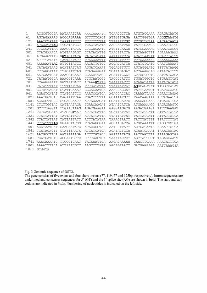
3.1.1 Sequence analysis
DST2 was discovered by screening the sequences of the D. discoideum cDNA project
database in Tsukuba, Japan (website: http://www.csm.biol.tsukuba.ac.jp/cDNAproject.html)
using DST1 as query. The genomic sequence of DST2 was amplified by PCR, cloned into the
pQE30-vector and sequenced. Analysis of the genomic structure of the DST2 gene showed
that it is composed of five exons separated by four introns and spans a genomic region of
approximately 1.8kb. Introns are usually very small and AT-rich in D. discoideum. These
features are also apparent in the four DST2 introns which have sizes of 77, 119, 77 and 175bp,
respectively. The sequences of all exon/intron borders follow the consensus 'GT-AG' rule
(Fig. 3).
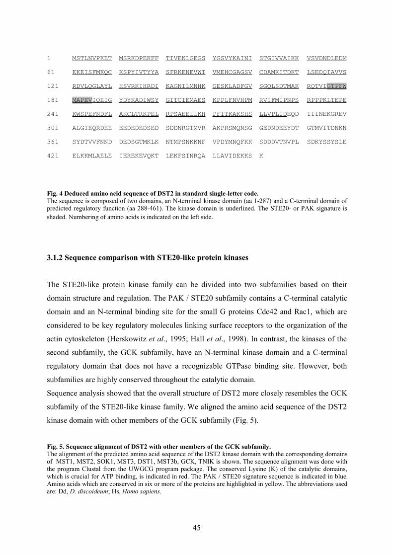
The full length cDNA clone of DST2 was kindly provided by the D. discoideum cDNA
project in Tsukuba. The cDNA contains an open reading frame of 1383bp that encodes a
protein of 461 amino acids with a predicted molecular mass of 52 kDa. Motif searches and
sequence comparison showed that DST2 consists of a 287-amino acid N-terminal kinase
domain and a 174-amino acid C-terminal regulatory domain. Database searches revealed that
DST2 is a member of the STE20 (sterile 20) or PAK (p21 activated kinase) family of protein
kinases. DST2 displays high homology throughout the kinase domain and contains the amino
acid sequence GTPYFWMAPEV in the kinase domain (Fig. 4). This sequence motif, the so-
called PAK signature is characteristic for the PAK / STE20 family of protein kinases and is
critical for kinase activity of STE20 (Wu et al., 1995).
43
1 ACGCGTCCGA AATAAATCAA AAAGAAAATG TCAACGCTCA ATGTACCAAA AGAGACAATG61 AGTAGAAAAG ACCCAGAAAA GTTTTTCACT ATTGTTGAGA AATTGGGTGA AGGGT AAGTG 121 AAATCTATTT TAAATTTTTT TTTTTTTTTT TTTTTTTTGC TCTGTTCTAA CACAATAATA181 ATAAATAT AG TTCATATGGT TCAGTATATA AAGCAATTAA TATTTCAACA GGAATTGTTG241 TTGCCATTAA AAAGGTATCA GTCGACAATG ATCTTGAAGA TATGGAAAAG GAAATCAGCT301 TTATGAAACA ATGTAAGAGT CCATACATTG TAACTTACTA TGCAAGCTTT AGAAAAGAAA361 ATGAAGTTTG GGT ACATATA TATATATATA AATATTTCTA ACATTAATAT ACATTTATTT421 ATTTTATATA TATTAATATT TTAAAAATTT ATTTCTTTTT TTTAAAAAAA AAAAAAAAAA481 AAAAAATT AG ATTGTTATGG AACATTGTGG AGCAGGATCA GTATGTGATG CAATGAAAAT541 TACAGATAAG ACATTATCAG AGGATCAAAT TGCAGTTGTT AGTAGGGATG TTTTACAAGG601 TTTAGCATAT TTACATTCAG TTAGAAAGAT TCATAGAGAT ATTAAAGCAG GTAACATTTT661 AATGAATCAT AAAGGTGAAT CGAAATTAGC AGATTTCGGT GTTAGTGGTC AATTATCAGA721 TACAATGGCA AAACGTCAAA CTGTAATCGG TACCCCATTT TGGATGGCTC CTGAAGTCAT781 TCAAGAAATT GGTTATGATT ATAAAGT ATG TAATTTATTT ATAGATAATA TATATATATA841 TATATTTTAT TTTTTATTAA TTTGATATTA TTATTATTAT AGGCAGATAT TTGGTCATAT901 GGTATTACAT GTATTGAAAT GGCAGAATCA AAACCACCAT TATTTAATGT TCATCCAATG961 AGAGTCATAT TTATGATTCC AAATCCATCA AGACCACCAC CAAAATTAAC AGAACCAGAG1021 AAATGGTCAC CAGAATTCAA TGACTTTTTA GCAAAATGTT TAACAAGAAA ACCAGAATTA1081 AGACCTTCCG CTGAGGAATT ATTAAAACAT CCATTCATTA CAAAAGCAAA ATCACATTCA1141 CTCTTGGTAC CATTAATAGA TGAACAAGAT ATAATCATCA ATGAAAAAGG TAGAGAAGTC1201 GCTTTAGGTA TTGAACAAAG AGATGAAGAA GAGGAAGATG AAGATGAAGA TTCTGAAGAT1261 TCTGATGATA ATAGAGT AAT ATTATCATTA TCATTATTAT TATTATTATT ATTATTATTA1321 TTATTATTAT TATTATTATT ATTATTATTA TTATTATTAT TATTATTATT ATTATTATTA1382 TTATTATTAT TATTATTATT ATTTATATAA TAAACTAATT TATTTATTTT TTATTTTTAT1441 TTTATTTT AG GGAACTATGG TTAGAGCGAA ACCAAGATCA ATGCAAAATT CAGGTGGTGA1501 AGATAATGAT GAAGAATATG ATACAGGTAC AATGGTTATT ACTGATAATA AGAATTCTTA1561 TGATACAGTT GTATTTAATA ATGATGATGA AGATAGTGGA ACAATGAAAT TAAAGAATAC1621 AATGCCTTCA AATAAAAAGA ATTTTGTACC AGATTATATG AATCAATTTA AAAAGAGTGA1681 TGATGATGTC ACCAATGTTC CTTTAAGTGA TAAATACTCT AGTTATTCCT TAGAGGAATT1741 AAAGAAAATG TTGGCTGAAT TAGAAATTGA AAGAGAAAAA GAAGTTCAAA AAACACTTGA1801 AAAATTTTCA ATTAATCGTC AAGCTTTATT AGCTGTAATT GATGAAAAGA AATCAAAGTA1861 GTAGTA
Fig. 3 Genomic sequence of DST2.The gene consists of five exons and four short introns (77, 119, 77 and 175bp, respectively). Intron sequences areunderlined and consensus sequences for 5’ (GT) and the 3’ splice site (AG) are shown in bold. The start and stopcodons are indicated in italic. Numbering of nucleotides is indicated on the left side.
44
1 MSTLNVPKET MSRKDPEKFF TIVEKLGEGS YGSVYKAINI STGIVVAIKK VSVDNDLEDM61 EKEISFMKQC KSPYIVTYYA SFRKENEVWI VMEHCGAGSV CDAMKITDKT LSEDQIAVVS121 RDVLQGLAYL HSVRKIHRDI KAGNILMNHK GESKLADFGV SGQLSDTMAK RQTVI GTPFW 181 MAPEV IQEIG YDYKADIWSY GITCIEMAES KPPLFNVHPM RVIFMIPNPS RPPPKLTEPE241 KWSPEFNDFL AKCLTRKPEL RPSAEELLKH PFITKAKSHS LLVPLIDEQD IIINEKGREV301 ALGIEQRDEE EEDEDEDSED SDDNRGTMVR AKPRSMQNSG GEDNDEEYDT GTMVITDNKN361 SYDTVVFNND DEDSGTMKLK NTMPSNKKNF VPDYMNQFKK SDDDVTNVPL SDKYSSYSLE421 ELKKMLAELE IEREKEVQKT LEKFSINRQA LLAVIDEKKS K
Fig. 4 Deduced amino acid sequence of DST2 in standard single-letter code.The sequence is composed of two domains, an N-terminal kinase domain (aa 1-287) and a C-terminal domain ofpredicted regulatory function (aa 288-461). The kinase domain is underlined. The STE20- or PAK signature isshaded. Numbering of amino acids is indicated on the left side.
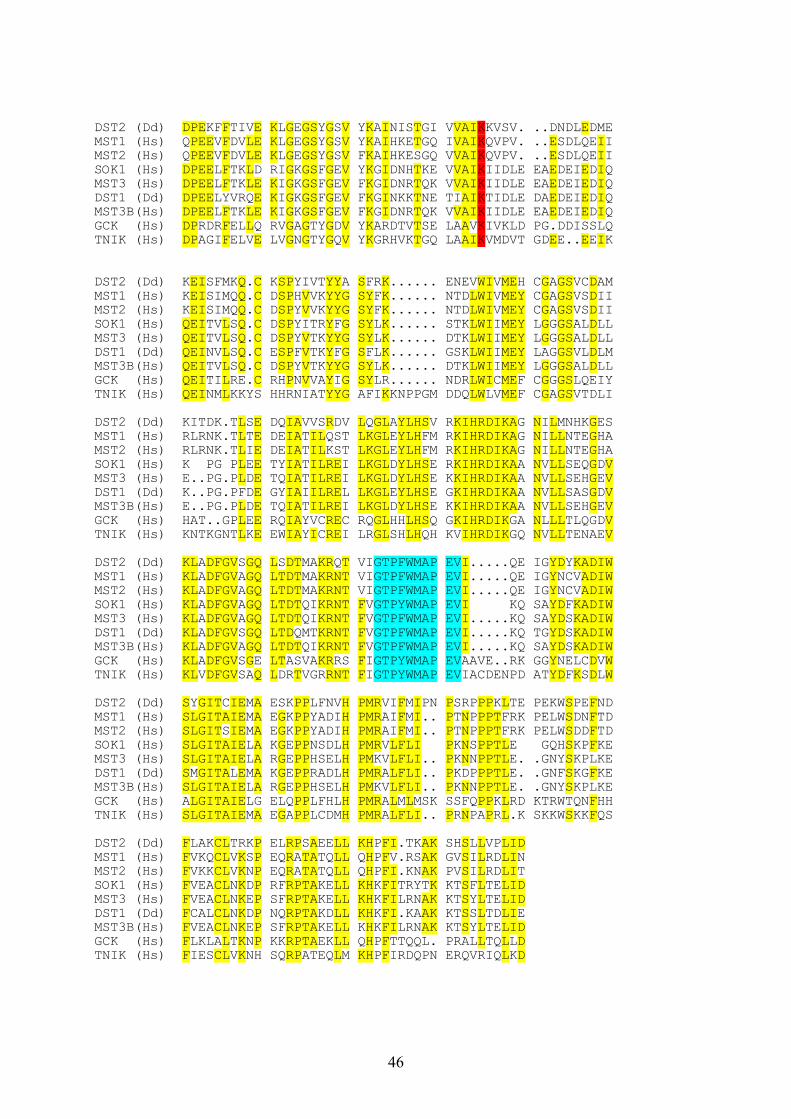
3.1.2 Sequence comparison with STE20-like protein kinases
The STE20-like protein kinase family can be divided into two subfamilies based on their
domain structure and regulation. The PAK / STE20 subfamily contains a C-terminal catalytic
domain and an N-terminal binding site for the small G proteins Cdc42 and Rac1, which are
considered to be key regulatory molecules linking surface receptors to the organization of the
actin cytoskeleton (Herskowitz et al., 1995; Hall et al., 1998). In contrast, the kinases of the
second subfamily, the GCK subfamily, have an N-terminal kinase domain and a C-terminal
regulatory domain that does not have a recognizable GTPase binding site. However, both
subfamilies are highly conserved throughout the catalytic domain.
Sequence analysis showed that the overall structure of DST2 more closely resembles the GCK
subfamily of the STE20-like kinase family. We aligned the amino acid sequence of the DST2
kinase domain with other members of the GCK subfamily (Fig. 5).
Fig. 5. Sequence alignment of DST2 with other members of the GCK subfamily. The alignment of the predicted amino acid sequence of the DST2 kinase domain with the corresponding domainsof MST1, MST2, SOK1, MST3, DST1, MST3b, GCK, TNIK is shown. The sequence alignment was done withthe program Clustal from the UWGCG program package. The conserved Lysine (K) of the catalytic domains,which is crucial for ATP binding, is indicated in red. The PAK / STE20 signature sequence is indicated in blue.Amino acids which are conserved in six or more of the proteins are highlighted in yellow. The abbreviations usedare: Dd, D. discoideum; Hs, Homo sapiens.
45
DST2 (Dd) DPEKFFTIVE KLGEGSYGSV YKAINISTGI VVAIKKVSV. ..DNDLEDMEMST1 (Hs) QPEEVFDVLE KLGEGSYGSV YKAIHKETGQ IVAIKQVPV. ..ESDLQEIIMST2 (Hs) QPEEVFDVLE KLGEGSYGSV FKAIHKESGQ VVAIKQVPV. ..ESDLQEIISOK1 (Hs) DPEELFTKLD RIGKGSFGEV YKGIDNHTKE VVAIKIIDLE EAEDEIEDIQMST3 (Hs) DPEELFTKLE KIGKGSFGEV FKGIDNRTQK VVAIKIIDLE EAEDEIEDIQDST1 (Dd) DPEELYVRQE KIGKGSFGEV FKGINKKTNE TIAIKTIDLE DAEDEIEDIQMST3B(Hs) DPEELFTKLE KIGKGSFGEV FKGIDNRTQK VVAIKIIDLE EAEDEIEDIQGCK (Hs) DPRDRFELLQ RVGAGTYGDV YKARDTVTSE LAAVKIVKLD PG.DDISSLQTNIK (Hs) DPAGIFELVE LVGNGTYGQV YKGRHVKTGQ LAAIKVMDVT GDEE..EEIK
DST2 (Dd) KEISFMKQ.C KSPYIVTYYA SFRK...... ENEVWIVMEH CGAGSVCDAMMST1 (Hs) KEISIMQQ.C DSPHVVKYYG SYFK...... NTDLWIVMEY CGAGSVSDIIMST2 (Hs) KEISIMQQ.C DSPYVVKYYG SYFK...... NTDLWIVMEY CGAGSVSDIISOK1 (Hs) QEITVLSQ.C DSPYITRYFG SYLK...... STKLWIIMEY LGGGSALDLLMST3 (Hs) QEITVLSQ.C DSPYVTKYYG SYLK...... DTKLWIIMEY LGGGSALDLLDST1 (Dd) QEINVLSQ.C ESPFVTKYFG SFLK...... GSKLWIIMEY LAGGSVLDLMMST3B(Hs) QEITVLSQ.C DSPYVTKYYG SYLK...... DTKLWIIMEY LGGGSALDLLGCK (Hs) QEITILRE.C RHPNVVAYIG SYLR...... NDRLWICMEF CGGGSLQEIYTNIK (Hs) QEINMLKKYS HHRNIATYYG AFIKKNPPGM DDQLWLVMEF CGAGSVTDLIDST2 (Dd) KITDK.TLSE DQIAVVSRDV LQGLAYLHSV RKIHRDIKAG NILMNHKGESMST1 (Hs) RLRNK.TLTE DEIATILQST LKGLEYLHFM RKIHRDIKAG NILLNTEGHAMST2 (Hs) RLRNK.TLIE DEIATILKST LKGLEYLHFM RKIHRDIKAG NILLNTEGHASOK1 (Hs) K PG PLEE TYIATILREI LKGLDYLHSE RKIHRDIKAA NVLLSEQGDVMST3 (Hs) E..PG.PLDE TQIATILREI LKGLDYLHSE KKIHRDIKAA NVLLSEHGEVDST1 (Dd) K..PG.PFDE GYIAIILREL LKGLEYLHSE GKIHRDIKAA NVLLSASGDVMST3B(Hs) E..PG.PLDE TQIATILREI LKGLDYLHSE KKIHRDIKAA NVLLSEHGEVGCK (Hs) HAT..GPLEE RQIAYVCREC RQGLHHLHSQ GKIHRDIKGA NLLLTLQGDVTNIK (Hs) KNTKGNTLKE EWIAYICREI LRGLSHLHQH KVIHRDIKGQ NVLLTENAEVDST2 (Dd) KLADFGVSGQ LSDTMAKRQT VIGTPFWMAP EVI.....QE IGYDYKADIWMST1 (Hs) KLADFGVAGQ LTDTMAKRNT VIGTPFWMAP EVI.....QE IGYNCVADIWMST2 (Hs) KLADFGVAGQ LTDTMAKRNT VIGTPFWMAP EVI.....QE IGYNCVADIWSOK1 (Hs) KLADFGVAGQ LTDTQIKRNT FVGTPYWMAP EVI KQ SAYDFKADIWMST3 (Hs) KLADFGVAGQ LTDTQIKRNT FVGTPFWMAP EVI.....KQ SAYDSKADIWDST1 (Dd) KLADFGVSGQ LTDQMTKRNT FVGTPFWMAP EVI.....KQ TGYDSKADIWMST3B(Hs) KLADFGVAGQ LTDTQIKRNT FVGTPFWMAP EVI.....KQ SAYDSKADIWGCK (Hs) KLADFGVSGE LTASVAKRRS FIGTPYWMAP EVAAVE..RK GGYNELCDVWTNIK (Hs) KLVDFGVSAQ LDRTVGRRNT FIGTPYWMAP EVIACDENPD ATYDFKSDLWDST2 (Dd) SYGITCIEMA ESKPPLFNVH PMRVIFMIPN PSRPPPKLTE PEKWSPEFNDMST1 (Hs) SLGITAIEMA EGKPPYADIH PMRAIFMI.. PTNPPPTFRK PELWSDNFTDMST2 (Hs) SLGITSIEMA EGKPPYADIH PMRAIFMI.. PTNPPPTFRK PELWSDDFTDSOK1 (Hs) SLGITAIELA KGEPPNSDLH PMRVLFLI PKNSPPTLE GQHSKPFKEMST3 (Hs) SLGITAIELA RGEPPHSELH PMKVLFLI.. PKNNPPTLE. .GNYSKPLKEDST1 (Dd) SMGITALEMA KGEPPRADLH PMRALFLI.. PKDPPPTLE. .GNFSKGFKEMST3B(Hs) SLGITAIELA RGEPPHSELH PMKVLFLI.. PKNNPPTLE. .GNYSKPLKEGCK (Hs) ALGITAIELG ELQPPLFHLH PMRALMLMSK SSFQPPKLRD KTRWTQNFHHTNIK (Hs) SLGITAIEMA EGAPPLCDMH PMRALFLI.. PRNPAPRL.K SKKWSKKFQSDST2 (Dd) FLAKCLTRKP ELRPSAEELL KHPFI.TKAK SHSLLVPLID MST1 (Hs) FVKQCLVKSP EQRATATQLL QHPFV.RSAK GVSILRDLIN MST2 (Hs) FVKKCLVKNP EQRATATQLL QHPFI.KNAK PVSILRDLITSOK1 (Hs) FVEACLNKDP RFRPTAKELL KHKFITRYTK KTSFLTELIDMST3 (Hs) FVEACLNKEP SFRPTAKELL KHKFILRNAK KTSYLTELIDDST1 (Dd) FCALCLNKDP NQRPTAKDLL KHKFI.KAAK KTSSLTDLIE MST3B(Hs) FVEACLNKEP SFRPTAKELL KHKFILRNAK KTSYLTELID GCK (Hs) FLKLALTKNP KKRPTAEKLL QHPFTTQQL. PRALLTQLLD TNIK (Hs) FIESCLVKNH SQRPATEQLM KHPFIRDQPN ERQVRIQLKD
46

The alignment clearly showed that the DST2 kinase domain is highly homologous to the
kinase domains of MST1 (Hs), MST2 (Hs), SOK1 (Hs), MST3 (Hs), DST1 (Dd), MST3b
(Hs), GCK (HS) and TNIK (Hs). These proteins share a similar structure and have 79, 76, 70,
69, 69, 68, 66 and 65% amino acid similarities in the catalytic domains, respectively. These
proteins have a highly conserved catalytic domain in common but differ in their C-terminal
regulatory domain, which does not contain any identifiable sequence motifs. Interestingly,
DST2 is more closely related to human MST1 and MST2 (79% and 76% similarity I in the
catalytic domain, respectively) than to D. discoideum DST1 (with only 69%) (Fig. 6A). To
further clarify the relationships between these protein kinases and DST2, we calculated
multiple sequence alignments of the catalytic domains. The derived evolutionary tree is split
into two main branches, one formed by MST1, MST2, DST2, GCK and TNIK, the other one
by MST3, MST3b, DST1 and SOK-1 (Fig. 6B)
A
47
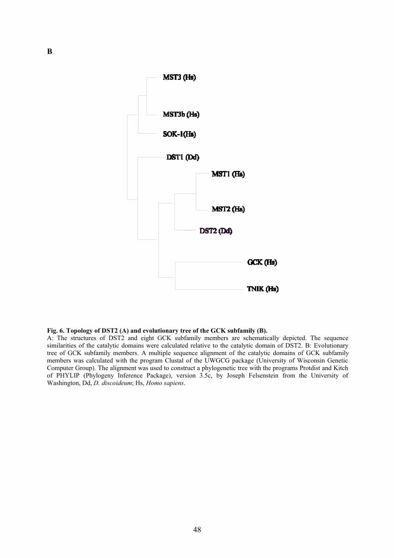
B
Fig. 6. Topology of DST2 (A) and evolutionary tree of the GCK subfamily (B).A: The structures of DST2 and eight GCK subfamily members are schematically depicted. The sequencesimilarities of the catalytic domains were calculated relative to the catalytic domain of DST2. B: Evolutionarytree of GCK subfamily members. A multiple sequence alignment of the catalytic domains of GCK subfamilymembers was calculated with the program Clustal of the UWGCG package (University of Wisconsin GeneticComputer Group). The alignment was used to construct a phylogenetic tree with the programs Protdist and Kitchof PHYLIP (Phylogeny Inference Package), version 3.5c, by Joseph Felsenstein from the University ofWashington, Dd, D. discoideum; Hs, Homo sapiens.
48
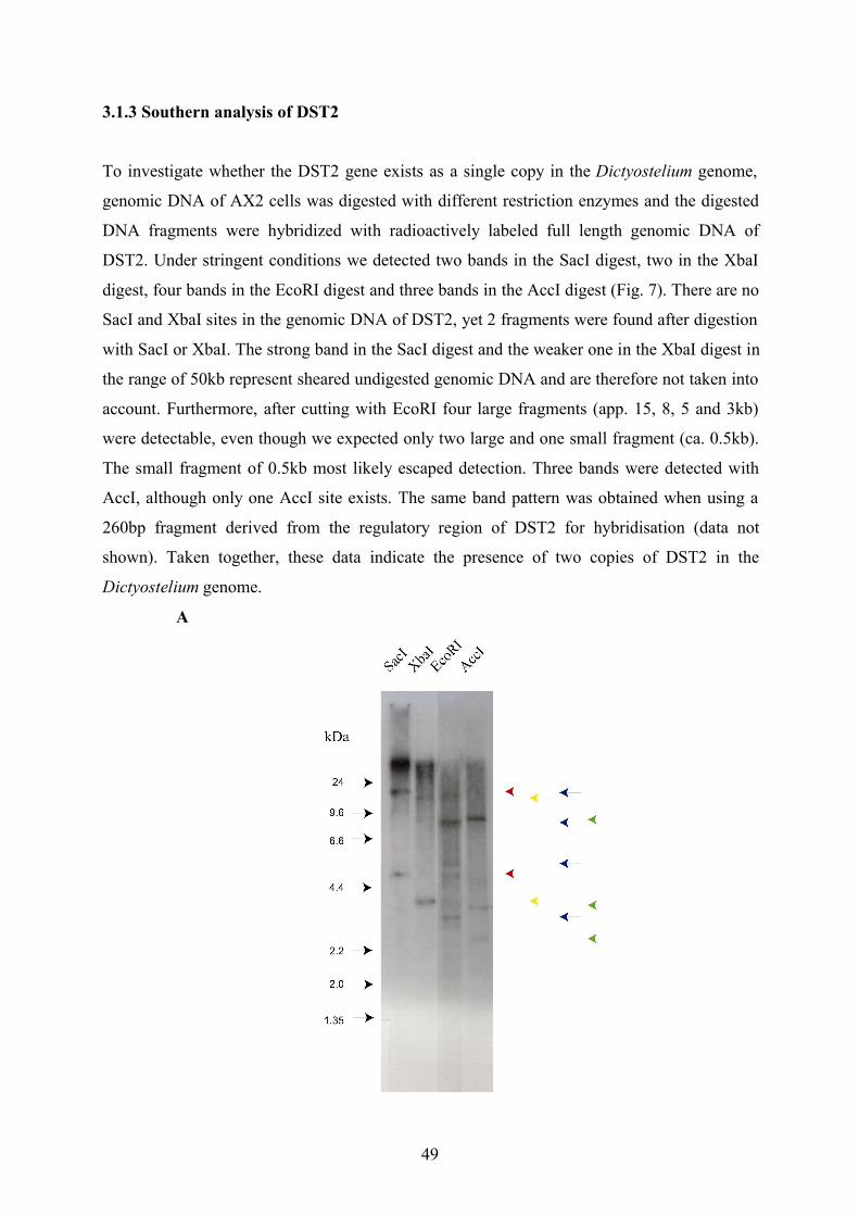
3.1.3 Southern analysis of DST2
To investigate whether the DST2 gene exists as a single copy in the Dictyostelium genome,
genomic DNA of AX2 cells was digested with different restriction enzymes and the digested
DNA fragments were hybridized with radioactively labeled full length genomic DNA of
DST2. Under stringent conditions we detected two bands in the SacI digest, two in the XbaI
digest, four bands in the EcoRI digest and three bands in the AccI digest (Fig. 7). There are no
SacI and XbaI sites in the genomic DNA of DST2, yet 2 fragments were found after digestion
with SacI or XbaI. The strong band in the SacI digest and the weaker one in the XbaI digest in
the range of 50kb represent sheared undigested genomic DNA and are therefore not taken into
account. Furthermore, after cutting with EcoRI four large fragments (app. 15, 8, 5 and 3kb)
were detectable, even though we expected only two large and one small fragment (ca. 0.5kb).
The small fragment of 0.5kb most likely escaped detection. Three bands were detected with
AccI, although only one AccI site exists. The same band pattern was obtained when using a
260bp fragment derived from the regulatory region of DST2 for hybridisation (data not
shown). Taken together, these data indicate the presence of two copies of DST2 in the
Dictyostelium genome.
A
49
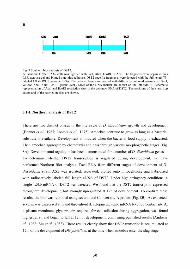
B
Fig. 7 Southern blot analysis of DST2.A: Genomic DNA of AX2 cells was digested with SacI, XbaI, EcoRI, or AccI. The fragments were separated in a0.8% agarose gel and blotted onto nitrocellulose. DST2 specific fragments were detected with the full length 32P-labeled 1.8 kb DST2 genomic DNA. The detected bands are marked with differently coloured arrows (red: SacI;yellow: XbaI; blue: EcoRI; green: AccI). Sizes of the DNA marker are shown on the left side. B: Schematicrepresentation of AccI and EcoRI restriction sites in the genomic DNA of DST2. The positions of the start, stopcodon and of the restriction sites are shown.
3.1.4. Northern analysis of DST2
There are two distinct phases in the life cycle of D. discoideum, growth and development
(Bonner et al., 1967; Loomis et al., 1975). Amoebae continue to grow as long as a bacterial
substrate is available. Development is initiated when the bacterial food supply is exhausted.
Then amoebae aggregate by chemotaxis and pass through various morphogenetic stages (Fig.
8A). Developmental regulation has been demonstrated for a number of D. discoideum genes.
To determine whether DST2 transcription is regulated during development, we have
performed Northern Blot analysis. Total RNA from different stages of development of D.
discoideum strain AX2 was isolated, separated, blotted onto nitrocellulose and hybridized
with radioactively labeled full length cDNA of DST2. Under high stringency conditions, a
single 1.5kb mRNA of DST2 was detected. We found that the DST2 transcript is expressed
throughout development, but strongly upregulated at 12h of development. To confirm these
results, the blot was reprobed using severin and Contact site A probes (Fig. 8B). As expected,
severin was expressed at t0 and throughout development, while mRNA level of Contact site A,
a plasma membrane glycoprotein required for cell adhesion during aggregation, was found
highest at 9h and began to fall at 12h of development, confirming published results (André et
al., 1988; Siu et al., 1988). These results clearly show that DST2 transcript is accumulated at
12 h of the development of Dictyostelium, at the time when amoebae enter the slug stage.
50
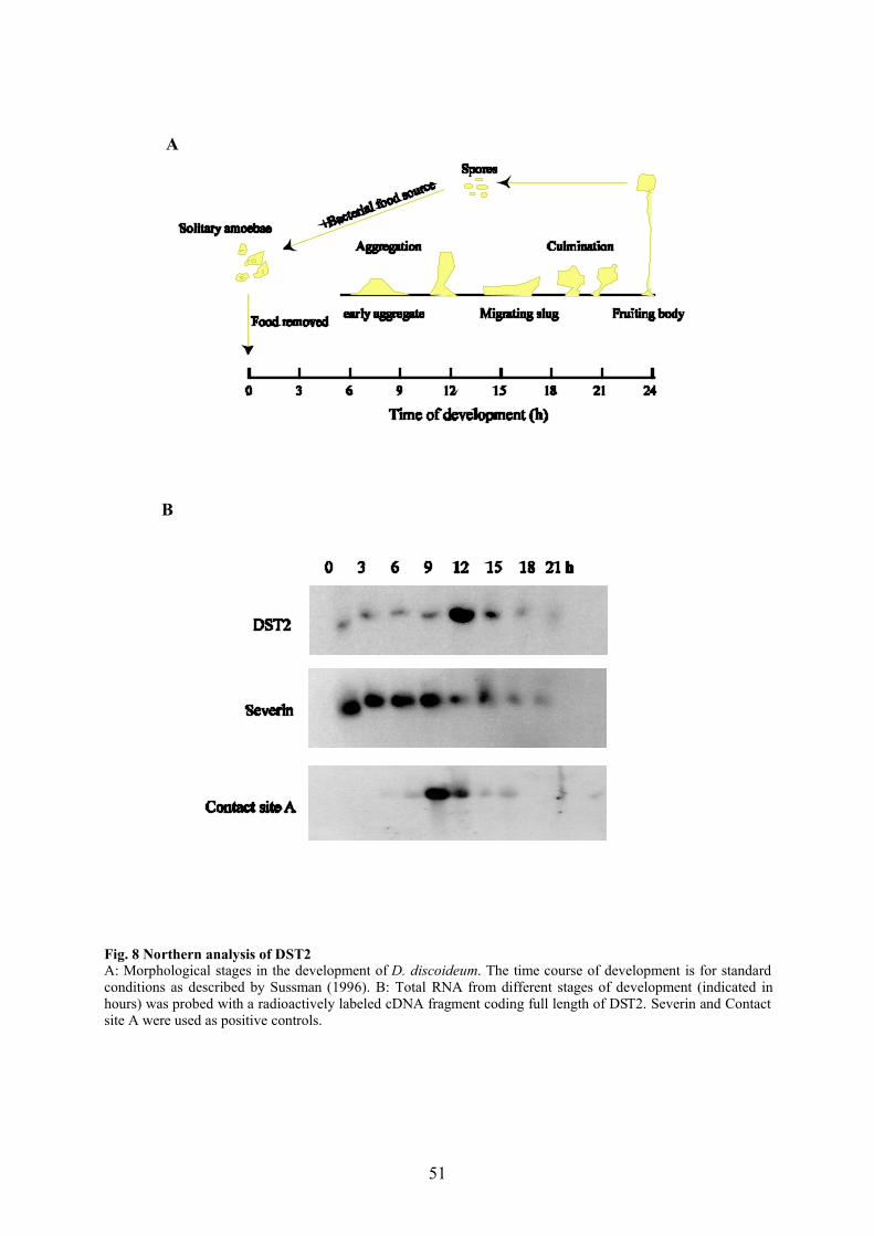
A
B
Fig. 8 Northern analysis of DST2A: Morphological stages in the development of D. discoideum. The time course of development is for standardconditions as described by Sussman (1996). B: Total RNA from different stages of development (indicated inhours) was probed with a radioactively labeled cDNA fragment coding full length of DST2. Severin and Contactsite A were used as positive controls.
51

3.2 Biochemical and cell biological characterization of DST2
3.2.1 Expression and purification of recombinant DST2
Full length DST2 (MBP-DST2), the catalytic domain (MBP-DST2-cat) and the regulatory
domain (MBP-DST2-reg) were expressed as maltose binding protein (MBP)-tagged fusion
proteins. After opening the bacteria, the recombinant proteins were found soluble and purified
using affinity chromatography. Recombinant MBP-DST2 showed a molecular mass of about
95 kDa, MBP-DST2-cat of about 80 kDa and MBP-DST2-reg of about 70 kDa (Fig. 9A, C,
E). Expression and purification worked very well for MBP-DST-reg, while considerable
degradation was observed in the case of MBP-DST2 and, less pronounced, MBP-DST2-cat.
The purified recombinant proteins were used for the generation of polyclonal antisera as well
as for the investigation of their catalytic activity in in vitro assays (see 3.2.3). The full length
proteins as well as the degradation products were also seen in immunoblots using the obtained
polyclonal antisera (Fig. 9 B, D). The polyclonal antiserum against MBP-DST2-reg was
superior and used for further investigations. However, the affinity and specificity of the anti
MBP-DST2-reg antibodies was not sufficient for immunoprecipitation of native DST2 from
whole Dictyostelium cell homogenates.
52
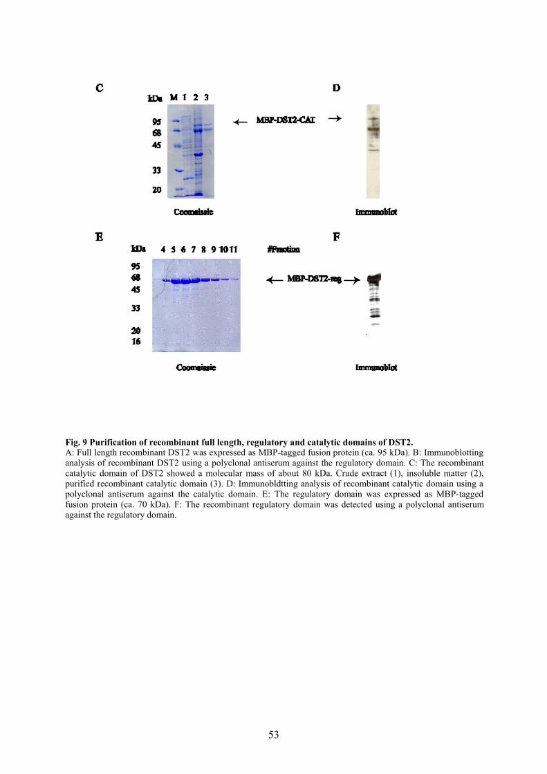
Fig. 9 Purification of recombinant full length, regulatory and catalytic domains of DST2.A: Full length recombinant DST2 was expressed as MBP-tagged fusion protein (ca. 95 kDa). B: Immunoblottinganalysis of recombinant DST2 using a polyclonal antiserum against the regulatory domain. C: The recombinantcatalytic domain of DST2 showed a molecular mass of about 80 kDa. Crude extract (1), insoluble matter (2),purified recombinant catalytic domain (3). D: Immunobldtting analysis of recombinant catalytic domain using apolyclonal antiserum against the catalytic domain. E: The regulatory domain was expressed as MBP-taggedfusion protein (ca. 70 kDa). F: The recombinant regulatory domain was detected using a polyclonal antiserumagainst the regulatory domain.
53
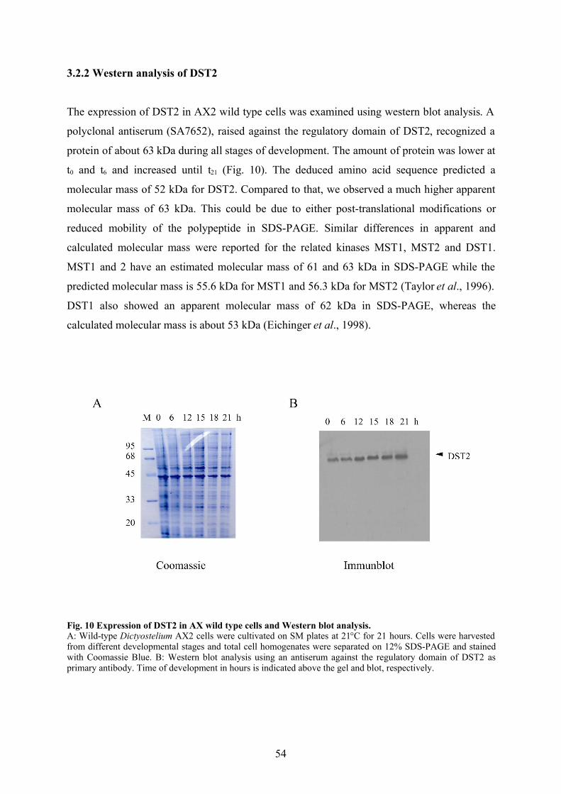
3.2.2 Western analysis of DST2
The expression of DST2 in AX2 wild type cells was examined using western blot analysis. A
polyclonal antiserum (SA7652), raised against the regulatory domain of DST2, recognized a
protein of about 63 kDa during all stages of development. The amount of protein was lower at
t0 and t6 and increased until t21 (Fig. 10). The deduced amino acid sequence predicted a
molecular mass of 52 kDa for DST2. Compared to that, we observed a much higher apparent
molecular mass of 63 kDa. This could be due to either post-translational modifications or
reduced mobility of the polypeptide in SDS-PAGE. Similar differences in apparent and
calculated molecular mass were reported for the related kinases MST1, MST2 and DST1.
MST1 and 2 have an estimated molecular mass of 61 and 63 kDa in SDS-PAGE while the
predicted molecular mass is 55.6 kDa for MST1 and 56.3 kDa for MST2 (Taylor et al., 1996).
DST1 also showed an apparent molecular mass of 62 kDa in SDS-PAGE, whereas the
calculated molecular mass is about 53 kDa (Eichinger et al., 1998).
Fig. 10 Expression of DST2 in AX wild type cells and Western blot analysis.A: Wild-type Dictyostelium AX2 cells were cultivated on SM plates at 21°C for 21 hours. Cells were harvestedfrom different developmental stages and total cell homogenates were separated on 12% SDS-PAGE and stainedwith Coomassie Blue. B: Western blot analysis using an antiserum against the regulatory domain of DST2 asprimary antibody. Time of development in hours is indicated above the gel and blot, respectively.
54

3.2.3 In vitro kinase assays with recombinant DST2 constructs
Bacterially expressed fusion proteins of MBP and full length DST2 (MBP-DST2), the
catalytic domain (MBP-DST2-cat) and the regulatory domain (MBP-DST2-reg) were used in
in vitro kinase assays either alone or with myelin basic protein (MBP’) as substrate. As shown
in Fig. 11, MBP-DST2 was able to phosphorylate itself (lane 1) and MBP’ (lane 2). However,
MBP-DST2-cat (lanes 3 and 4) and MBP-DST2-reg (lanes 5 and 6) did not show any kinase
activity. The loss of catalytic activity of the recombinant catalytic domain was unexpected and
could be due to a protein-folding problem.
Since full length recombinant DST2 protein was tagged with maltose binding protein, we
examined whether maltose binding protein might be phosphorylated by DST2 or have any
effect on kinase activity. As shown in Fig. 12, MBP was not phosphorylated by MBP-DST2
and no difference in the autophosphorylation of DST2 was seen in the presence of MBP (lane
1, lane 2), while MBP-DST2 still phosphorylated MBP’ (lane 3). These results show that
added MBP is not phosphorylated by MBP-DST2 and does not influence the catalytic activity
of MBP-DST2. MBP-DST2 phosphorylated itself, but it was not clear where the
autophosphorylation occurs. To find out which domain of DST2 is phosphorylated during
autophosphorylation, the recombinant regulatory and catalytic domains of DST2 were used as
potential substrates in the in vitro kinase assay. While the regulatory domain was heavily
phosphorylated by MBP-DST2 (lane 4), we observed no phosphorylation of the catalytic
domain (lane 5), indicating that the phosphorylation site(s) of autophosphorylation are located
on the regulatory domain.
In addition to MBP’, we tested several additional potential substrates of MBP-DST2 in in
vitro kinase assays. H1 and -casein turned out to be poor substrates for MBP-DST2 (data
not shown). We also tested Dictyostelium severin as a potential substrate for MBP-DST2,
because previously it was shown that severin was very efficiently phosphorylated by DST1
(Eichinger et al., 1998) and found that severin is also a very good substrate for MBP-DST2
(Fig. 13). All further kinase assays were carried out with either no substrate or using MBP’ or
severin as a substrate.
To address the question whether autophosphorylation on the regulatory domain could regulate
the kinase activity, we incubated MBP-DST2 in the presence of 32P-ATP for 0 or 20 min
followed by the addition of severin as a substrate.
55
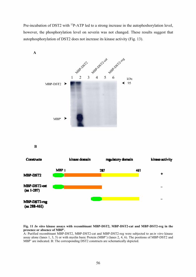
Pre-incubation of DST2 with 32P-ATP led to a strong increase in the autophoshorylation level,
however, the phosphorylation level on severin was not changed. These results suggest that
autophosphorylation of DST2 does not increase its kinase activity (Fig. 13).
A
B
Fig. 11 In vitro kinase assays with recombinant MBP-DST2, MBP-DST2-cat and MBP-DST2-reg in thepresence or absence of MBP’.A: Purified recombinant MBP-DST2, MBP-DST2-cat and MBP-DST2-reg were subjected to an in vitro kinaseassay alone (lanes 1, 3, 5) or with myelin basic Protein (MBP’) (lanes 2, 4, 6). The positions of MBP-DST2 andMBP’ are indicated. B: The corresponding DST2 constructs are schematically depicted.
56
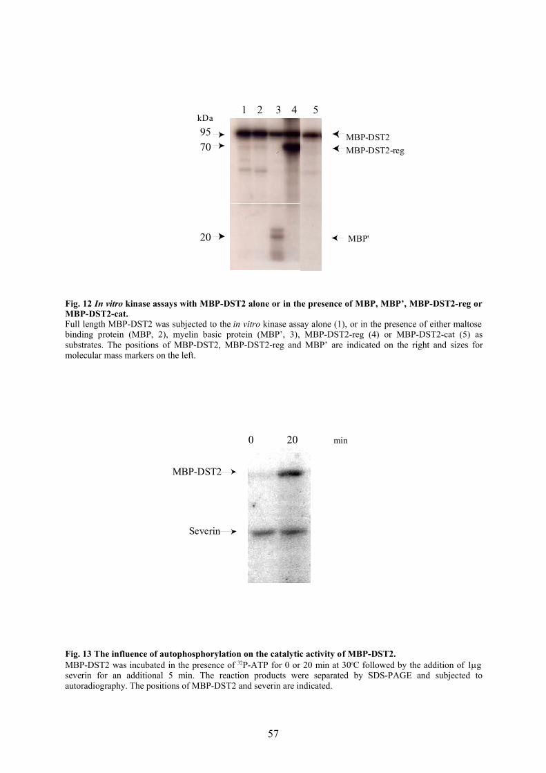
9570
MBP-DST2MBP-DST2-reg
MBP'
2 3 4 5kDa
20
1
Fig. 12 In vitro kinase assays with MBP-DST2 alone or in the presence of MBP, MBP’, MBP-DST2-reg orMBP-DST2-cat.Full length MBP-DST2 was subjected to the in vitro kinase assay alone (1), or in the presence of either maltosebinding protein (MBP, 2), myelin basic protein (MBP’, 3), MBP-DST2-reg (4) or MBP-DST2-cat (5) assubstrates. The positions of MBP-DST2, MBP-DST2-reg and MBP’ are indicated on the right and sizes formolecular mass markers on the left.
0 20
MBP-DST2
Severin
min
Fig. 13 The influence of autophosphorylation on the catalytic activity of MBP-DST2.MBP-DST2 was incubated in the presence of 32P-ATP for 0 or 20 min at 30oC followed by the addition of 1gseverin for an additional 5 min. The reaction products were separated by SDS-PAGE and subjected toautoradiography. The positions of MBP-DST2 and severin are indicated.
57
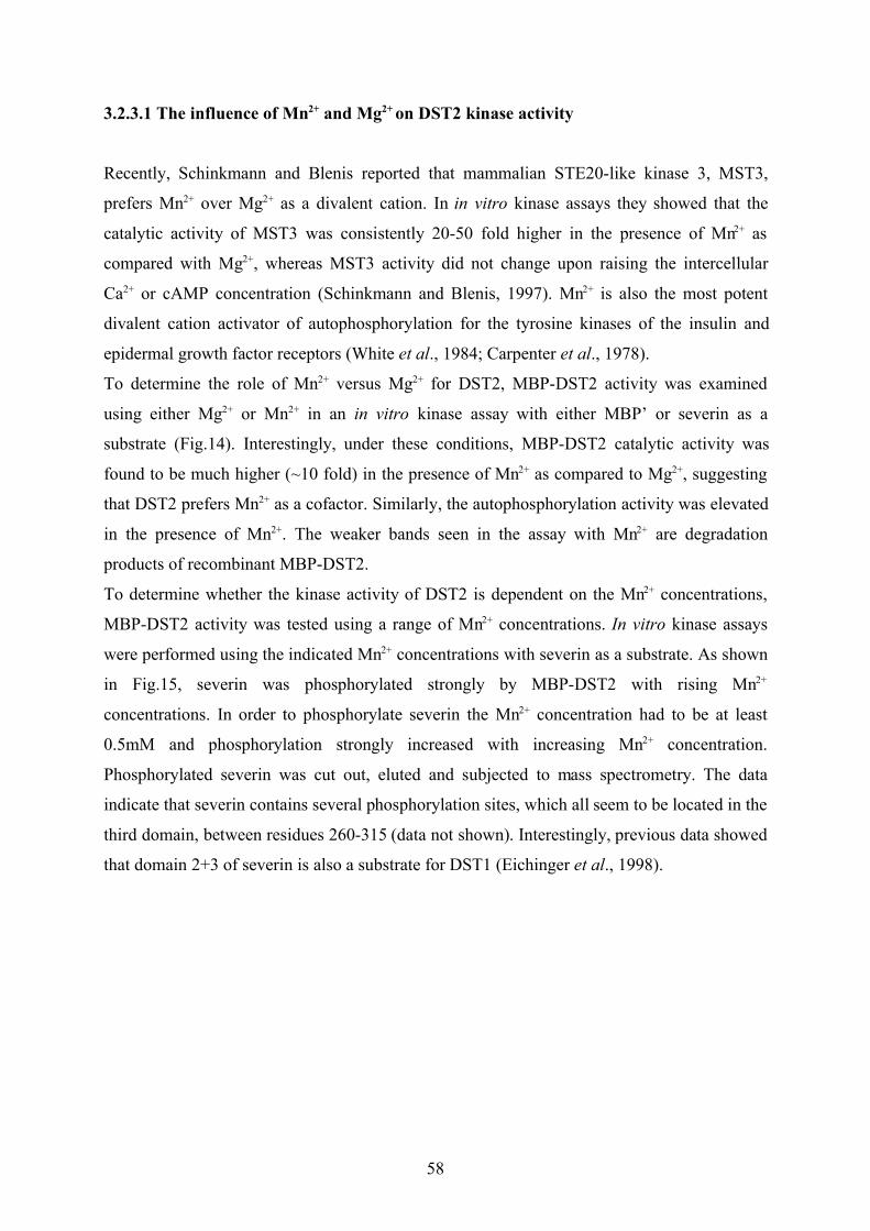
3.2.3.1 The influence of Mn2+ and Mg2+ on DST2 kinase activity
Recently, Schinkmann and Blenis reported that mammalian STE20-like kinase 3, MST3,
prefers Mn2+ over Mg2+ as a divalent cation. In in vitro kinase assays they showed that the
catalytic activity of MST3 was consistently 20-50 fold higher in the presence of Mn2+ as
compared with Mg2+, whereas MST3 activity did not change upon raising the intercellular
Ca2+ or cAMP concentration (Schinkmann and Blenis, 1997). Mn2+ is also the most potent
divalent cation activator of autophosphorylation for the tyrosine kinases of the insulin and
epidermal growth factor receptors (White et al., 1984; Carpenter et al., 1978).
To determine the role of Mn2+ versus Mg2+ for DST2, MBP-DST2 activity was examined
using either Mg2+ or Mn2+ in an in vitro kinase assay with either MBP’ or severin as a
substrate (Fig.14). Interestingly, under these conditions, MBP-DST2 catalytic activity was
found to be much higher (~10 fold) in the presence of Mn2+ as compared to Mg2+, suggesting
that DST2 prefers Mn2+ as a cofactor. Similarly, the autophosphorylation activity was elevated
in the presence of Mn2+. The weaker bands seen in the assay with Mn2+ are degradation
products of recombinant MBP-DST2.
To determine whether the kinase activity of DST2 is dependent on the Mn2+ concentrations,
MBP-DST2 activity was tested using a range of Mn2+ concentrations. In vitro kinase assays
were performed using the indicated Mn2+ concentrations with severin as a substrate. As shown
in Fig.15, severin was phosphorylated strongly by MBP-DST2 with rising Mn2+
concentrations. In order to phosphorylate severin the Mn2+ concentration had to be at least
0.5mM and phosphorylation strongly increased with increasing Mn2+ concentration.
Phosphorylated severin was cut out, eluted and subjected to mass spectrometry. The data
indicate that severin contains several phosphorylation sites, which all seem to be located in the
third domain, between residues 260-315 (data not shown). Interestingly, previous data showed
that domain 2+3 of severin is also a substrate for DST1 (Eichinger et al., 1998).
58
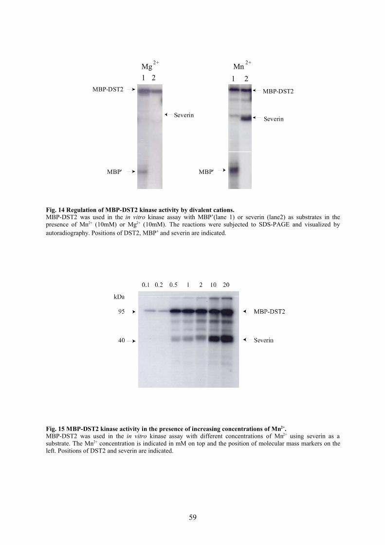
Mn Mg
MBP-DST2
MBP'
MBP-DST2
Severin
MBP'
1 2 1 2
2+ 2+
Severin
Fig. 14 Regulation of MBP-DST2 kinase activity by divalent cations.MBP-DST2 was used in the in vitro kinase assay with MBP’(lane 1) or severin (lane2) as substrates in thepresence of Mn2+ (10mM) or Mg2+ (10mM). The reactions were subjected to SDS-PAGE and visualized byautoradiography. Positions of DST2, MBP’ and severin are indicated.
Fig. 15 MBP-DST2 kinase activity in the presence of increasing concentrations of Mn2+.MBP-DST2 was used in the in vitro kinase assay with different concentrations of Mn2+ using severin as asubstrate. The Mn2+ concentration is indicated in mM on top and the position of molecular mass markers on theleft. Positions of DST2 and severin are indicated.
59
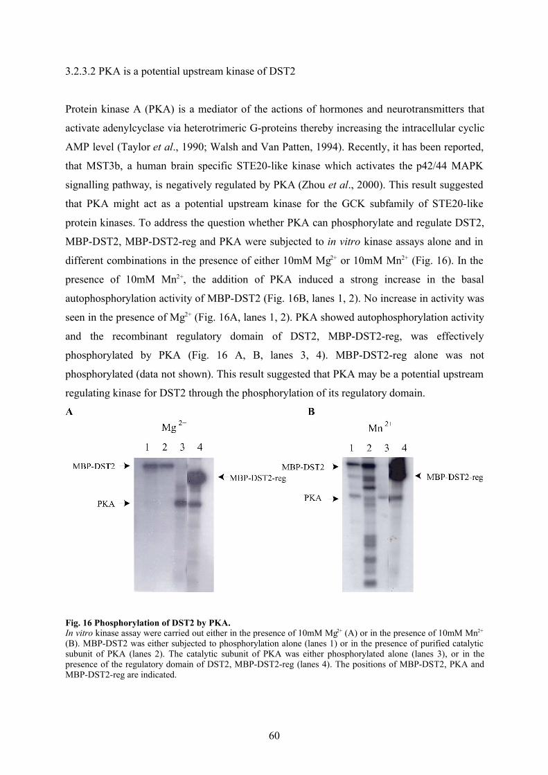
3.2.3.2 PKA is a potential upstream kinase of DST2
Protein kinase A (PKA) is a mediator of the actions of hormones and neurotransmitters that
activate adenylcyclase via heterotrimeric G-proteins thereby increasing the intracellular cyclic
AMP level (Taylor et al., 1990; Walsh and Van Patten, 1994). Recently, it has been reported,
that MST3b, a human brain specific STE20-like kinase which activates the p42/44 MAPK
signalling pathway, is negatively regulated by PKA (Zhou et al., 2000). This result suggested
that PKA might act as a potential upstream kinase for the GCK subfamily of STE20-like
protein kinases. To address the question whether PKA can phosphorylate and regulate DST2,
MBP-DST2, MBP-DST2-reg and PKA were subjected to in vitro kinase assays alone and in
different combinations in the presence of either 10mM Mg2+ or 10mM Mn2+ (Fig. 16). In the
presence of 10mM Mn2+, the addition of PKA induced a strong increase in the basal
autophosphorylation activity of MBP-DST2 (Fig. 16B, lanes 1, 2). No increase in activity was
seen in the presence of Mg2+ (Fig. 16A, lanes 1, 2). PKA showed autophosphorylation activity
and the recombinant regulatory domain of DST2, MBP-DST2-reg, was effectively
phosphorylated by PKA (Fig. 16 A, B, lanes 3, 4). MBP-DST2-reg alone was not
phosphorylated (data not shown). This result suggested that PKA may be a potential upstream
regulating kinase for DST2 through the phosphorylation of its regulatory domain.
Fig. 16 Phosphorylation of DST2 by PKA.In vitro kinase assay were carried out either in the presence of 10mM Mg2+ (A) or in the presence of 10mM Mn2+
(B). MBP-DST2 was either subjected to phosphorylation alone (lanes 1) or in the presence of purified catalyticsubunit of PKA (lanes 2). The catalytic subunit of PKA was either phosphorylated alone (lanes 3), or in thepresence of the regulatory domain of DST2, MBP-DST2-reg (lanes 4). The positions of MBP-DST2, PKA andMBP-DST2-reg are indicated.
60

3.2.4 Expression, purification and biochemical characterisation of recombinant C-
terminally truncated DST2 constructs
The catalytic domain of DST2 is highly homologous with the kinase domains of the GCK
subfamily. However, the putative regulatory domain of DST2 has no significant similarities to
other known proteins. To understand its function, C-terminally truncated constructs were
created. In addition to the previously described construct MBP-DST2-cat (see Fig. 11), the
catalytic domain of DST2, we generated three additional deletion constructs within the DST2
carboxyl terminus by PCR. These code for MBP-DST2(1-421), MBP-DST2(1-368) and MBP-
DST2(1-326) and end at the indicated amino acid of DST2. The corresponding products were
expressed in E. coli and purified as maltose binding proteins (see 2.4.7.2). Their purification
is shown in Fig. 17. Immunoblots with an antibody against the regulatory domain of DST2
showed that most of the additional bands in the eluted fractions are degradation products (data
not shown).
6 7 8 9 10 116 7 8 9 10 118 9 10 11 12
9568
45
A B C
kDa#
Fig. 17 Purification of C-terminally truncated constructs of DST2.Three C-terminally truncated constructs were generated via PCR, inserted into the pMal-vector, and expressed.The recombinant DST2 deletion constructs were purified by affinity chromatography with an amylose resin. A:MBP-DST2(1-421), B: MBP-DST2(1-368), C: MBP-DST2(1-326). The positions of the purified constructs areindicated by an arrow. The fraction size was 0.5 ml and the fraction numbers are give above the gel. Thepositions of the molecular mass markers are indicated on the left.
61

3.2.5.1 In vitro kinase assays with C-terminally truncated constructs of DST2
For MST1, MST2 and SOK-1, all members of the GCK subfamily, it was shown that the
putative regulatory domain apparently has an inhibitory function, because its removal resulted
in an increase in protein kinase activity (Pombo et al., 1996; Creasy et al., 1996). Since MST1
is the protein kinase with the highest homology to DST2 we decided to map the potential
inhibitory region of DST2 and generated three C-terminal deletion constructs, MBP-DST2 (1-
421), MBP-DST2 (1-368) and MBP-DST2 (1-326). These were used in comparison to full
length DST2 (MBP-DST2) in an in vitro kinase assay with or without MBP’ as a substrate.
MBP-DST2 (1-421), the deletion construct lacking the last 40 amino acids of DST2 showed
an app. 2 fold increase in kinase activity. A further deletion of an additional 53 amino acids in
MBP-DST2(1-368) resulted in an app. 10 fold increase in kinase activity, and a deletion of 42
amino acids more MBP-DST2(1-326) increased the activity by another factor of 5. We also
detected an increase in the autophosphorylation levels of the truncated constructs. This result
suggests that within amino acids 326-461 of DST2 an inhibitory region is present that
negatively regulates its kinase activity (Fig. 18). Surprisingly, deletion of the complete
regulatory domain (MBP-DST2-cat, comprising aa 1-287 of DST2) resulted in a complete
loss of catalytic activity (see Fig. 11). This could be either due to folding problems or,
alternatively, part of the regulatory domain is needed for the catalytic activity of DST2.
62
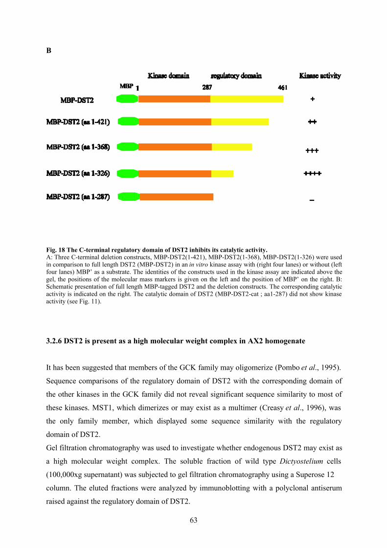
B
Fig. 18 The C-terminal regulatory domain of DST2 inhibits its catalytic activity.A: Three C-terminal deletion constructs, MBP-DST2(1-421), MBP-DST2(1-368), MBP-DST2(1-326) were usedin comparison to full length DST2 (MBP-DST2) in an in vitro kinase assay with (right four lanes) or without (leftfour lanes) MBP’ as a substrate. The identities of the constructs used in the kinase assay are indicated above thegel, the positions of the molecular mass markers is given on the left and the position of MBP’ on the right. B:Schematic presentation of full length MBP-tagged DST2 and the deletion constructs. The corresponding catalyticactivity is indicated on the right. The catalytic domain of DST2 (MBP-DST2-cat ; aa1-287) did not show kinaseactivity (see Fig. 11).
3.2.6 DST2 is present as a high molecular weight complex in AX2 homogenate
It has been suggested that members of the GCK family may oligomerize (Pombo et al., 1995).
Sequence comparisons of the regulatory domain of DST2 with the corresponding domain of
the other kinases in the GCK family did not reveal significant sequence similarity to most of
these kinases. MST1, which dimerizes or may exist as a multimer (Creasy et al., 1996), was
the only family member, which displayed some sequence similarity with the regulatory
domain of DST2.
Gel filtration chromatography was used to investigate whether endogenous DST2 may exist as
a high molecular weight complex. The soluble fraction of wild type Dictyostelium cells
(100,000xg supernatant) was subjected to gel filtration chromatography using a Superose 12
column. The eluted fractions were analyzed by immunoblotting with a polyclonal antiserum
raised against the regulatory domain of DST2.
63
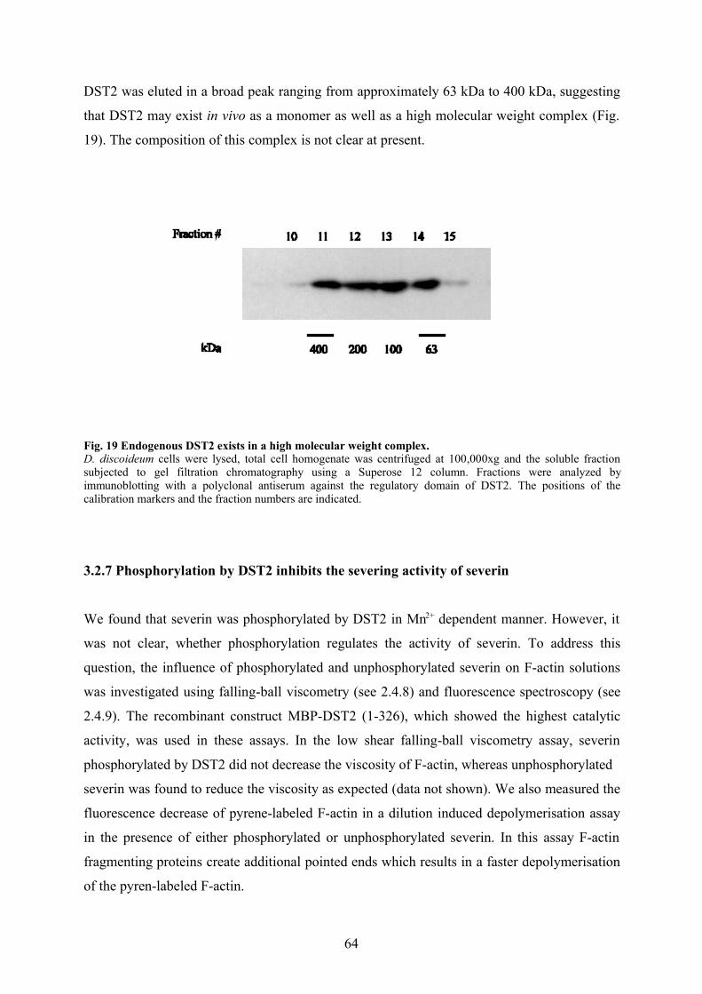
DST2 was eluted in a broad peak ranging from approximately 63 kDa to 400 kDa, suggesting
that DST2 may exist in vivo as a monomer as well as a high molecular weight complex (Fig.
19). The composition of this complex is not clear at present.
Fig. 19 Endogenous DST2 exists in a high molecular weight complex.D. discoideum cells were lysed, total cell homogenate was centrifuged at 100,000xg and the soluble fractionsubjected to gel filtration chromatography using a Superose 12 column. Fractions were analyzed byimmunoblotting with a polyclonal antiserum against the regulatory domain of DST2. The positions of thecalibration markers and the fraction numbers are indicated.
3.2.7 Phosphorylation by DST2 inhibits the severing activity of severin
We found that severin was phosphorylated by DST2 in Mn2+ dependent manner. However, it
was not clear, whether phosphorylation regulates the activity of severin. To address this
question, the influence of phosphorylated and unphosphorylated severin on F-actin solutions
was investigated using falling-ball viscometry (see 2.4.8) and fluorescence spectroscopy (see
2.4.9). The recombinant construct MBP-DST2 (1-326), which showed the highest catalytic
activity, was used in these assays. In the low shear falling-ball viscometry assay, severin
phosphorylated by DST2 did not decrease the viscosity of F-actin, whereas unphosphorylated
severin was found to reduce the viscosity as expected (data not shown). We also measured the
fluorescence decrease of pyrene-labeled F-actin in a dilution induced depolymerisation assay
in the presence of either phosphorylated or unphosphorylated severin. In this assay F-actin
fragmenting proteins create additional pointed ends which results in a faster depolymerisation
of the pyren-labeled F-actin.
64
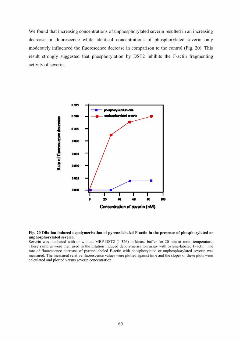
We found that increasing concentrations of unphosphorylated severin resulted in an increasing
decrease in fluorescence while identical concentrations of phosphorylated severin only
moderately influenced the fluorescence decrease in comparison to the control (Fig. 20). This
result strongly suggested that phosphorylation by DST2 inhibits the F-actin fragmenting
activity of severin.
Fig. 20 Dilution induced depolymerisation of pyrene-lebaled F-actin in the presence of phosphorylated orunphosphorylated severin.Severin was incubated with or without MBP-DST2 (1-326) in kinase buffer for 20 min at room temperature.These samples were then used in the dilution induced depolymerisation assay with pyrene-labeled F-actin. Therate of fluorescence decrease of pyrene-labeled F-actin with phosphorylated or unphosphorylated severin wasmeasured. The measured relative fluorescence values were plotted against time and the slopes of these plots werecalculated and plotted versus severin concentration.
65
Chapter 4
Discussion
4.1 DST2 is a new member of the STE20-like protein kinase family
We screened the sequences of the D. discoideum cDNA project database in Tsukuba, Japan
(Tsukuba website: http://www.csm.biol.tsukuba.ac.jp/cDNAproject.html), using DST1
(Dictyostelium STE20-like kinase 1) as query and discovered a cDNA clone coding for a
highly homologous protein kinase. Pending the identification of a physiological function, this
kinase was named DST2 (Dictyostelium STE20-like kinase 2). The 1.5 kilobase cDNA insert
contained an open reading frame coding for a protein of 461 amino acids with a predicted
molecular mass of 52 kDa. Based on sequence homologies, DST2 was identified as a new
member of the GCK subfamily of STE20-like kinases with an N-terminal catalytic 287-amino
acids and a C-terminal regulatory domain 174-amino acids. DST2 displays 69% amino acid
similarity to the kinase domain of DST1. DST1 is activated by osmotic stress, phosphorylates
the F-actin fragmenting protein severin and is most closely related to human SOK1, which is
activated by oxidant stress (Pombo et al., 1996; Eichinger et al., 1998; unpublished results).
Further sequence comparisons showed that DST2 is most closely related to human MST1 with
79% amino acid similarity in the kinase.
The strong similarity of DST2 and MST1 over their entire length suggests that these two
protein kinases are orthologues and might have similar or identical in vivo function. MST1
dimerizes and exists in a high molecular weight complex. Its catalytic activity is inhibited by
the regulatory domain. MST1 is activated upon treatment of cells with staurosporine, okadaic
acid, high concentrations of sodium arsenite, and extreme heat shock at 55°C (Creasy and
Chernoff, 1995; Creasy et al., 1996; Taylor et al., 1996). Furthermore, it was found that
MST1 is specifically cleaved by a caspase-3 like activity during apoptosis, induced by
CD95/Fas. Mutational analysis indicated that the caspase recognition and cleavage site in
MST1 is DEMD, which is similar to the consensus sequence for caspase 3 (DEVD) (Graves
et al., 1998). Interestingly, DST2 contains the sequence motif DEQD at the border between
the N-terminal kinase domain and the C-terminal regulatory domain (Fig. 21).
66
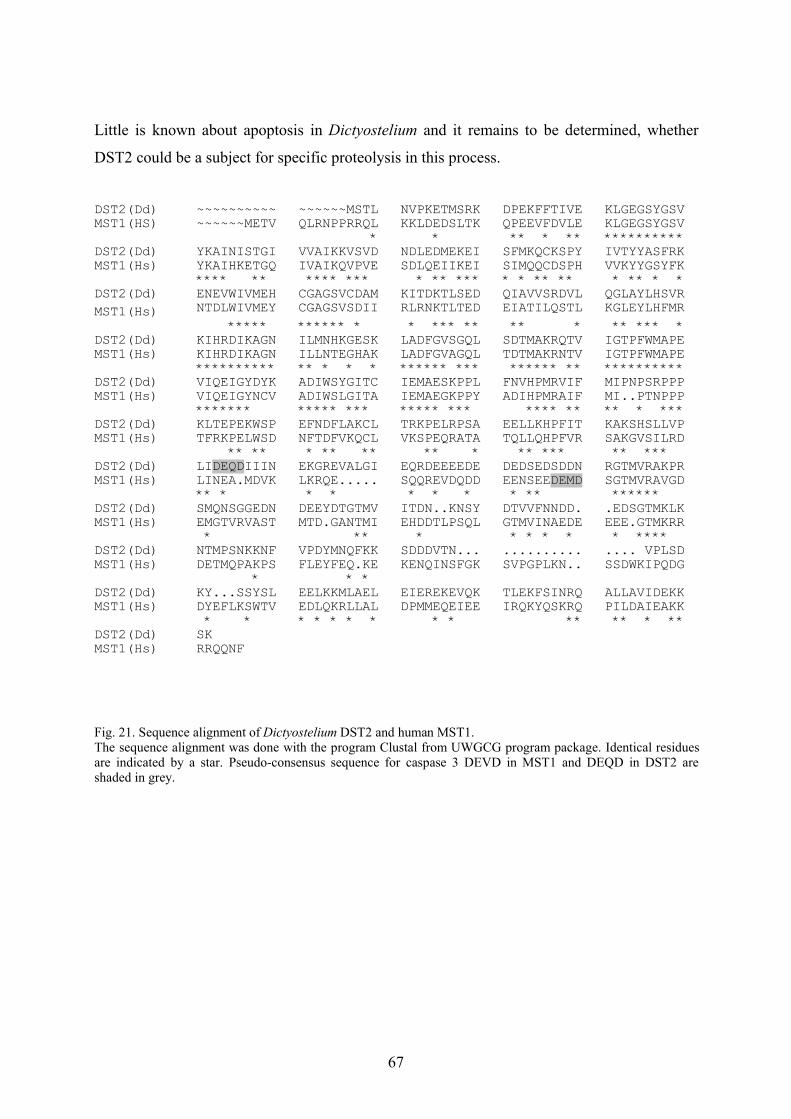
Little is known about apoptosis in Dictyostelium and it remains to be determined, whether
DST2 could be a subject for specific proteolysis in this process.
DST2(Dd) ~~~~~~~~~~ ~~~~~~MSTL NVPKETMSRK DPEKFFTIVE KLGEGSYGSVMST1(HS) ~~~~~~METV QLRNPPRRQL KKLDEDSLTK QPEEVFDVLE KLGEGSYGSV
* * ** * ** **********DST2(Dd) YKAINISTGI VVAIKKVSVD NDLEDMEKEI SFMKQCKSPY IVTYYASFRKMST1(Hs) YKAIHKETGQ IVAIKQVPVE SDLQEIIKEI SIMQQCDSPH VVKYYGSYFK
**** ** **** *** * ** *** * * ** ** * ** * *DST2(Dd) ENEVWIVMEH CGAGSVCDAM KITDKTLSED QIAVVSRDVL QGLAYLHSVRMST1(Hs) NTDLWIVMEY CGAGSVSDII RLRNKTLTED EIATILQSTL KGLEYLHFMR
***** ****** * * *** ** ** * ** *** *DST2(Dd) KIHRDIKAGN ILMNHKGESK LADFGVSGQL SDTMAKRQTV IGTPFWMAPEMST1(Hs) KIHRDIKAGN ILLNTEGHAK LADFGVAGQL TDTMAKRNTV IGTPFWMAPE
********** ** * * * ****** *** ****** ** **********DST2(Dd) VIQEIGYDYK ADIWSYGITC IEMAESKPPL FNVHPMRVIF MIPNPSRPPPMST1(Hs) VIQEIGYNCV ADIWSLGITA IEMAEGKPPY ADIHPMRAIF MI..PTNPPP
******* ***** *** ***** *** **** ** ** * ***DST2(Dd) KLTEPEKWSP EFNDFLAKCL TRKPELRPSA EELLKHPFIT KAKSHSLLVPMST1(Hs) TFRKPELWSD NFTDFVKQCL VKSPEQRATA TQLLQHPFVR SAKGVSILRD
** ** * ** ** ** * ** *** ** *** DST2(Dd) LIDEQDIIIN EKGREVALGI EQRDEEEEDE DEDSEDSDDN RGTMVRAKPRMST1(Hs) LINEA.MDVK LKRQE..... SQQREVDQDD EENSEEDEMD SGTMVRAVGD
** * * * * * * * ** ****** DST2(Dd) SMQNSGGEDN DEEYDTGTMV ITDN..KNSY DTVVFNNDD. .EDSGTMKLKMST1(Hs) EMGTVRVAST MTD.GANTMI EHDDTLPSQL GTMVINAEDE EEE.GTMKRR
* ** * * * * * * ****DST2(Dd) NTMPSNKKNF VPDYMNQFKK SDDDVTN... .......... .... VPLSDMST1(Hs) DETMQPAKPS FLEYFEQ.KE KENQINSFGK SVPGPLKN.. SSDWKIPQDG
* * *DST2(Dd) KY...SSYSL EELKKMLAEL EIEREKEVQK TLEKFSINRQ ALLAVIDEKKMST1(Hs) DYEFLKSWTV EDLQKRLLAL DPMMEQEIEE IRQKYQSKRQ PILDAIEAKK
* * * * * * * * * ** ** * **DST2(Dd) SKMST1(Hs) RRQQNF
Fig. 21. Sequence alignment of Dictyostelium DST2 and human MST1.The sequence alignment was done with the program Clustal from UWGCG program package. Identical residuesare indicated by a star. Pseudo-consensus sequence for caspase 3 DEVD in MST1 and DEQD in DST2 areshaded in grey.
67
4.2. DST2 is autophosphorylated and activated in Mn2+ dependent manner
We used MBP-tagged recombinant DST2 in in vitro kinase assays and found that DST2
possesses unusual cofactor requirements. The ability to phosphorylate itself and exogenous
substrates was consistently more than 10-fold higher in the presence of Mn2+ versus Mg2+ .
The increase of DST2 activity observed in response to increasing Mn2+ concentration
suggested that besides formation of the substrate MnATP, also binding of free Mn2+ to a
distinct site on DST2 might be required for full kinase activity. Mn2+ is the most potent
divalent cation activator of autophosphorylation for the tyrosine kinases of the insulin and
epidermal growth factor receptors (White et al., 1984; Carpenter et al., 1978). Recently, it was
shown that mammalian STE20-like kinase 3 (MST3), a close homologue of MST1, MST2
and DST2, also prefers Mn2+ to Mg2+ as a cofactor. MST3 catalytic activity was found to be
20-50 fold higher in the presence of Mn2+ compared with Mg2+ (Schinkmann et al., 1997).
However, the physiological role of Mn2+ in the activation of DST2 and MST3 needs to be
elucidated.
4.3 Regulation of DST2 activity
The protein kinase activity of PAKs can be stimulated by binding of activated GTP-bound
Cdc42 and Rac (Manser et al., 1994). Accumulated data suggest that PAK kinase activity is
repressed by an intramolecular interaction between the regulatory and catalytic domains.
Binding of GTP-bound Cdc42 or Rac disrupts this interaction resulting in a stimulation of
kinase activity (Bagrodia and Cerione, 1999).
For members of the GCK subfamily the putative regulatory role of the C-terminal non-
catalytic domain is not clear. In the case of MST1, MST2, and SOK-1 it apparently has an
inhibitory function because its removal resulted in an increase in kinase activity (Pombo et al.,
1996; Creasy et al., 1996). Furthermore, it has been shown that the C-terminal domains of
MST1 and MST2 mediate homo- and heterodimerization (Creasy et al., 1996). In order to
investigate the role of the putative regulatory domain of DST2 we performed a series of C-
terminal deletions and also looked for potential upstream effectors. The C-terminal deletion
results showed that DST2 contains a region between amino acids 326-461 that inhibits its
catalytic activity (3.2.5).
68

There are several possible mechanisms by which this region may regulate kinase activity.
Firstly, an inhibitory protein may bind to this region, as has been suggested for the related
GCK (Pombo et al., 1995). Inactivation of the inhibitory molecule would in turn activate the
kinase. Our in vitro kinase assay results argue against this scenario. Secondly, similar to the
PAKs, the C-terminal domain per se might inhibit the catalytic activity. Our results support
this model since several C-terminal deletions of DST2 were found to be much more active
than the full length molecule (Fig. 22). If this model is correct we are faced with the question
how the inhibitory role of the regulatory domain might be overcome in vivo. One possibility is
regulation by autophosphorylation and in in vitro kinase assays we found that the regulatory
domain was phosphorylated by full length DST2, suggesting that there might be a
pseudosubstrate site within this domain. However, no change in activity was observed.
Another possibility is activation through an upstream effector. We found that the regulatory
domain contains several potential phosphorylation sites for PKA, it was phosphorylated by
PKA in vitro and phosphorylation of recombinant DST2 by PKA significantly increased its
activity (3.2.3.2). Taken together, these data support a model, in which phosphorylation of the
regulatory domain by either PKA or another as yet unknown protein kinase, triggers a
conformational change that leads to activation of DST2.
Fig. 22 Schematic representation of human PAK1 regulation.Depiction of the Cdc42/Rac stimulated activation of PAK. The binding of Cdc42/Rac reverses an autoinhibitoryintramolecular interaction. The following domains or sequence motifs are indicated. P1, P2, P3, P4: potentialSH3 (Src-homology 3) binding motifs; PB: P21-binding domain; CP: cool-pix-binding region; STDK: serine-threonine kinase domain (adapted from Bagrodia and Cerione, 1999).
69
4.4. DST2 exists as a high molecular weight complex
In gel filtration chromatography (3.2.6), DST2 was eluted in a broad peak ranging from
approximately 63 kDa to 400 kDa, suggesting that DST2 may be part of a high molecular
weight complex in vivo. However, the composition of this complex is not clear at all. It could
be either composed of DST2 only or of DST2 plus additional proteins. Human MST1, which
is the closest homologue to DST2 and the only group II GCK subfamily member with
similarity to the regulatory domain of DST2, homodimerizes and seems to be associated with
additional proteins (Creasy et al., 1996). Therefore, we favor a model in which also in D.
discoideum a DST2 dimer is associated with additional proteins. Purification of native DST2
kinase from D. discoideum should give insight into the composition and the stoichiometry of
the proposed complex. The functional significance of complex formation of MST1 and
possibly DST2 and other kinases of the GCK subfamily is not clear. For some protein kinases
dimerization seems to be important for recognition by an effector molecule or phosphorylation
specificity. One such example is the monomeric cGMP-dependent protein kinase, which
phosphorylated histone and peptides, whereas only the dimer phosphorylated vimentin, the
protein suspected to be the real in vivo substrate (MacMillan-Crow and Lincoln, 1994).
4.5. Binding partners of PAK family kinases
Recently, several PAK-binding proteins were identified. These PAK binding partners are
p50Cool-1, p85Cool-1/Pix, Cool-2/Pix, which contain SH3 (src-homology), DH (Dbl-
homology) and PH (Pleckstrin-homology) domains (Manser et al., 1998). The binding of
Cool/Pix to PAK seems to activate PAK either by cooperating with the binding of activated
Cdc42 or Rac or by altering PAK confirmation (Bagrodia and Cerione, 1999).
Some of the group I GCKs might be downstream effectors of TNF (tumor necrosis factor).
Binding of TNF to the TNF receptor recruits TRAF2 (TNF receptor-associated factor 2) and it
was found that GCK and GCLK can be associated in vivo with TRAF2. This result suggested
that GCK and GCKR may be important effectors for TRAF2 (Pombo et al., 1995; Yuasa et
al., 1998; Arch et al., 1998). So far no binding partners have been identified for the Group II
GCK subfamily, to which DST2 belongs to.
70

However, it was found that the Group II GCK subfamily can be activated by different
environmental stresses. MST1 and MST2 can be activated by treatment of cells with
staurosporine, okadaic acid, high concentrations of sodium arsenite, and extreme heat shock at
55°C (Creasy et al., 1996). MST1 could also be activated in vitro by phosphatase 2A
suggesting that MST1 might be activated by dephosphorylation (Taylor et al., 1996). SOK1 is
strongly activated by oxidant stress and also by ischemic injury (Pombo et al., 1996;
Schinkmann and Blenis, 1997). Nothing is known about environmental conditions that might
trigger activation of DST2. However, its pronounced similarity to human MST1 suggests a
possible role of DST2 in an as yet unknown stress response pathway in D. discoideum.
Knock-out experiments as well as treatment of cells with various stresses in combination with
in vivo labelling and immunoprecipitation should provide insight into the physiological role of
DST2.
4.6. Signalling to the cytoskeleton
Several members of the PAK family have recently been implicated in cytoskeletal
reorganization or the regulation of cytoskeletal proteins. PAK1 kinase activity is essential for
disassembly of focal adhesion (Frost et al., 1998). LIMK1 (LIM-motif-containing protein
kinase) phosphorylates the actin-depolymerizing factor cofilin and serves as a target for PAK1
(Edwards et al.,1999). MIHCK from Dictyostelium and its homologue from Acanthamoeba
phosphorylate the heavy chain of some of the myosinI isozymes on a single serine or
threonine residue and thereby stimulate their actin-activated MgATPase activity 30 to 50 fold
(Lee and Côté, 1995; Brzeska and Korn, 1996). Cloning of the corresponding genes revealed
that MIHCK is a member of the PAK family and closely related to mammalian PAK and yeast
Ste20p molecules (Lee et al., 1996; Brzeska et al., 1996). In gel overlay assays and affinity
chromatography experiments MIHCK from Dictyostelium interacted with GTPS-labeled
Rac1 and Cdc42, which probably bind to a conserved PBD commonly found in the N-terminal
regulatory domain of true PAKs. Interestingly, in the presence of active Rac1 and Cdc42
autophosphorylation of MIHCK increased from 1 up to 9 moles of phosphate per mol of
kinase concomitant with an approximately 10-fold stimulation of the rate of myosin ID
phosphorylation. These results suggest that MIHCK directly links Cdc42/Rac signalling
pathways to motile processes driven by myosin I molecules (Lee et al., 1996).
71

The role of GCK subfamily members in signalling to the cytoskeleton is less clear. Recently it
was shown that overexpression of either wild type MST1 or a truncated mutant induced
morphological changes (Graves et al., 1998). DST1 was found to phosphorylate the actin
fragmenting protein severin (Eichinger et al., 1998). In vitro TNIK could phosphorylate
gelsolin, the mammalian severin homologue and overexpression of wild type TNIK resulted
in the disruption of F-actin structure and the inhibition of cell spreading. (Fu et al., 1999).
Here we showed that native severin was also a substrate for DST2 in vitro (3.2.3.1). Severin is
a Ca2+- activated actin-binding protein that nucleates actin assembly, fragments F-actin and
caps the newly created barbed end. It consists of three highly conserved domains. Domain 1 of
severin is responsible for F-actin capping, domains 1/2 cooperate in F-actin fragmenting
whereas domains 2/3 possess two F-actin binding sites (Eichinger et al., 1992).
Phosphorylation of severin by DST2 resulted in an inhibition of severin’s F-actin fragmenting
activity (see 3.2.7), suggesting that DST2 may play a role in the remodeling of the actin
cytoskeleton. Mass spectrometry data suggested that all phosphorylation sites on severin seem
to be in the third domain, between residues 260-315. Phoshorylation of domain 3 might lead
to a conformational change and thus inhibit the F-actin fragmenting activity of severin.
Several recent findings indicate that PKA functions in regulating STE20-like kinases.
Functional connections between PKA and PAKs have been demonstrated in the regulation of
oocyte maturation in Xenopus and chemotaxis in Dictyostelium (Faure et al., 1999; Chung et
al., 1999). MST3b was directly phosphorylated and inhibited by PKA in vivo and in vitro
(Zhou et al., 2000). We have shown (3.2.3.2) that exposure of DST2 in in vitro kinase assays
to the purified catalytic subunit of PKA resulted in the phosphorylation of the regulatory
domain and we also observed a strong increase in the basal autophosphorylation activity of
DST2.
Taken together our data suggest that PKA might be a potential upstream regulatory kinase for
DST2 which in turn might phosphorylate and regulate the F-actin fragmenting protein severin.
The inhibition of severin’s F-actin fragmenting activity might play a role in the reorganization
of the actin cytoskeleton. Further studies are needed in order to unravel upstream and
downstream components of signalling via DST2.
72

4.7. Future prospects
To learn more about the in vivo function of DST2, the following studies need to be done in the
future:
1. Disruption of the Dictyostelium DST2 gene by homologous recombination.
2. Identification of possible binding partners of DST2 using the yeast two-hybrid system.
3. Expression of GFP-DST2 fusion proteins to investigate the intracellular localisation.
4. Production of better polyclonal and/or monoclonal antibodies against DST2.
5. Purification and characterisation of endogenous DST2 using affinity chromatography.
6. In vivo labelling experiments with cells treated with various stresses followed by
immunoprecipitation of DST2 and/or severin.
73
Chapter 5
References
André, E., Lottspeich, F., Schleicher, M. and Noegel, A. (1988) Severin, gelsolin, and villin
share a homologous sequence in regions presumed to contain F-actin severing domains. J.
Biol. Chem. 15, 722-727
Arber, S., Barbayannis, F. A., Hanser, H., Schneider, C., Stanyon, C. A., Bernard, O. and
Caroni P. (1998) Regulation of actin dynamics through phosphorylation of cofilin by LIM-
Kinase. Nature. 393, 805-809
Arch, R. H., Gedrich, R. W. and Thompson, C. B. (1998) Tumor necrosis factor receptor-
associated factors (TRAFs)--a family of adapter proteins that regulates life and death. Genes.
Dev. 12, 2821-2830
Bagrodia, S., Derijard, B., Davis, R. J. and Cerione, R. A. (1995) Cdc42 and PAK-mediated
signalling leads to Jun kinase and p38 mitogen-activated protein kinase activation. J. Biol.
Chem. 270, 27995-27998
Bagrodia, S. and Cerione, R. A. (1999) PAK to the future. Trends in Cell Biol. 9, 350-355.
Bear, J. E., Svitkina, T. M., Krause, M., Schafer, D. A., Loureiro, J. J., Strasser, G. A., Maly,
I. V., Chaga, O. Y., Cooper, J. A., Borisy, G. G. and Gertler, F. B. (2002) Antagonism
between Ena/VASP Proteins and Actin Filament Capping Regulates Fibroblast Motility. Cell
109, 509-521
Benner, G. E., Dennis, P. B., and Masaracchia, R. A. (1995) Activation of an S6/H4 kinase
(PAK 65) from human placenta by intramolecular and intermolecular autophosphorylation.
J. Biol. Chem. 270, 21121-21128
74

Bokoch, G. M., Reilly, A. M., Daniels, R. H., King, C. C., Olivera, A., Spiegel, S. and Knaus,
U. G. (1998) A GTPase-independent mechanism of p21-activated kinase activation.
Regulation by sphigosine and other biologically active lipids. J. Biol. Chem. 273, 8137-8144
Bonner, J. T. (1967) The cellular silm molds, Princeton university
Bradford, M. M (1976) A rapid and sensitive method for the quantification of microgram
quantities of protein utilizing the principle of protein dye-binding. Anal. Biochem. 72:248-254
Bretscher, A. and Weber, K. (1979) Villin: the major microfilament-associated protein of the
intestinal microvillus. Proc Natl. Acad. Sci. U S A 76, 2321-2325
Brown, S. S., Yamamoto, K. and Spudich, J. A. (1982) A 40,000-dalton protein from
Dictyostelium discoideum affects assembly properties of actin in a Ca2+-dependent manner. J.
Cell. Biol. 93, 205-210
Brzeska, H. and Korn, E. D. (1996) Regulation of class I and class II myosins by heavy chain
phosphorylation. J. Biol. Chem. 271, 16983-16986
Brzeska, H., Szczepanowska, J., Hoey, J. and Korn, E. D. (1996) The catalytic domain of
acanthamoeba myosin I heavy chain kinase. II. Expression of active catalytic domain and
sequence homology to p21-activated kinase (PAK). J. Biol. Chem. 271, 27056-27062
Caron, E. (2002) Regulation of Wiskott-Aldrich syndrome protein and related molecules.
Curr Opin Cell Biol. 14, 82-7.
Carpenter, G., King, L. Jr. and Cohen, S. (1978) Epidermal growth factor stimulates
phosphorylation in membrane preparations in vitro. Nature 276, 409-410
Chung, C. Y., and Firtel, R. A. (1999) PAKa, a putative PAK family member, is required for
cytokinesis and the regulation of the cytoskeleton in Dictyostelium discoideum cells during
chemotaxis. J. Biol. Chem. 147, 559-576
75
Claviez, M., Pagh, K., Maruta, H., Baltes, W., Fisher, P. and Gerisch, G. (1982). Electron
microscopic mapping of monoclonal antibodies on the tail region of Dictyostelium myosin.
EMBO J. 1, 1017-1022
Creasy, C. L. and Chernoff, J. (1995a) Cloning and charaterisation of a human protein kinase
with homology to STE20. J. Biol. Chem. 37, 21695-21700.
Creasy, C. L and Chernoff, J. (1995b) Cloning and of a member of the MST sunfamily of
STE`20-like kinases. Gene. 167, 303-306.
Creasy, C. L., Ambrose, D. M. and Chernoff, J. (1996) The STE20-like protein kinase, MST1,
dimerizes and contains an inhibitory domain. J. Biol. Chem. 271, 21049-21053
Cunningham, C. C., Stossel, T. P. and Kwiatkowski, D. J. (1991) Enhanced motility in NIH
3T3 fibroblasts that overexpress gelsolin. Science 251, 1233-1236
Daniels, R. H. and Bokoch, G. M. (1999) p21-activated protein kinase: a crucial component of
morphological signalling? Trends Biochem Sci 24, 350-355.
Diener, K., Wang, X. S., Chen, C., Meyer, C. F., Keesler, G., Zukowski, M., Tan, T. H. and
Yao, Z (1997) Activation of the c-Jun N-terminal kinase pathway by a novel protein kinase
related to human germinal center kinase. Proc. Natl. Acad. Sci. U. S. A. 94, 9687-9692
Edwards, D. C., Sanders, L. C., Bokoch, G. M. and Gill, G. N. (1999) Activation of LIM-
kinase by Pak1 couples Rac/Cdc42 GTPase signalling to actin cytoskeletal dynamics. Nat.
Cell. Biol.1, 253-259.
Eichinger, L., Noegel, A. A. and Schliecher, M. (1990) Ca2(+)-binding proteins as
components of the cytoskeleton. Adv. Exp. Med. Biol. 269, 99-102.
Eichinger, L., Noegel, A. A. and Schleicher, M. (1991) Domain structure in actin-binding
proteins: expression and functional of truncated severin. J. Cell. Biol. 112, 665-676
76
Eichinger, L. and Schliecher, M. (1992) Characterisation of actin- and lipid-binding domains
in severin, a Ca(2+)-dependent F-actin fragmenting protein. Biochemistry. 31, 4779-4787
Eichinger, L., Bomblies, L., Vandekerckhove., Schleicher, M. and Gettemans, J (1996) A
novel type of protein kinase phosphorylates actin in the actin fragmin complex. EMBO. J. 15,
5547-5556
Eichinger, L., Baehler, M., Diez, M., Eckerskorn, C. and Schleicher, M. (1998)
Characterisation and cloning of a Dictyostelium STE20-like protein kinase that
phosphorylates the actin binding protein severin. J. Biol. Chem. 273, 12952-12959
Eichinger, L., Lee, S. S. and Schleicher, M. (1999) Dictyostelium as model system for studies
of the actin cytoskeleton by molecular genetics Micro Res and Tech. 47, 124-134
Every, D. and Ashworth, J. M. (1975) Rates of degradation and synthesis of glycosidases de
novo during growth and differentiation of the Dictyostelium discoideum. Biochem. J. 148,
169-177
Faure, S., Vigneron, S., Galas, S., Brassac, T., Delsert, C. and Morin N. (1999) Control of
G2/M transition in Xenopus by a member of the p21-activated kinase (PAK) family; a link
between protein kinase A and PAK signalling pathways? J. Biol. Chem. 274, 3573-3579
Franke, J. and Sussman, M. (1971) Synthesis of uridine diphosphate glucose
pyrophosphorylase during the development of Dictyostelium discoideum. J. Biol. Chem. 326,
6381-6388
Frost, J. A., Khokhlatchev. A, Stippec, S., White, M. A. and Cobb, M. H. (1998) Differential
effects of PAK1-activating mutations reveal activity-dependent and -independent effects on
cytoskeletal regulation. J. Biol. Chem. 273, 28191-28198.
Friesen, H., Lunz, R., Doyle, S. and Segall, J. (1994) Mutation of the SPS1-encoded protein
kinase of Saccharomyces cerevisiae leads to defects in transcription and morphology during
spore formation. Genes Dev. 8, 2162-2175
77

Fu, C. A., Shen, M., Huang, B. C. B., Lasaga, J., Payan, D. G. and Luo, Y. (1999) TNIK, a
novel member of the germinal center kinase family that activates the c-Jun N-terminal kinase
pathway and regulates the cytoskeleton. J. Biol. Chem. 274, 30729-30737.
Garrity, P. A., Rao, Y., Salecker, I., McGlade, J., Pawson, T. and Zipursky, S. L (1996)
Drosophila photoreceptor axon guidance and targeting requires the dreadlocks SH2/SH3
adapter protein. Cell 85, 639-650
Gettemans, J., De Ville, Y., Waelkens, E. and Vandekerckhove, J. (1995) The actin-binding
properties of the Physarum actin-binding complex. Regulation by calcium, phospholipids and
by phosphorylation. J. Biol. Chem. 270, 2644-2651
Graves, J. D., Gotoh, Y., Draves, K. E., Ambrose, D., Chenoff, J., Clark, E. A. and Krebs, E.
G. (1998) Caspase-mediated activation and induction of apoptosis by the mammalian STE20-
like kinase MST1, EMBO. J. 17, 2224-2234.
Gurdon, J. B. (1974) The control of gene expression in animal development. Clarendon Press,
Oxford
Hall, A. (1998) Rho GTPases and the actin cytoskeleton. Science. 279, 509-514
Hartwig, J. H., Bokoch, G. M., Carpenter, C. L., Janmey, P. A., Taylor, L. A., Toker, A. and
Stossel, T. P. (1995) Thrombin receptor ligation and activated Rac uncap actin filament
barbed ends through phosphoinositide synthesis in permeabilized human platelets. Cell. 82,
643-53
Hasegawa, T., Takahashi, S., Hayashi, H. and Hatano, S. (1980) Fragmin : A calcium ion
sensitive regulatory factor on the formation of actin filaments. Biochemisty 19, 2677-2683
Herskowitz, I. (1995) MAP kinase pathways in yeast: for mating and more. Cell. 80, 187-197
Hinssen, H. (1981) An actin modulating protein from Physarum polycephalum. Eur. J. Cell.
Biol. 23, 225-240
78
Holmes, D. S and Quigley, M. (1981) A rapid boiling method for the preparation of bacterial
plasmids. Anal. Biochem. 114, 193-197
Housewart, M. and Cleveland, D. (1998) Intermediate filaments and their associated proteins:
multiple dynamic personalities. Curr. Opin. Cell Biol. 10, 93-101
Howe, A. K. and Juliano, R. L. (2000) Regulation of anchorge-dependent signal transduction
by protein kinase A and p21-activated kinase Nat. Cell. Biol. 2, 593-600
Hu, M. C. T., Qie, W. R., Wang, X., Meyer, C. F. and Tan, T. H (1996) Human HPK1, a
novel human hematopoietic progenitor kinase that activates the JNK/SAPK kinase cascade.
Genes Dev. 10, 2251-2264
Janmy, P. A., Iida, K., Yin, H. L. and Stossel, T. P. (1987) Polyphosphoinositide micelles and
polyphosphoinositide-containing vesicles dissociate endogenous gelsolin-actin complexes and
promote actin assembly from the fast-growing end of actin filaments blocked by gelsolin. J.
Bio. Chem. 262, 12228-12236.
Janmey, P. A. and Stossel, T. P. (1989) Gelsolin-polyphosphoinositide interaction. Full
expression of gelsolin-inhibiting function by polyphosphoinositides in vesicular form and
inactivation by dilution, aggregation, or masking of the inositol head group. J. Biol. Chem.
264, 4825-4831
Karakesisoglou, I., Janssen, K. P., Eichinger, L., Noegel, A. A. and Schleicher, M. (1999)
Identification of a suppressor of the Dictyostelium profilin-minus phenotype as a CD36/LIMP-
II homologue. J. Cell. Biol. 145, 167-181
Katz, P., Whalen, G. and Kehrl, J. H. (1994) Differential expression of a novel protein kinase
in human B lymphocytes. Preferential localization in the germinal center. J. Biol. Chem. 269,
16802-16809
Kiefer, F., Tibbles, L. A., Anafi, M., Janssen, A., Zanke, B. W., Lassam, N., Pawson, T.,
Woodgett, J. R. and Iscove, N. R. (1996) HPK1, a hematopoietic protein kinase activating the
SAPK/JNK pathway. EMBO J. 15, 7013-7025
79
Kiuru, S., Javela, K., Somer, H. and Kekomaki, R. (2000) Altered platelet shape change in
hereditary gelsolin Asp187Asn-related amyloidosis. Thromb. Haemost 83, 491-495.
Knecht, D. A. and Loomis, W. F. (1987) Antisense RNA inactivation of myosin heavy chain
gene expression in Dictyostelium discoideum. Science 236, 1081-1086
Korn, E. D., Carlier, M-F. and Pantaloni, D. (1987) Actin polymerization and ATP hydrolysis.
Science 238, 638-644
Kuspa, A. and Loomis, W. F. (1992) Tagging developmental genes in Dictyostelium by
restriction enzyme-mediated integration of plasmid DNA. Proc. Natl. Acad. Sci. U S A 89,
8803-8807
Kwiatkowski, D. J., Stossel, T. P., Orkin, S. H., Mole, J. E., Colten, H. R. and Yin, H. L.
(1986) Plasma and cytoplasmic gelsolins are encoded by a single gene and contain a
duplicated actin-binding domain. Nature. 323, 1986, 455-9
Kwiatkowski, D. J. (1999) Functions of gelsolin: motility, signalling, apoptosis, cancer. Curr.
Opin. Cell Biol. 11, 103
Kyriakis, J. M. and Avruch, J. (1996) Sounding the alarm: protein kinase cascades activated
by stress and inflammation. J. Biol. Chem. 271, 24313-24316
Lammli, UK. (1970) Clevage of structure proteins during assembly of the head of
bacteriophage T4. Nature 227; 680-685
Leberer, E., Dignard, D., Harcus, D., Thomas, D. Y. and Whitway, M (1992) The protein
kinase homologue STE20p is required to link the yeast pheromone response G-protein beta
gamma subunits to downstream signalling components. EMBO J. 11, 4815-4824
Lee, S. F. and Côté, G. P. (1995) Purification and of a Dictyostelium protein kinase required
for actin activation of the Mg2+ ATPase activity of Dictyostelium myosin ID. J. Biol. Chem.
270, 11776-11782
80
Lee, S. F, Egelhoff, T. T., Mahasneh, A. and Cote, G. P. (1996) Cloning and of a
Dictyostelium myosin I heavy chain kinase activated by Cdc42 and Rac. J. Biol. Chem. 271,
27044-27048
Liu, T., Williams, J. G. and Clarke, M. (1992) Inducible expression of calmodulin antisense
RNA in Dictyostelium cells inhibits the completion of cytokinesis. Mol. Biol. Cell. 3, 1403-
1413
Loomis, W. F. (1975) Dictyostelium discoideum : A developmental system, Academic press,
New York
Loomis, W. F (1996) Genetic networks that regulate development in Dictyostelium cells.
Microbiol. Rev. 60, 135-150
Machesky. L. M., Reeves, E., Wientjes, F., Mattheyse, F., Grogan, A., Totty, N., Burlingame,
A.L., Hsuan,J. J. and Segel, A.W. (1997) Mammalian actin-related protein 2/3 complex
localizes to regions of lamellipodial protrusion and is composed of evolutionarily conserved
proteins. Biochem. J. 328, 105-112
Machesky, L. M. and Insall, R. H. (1998) Scar1 and the related Wiskott-Aldrich syndrome
protein, WASP, regulate the actin cytoskeleton through the Arp2/3 complex. Curr. Biol. 8,
1347-1356
MacMillan-Crow, L. A. and Lincoln, T. M. (1994) High-affinity binding and localization of
the cyclic GMP-dependent protein kinase with the intermediate filament protein vimentin.
Biochemistry 33, 8035-8043
Manser, E., Leung, T., Salihuddin, H., Zhao, Z. S. and Lim, L. (1994) A brain serine/threonine
protein kinase activated by Cdc42 and Rac1. Nature. 367, 40-46
Manser, E., Loo, T. H., Koh, C. G., Zhao, Z. S., Chen, X. Q., Tan, L., Tan, I., Leung, T. and
Lim, L. (1998) PAK kinases are directly coupled to the PIX family of nucleotide exchange
factors. Mol. Cell. 1, 183-92
81
Manstein, D. J., Titus, M. A., De Lozanne, A. and Spudich, J. A. Gene replacement in
Dictyostelium: generation of myosin null mutants. EMBO. J. 8, 923-32
Maruta, H. and Isenberg, G. (1983) Ca2+ dependent actin-binding phosphoprotein in
Physarum polycephalum. J. Biol. Chem. 258, 10151-10158.
McGrath, J. L., Osborn, E. A., Tardy, Y. S., Dewey, C. F. and Hartwig, J. H. (2000)
Regulation of the actin cycle in vivo by actin filament severing. PNAS. 12, 6532-6537
McLean-Fletcher, S. and Pollard, T. D. (1980) Visometric analysis of the gelation of
Acanthamoeba extracts and purification of two gelation factors. J. Cell. Biol. 85, 414-428
Mischak, H., Seitz, T., Janosch, P., Eulitz, M., Steen, H., Schellerer, M., Philipp, A. and
Kolch, W. (1996) Negative regulation of Raf-1 by phosphorylation of serine 621. Mol Cell
Biol. 16, 5409-18
Mueller, K. and Gerisch, G. (1978) A specific glycoprotein as the target site of adhesion
blocking Fab in aggregating Dictyostelium cells. Nature 274, 445-449
Mullins, R.D., Stafford, W.F. and Pollard, T. D. (1997) Structure, subunit topology, and actin-
binding activity of the Arp2/3 complex from Acanthamoeba. J. Cell. Biol. 136, 331-343
Murray, B. A., Yee, L. D. and Loomis, W. F. (1981) Immunological analysis of glycoprotein
(contact sites A) involved in intercellular adhesion of Dictyostelium discoideum. J. Suparmol.
Struct. Cell. Biochem. 17, 197-211
Noegel, A. A. and Luna, E. J. (1995) The Dictyostelium cytoskeleton. Experientia 51, 1135-
1143
Pardee, J. D. and Spudich, J. A. (1982) Purification of muscle actin. Meth. Enzym. 85: 164-
181
82
Paul, A., Wilson, S., Belham, C. M., Robinson, C. J., Scott, P. H., gould, G. W. and Plevin, R.
(1997) Stress-activated protein kinases: activation, regulation and function. Cell. Signal. 9,
403-410
Pombo, C. M., Kehrl, J. H., Sanchez, I. Katz, P., Avruch, J., Zon, L. I., Woodget, J. R., Force,
T. and Kyriakis, J. M. (1995) Activation of the SAPK pathway by the human STE20
homologue germinal center kinase. Nature 377, 750-754
Pombo, C. M., Bonventre, J. V., Molner, A., Kyriakis, J. and Force, T. (1996) Activation of a
human STE20-like kinase by oxidant stress defines a novel response pathway EMBO. J. 15,
4537-4546
Pope, B., Way, M., Matsudaira, P. T. and Weeds, A. (1994) of the F-actin binding domains of
villin: classification of F-actin binding proteins into two groups according to their binding
sites on actin. FEBS Lett, 338, 58-62
Pringault, E., Arpin, M., Garcia, A., Finidori, J. and Louvard D. (1986) A human villin cDNA
clone to investigate the differentiation of intestinal and kidney cells in vivo and in culture.
EMBO J 5, 3119-24
Pruyne D, Evangelista M, Yang C, Bi E, Zigmond S, Bretscher A, Boone C. (2002) Role of
Formins in Actin Assembly: Nucleation and Barbed End Association. Science in Press
Qian, Z., Lin, C., Espinosa, R., LeBeau, M. and Rosner, R. (2001) Cloning and
characterization of MST4, a novel STE20-like kinase. J. Biol. Chem 276, 22439-22445
Raper, K. B (1935) Dictyostelium discoideum, a new species of slim mold decaying forest
leaves. J. Agric. Res. 50, 133-147
Robinson, M. J. and Cobb, M. H. (1997) Mitogen-activated protein kinase pathways. Curr.
Opin. Cell Biol. 9, 180-186
Sambrook, J., Fritsch, E. F. and Maniatis, T. (1989) Molecular cloning: a laboratory manual.
Cold Spring Harbor Laboratory Press.
83
Sandoz, D., Chailley, B., Boisvieux-Ulrich, E., Lemullois, M., Laine, M. C. and Bautista-
Harris, G. (1988) Organization and functions of cytoskeleton in metazoan ciliated cells. Biol
Cell 63, 183-93.
Saxena, M., Williams, S. and Mustelin, T. (1999) Crosstalk between cAMP-dependent kinase
and MAP kinase through a protein tyrosine phosphatase. Nat. Cell. Biol, 1, 305-311
Schinkmann, K. and Blenis, J. (1997) Cloning and characterisation of a human STE20-like
protein kinase with unusual cofactor requirements. J. Biol. Chem 272, 28695-28703
Schleicher, M., Andre, E., Hartmann, H. and Noegel, A. A. (1988) Actin-binding proteins are
Conserved from slime molds to man. Dev Genet 9, 521-530
Schleicher, M. and Noegel, A. A. (1992) Dynamics of the Dictyostelium cytoskeleton during
chemotaxis. New. Biol. 4, 461-472
Schleicher, M., Andre, B., Andreoli, C., Eichinger, L., Haugwitz, M., Hofmann, A.,
Karakesisoglou, J., Stockelhuber, M. and Noegel, A. A. (1995) Structure/function studies on
cytoskeletal proteins in Dictyostelium amoebae as a paradigm. FEBS. Lett. 369, 38-42
Schmidt, A. and Hall, M.N. (1998). Signalling to the actin cytoskeleton. Ann. Rev. Cell.
Devel. Biol. 14, 305-338
Schoenwaelder, S. M. and Burridge, K. (1999) Bidirectional signalling between the
cytoskeleton and integrins. Curr. Opin. Cell boil. 11, 274-286
Schweitzer, B. and Philippsen, P. (1991) CDC15, an essential cell cycle gene in
Saccharomyces cerevisiae, encodes a protein kinase domain. Yeast 7, 265-73.
Seastone, D. J., Harris, E., Temesvari, L. A., Bear, J. E., Saxe, C. L. and Cardelli, J. (2001)
The WASp-like protein scar regulates macropinocytosis, phagocytosis and endosomal
membrane flow in Dictyostelium. J. Cell. Sci. 114, 2673-2683.
84
Sells, M. A. and Chernoff, J. (1997) Human p21-activated kinase (Pak1) regulates actin
organization in mammalian cells. Curr Biol. 7, 202-210.
Shi, C. S. and Kehrl, J. H. (1997) Activation of stress-activated protein kinase/c-Jun N-
terminal kinase, but not NF-kappaB, by the tumor necrosis factor (TNF) receptor 1 through a
TNF receptor-associated factor 2- and germinal center kinase related-dependent pathway. J.
Biol. Chem. 272, 32102-32107
Simpson, P. A., Spudich, J. A. and Parham, P. (1984) Monoclonal antibodies prepared against
Dictyostelium actin : and interaction with actin. J. Cell Biol. 99: 287-295
Siu, C. H, Lam, T. Y, Wong, L. M. (1988) Expression of the contact site A glycoprotein in
Dictyostelium discoideum: quantitation and developmental regulation. Biochim Biophys Acta.
968, 283-290.
Spudich, J. A. and Watt, S. (1971) The regulation of rabbit societal muscle contraction.
Biochemical studies on the interaction of tropomyosin-troponin complex with actin and the
proteolytic fragments of myosin. J. Biol. Chem. 246: 4866-4871
Stossel, T. P (1989) From signal to pseudopod. How cells control cytoplasmic actin assembly.
J. Biol. Chem. 264, 18261-18264
Su, Y. C., Han, J., Xu, S., Cobb, M. and Skolink, E. Y (1997) NIK is a new STE20-related
kinase that binds NCK and MEKK1 and activates the SAPK/JNK cascade via a conserved
regulatory domain. EMBO J. 16, 1279-1290
Svitkina, T. M. and Borisy, G. G. (1999) Arp2/3 complex and actin depolymerizing
factor/cofilin in dendritic organization and treadmilling of actin filament array in
lamellipodia. J. Cell. Biol. 145, 1009-26
Taylor, S. S., Buechler, J. A. and Yonemoto, W. (1990) cAMP-dependent protein kinase:
framework for adiverse family of regulatory enzymes. Annu. Rev. Biochem. 59, 971-1005
Taylor, L. K., Wang, H. C. and Erikson, R. L. (1996) Newly identified stress-responsive
protein kinases, Krs-1 and Krs-2. Proc. Natl. Acad. Sci. U S A. 93, 10099-10104.
85
Thomas, D. A. and Wright, B. E. (1976) Glycogen phosphorylase in Dictyostelium
discoideum. II. Synthesis and degradation during differentiation. J. Biol. Chem. 251, 1258-
1263
T'Jampens, D., Meerschaert, K., Constantin, B., Bailey, J., Cook, L. J., De Corte, V., De Mol,
H., Goethals, M., Van Damme, J., Vandekerckhove, J. and Gettemans, J. (1997) Molecular
cloning, over-expression, developmental regulation and immunolocalization of fragmin, a
gelsolin-related actin-binding protein from Physarum polycephalum plasmodia. J Cell Sci
110, 1215-26
Towbin, H., Staehelin, T. and Gordon, J. (1979) Electrophoretic transfer of proteins from
polyacrylamide gels to nitrocellulose sheets: procedure and some applications. Proc. Natl.
Acad. Sci. USA 76:4350-4354
Tung, R. M. and Blenis, J. (1997) A novel human SPS1/STE20 homologue, KHS, activates
Jun N-terminal kinase. Oncogene 14, 653-659
Yang, N., Higuchi, O., Ohashi, K., Nagata, K., Wada, A., Kangawa, K., Nishida, E. and
Mizuno, K. (1998) Cofilin phosphorylation by LIM-kinase1 and its role in Rac-mediated actin
Recognitation. Nature. 393, 809-812.
Valiron, O., Caudron, N. and Job D. (2001) Microtubule dynamics. Cell Mol Life Sci 258,
2069-2084.
Vardar, D., Chishti, A. H., Frank, B. S., Luna, E. J., Noegel, A. A., Oh, S. W., Schleicher,
M. and McKnight, C. J. (2002) Villin-type headpiece domains show a wide range of F-actin-
binding affinities. Cell. Motil. Cytoskeleton. 52, 9-21.
Vossler, M. R., Yao, H., York, R. D., Pan, M. G. and Stork, P. J. (1997) cAMP activates MAP
kinase and Elk-1 through a B-Raf- and Rap1-dependent pathway. Cell, 89, 73-82
Walsh, D. A. and Van Patten, S. M. (1994) Multiple pathway signal transduction by the
cAMP-dependent protein kinase FASEB J. 8, 1227-36
86
Watts, D. J. (1984) Protein synthesis during development and differentiation in the cellular
slime mould Dictyostelium discoideum. Biochem. J. 220, 1-14
Way, M. and Weeds, A. G. (1988) Nucleotide sequence of pig plasma gelsolin. Comparison
of protein sequence with human gelsolin and other actin-severing proteins shows strong
homologies and evidence for large internal repeats. J. Mol. Biol. 203, 1127-33
Wegner, A. (1976) Head-to-tail polymerization of actin. J. Mol. Biol. 108, 139-150
White, M. F., Haring, H. U., Kasuga, M. and Kahn, C. R. (1984) Kinetic properties and sites
of autophosphorylation of the partially purified insulin receptor from hepatoma cells. J. Biol.
Chem. 259, 255-264
Witke, W., Sharpe, A. H., Hartwig, J. H., Azuma, T., Stossel, T. P. and Kwiatkowski, D. J.
(1995) Hemostatic, inflammatory, and fibroblast responses are blunted in mice lacking
gelsolin. Cell. 81, 41-51
Wu, C., Whiteway, M., Thomas, D. Y. and Leberer, E. (1995) Molecular characterisation of
STE20p, a potential mitogen-activated protein or extracellular signal-regulated kinase kinase
(MEK) kinase kinase from Saccharomyces cerevisiae. J. Biol. Chem. 270, 15984-15992
Yao, Z., Zhou, G., Wang, X. S., Brown, A., Diener, K., Gan, H., and Tan, T. H. (1999) A
novel human STE20-related protein kinase, HGK, that specifically activates the c-Jun N-
terminal kinase signalling pathway. J. Biol. Chem. 274, 2118-2125
Yin, H. L., and Stossel, T. P. (1979) Control of cytoplasmic actin gel-sol transformation by
gelsolin, a calcium-dependent protein. Nature. 218, 583-586
Yin, H. L., Janmey, P. A. and Schleicher, M. (1990) Severin is a gelsolin prototype. FEBS.
Lett, 264, 78-80
Yu, F-X., Johnston, T. C., Sudhof, T. C. and Yin, H. L. (1990) gCap39, a calcium ion- and
polyphosphoinositide-regulated actin capping protein. Science. 250, 1413-1415
87
Yu, J. S., Chen, W. J., Ni, M. H., Chan, W. H. and Yang, S. D. (1998) Identification of the
regulatory autophosphorylation site of autophosphorylation-dependent protein kinase (auto-
kinase). Evidence that auto-kinase belongs to a member of the p21-activated kinase family.
Biochem. J. 334, 121-131
Yuasa, T., Ohno, S., Kehrl, J. H. and Kyriakis, J. M. (1998) Tumor necrosis factor signalling
to stress-activated protein kinase (SAPK)/Jun NH2-terminal kinase (JNK) and p38. Germinal
center kinase couples TRAF2 to mitogen-activated protein kinase/ERK kinase kinase 1 and
SAPK while receptor interacting protein associates with a mitogen-activated protein kinase
kinase kinase upstream of MKK6 and p38. J. Biol. Chem. 273, 22681-22692
Zhou, T-H., Ling, K., Guo, J., Zhou, H., Wu, Y-L., Jing, Q., Ma, L. and Pei, G. (2000)
Identification of a human brain-specific isoform of mammalian STE20-like kinase 3 that is
regulated by cAMP-dependent protein kinase. J. Biol. Chem. 275, 2513-2519
88
CURRICULUM VITAE
Name : Hyun-Ju Son
Experience:
Molecular Biolgist, Adolf Butenandt Institute, Muenchen, Germany, 5/99 – Present
Currently enrolled in program leading to a Ph.D in Biology. Area of concentration is in actin
cytoskeleton with special focus on the study of a kinase involved in cytoskeleton remodelling.
Research Associate, KRIBB (Korean Research Institute of Bioscience & Biotechnology),
Taejon, Daeduck Research Park, South Korea, 1997-1999
As a research associate, worked on a project concerning tumor suppressor p53 and telomerase
to find out which signalling molecules are involved in suppressing telomerase activity during
differentiation in order to develop an anti-cancer drug.
Internship, Harvard medical school, Boston, USA, 1997
Was a member of research staff carrying out cloning and characterisation of cdk5/p35
associated protein involved in neuronal differentiation.
Graduate student, German Cancer Research Center, Heidelberg, Germany, 1996
Worked on a project about genomic analysis and gene regulation of the keratinocyte lipid
binding protein which was over expressed during tumour development.
Education:M.S. Biology, Ruprecht-karls University, Heidelberg, Germany, 1996
Major: Molecular biology and Biochemistry
Grade Average: 1.6 (scale 1.0-4.0; 1.0 is best, 4.0 is worst)
B.A. Biology, Dongguk University, Seoul, South Korea, 1990
Fellowships: Dongguk University Fellowship, 1985-1989
Doctoral student Grant, Ludwig Maximilians University, 1999- Present
Languages: Fluency in English, German and Korean.
89

ACKNOWLEDGEMENTS
I am very grateful to Prof. Dr. Charles N. David from the Zoological Institute, LMU, for hiscare of my thesis.
I would like to thank Prof. Dr. Michael Schleicher for the wonderful opportunity to work in
his group and for his guidance and support throughout my doctorate work.
Especially, I would like to express my gratitude to Dr. Ludwig Eichinger from the
Biochemstry Institute, University Köln, for his interest in my work and for his valuable advice
and support concerning it.
I would also like to thank Prof. Dr. Angelika Noegel for her interest in my work and
generosity in sharing things from her lab.
My appreciation to Prof. Dr. Manfred Schliwa for his support and generosity in sharing thingsfrom his lab.
I would like to especially thank Dr. Ralph Gräf for his helpful suggestions and the technical
expertise he offered during my work.
90