Embed Size (px)
Citation preview

Biochemical and Structural Characterizations of Two DictyosteliumCellobiohydrolases from the Amoebozoa Kingdom Reveal a HighLevel of Conservation between Distant Phylogenetic Trees of Life
Sarah E. Hobdey,a* Brandon C. Knott,b Majid Haddad Momeni,c* Larry E. Taylor II,a Anna S. Borisova,c Kara K. Podkaminer,a
Todd A. VanderWall,a Michael E. Himmel,a Stephen R. Decker,a Gregg T. Beckham,b Jerry Ståhlbergc
Biosciences Center, National Renewable Energy Laboratory, Golden, Colorado, USAa; National Bioenergy Center, National Renewable Energy Laboratory, Golden,Colorado, USAb; Department of Chemistry and Biotechnology, Swedish University of Agricultural Sciences, Uppsala, Swedenc
ABSTRACT
Glycoside hydrolase family 7 (GH7) cellobiohydrolases (CBHs) are enzymes commonly employed in plant cell wall degradationacross eukaryotic kingdoms of life, as they provide significant hydrolytic potential in cellulose turnover. To date, many fungalGH7 CBHs have been examined, yet many questions regarding structure-activity relationships in these important natural andcommercial enzymes remain. Here, we present the crystal structures and a biochemical analysis of two GH7 CBHs from socialamoeba: Dictyostelium discoideum Cel7A (DdiCel7A) and Dictyostelium purpureum Cel7A (DpuCel7A). DdiCel7A andDpuCel7A natively consist of a catalytic domain and do not exhibit a carbohydrate-binding module (CBM). The structures ofDdiCel7A and DpuCel7A, resolved to 2.1 Å and 2.7 Å, respectively, are homologous to those of other GH7 CBHs with an enclosedactive-site tunnel. Two primary differences between the Dictyostelium CBHs and the archetypal model GH7 CBH, Trichodermareesei Cel7A (TreCel7A), occur near the hydrolytic active site and the product-binding sites. To compare the activities of theseenzymes with the activity of TreCel7A, the family 1 TreCel7A CBM and linker were added to the C terminus of each of the Dic-tyostelium enzymes, creating DdiCel7ACBM and DpuCel7ACBM, which were recombinantly expressed in T. reesei. DdiCel7ACBM
and DpuCel7ACBM hydrolyzed Avicel, pretreated corn stover, and phosphoric acid-swollen cellulose as efficiently as TreCel7A whenhydrolysis was compared at their temperature optima. The Ki of cellobiose was significantly higher for DdiCel7ACBM andDpuCel7ACBM than for TreCel7A: 205, 130, and 29 �M, respectively. Taken together, the present study highlights the remarkable de-gree of conservation of the activity of these key natural and industrial enzymes across quite distant phylogenetic trees of life.
IMPORTANCE
GH7 CBHs are among the most important cellulolytic enzymes both in nature and for emerging industrial applications for cellu-lose breakdown. Understanding the diversity of these key industrial enzymes is critical to engineering them for higher levels ofactivity and greater stability. The present work demonstrates that two GH7 CBHs from social amoeba are surprisingly quite sim-ilar in structure and activity to the canonical GH7 CBH from the model biomass-degrading fungus T. reesei when tested underequivalent conditions (with added CBM-linker domains) on an industrially relevant substrate.
Dictyostelia are a class of social amoebae (historically known asslime molds) containing four different groups of terrestrial
bacterivores from the kingdom Amoebozoa. During vegetativegrowth, dictyostelia exist as single-celled organisms; upon starva-tion, a lack of nutrients becomes preventive for vegetative growthand the cells aggregate into a multicellular slug. Slugs have a de-fined posterior and anterior, have the ability to migrate, are sen-sitive to light and temperature, and exhibit an innate immunesystem. When conditions are sufficiently severe, the slug can forma fruiting body, where cells differentiate into a spore and stalk.During the formation of the slug and fruiting body, proteins andcellulose are deposited as an extracellular matrix, providing theorganism with environmental protection and structural rigidity(1–4). Cellulose is also found in the sheath that surrounds the cellaggregates and is deposited in the stalk, stalk cell walls, and sporecoats (2, 5). Thus, the deposition and reorganization of celluloseupon morphogenesis into the fruiting body are crucial to the de-velopment and propagation of the organism.
Cellulose is the homopolymer of �-(1,4)-D-glucose, and indi-vidual cellulose chains pack into dense, recalcitrant crystalline mi-crofibrils in plant cell walls and other biological tissues (6). Innature, the intrinsic crystallinity and recalcitrance of cellulose are
of significant benefit to plants and other organisms that employ itas a structural material, such as those in the genus Dictyostelium.
Received 17 January 2016 Accepted 25 March 2016
Accepted manuscript posted online 1 April 2016
Citation Hobdey SE, Knott BC, Haddad Momeni M, Taylor LE, II, Borisova AS,Podkaminer KK, VanderWall TA, Himmel ME, Decker SR, Beckham GT, Ståhlberg J.2016. Biochemical and structural characterizations of two Dictyosteliumcellobiohydrolases from the Amoebozoa kingdom reveal a high level ofconservation between distant phylogenetic trees of life. Appl Environ Microbiol82:3395–3409. doi:10.1128/AEM.00163-16.
Editor: D. Cullen, USDA Forest Products Laboratory
Address correspondence to Gregg T. Beckham, [email protected], orJerry Ståhlberg, [email protected].
* Present address: Sarah E. Hobdey, Idaho Veterans Research and EducationFoundation, VA Medical Center, Boise, Idaho, USA; Majid Haddad Momeni,Enzyme and Protein Chemistry, Department of Systems Biology, TechnicalUniversity of Denmark, Kongens Lyngby, Denmark.
Supplemental material for this article may be found at http://dx.doi.org/10.1128/AEM.00163-16.
Copyright © 2016, American Society for Microbiology. All Rights Reserved.
crossmark
June 2016 Volume 82 Number 11 aem.asm.org 3395Applied and Environmental Microbiology
on February 28, 2018 by guest
http://aem.asm
.org/D
ownloaded from
Besides providing a structural framework for biological tissues,cellulose also provides significant protection from physical, chem-ical, and biological damage. The genomes of Dictyostelium discoi-deum and Dictyostelium purpureum exhibit �40 genes related tocellulose synthesis and hydrolysis, including glycoside hydrolase(GH) family 3 (GH3) �-glucosidases, glycoside hydrolase family 5(GH5) and 9 (GH9) endoglucanases, and a single gene with se-quence homology to the gene for a GH family 7 (GH7) cellobio-hydrolase (CBH) (7). GH7 CBHs are of particular interest, in thatthey exhibit significant hydrolytic potential and are commonlyused by fungi and other biomass-degrading eukaryotes for cellu-lose hydrolysis (8). GH7 CBHs also form the basis of most indus-trial cellulase cocktails for industrial lignocellulosic biomass con-version (8–10). The D. discoideum GH7 CBH, Cel7A (DdiCel7A;previously CbhA) (7, 11), is a developmentally regulated enzyme,with the highest levels of expression occurring during formationof the fruiting body (11). The deletion of DdiCel7A results innormal phenotypes but delayed morphogenesis from slug to fruit-ing body (11), suggesting that DdiCel7A is required for the break-down and rearrangement of cellulose during these phases of de-velopment. Given the similarities in organism developmentbetween D. discoideum and D. purpureum, it is likely that D. pur-pureum Cel7A (DpuCel7A) serves a similar function.
Structural and biochemical data for GH7s obtained over thelast several decades have revealed that they have similar overallfolds and catalytic arrangements (8). The structures of GH7 cata-lytic domains are known from 10 CBHs, Trichoderma reesei Cel7A(TreCel7A) (12–14), Heterobasidion irregulare Cel7A (HirCel7A)(15), Phanerochaete chrysosporium Cel7D (PchCel7D) (16), Tala-romyces emersonii Cel7A (RemCel7A) (17), Trichoderma harzia-num Cel7A (ThaCel7A) (18), Melanocarpus albomyces Cel7B(MalCel7B) (19), Aspergillus fumigatus Cel7A (AfuCel7A) (20),Humicola grisea var. thermoidea Cel7A (HgtCel7A) (21) Limnoriaquadripunctata Cel7B (LquCel7B) (22), and Geotrichum candi-dum Cel7A (23), and three endoglucanases (EGs), T. reesei Cel7B(24), Humicola insolens Cel7B (25), and Fusarium oxysporumCel7B (26). All GH7s share a �-jelly roll fold, with two antiparallel�-sheets packing face to face to form a curved �-sandwich. Longloops extend the edges of the �-sandwich and form an �45-Å-long binding groove along the entire length of the enzyme. InCBHs, loops A1 to A4 and B1 to B4 (the nomenclature is definedin reference 8) are further elongated and enclose the cellulosechain in a tunnel using a threading-like mechanism, whereas EGsexhibit a more open cleft. Many GH7 enzymes are bimodular innature, having a family 1 carbohydrate-binding module (CBM)connected to the catalytic domain (CD) by a glycosylated, flexiblelinker comprised of about 30 amino acids (27–29).
Deconstruction of cellulose by GH7 CBHs is a multistep pro-cess that includes substrate binding, formation of the catalyticallyactive complex, hydrolysis, product release, and processive trans-lation along the substrate chain (8, 9, 30, 31). These cellulases actfrom the reducing end of cellulose chains and perform multiple,processive hydrolytic events before disassociating from a cellulosechain (32). For cellulases, such as the model GH7 CBH from T.reesei (TreCel7A), this process continues until the enzyme eitherruns into an obstruction where the enzyme is stalled and eventu-ally releases the substrate or the end of the cellulose chain isreached (33–35). TreCel7A exhibits the most extensively enclosedtunnel among known GH7 CBH structures, while PchCel7D dis-plays the most open active site due to several loop deletions and
residue size reductions on the tips of tunnel-enclosing loops (36).It has been suggested that this more open active site can haveincreased activity on microcrystalline cellulose by increasing therate of endoinitiation while decreasing the effect of product inhi-bition (34, 36).
While the posthydrolysis release of cellobiose is important forprocessivity, it also has an undesired consequence of becoming astrong inhibitor of hydrolysis. Product inhibition slows the overallconversion rate of cellulose to glucose, particularly when substrateconcentrations are high (�10%) (37, 38). In commercial cellulasecocktails and natural organismal secretomes, this product inhibi-tion is relieved by the action of �-glucosidases, glycoside hydro-lases that cleave cellobiose into two glucose molecules. As GH7CBHs comprise the majority of industrial cellulase mixtures andare significantly inhibited by cellobiose, overcoming product in-hibition in GH7 CBHs is of paramount importance for achievinghigh product yields in the enzymatic hydrolysis of biomass.
Although the study of GH7 CBHs has now spanned severaldecades, our collective understanding of their action is still beingrevealed (8). The development of accurate structure-activity rela-tionships of these enzymes, especially given their significant soci-etal importance in the renewable fuels industry as well as theirnatural importance in the turnover of the most abundant biolog-ical material on Earth, warrants concerted efforts to elucidate,understand, and improve their function. With the wealth of newgenomics and metagenomics data, new GH7 CBHs have recentlybeen revealed, as described above, outside the fungal kingdom (7,39, 40), thus presenting new opportunities to study these key en-zymes in branches of life phylogenetically distant from fungi (41).To that end, we present here the structures of two GH7 CBHsfrom the social amoebas D. discoideum and D. purpureum fromthe Amoebozoa kingdom and a corresponding biochemical char-acterization of these CBHs. We also constructed and expressedchimeras of these GH7 CBHs in an industrially relevant fungalhost, T. reesei, with the family 1 CBM and linker from TreCel7Asuch that performance characterizations could be conducted onthe basis of equivalence to the full-length model GH7 CBHTreCel7A. Approaches such as this, wherein full-length enzymesare produced in the same expression host to ensure similar glyco-sylation patterns (42), will be essential to conduct comparisons ofcellulase activities on a consistent basis (43). Using this approach,we demonstrate that both Dictyostelium GH7 CBHs are equally asefficient as TreCel7A in the hydrolysis of crystalline cellulose attheir relative temperature and pH optima and also exhibit in-creased tolerance to product inhibition.
MATERIALS AND METHODSCloning and protein expression. The genomes of D. discoideum and D.purpureum have been fully sequenced (7, 40), and each has been shown tocontain one GH7 CBH (Cel7A, previously CbhA) (7, 11), herein referredto as DdiCel7A and DpuCel7A, respectively, to conform with current no-menclature (8). The DdiCel7A and DpuCel7A sequences were obtainedfrom the Joint Genome Institute genome database (44, 45). DictyosteliumGH7 CBH genes were codon optimized and synthesized (GeneArt) with-out introns and cloned into and expressed in the linearized pTrEno plas-mid, and plasmid pTrEno was transformed into T. reesei strain QM6afrom which cbh1 was deleted as described previously (46). In nature,DdiCel7A and DpuCel7A do not exhibit a CBM; thus, the TreCel7A linker,which connects the CD and family 1 CBM, was added to the 3= end togenerate a chimeric Cel7A, referred to here as DdiCel7ACBM and
Hobdey et al.
3396 aem.asm.org June 2016 Volume 82 Number 11Applied and Environmental Microbiology
on February 28, 2018 by guest
http://aem.asm
.org/D
ownloaded from

DpuCel7ACBM, respectively. The linker sequence starts at the GNPPGGNPP motif from the TreCel7A linker.
Expression medium was inoculated with spores from transformedspore stocks, and the production volume was scaled up stepwise from 0.1liter to a final volume of 8 liters in a 14-liter bioreactor (BioFlo3000; NewBrunswick) as previously described (46).
Protein purification. Prior to protein purification, expression of theDictyostelium GH7 CBHs with and without the TreCel7A CBM-linkerdomain was verified by determination of their activity on p-nitrophenyl-�-D-lactopyranoside (pNPL; Sigma-Aldrich, St. Louis, MO) and visual-ization on SDS-polyacrylamide gels (see Fig. S1 in the supplemental ma-terial) and Western blots with rabbit anti-Cel7A polyclonal IgG.
For protein purification, fermentation broths were filtered through a0.45-�m-pore-size filter, concentrated �40 times, and exchanged into 20mM bis-Tris buffer, pH 6.5, by ultrafiltration through a hollow fiber witha 10,000-Da-molecular-mass cutoff. The filtrate was adjusted to 1.5 M(NH4)2SO4 by the addition of 4.0 M (NH4)2SO4 in 20 mM bis-Tris buffer,pH 6.5, and filtered through a 0.2-�m-pore-size polyethersulfone mem-brane prior to hydrophobic interaction chromatography on a 26/10 phe-nyl Sepharose fast-flow column eluted with a 1.5 M to 0.0 M (NH4)2SO4
gradient in 20 mM bis-Tris buffer, pH 6.5, over 8 column volumes. Frac-tions containing Cel7s, identified by activity on pNPL, were pooled, de-salted, and separated on a Source 15Q 10/100 Tricorn anion-exchangecolumn with a gradient of 0 to 500 mM NaCl in 20 mM bis-Tris buffer, pH6.5, for 30 column volumes. Purity was assessed by SDS-PAGE and West-ern blotting. The anion-exchange chromatography was completed twicefor the native recombinant DdiCel7A and DpuCel7A enzymes only. Fi-nally, the enzymes were size separated and buffer exchanged over a 26/60Superdex 75 size exclusion column in 20 mM acetate buffer, pH 5.0, 100mM NaCl containing 0.02% NaN3. Purity was determined by SDS-PAGE.Trypsin digestion, followed by liquid chromatography-mass spectrome-try (MS) and peptide mapping, confirmed the protein identities.
DSC. Thermal stability was evaluated by differential scanning micro-calorimetry (DSC) using a MicroCal model VP-DSC calorimeter (Micro-Cal, Inc., Northampton, MA). Data were collected by Origin for DSCsoftware (MicroCal). Samples contained 50 �g/ml protein at pH 5.0 in 20mM acetate buffer, 100 mM NaCl. The calorimeter scan rate was 60°C/hover a temperature range of from 30°C to 110°C.
pH and temperature optima. pH profiles for enzymatic activity weregenerated in duplicate by incubating 1.6 mM pNPL with 0.66 �M theindicated enzyme in 150 �l of MacIlvaine buffer (citrate, phosphate,NaCl) with a constant conductivity of between 19 and 21 mS/cm in a96-well plate at pH 2.9, 3.9, 5.0, 6.1, 7.1, and 8.3 at 30°C for 0, 1, 5, 10, 20,and 30 min. The reaction was stopped by the addition of 25 �l of 1.0 Msodium carbonate, and the A405 was determined by use of a colorimetricmicrotiter plate reader. The absorbance values were correlated to the p-nitrophenol (pNP) concentration by the use of pNP standards, and the pHprofile graphs were generated by plotting the linear rate of pNP produc-tion as a function of pH.
Temperature profiles were generated in duplicate by incubating 1.6mM pNPL with 0.66 �M enzyme in 2.0 ml of 20 mM sodium acetatebuffer, pH 5.0, 100 mM NaCl, and 0.02% NaN3. Reaction mixtures wereincubated at 30, 35, 40, 45, 50, 55, 60, and 65°C in a water bath. At 0, 1, 5,10, 20, and 30 min, 150 �l was removed from the reaction vial and placedin a 96-well plate containing 25 �l of 1.0 M sodium carbonate. The absor-bance values were determined as described above for pH, and temperatureprofile graphs were generated by plotting the linear rate of pNPL turnoveras a function of temperature.
Enzyme kinetics. Vmax and apparent Km (Kmapp) values were deter-
mined in pNPL solutions of 0.0, 0.8, 1.6, 2.5, 4.2, 5.8 6.6, and 8.3 mM.pNPL was incubated with 0.66 �M CBH at 40°C or 45°C in 20 mM acetatebuffer, pH 5.0, 100 mM NaCl in 96-well microtiter plates. The reactionswere quenched by the addition of 25 �l of 1.0 M sodium carbonate at 0, 1,5, 10, 15, 20, and 30 min. The absorbance values were converted to molarproduct concentrations using pNP standard curves. The data were fit to
the Michaelis-Menten expression. Fitting and plots were generated usingKaleidagraph software (Synergy Software).
Inhibition experiments were completed with 0, 0.16.6, 41.6, 75.0, 91.6,and 108.3 �M cellobiose and 0.0, 0.8, 1.7, 2.5, and 4.2 mM pNPL incu-bated with 0.6 �M Cel7A for 0, 5, 10, 15, and 20 min. The reactions werequenched by the addition of 25 �l of 1.0 M sodium carbonate, and theA405 was recorded. To fit kinetic parameters, theoretical rates were calcu-lated from starting input values of Vmax, Km
app, and Ki using the Michae-lis-Menten expression for competitive enzyme inhibition. Data were alsoevaluated for noncompetitive, uncompetitive, and mixed inhibition.
Nonlinear regression fitting was accomplished using the Excel Solveradd-in (Microsoft, Redmond WA). Weighted squared residuals were cal-culated for each data point using three different weighting schemes—simple, (�obs � �calc)
2; proportional, [(�obs � �calc)/�calc]2; and statistical,
[(�obs � �calc)2/�calc)], where �obs is the observed value of � and �calc is the
calculated value of �—and the sum of residuals was minimized. The bestfit to the experimental data obtained was used for the reported kineticparameters derived.
Hydrolysis of crystalline and amorphous cellulose. Hydrolysis reac-tion mixtures contained 5.0 mg/ml of the following cellulose substrates:Avicel (Sigma-Aldrich PH-101), pretreated corn stover (PCS; NREL di-lute acid-pretreated corn stover [P050921]), or phosphoric acid-swollencellulose (PASC; from cotton linter). The substrates were incubated with20 mg CBH per gram of glucan, 1.0 mg �-glucosidase (Aspergillus niger;Novozymes 188) per gram of glucan, and 1.0 mg Cel5A CD with a Y245Gsubstitution (Acidothermus cellulolyticus) per gram of glucan in 20 mMsodium acetate buffer, pH 5.0, 100 mM NaCl, and 0.02% NaN3. Reactionswere conducted in triplicate in 1.8-ml sealed cryogenic vials with contin-uous mixing by rotational inversion at �10 rpm and incubation at 50°Cor 40°C, as specified below. At the time points indicated below, 100-�lsamples were removed from the reaction vials and diluted in water, andthe mixture was boiled for 5 min to quench the reaction. Boiled sampleswere filtered through a 0.22-�m-pore-size syringe filter into glass high-performance liquid chromatography (HPLC) vials. Glucose and cellobi-ose were determined by HPLC analysis on an Aminex HPX-87P columnoperated at 65°C with water as the mobile phase.
Crystallization and X-ray data collection. The purified DdiCel7A andDpuCel7A proteins (without CBM) were desalted to 20 mM sodium ace-tate buffer, pH 5.0, prior to crystallization. Initial screens for crystalliza-tion conditions were performed by sitting-drop vapor diffusion in 96-wellplates and were set up using a Mosquito crystallization robot (TTPLabtech, Cambridge, United Kingdom). Equal amounts of protein (12mg/ml) and well solution were mixed in 0.3-�l crystallization drops. Themost promising crystallization hits for DdiCel7A and DpuCel7A wereobtained with the Morpheus (Molecular Dimensions) and PEG Ion II(Hampton Research) screens, respectively.
Crystals used for data collection were grown at 20°C by the hanging-drop vapor diffusion method (47) by mixing protein (7 mg/ml in 20 mMsodium acetate, pH 5.0) and precipitant solutions at a 1:1 ratio, as follows:DdiCel7A with 12.5% (wt/vol) polyethylene glycol (PEG) 1000, 12.5%(wt/vol) PEG 3350, 12.5% (vol/vol) MPD (2-methyl-2,4-pentanediol),0.1 M sodium HEPES-MOPS (morpholinepropanesulfonic acid), pH 7.5,0.03 M MgCl2, and 0.03 M CaCl2 and DpuCel7A with 0.1 M sodiummalonate, pH 7.0, and 12% (wt/vol) PEG 3350.
Data collection and processing and structure solution. X-ray diffrac-tion data were collected at 100 K from single crystals of DdiCel7A andDpuCel7A at the MAX-Lab I911-3 (Lund, Sweden) and the EuropeanSynchrotron Radiation Facility (ESRF) ID23-1 (Grenoble, France)beam lines, respectively. The data were indexed and integrated usingthe IMOSFLM2 program and scaled with the SCALA3 tool in the CCP4program suite (48). Five percent of the reflections were set aside for cal-culation of Rfree factors. The crystal structures were solved by molecularreplacement using the Phaser program (49) in the CCP4 package. A struc-ture of TreCel7A (Protein Data Bank [PDB] accession number 4D5J) (50)was used as the search model for DdiCel7A, which was then used as the
Dictyostelium Cellobiohydrolases
June 2016 Volume 82 Number 11 aem.asm.org 3397Applied and Environmental Microbiology
on February 28, 2018 by guest
http://aem.asm
.org/D
ownloaded from
model for DpuCel7A. The DdiCel7A structure was solved in space groupP212121 with a single molecule in the asymmetric unit, and the DpuCel7Astructure was solved in space group P212121 with two molecules in theasymmetric unit. Both structures were refined by alternating cycles ofmaximum likelihood refinement with the REFMAC5 program (51) andmanual rebuilding using the Coot program (52). Statistics from diffrac-tion data processing and structure refinement are summarized in Results.
Sequence alignments and analyses. A structure-based sequencealignment was first generated using the Swiss-PdbViewer (v. 4.1) applica-tion (53; http://www.expasy.org/spdbv/) by superimposing the structuresof DdiCel7A (PDB accession number 4ZZQ), DpuCel7A (PDB accessionnumber 4ZZP), and GH7 CBHs from PDB (TreCel7A, PDB accessionnumber 4C4C; ThaCel7A, PDB accession number 2YOK; AfuCel7A, PDBaccession number 4V20; RemCel7A, PDB accession number 3PL3;HgtCel7A, PDB accession number 4CSI; MalCel7B, PDB accession num-ber 2RFZ; HirCel7A, PDB accession number 2XSP; PchCel7D, PDB ac-cession number 1Z3V; LquCel7B, PDB accession number 4IPM). Thisalignment was opened in the Jalview (v. 2.9.0b2) program (54), and full-length sequences corresponding to the PDB entries and a collection of 38nonfungal GH7 sequences were added, followed by multiple-sequencealignment using the MUSCLE web service with default settings (55). Re-gions flanking the GH7 domain (e.g., the signal peptide, CBM, linker)were trimmed off. The sequences selected for use in Fig. 7 and Table 4 wererealigned with the MUSCLE web service, and the program Indonesia (D.Madsen, P. Johansson, N. Johansson, S. Arent, M. Harris, and G. Kley-wegt, unpublished) was used to calculate pairwise sequence identities andsimilarities using the Gonnet substitution matrix (56). Phylogenetic anal-yses of an extended set of 113 GH7 protein sequences were conducted withthe program MEGA (v. 7) (57). Sequences were collected by a pBLASTsearch of the sequence of each nonfungal GH7 against the sequences in theNCBI and UniProt databases, and a selection of taxonomically diverse hitsamong the many fungal entries was retrieved. The sequences were alignedby use of MUSCLE (55), and regions flanking the GH7 domain weretrimmed off. A 90% identity threshold was applied to remove redun-dancy, and endoglucanases lacking the B4 loop (marked in Fig. 1 and Fig.4A) were excluded. The evolutionary history was inferred using the min-imum evolution method (58) and bootstrap phylogeny testing with 700replicates (59). The optimal tree with the sum of the branch length of 19.8is shown below. Branch lengths are drawn to scale, with evolutionarydistances, computed using the Dayhoff matrix-based method (60), beingin units of number of amino acid substitutions per site. The minimumevolution tree was searched using the close-neighbor-interchange (CNI)algorithm (61) at a search level of 1. The neighbor-joining algorithm (62)was used to generate the initial tree. All ambiguous positions were re-moved for each sequence pair. There were a total of 520 positions in thefinal data set.
Protein structure accession numbers. The coordinates and structurefactors for the DdiCel7A and DpuCel7A structures have been deposited inPDB under accession numbers 4ZZQ and 4ZZP, respectively.
RESULTSExpression and purification of enzymes. DdiCel7A, DdiCel7ACBM,DpuCel7A, DpuCel7ACBM, and TreCel7A were recombinantly ex-pressed in a newly developed T. reesei eno expression system (46).The constitutive eno promoter drives expression of Cel7A to allowgrowth on glucose, which represses the native cellulase system.Coupling of this system with the �cbh strain of T. reesei ensuresthat only the recombinant Cel7A protein is expressed, as previ-ously described (46). For evaluation of cellulose digestion on var-ious solid substrates, we added the family 1 CBM from TreCel7Ato the CD of native DdiCel7A and DpuCel7A (dubbed DdiCel7CBM
and DpuCel7ACBM, respectively). Fed-batch fermentations typi-cally yielded 5 to 25 mg/liter recombinant protein after processingand purification. All proteins were purified to electrophoretic ho-
mogeneity, with the exception of DpuCel7A, which exhibited adoublet band by SDS-PAGE (see Fig. S1 in the supplemental ma-terial). Sequencing mass spectrometry identified both bands to benative DpuCelA, and it is thus unclear why there were two popu-lations. However, as we were not able to separate the two popula-tions, the mixed population was provided for crystallization.
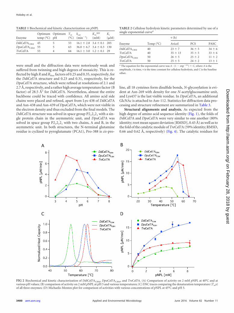
Temperature, pH, and thermostability profiles. The depen-dence of activity on temperature and pH for DdiCel7ACBM,DpuCel7ACBM, and, for comparison, TreCel7A was determinedusing pNPL as the substrate. Activities were measured at eighttemperatures at pH 5.0, and the data point with the highestactivity was recorded as the temperature optimum in Table 1.DdiCel7ACBM exhibited a lower temperature optimum, 45°C,than DpuCel7ACBM and TreCel7A, which exhibited optimal activ-ity at about 55°C, as shown in Fig. 2. For consistency, the optimalpH was determined in MacIlvaine buffer with a constant ionicstrength. Activity was evaluated at 40°C for DdiCel7ACBM and45°C for DpuCel7ACBM and TreCel7A to account for the differ-ence in temperature optima. The pH of maximum pNPL turnoverfor DpuCel7A and DdiCel7A, pH 5, was higher than that ofTreCel7A, pH 4, as shown in Fig. 2.
The denaturation temperature was measured by DSC inthe absence of substrate. The melting temperatures (Tms) ofDdiCel7ACBM, DpuCel7ACBM, and TreCel7A were 53°C, 62°C, and64°C, respectively, as shown in Table 1 and Fig. 2.
Enzyme kinetics on pNPL. The kinetic properties ofDdiCel7ACBM, DpuCel7ACBM, and TreCel7A on pNPL were com-pared (Table 1). Vmax and Km
app were determined by plotting theturnover rate on pNPL for increasing substrate concentrationsand evaluated by use of the Michaelis-Menten model, as shown inFig. 2. The catalytic rate constant (kcat) of DpuCel7ACBM was sig-nificantly higher at 36.2 min�1 than the kcats of DdiCel7ACBM andTreCel7A, which were nearly equal at 16.2 and 15.9 min�1, respec-tively. However, the Km
app values of both DpuCel7ACBM andDdiCel7ACBM were 3.4 mM, which was higher than the Km
app
value of TreCel7A, 1.2 mM, indicating a lower binding affinity forpNPL.
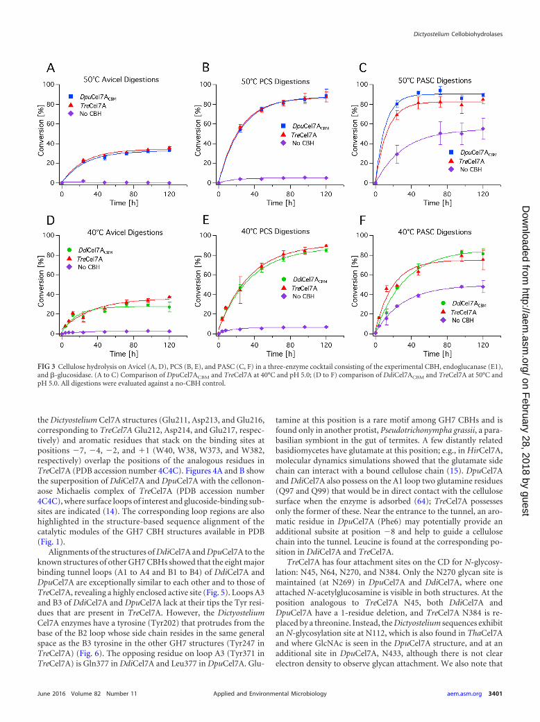
Cellulose hydrolysis. Enzymatic activity comparisons wereconducted on 0.5% Avicel, pretreated corn stover (PCS), andphosphoric acid-swollen cellulose (PASC) substrates. The en-zyme cocktails comprised �-glucosidase from Novozymes 188,the E1 endoglucanase from A. cellulolyticus, and DpuCel7ACBM,DdiCel7ACBM, or TreCel7A (63). Assays were completed nearthe previously determined pH and temperature optima forDpuCel7ACBM and DdiCel7ACBM, at pH 5.0 and 50°C and 40°C,respectively. Glucose and cellobiose concentrations were deter-mined by HPLC and plotted as a function of time. Curves were fitto a single exponential; the time constant () for cellulose hydro-lysis is reported in Table 2 and indicates that cocktails with bothDpuCel7ACBM and DdiCel7ACBM convert cellulose to glucose ina manner equivalent to that for TreCel7A on all three cellulosesubstrates. All hydrolysis profiles were strikingly similar(Fig. 3), with only one minor difference in the hydrolysis of Avicelby DpuCel7ACBM being seen (Fig. 3), where the enzyme reached alower maximum conversion than TreCel7A but did so faster thanTreCel7A.
DdiCel7A and DpuCel7A structures. After extensive screen-ing, crystals of the T. reesei-expressed DdiCel7A and DpuCel7Aproteins that could be used for synchrotron radiation diffractiondata collection were obtained. However, in both cases the crystals
Hobdey et al.
3398 aem.asm.org June 2016 Volume 82 Number 11Applied and Environmental Microbiology
on February 28, 2018 by guest
http://aem.asm
.org/D
ownloaded from

FIG 1 Sequence alignment of the GH7 CBHs that have been structurally characterized to date. The regions highlighted in blue denote N-glycosylation motifs,the residues in red (EXDXXE motif) denote the catalytic residues, and the residues enclosed in boxes represent the A1 to A4 and B1 to B4 loops enclosing theactive-site tunnel.
Dictyostelium Cellobiohydrolases
June 2016 Volume 82 Number 11 aem.asm.org 3399Applied and Environmental Microbiology
on February 28, 2018 by guest
http://aem.asm
.org/D
ownloaded from
were small and the diffraction data were notoriously weak andsuffered from twinning and high degrees of mosaicity. This is re-flected by high R and Rfree factors of 0.25 and 0.35, respectively, forthe DdiCel7A structure and 0.23 and 0.31, respectively, for theDpuCel7A structure, which were refined at resolutions of 2.1 and2.7 Å, respectively, and a rather high average temperature factor (Bfactor) of 28.5 Å2 for DdiCel7A. Nevertheless, almost the entirebackbone could be traced with confidence. All amino acid sidechains were placed and refined, apart from Lys-438 of DdiCel7Aand Asn-438 and Asn-439 of DpuCel7A, which were not visible inthe electron density and thus excluded from the final models. TheDdiCel7A structure was solved in space group P212121 with a sin-gle protein chain in the asymmetric unit, and DpuCel7A wassolved in space group P212121 with two chains, A and B, in theasymmetric unit. In both structures, the N-terminal glutamineresidue is cyclized to pyroglutamate (PCA1), Pro-388 is cis-pro-
line, all 18 cysteines form disulfide bonds, N-glycosylation is evi-dent at Asn-269 with density for one N-acetylglucosamine unit,and Lys437 is the last visible residue. In DpuCel7A, an additionalGlcNAc is attached to Asn-112. Statistics for diffraction data pro-cessing and structure refinement are summarized in Table 3.
Structural alignments and analysis. As expected from thehigh degree of amino acid sequence identity (Fig. 1), the folds ofDdiCel7A and DpuCel7A were very similar to one another (80%identity; root mean square deviation [RMSD], 0.45 Å) as well as tothe fold of the catalytic module of TreCel7A (59% identity; RMSD,0.66 and 0.62 Å, respectively) (Fig. 4). The catalytic residues for
TABLE 1 Biochemical and kinetic characterization on pNPL
EnzymeOptimumtemp (°C)
OptimumpH
Tm
(°C)kcat
(min�1)Km
app
(mM)Ki
(�M)
DdiCel7ACBM 45 5 53 16.1 2.8 3.4 0.1 205DpuCel7ACBM 55 5 63 36.0 6.7 3.4 0.3 130TreCel7A 55 4 64 16.1 3.0 1.2 0.1 29
FIG 2 Biochemical and kinetic characterization of DdiCel7ACBM, DpuCel7ACBM, and TreCel7A. (A) Comparison of activity on 2 mM pNPL at 40°C and atvarious pH values; (B) comparison of activity on 2 mM pNPL at pH 5 and various temperatures; (C) DSC traces comparing the denaturation temperature (Tms)of all three enzymes; (D) Michaelis-Menten plot for comparison of activities with various concentrations of pNPL at 45°C and pH 5.
TABLE 2 Cellulose hydrolysis kinetic parameters determined by use of asingle exponential curvea
Enzyme Temp (°C)
(h)
Avicel PCS PASC
DdiCel7ACBM 40 23 7 36 5 34 6TreCel7A 40 35 13 35 5 33 6DpuCel7ACBM 50 26 5 25 2 11 2TreCel7A 50 25 5 24 2 13 1a The equation for the exponential curve was A · (1 � exp�t/) � C, where A is theamplitude, t is time, is the time constant for cellulose hydrolysis, and C is the baselineoffset.
Hobdey et al.
3400 aem.asm.org June 2016 Volume 82 Number 11Applied and Environmental Microbiology
on February 28, 2018 by guest
http://aem.asm
.org/D
ownloaded from
the Dictyostelium Cel7A structures (Glu211, Asp213, and Glu216,corresponding to TreCel7A Glu212, Asp214, and Glu217, respec-tively) and aromatic residues that stack on the binding sites atpositions �7, �4, �2, and �1 (W40, W38, W373, and W382,respectively) overlap the positions of the analogous residues inTreCel7A (PDB accession number 4C4C). Figures 4A and B showthe superposition of DdiCel7A and DpuCel7A with the cellonon-aose Michaelis complex of TreCel7A (PDB accession number4C4C), where surface loops of interest and glucoside-binding sub-sites are indicated (14). The corresponding loop regions are alsohighlighted in the structure-based sequence alignment of thecatalytic modules of the GH7 CBH structures available in PDB(Fig. 1).
Alignments of the structures of DdiCel7A and DpuCel7A to theknown structures of other GH7 CBHs showed that the eight majorbinding tunnel loops (A1 to A4 and B1 to B4) of DdiCel7A andDpuCel7A are exceptionally similar to each other and to those ofTreCel7A, revealing a highly enclosed active site (Fig. 5). Loops A3and B3 of DdiCel7A and DpuCel7A lack at their tips the Tyr resi-dues that are present in TreCel7A. However, the DictyosteliumCel7A enzymes have a tyrosine (Tyr202) that protrudes from thebase of the B2 loop whose side chain resides in the same generalspace as the B3 tyrosine in the other GH7 structures (Tyr247 inTreCel7A) (Fig. 6). The opposing residue on loop A3 (Tyr371 inTreCel7A) is Gln377 in DdiCel7A and Leu377 in DpuCel7A. Glu-
tamine at this position is a rare motif among GH7 CBHs and isfound only in another protist, Pseudotrichonympha grassii, a para-basilian symbiont in the gut of termites. A few distantly relatedbasidiomycetes have glutamate at this position; e.g., in HirCel7A,molecular dynamics simulations showed that the glutamate sidechain can interact with a bound cellulose chain (15). DpuCel7Aand DdiCel7A also possess on the A1 loop two glutamine residues(Q97 and Q99) that would be in direct contact with the cellulosesurface when the enzyme is adsorbed (64); TreCel7A possessesonly the former of these. Near the entrance to the tunnel, an aro-matic residue in DpuCel7A (Phe6) may potentially provide anadditional subsite at position �8 and help to guide a cellulosechain into the tunnel. Leucine is found at the corresponding po-sition in DdiCel7A and TreCel7A.
TreCel7A has four attachment sites on the CD for N-glycosy-lation: N45, N64, N270, and N384. Only the N270 glycan site ismaintained (at N269) in DpuCel7A and DdiCel7A, where oneattached N-acetylglucosamine is visible in both structures. At theposition analogous to TreCel7A N45, both DdiCel7A andDpuCel7A have a 1-residue deletion, and TreCel7A N384 is re-placed by a threonine. Instead, the Dictyostelium sequences exhibitan N-glycosylation site at N112, which is also found in ThaCel7Aand where GlcNAc is seen in the DpuCel7A structure, and at anadditional site in DpuCel7A, N433, although there is not clearelectron density to observe glycan attachment. We also note that
FIG 3 Cellulose hydrolysis on Avicel (A, D), PCS (B, E), and PASC (C, F) in a three-enzyme cocktail consisting of the experimental CBH, endoglucanase (E1),and �-glucosidase. (A to C) Comparison of DpuCel7ACBM and TreCel7A at 40°C and pH 5.0; (D to F) comparison of DdiCel7ACBM and TreCel7A at 50°C andpH 5.0. All digestions were evaluated against a no-CBH control.
Dictyostelium Cellobiohydrolases
June 2016 Volume 82 Number 11 aem.asm.org 3401Applied and Environmental Microbiology
on February 28, 2018 by guest
http://aem.asm
.org/D
ownloaded from

TreCel7A has 10 disulfide bridges, whereas the DictyosteliumCel7A enzymes have only 9. The more common scenario in GH7cellulases is for them to lack this extra disulfide bridge betweenCys4 and Cys72 (TreCel7A numbering). In fact, TreCel7A and T.harzianum Cel7A are the only GH7 members with a solved struc-ture that possess the 10th disulfide bridge.
Lastly, the product-binding sites of DdiCel7A and DpuCel7Aare quite similar to those of TreCel7A, with the exception of a fewnotable differences among residues that have been extensivelystudied and discussed in past GH7 studies (8). Thr246 at the tip ofloop B3 in TreCel7A is replaced by Ala245 in DdiCel7A and
DpuCel7A, resulting in the loss of a hydrogen bond to the sub-strate at the subsite at position �1 (Fig. 6A). At the same time, theside chain of Asp345 in loop B4 of DdiCel7A and DpuCel7A issuitably positioned for H bonding to OH1 of the glucosyl unit atposition �2 of the reducing end. Actually, most GH7 CBHs haveaspartate at the corresponding position, but TreCel7A and otherTrichoderma species lack this interaction due to a 1-residue dele-tion in loop B4 (15, 50, 65).
Cellobiose inhibition. In light of the structural differencesfound in the product-binding site, Ki studies for product inhibi-tion were carried out using pNPL as the substrate. Simultaneousnonlinear regression was used to evaluate the events for compet-itive, uncompetitive, noncompetitive, and mixed inhibition. Thedata collected suggest that these enzymes are competitively inhib-ited by cellobiose, and the Kis for competitive inhibition are re-ported in Table 1. Cellobiose is 5 to 7 times less inhibitory toDdiCel7ACBM and DpuCel7ACBM than to TreCel7A, with Ki valuesof 205, 130, and 29 �M, respectively. This is consistent withthe findings of previous studies investigating similar structuralchanges in the GH7 CBH active site (36).
DISCUSSION
GH7 CBHs are complex, important enzymes in both nature andthe growing biofuels industry. Continued structural and bio-chemical studies of these enzymes are essential to developing de-tailed structure-function relationships, given their importance.The GH7 CBHs from D. discoideum and D. purpureum are nowthe second and third known nonfungal GH7 enzyme structures,with the first one being from a salt water-inhabiting, wood-boringisopod, L. quadripunctata (22).
Although many GH7 CBHs are bimodular, exhibiting a cata-lytic domain (CD) connected to a carbohydrate-binding module(CBM) by an �30-amino-acid flexible linker, the native se-quences of DdiCel7A and DpuCel7A do not exhibit a CBM-linkerdomain. It has long been known that the CBM aids the enzyme byincreasing the binding to its crystalline cellulose substrate (66);however, it is likely that there are evolutionary reasons for the lackof a CBM, such as environments of high cellulose density wherethe substrate concentration greatly surpasses the Km of the CBMor CD. Indeed, a recent study by Varnai et al. demonstrated that
TABLE 3 Data collection and refinement statistics for DdiCel7A andDpuCel7A
Parameter
Value(s) fora:
DdiCel7A DpuCel7A
Data collection statisticsPDB accession no. 4ZZQ 4ZZPBeam line I911-3, MAX-Lab ID23-1, ESRFSpace group P212121 P212121
a, b, c (Å) unit celldimensions
44.2, 62.9, 132.9 55.6, 85.2, 168.5
Wavelength (Å) 1.0 0.87Resolution (Å) 45.7–2.10 (2.21–2.10) 46.9–2.70 (2.85–2.70)No. of unique reflections 22,434 22,787Multiplicity 8.3 (8.3) 6.6 (6.3)Completeness (%) 100 (100) 99.9 (99.9)I/�I 5.5 (1.3) 5.3 (3.2)Rmerge
b 0.33 (1.6) 0.25 (0.5)
Refinement statisticsRwork/Rfree (%) 25.6/35.1 23.5/31.4Mean B factor (Å2) 28.5 15.16No. of amino acids 437 437Completeness (%) 99.9 99.8RMSD
Bond angle (°) 1.26 1.27Bond length (Å) 0.0073 0.0058
a Numbers in parentheses correspond to the highest-resolution bins.b Rmerge �h�i|I(h)i � �I(h)�|/�h�i I(h)i, where I(h)i is the intensity (I) of reflection hand �I(h)� is the average value over multiple measurements.
FIG 4 Superposition of the structures of DpuCel7A (A) and DdiCel7A (B) with the structure of the cellononaose Michaelis complex of TreCel7A (PDB accessionnumber 4C4C). The key substrate-binding loops are labeled in panel A, and the substrate-binding sites are labeled in panel B.
Hobdey et al.
3402 aem.asm.org June 2016 Volume 82 Number 11Applied and Environmental Microbiology
on February 28, 2018 by guest
http://aem.asm
.org/D
ownloaded from

TreCel7A without the CBM-linker is able to achieve the same ex-tent of conversion on Avicel and pretreated wheat straw at a highlevel (20%) of solids loadings, while at a lower level of solids load-ing, such as the 0.5% used here, the CBM-linker aids in conversionby binding crystalline cellulose and increasing the local concen-tration (67). Consequently, for biochemical studies, we haveadded the family 1 CBM and linker from TreCel7A to the CD ofnative DdiCel7A and DpuCel7A (DdiCel7CBM and DpuCel7ACBM,respectively) to be able to compare more directly GH7 CBH con-version at low levels of solids loadings. Going forward, self-con-sistent comparisons of GH7 CBHs with (or without) CBM-link-ers, enabled by the development of a T. reesei expression host fromwhich Cel7A is deleted (46), will be important to the continueddevelopment of structure-activity relationships and the develop-ment of the baseline activity of GH7 CBHs from natural diversityin the context of a background enzyme cocktail on process-rele-vant substrates.
Since T. reesei is known to be one of the most cellulolytic or-ganisms, it is surprising that both Dictyostelium GH7s wereequally active on biomass and crystalline cellulose at their respec-tive temperature optima (Table 1). However, previous investiga-tions of the cellulose synthase in D. discoideum have shown thatthe cellulose deposited in the sheath is highly crystalline cellulose I(68) and that the high crystallinity is positively correlated with anincreased strength and rigidity of the stalk (69). Thus, it is notunreasonable to surmise that the Dictyostelium Cel7s have evolveda highly efficient mechanism for rearrangement of crystalline cel-lulose I to resourcefully modify the cell wall and sheath structureand potentially recycle glucose during formation of the stalk andfruiting body. Additionally, Eichinger et al. speculate that dictyo-stelia may also utilize the cellulose- and hemicellulose-degradingenzymes in their genomes to degrade plant tissue for food (40).Immunolabeling of GH7 CBHs is a well-known technique to vi-sualize these enzymes (70) and could potentially be used to exam-ine the distribution of these enzymes when they are expressed inthe presence and absence of either self-generated cellulose or ex-ogenous sources to determine the function of GH7 CBHs in thelife cycle of dictyostelia.
In a previous report, following the in vivo expression of GH7CBH in D. discoideum, the enzyme was found to be highly con-
FIG 5 Space-fill GH7 structures comparing the substrate tunnel enclosures of CBHs from T. reesei (PDB accession number 4C4C), D. discoideum, D. purpureum,H. irregulare (PDB accession number 2YG1), P. chrysosporium (PDB accession number 1GPI), and the endoglucanase (EG) Cel7B from T. reesei (PDB accessionnumber 1EG1). In all frames, the cellononaose ligand from the TreCel7A Michaelis complex is shown as gray sticks.
FIG 6 Alignments of the structures of DdiCel7A and DpuCel7A with theknown structures of other GH7 CBHs. (A) Alignment around the active siteillustrating the interactions of the B2, B3, and A3 loops discussed in the text.(B) Product-binding sites illustrating the relative positioning of the commonlyfound aspartate residue that interacts with the glucosyl unit at position �2 butwhich is replaced by glycine in TreCel7A and ThaCel7A. Both the Michaelis(PDB accession number 4C4C) and primed glycosyl-enzyme intermediate(GEI; PDB accession number 3CEL) ligands are from the TreCel7A structure.The aspartic acid side chains of PchCel7D, HirCel7A, RemCel7A, AfuCel7A,and LquCel7B essentially overlay those of DpuCel7A and DdiCel7A and are notshown for clarity.
Dictyostelium Cellobiohydrolases
June 2016 Volume 82 Number 11 aem.asm.org 3403Applied and Environmental Microbiology
on February 28, 2018 by guest
http://aem.asm
.org/D
ownloaded from

trolled by the developmental phase of the organism (11). Whilethe actual quantity of GH7 CBH at each stage was not reported, itmay be that this organism expresses only a small amount of Cel7 atcritical developmental phases and that the expression of largequantities, like those secreted by T. reesei during biomass degra-dation, could perhaps be detrimental to the organism. It is alsopossible that the hydrolytic ability of these enzymes is advanta-geous, in that it enables the organism to progress through mor-phogenesis quickly, therefore allowing it to rapidly escape ill-fatedenvironments. Given this tight regulation of expression, it wouldalso be interesting to investigate the regulation of the GH7 CBHpromoter during morphogenesis to better understand the processof morphogenesis or the mechanism of transcription regulation,as well as for its potential use in future GH7 expression systems forindustrial applications.
The crystal structures of native DdiCel7A and DpuCel7A areexceptionally similar to each other and to the crystal structure ofTreCel7A (Fig. 4). Indeed, the degree of binding tunnel closure ofnative DdiCel7A and DpuCel7A is the closest to that of TreCel7Ato have been observed among GH7 structures to date (Fig. 5).Functionally, this suggests increased processivity and a reduced dis-sociation constant that are perhaps similar to those of TreCel7A. Ofparticular significance is the lack of tyrosine in the B3 and A3loops, whose side chains maintain some degree of flexibility (ex-emplified by their different positions in the TreCel7A structureswith PDB accession numbers 1CEL and 4C4C) and partially en-close the binding tunnel at the catalytic center (8). To compensate,the Dictyostelium Cel7A enzymes have a unique tyrosine (Y202)that protrudes from the base of the B2 loop, whose side chainresides in the same general space as the side chains of the A3 andB3 tyrosines in the other GH7 structures (Fig. 6A). The result isthat the degree of binding tunnel enclosure in DpuCel7A andDdiCel7A in this region is comparable to or greater than that inthe other GH7 CBHs. In addition to the aforementioned implica-tions for processivity and the dissociation constant, this may sug-gest that the Dictyostelium Cel7A enzymes may have elevatedprobabilities of endoinitiation compared with the probability forTreCel7A (34).
The product-binding sites of DdiCel7A and DpuCel7A are sim-ilar to those of TreCel7A with a few notable, potentially importantdifferences, namely, T246 (TreCel7A numbering) and D345(DpuCel7A and DdiCel7A numbering) (Fig. 6A). Ståhlberg et al.(71) speculated on the flexibility of the T246 side chain, as evi-denced by elevated temperature factors and weaker electron den-sity when the binding site at position �1 was vacant. However,when the binding site at position �1 was filled, the position ofT246 was as stable as the rest of the protein, thus providing furtherevidence for the importance of this hydrogen bond. von Ossowskiet al. (36) discussed the impact of this residue’s absence inPchCel7D, as did Ubhayasekera et al. (65). Ubhayasekera et al.(65) concluded that the two most significant differences in ligandbinding between TreCel7A and PchCel7D are the direct hydrogenbond with T246 (which is absent in PchCel7D) and the D336interaction at the binding tunnel exit, in which the interaction ispresent in PchCel7D but in which D336 is replaced by glycine inTreCel7A. Like PchCel7D, both Dictyostelium structures possessan aspartate at this position (D345 in DdiCel7A and DpuCel7A),as illustrated in Fig. 6B. The conservation of this interaction inGH7 cellulases has been discussed (65); as mentioned in Results,the aspartate residue here is rather well conserved in all GH7CBHs except those in the Trichoderma genus and a few others,where the B4 loop is 1 residue shorter. This aspartate is addition-ally absent in GH7 EGs due to deletion of the B4 loop, implyingthat this residue may play a role in cellulose chain processivity insome GH7 CBHs.
To verify the functional significance of the unique product sitemotif, we investigated the biochemical characteristics and activi-ties of DdiCel7ACBM and DpuCel7ACBM on soluble (Table 1) andinsoluble (Table 2) substrates. We found that on the soluble sub-strate pNPL, DpuCel7ACBM has a higher specific activity than ei-ther DdiCel7ACBM or TreCel7A. Interestingly, we could find nostructural difference between the CDs of DdiCel7A and DpuCel7Ato fully explain the differences in activities. Evolutionarily, it isunclear why the Dictyostelium Cel7s are more cellobiose tolerant.Perhaps the high viscosity of the extracellular matrix amplifies theeffective cellobiose concentration due to limited diffusion. Both
FIG 7 Eukaryote evolution timeline of branches where GH7 genes have been found and the percent identities (ID%) and similarities (Sim%) of the proteinsequences within the GH7 domain to the D. discoideum Cel7A sequence. Branch names and time points of divergence are from reference 72. Only points of earlydivergence (�600 million years ago) are included; the later divergence among stramenopiles, amoebozoa, and metazoans is not resolved here. The multiple-sequence alignment of selected GH7 sequences was done with the MUSCLE web service, and flanking regions (e.g., the signal peptide, CBM) were trimmed offbefore calculation of pairwise sequence identities and similarities using the Gonnet substitution matrix. UniProt accession numbers are provided, except in thecase of Schizochytrium aggregatum, for which the U.S. patent number is indicated (76). *, the Aureococcus anophagefferens GH7 sequence (UniProt accessionnumber F0YSW7) appears to be a fragment containing only 135 residues of the C-terminal part of the GH7 domain in the alignment.
Hobdey et al.
3404 aem.asm.org June 2016 Volume 82 Number 11Applied and Environmental Microbiology
on February 28, 2018 by guest
http://aem.asm
.org/D
ownloaded from

TA
BLE
4P
airwise
proteinsequ
ence
identities
and
similarities
with
inth
eG
H7
CD
a
Organ
ismgrou
pSpecies
b
%iden
tityor
similarity
a
Phytophthora
infestansSchizochytriumaggregatum
Aureococcus
anophagefferensT
halassiosiraoceanica
Pyrocystis
lunulaE
miliania
huxleyiP
seudotrichonympha
grassiiD
ictyosteliumdiscoideum
Dictyostelium
purpureumT
richoderma
reeseiP
hanerochaetechrysosporium
Am
phimedon
queenslandicaD
aphniapulex
Oom
ycetesP
hytophthorainfestans(D
0N841)
44156
4848
5148
5352
5252
5757
55
Labyrin
thu
lomycetes
Schizochytriumaggregatum(U
SP8470592)
73432
5448
5349
5957
5858
6259
53
Pelagoph
yceaeA
ureococcusanophagefferens(F0Y
SW7)
6671
13537
5042
5349
5145
4956
48
Bacillarioph
ytaT
halassiosiraoceanica( K
0T5R
6)
6868
58468
5251
4946
4843
5150
47
Din
ophyceae
Pyrocystis
lunula(D
8UX
L6)
7069
6769
43050
5550
5249
5556
53
Haptoph
yceaeE
miliania
huxleyi(R
1C7R
5)63
6558
6567
43351
4647
4648
4947
Parabasilia
Pseudotrichonym
phagrassii(Q
95YH
1)73
7675
7073
65436
5555
5764
6657
Am
oebozoaD
ictyosteliumdiscoideumC
el7A( Q
55FE6)
7274
7366
6963
75436
8060
5655
49
Am
oebozoaD
ictyosteliumpurpureumC
el7A(F0Z
JZ1)
7375
7366
7064
7593
43660
5855
50
Ascom
ycotaT
richoderma
reeseiC
el7A(P
62694)72
7265
6066
6175
7776
43056
5652
Basidiom
ycotaP
hanerochaetechrysosporiumC
el7D( Q
7LIJ0)
7879
7070
7263
7777
7874
42764
59
Porifera
Am
phimedon
queenslandica( I1FU
52)
7777
7371
7468
7975
7473
80440
59
Cru
staceaD
aphniapulex
Cel7A
(E9G
5J5)74
7368
6369
6577
7171
6976
78445
aP
airwise
proteinsequ
ence
identities
areto
the
upper
right
and
similarities
areto
the
lower
leftofth
ediagon
alofboldfacen
um
bers.Th
eboldface
nu
mbers
areth
en
um
berofresidu
esin
the
alignm
ent
foreach
sequen
ce.Th
esh
ading
high
lights
values
forth
eD
ictyosteliumsequ
ences.
bD
esignation
sin
parenth
esesare
Un
iProt
accessionn
um
bersfor
the
GH
7sequ
ences
exceptin
the
caseofSchizochytrium
aggregatum,w
hich
isa
U.S.paten
tn
um
berfrom
reference
76.
Dictyostelium Cellobiohydrolases
June 2016 Volume 82 Number 11 aem.asm.org 3405Applied and Environmental Microbiology
on February 28, 2018 by guest
http://aem.asm
.org/D
ownloaded from
Dictyostelium organisms studied here contain multiple genes pre-dicted to have �-glucosidase activity (seven for D. discoideum andeight for D. purpureum) (7); however, to our knowledge, it has notbeen reported if the organism utilizes the glucose for energy or forsome other purpose, such as feeding a bacterial symbiote, as hasbeen suggested previously (40). The cosecretion of �-glucosidasesalongside the GH7 CBHs has also not been examined, to ourknowledge.
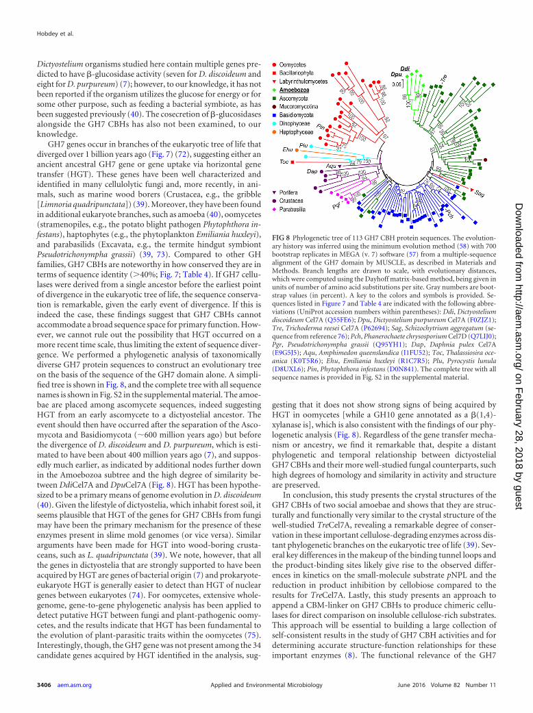
GH7 genes occur in branches of the eukaryotic tree of life thatdiverged over 1 billion years ago (Fig. 7) (72), suggesting either anancient ancestral GH7 gene or gene uptake via horizontal genetransfer (HGT). These genes have been well characterized andidentified in many cellulolytic fungi and, more recently, in ani-mals, such as marine wood borers (Crustacea, e.g., the gribble[Limnoria quadripunctata]) (39). Moreover, they have been foundin additional eukaryote branches, such as amoeba (40), oomycetes(stramenopiles, e.g., the potato blight pathogen Phytophthora in-festans), haptophytes (e.g., the phytoplankton Emiliania huxleyi),and parabasilids (Excavata, e.g., the termite hindgut symbiontPseudotrichonympha grassii) (39, 73). Compared to other GHfamilies, GH7 CBHs are noteworthy in how conserved they are interms of sequence identity (�40%; Fig. 7; Table 4). If GH7 cellu-lases were derived from a single ancestor before the earliest pointof divergence in the eukaryotic tree of life, the sequence conserva-tion is remarkable, given the early event of divergence. If this isindeed the case, these findings suggest that GH7 CBHs cannotaccommodate a broad sequence space for primary function. How-ever, we cannot rule out the possibility that HGT occurred on amore recent time scale, thus limiting the extent of sequence diver-gence. We performed a phylogenetic analysis of taxonomicallydiverse GH7 protein sequences to construct an evolutionary treeon the basis of the sequence of the GH7 domain alone. A simpli-fied tree is shown in Fig. 8, and the complete tree with all sequencenames is shown in Fig. S2 in the supplemental material. The amoe-bae are placed among ascomycete sequences, indeed suggestingHGT from an early ascomycete to a dictyostelial ancestor. Theevent should then have occurred after the separation of the Asco-mycota and Basidiomycota (�600 million years ago) but beforethe divergence of D. discoideum and D. purpureum, which is esti-mated to have been about 400 million years ago (7), and suppos-edly much earlier, as indicated by additional nodes further downin the Amoebozoa subtree and the high degree of similarity be-tween DdiCel7A and DpuCel7A (Fig. 8). HGT has been hypothe-sized to be a primary means of genome evolution in D. discoideum(40). Given the lifestyle of dictyostelia, which inhabit forest soil, itseems plausible that HGT of the genes for GH7 CBHs from fungimay have been the primary mechanism for the presence of theseenzymes present in slime mold genomes (or vice versa). Similararguments have been made for HGT into wood-boring crusta-ceans, such as L. quadripunctata (39). We note, however, that allthe genes in dictyostelia that are strongly supported to have beenacquired by HGT are genes of bacterial origin (7) and prokaryote-eukaryote HGT is generally easier to detect than HGT of nucleargenes between eukaryotes (74). For oomycetes, extensive whole-genome, gene-to-gene phylogenetic analysis has been applied todetect putative HGT between fungi and plant-pathogenic oomy-cetes, and the results indicate that HGT has been fundamental tothe evolution of plant-parasitic traits within the oomycetes (75).Interestingly, though, the GH7 gene was not present among the 34candidate genes acquired by HGT identified in the analysis, sug-
gesting that it does not show strong signs of being acquired byHGT in oomycetes [while a GH10 gene annotated as a �(1,4)-xylanase is], which is also consistent with the findings of our phy-logenetic analysis (Fig. 8). Regardless of the gene transfer mecha-nism or ancestry, we find it remarkable that, despite a distantphylogenetic and temporal relationship between dictyostelialGH7 CBHs and their more well-studied fungal counterparts, suchhigh degrees of homology and similarity in activity and structureare preserved.
In conclusion, this study presents the crystal structures of theGH7 CBHs of two social amoebae and shows that they are struc-turally and functionally very similar to the crystal structure of thewell-studied TreCel7A, revealing a remarkable degree of conser-vation in these important cellulose-degrading enzymes across dis-tant phylogenetic branches on the eukaryotic tree of life (39). Sev-eral key differences in the makeup of the binding tunnel loops andthe product-binding sites likely give rise to the observed differ-ences in kinetics on the small-molecule substrate pNPL and thereduction in product inhibition by cellobiose compared to theresults for TreCel7A. Lastly, this study presents an approach toappend a CBM-linker on GH7 CBHs to produce chimeric cellu-lases for direct comparison on insoluble cellulose-rich substrates.This approach will be essential to building a large collection ofself-consistent results in the study of GH7 CBH activities and fordetermining accurate structure-function relationships for theseimportant enzymes (8). The functional relevance of the GH7
FIG 8 Phylogenetic tree of 113 GH7 CBH protein sequences. The evolution-ary history was inferred using the minimum evolution method (58) with 700bootstrap replicates in MEGA (v. 7) software (57) from a multiple-sequencealignment of the GH7 domain by MUSCLE, as described in Materials andMethods. Branch lengths are drawn to scale, with evolutionary distances,which were computed using the Dayhoff matrix-based method, being given inunits of number of amino acid substitutions per site. Gray numbers are boot-strap values (in percent). A key to the colors and symbols is provided. Se-quences listed in Figure 7 and Table 4 are indicated with the following abbre-viations (UniProt accession numbers within parentheses): Ddi, Dictyosteliumdiscoideum Cel7A (Q55FE6); Dpu, Dictyostelium purpureum Cel7A (F0ZJZ1);Tre, Trichoderma reesei Cel7A (P62694); Sag, Schizochytrium aggregatum (se-quence from reference 76); Pch, Phanerochaete chrysosporium Cel7D (Q7LIJ0);Pgr, Pseudotrichonympha grassii (Q95YH1); Dap, Daphnia pulex Cel7A(E9G5J5); Aqu, Amphimedon queenslandica (I1FU52); Toc, Thalassiosira oce-anica (K0T5R6); Ehu, Emiliania huxleyi (R1C7R5); Plu, Pyrocystis lunula(D8UXL6); Pin, Phytophthora infestans (D0N841). The complete tree with allsequence names is provided in Fig. S2 in the supplemental material.
Hobdey et al.
3406 aem.asm.org June 2016 Volume 82 Number 11Applied and Environmental Microbiology
on February 28, 2018 by guest
http://aem.asm
.org/D
ownloaded from
CBHs in dictyostelia, likely intimately related to the reshaping ofthe extracellular matrix important to the slime mold life cycle in itsmorphogenesis from slug to fruiting body (11) or in its ability todigest cellulose-containing organisms in the forest soil (40), re-mains a question of particular interest to understand the evolu-tionary pressures on the activities of slime mold GH7 CBHs.
ACKNOWLEDGMENT
We are grateful to Nils Högberg, Swedish University of Agricultural Sci-ences, for advice on phylogenetic analysis.
FUNDING INFORMATIONS.E.H., B.C.K., L.E.T., K.K.P., T.A.V., M.E.H., S.R.D., and G.T.B. ac-knowledge the U.S. Department of Energy BioEnergy Technologies Officefor funding. M.H.M., A.S.B., and J.S. acknowledge the Swedish ResearchCouncil Formas and the Faculty for Natural Resources and Agriculture atthe Swedish University of Agricultural Sciences through the research pro-gram MicroDrivE. B.C.K. also thanks the National Renewable EnergyLaboratory’s Director’s Fellowship Program for funding.
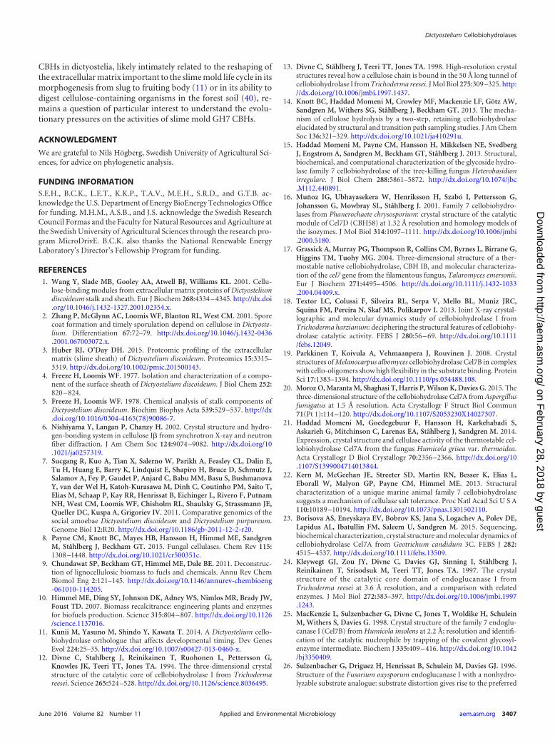
REFERENCES1. Wang Y, Slade MB, Gooley AA, Atwell BJ, Williams KL. 2001. Cellu-
lose-binding modules from extracellular matrix proteins of Dictyosteliumdiscoideum stalk and sheath. Eur J Biochem 268:4334 – 4345. http://dx.doi.org/10.1046/j.1432-1327.2001.02354.x.
2. Zhang P, McGlynn AC, Loomis WF, Blanton RL, West CM. 2001. Sporecoat formation and timely sporulation depend on cellulose in Dictyoste-lium. Differentiation 67:72–79. http://dx.doi.org/10.1046/j.1432-0436.2001.067003072.x.
3. Huber RJ, O’Day DH. 2015. Proteomic profiling of the extracellularmatrix (slime sheath) of Dictyostelium discoideum. Proteomics 15:3315–3319. http://dx.doi.org/10.1002/pmic.201500143.
4. Freeze H, Loomis WF. 1977. Isolation and characterization of a compo-nent of the surface sheath of Dictyostelium discoideum. J Biol Chem 252:820 – 824.
5. Freeze H, Loomis WF. 1978. Chemical analysis of stalk components ofDictyostelium discoideum. Biochim Biophys Acta 539:529 –537. http://dx.doi.org/10.1016/0304-4165(78)90086-7.
6. Nishiyama Y, Langan P, Chanzy H. 2002. Crystal structure and hydro-gen-bonding system in cellulose I� from synchrotron X-ray and neutronfiber diffraction. J Am Chem Soc 124:9074 –9082. http://dx.doi.org/10.1021/ja0257319.
7. Sucgang R, Kuo A, Tian X, Salerno W, Parikh A, Feasley CL, Dalin E,Tu H, Huang E, Barry K, Lindquist E, Shapiro H, Bruce D, Schmutz J,Salamov A, Fey P, Gaudet P, Anjard C, Babu MM, Basu S, BushmanovaY, van der Wel H, Katoh-Kurasawa M, Dinh C, Coutinho PM, Saito T,Elias M, Schaap P, Kay RR, Henrissat B, Eichinger L, Rivero F, PutnamNH, West CM, Loomis WF, Chisholm RL, Shaulsky G, Strassmann JE,Queller DC, Kuspa A, Grigoriev IV. 2011. Comparative genomics of thesocial amoebae Dictyostelium discoideum and Dictyostelium purpureum.Genome Biol 12:R20. http://dx.doi.org/10.1186/gb-2011-12-2-r20.
8. Payne CM, Knott BC, Mayes HB, Hansson H, Himmel ME, SandgrenM, Ståhlberg J, Beckham GT. 2015. Fungal cellulases. Chem Rev 115:1308 –1448. http://dx.doi.org/10.1021/cr500351c.
9. Chundawat SP, Beckham GT, Himmel ME, Dale BE. 2011. Deconstruc-tion of lignocellulosic biomass to fuels and chemicals. Annu Rev ChemBiomol Eng 2:121–145. http://dx.doi.org/10.1146/annurev-chembioeng-061010-114205.
10. Himmel ME, Ding SY, Johnson DK, Adney WS, Nimlos MR, Brady JW,Foust TD. 2007. Biomass recalcitrance: engineering plants and enzymesfor biofuels production. Science 315:804 – 807. http://dx.doi.org/10.1126/science.1137016.
11. Kunii M, Yasuno M, Shindo Y, Kawata T. 2014. A Dictyostelium cello-biohydrolase orthologue that affects developmental timing. Dev GenesEvol 224:25–35. http://dx.doi.org/10.1007/s00427-013-0460-x.
12. Divne C, Stahlberg J, Reinikainen T, Ruohonen L, Pettersson G,Knowles JK, Teeri TT, Jones TA. 1994. The three-dimensional crystalstructure of the catalytic core of cellobiohydrolase I from Trichodermareesei. Science 265:524 –528. http://dx.doi.org/10.1126/science.8036495.
13. Divne C, Ståhlberg J, Teeri TT, Jones TA. 1998. High-resolution crystalstructures reveal how a cellulose chain is bound in the 50 Å long tunnel ofcellobiohydrolase I from Trichoderma reesei. J Mol Biol 275:309 –325. http://dx.doi.org/10.1006/jmbi.1997.1437.
14. Knott BC, Haddad Momeni M, Crowley MF, Mackenzie LF, Götz AW,Sandgren M, Withers SG, Ståhlberg J, Beckham GT. 2013. The mecha-nism of cellulose hydrolysis by a two-step, retaining cellobiohydrolaseelucidated by structural and transition path sampling studies. J Am ChemSoc 136:321–329. http://dx.doi.org/10.1021/ja410291u.
15. Haddad Momeni M, Payne CM, Hansson H, Mikkelsen NE, SvedbergJ, Engstrom A, Sandgren M, Beckham GT, Ståhlberg J. 2013. Structural,biochemical, and computational characterization of the glycoside hydro-lase family 7 cellobiohydrolase of the tree-killing fungus Heterobasidionirregulare. J Biol Chem 288:5861–5872. http://dx.doi.org/10.1074/jbc.M112.440891.
16. Muñoz IG, Ubhayasekera W, Henriksson H, Szabó I, Pettersson G,Johansson G, Mowbray SL, Ståhlberg J. 2001. Family 7 cellobiohydro-lases from Phanerochaete chrysosporium: crystal structure of the catalyticmodule of Cel7D (CBH58) at 1.32 Å resolution and homology models ofthe isozymes. J Mol Biol 314:1097–1111. http://dx.doi.org/10.1006/jmbi.2000.5180.
17. Grassick A, Murray PG, Thompson R, Collins CM, Byrnes L, Birrane G,Higgins TM, Tuohy MG. 2004. Three-dimensional structure of a ther-mostable native cellobiohydrolase, CBH IB, and molecular characteriza-tion of the cel7 gene from the filamentous fungus, Talaromyces emersonii.Eur J Biochem 271:4495– 4506. http://dx.doi.org/10.1111/j.1432-1033.2004.04409.x.
18. Textor LC, Colussi F, Silveira RL, Serpa V, Mello BL, Muniz JRC,Squina FM, Pereira N, Skaf MS, Polikarpov I. 2013. Joint X-ray crystal-lographic and molecular dynamics study of cellobiohydrolase I fromTrichoderma harzianum: deciphering the structural features of cellobiohy-drolase catalytic activity. FEBS J 280:56 – 69. http://dx.doi.org/10.1111/febs.12049.
19. Parkkinen T, Koivula A, Vehmaanpera J, Rouvinen J. 2008. Crystalstructures of Melanocarpus albomyces cellobiohydrolase Cel7B in complexwith cello-oligomers show high flexibility in the substrate binding. ProteinSci 17:1383–1394. http://dx.doi.org/10.1110/ps.034488.108.
20. Moroz O, Maranta M, Shaghasi T, Harris P, Wilson K, Davies G. 2015. Thethree-dimensional structure of the cellobiohydrolase Cel7A from Aspergillusfumigatus at 1.5 Å resolution. Acta Crystallogr F Struct Biol Commun71(Pt 1):114 –120. http://dx.doi.org/10.1107/S2053230X14027307.
21. Haddad Momeni M, Goedegebuur F, Hansson H, Karkehabadi S,Askarieh G, Mitchinson C, Larenas EA, Ståhlberg J, Sandgren M. 2014.Expression, crystal structure and cellulase activity of the thermostable cel-lobiohydrolase Cel7A from the fungus Humicola grisea var. thermoidea.Acta Crystallogr D Biol Crystallogr 70:2356 –2366. http://dx.doi.org/10.1107/S1399004714013844.
22. Kern M, McGeehan JE, Streeter SD, Martin RN, Besser K, Elias L,Eborall W, Malyon GP, Payne CM, Himmel ME. 2013. Structuralcharacterization of a unique marine animal family 7 cellobiohydrolasesuggests a mechanism of cellulase salt tolerance. Proc Natl Acad Sci U S A110:10189 –10194. http://dx.doi.org/10.1073/pnas.1301502110.
23. Borisova AS, Eneyskaya EV, Bobrov KS, Jana S, Logachev A, Polev DE,Lapidus AL, Ibatullin FM, Saleem U, Sandgren M. 2015. Sequencing,biochemical characterization, crystal structure and molecular dynamics ofcellobiohydrolase Cel7A from Geotrichum candidum 3C. FEBS J 282:4515– 4537. http://dx.doi.org/10.1111/febs.13509.
24. Kleywegt GJ, Zou JY, Divne C, Davies GJ, Sinning I, Ståhlberg J,Reinikainen T, Srisodsuk M, Teeri TT, Jones TA. 1997. The crystalstructure of the catalytic core domain of endoglucanase I fromTrichoderma reesei at 3.6 Å resolution, and a comparison with relatedenzymes. J Mol Biol 272:383–397. http://dx.doi.org/10.1006/jmbi.1997.1243.
25. MacKenzie L, Sulzenbacher G, Divne C, Jones T, Woldike H, SchuleinM, Withers S, Davies G. 1998. Crystal structure of the family 7 endoglu-canase I (Cel7B) from Humicola insolens at 2.2 Å; resolution and identifi-cation of the catalytic nucleophile by trapping of the covalent glycosyl-enzyme intermediate. Biochem J 335:409 – 416. http://dx.doi.org/10.1042/bj3350409.
26. Sulzenbacher G, Driguez H, Henrissat B, Schulein M, Davies GJ. 1996.Structure of the Fusarium oxysporum endoglucanase I with a nonhydro-lyzable substrate analogue: substrate distortion gives rise to the preferred
Dictyostelium Cellobiohydrolases
June 2016 Volume 82 Number 11 aem.asm.org 3407Applied and Environmental Microbiology
on February 28, 2018 by guest
http://aem.asm
.org/D
ownloaded from
axial orientation for the leaving group. Biochemistry 35:15280 –15287.http://dx.doi.org/10.1021/bi961946h.
27. Stals I, Sandra K, Geysens S, Contreras R, Van Beeumen J, ClaeyssensM. 2004. Factors influencing glycosylation of Trichoderma reesei cellu-lases. I. Postsecretorial changes of the O- and N-glycosylation pattern ofCel7A. Glycobiology 14:713–724.
28. Beckham GT, Bomble YJ, Matthews JF, Taylor CB, Resch MG, Yar-brough JM, Decker SR, Bu L, Zhao X, McCabe C, Wohlert J, Bergen-stråhle M, Brady JW, Adney WS, Himmel ME, Crowley MF. 2010. TheO-glycosylated linker from the Trichoderma reesei family 7 cellulase is aflexible, disordered protein. Biophys J 99:3773–3781. http://dx.doi.org/10.1016/j.bpj.2010.10.032.
29. Sammond DW, Payne CM, Brunecky R, Himmel ME, Crowley MF,Beckham GT. 2012. Cellulase linkers are optimized based on domain typeand function: insights from sequence analysis, biophysical measurements,and molecular simulation. PLoS One 7:e48615. http://dx.doi.org/10.1371/journal.pone.0048615.
30. Beckham GT, Bomble YJ, Bayer EA, Himmel ME, Crowley MF. 2011.Applications of computational science for understanding enzymatic de-construction of cellulose. Curr Opin Biotechnol 22:231–238. http://dx.doi.org/10.1016/j.copbio.2010.11.005.
31. Bu L, Beckham GT, Shirts MR, Nimlos MR, Adney WS, Himmel ME,Crowley MF. 2011. Probing carbohydrate product expulsion from a pro-cessive cellulase with multiple absolute binding free energy methods. J BiolChem 286:18161–18169. http://dx.doi.org/10.1074/jbc.M110.212076.
32. Vrsanska M, Biely P. 1992. The cellobiohydrolase-I from Trichodermareesei Qm-9414 —action on cello-oligosaccharides. Carbohydr Res 227:19 –27. http://dx.doi.org/10.1016/0008-6215(92)85058-8.
33. Igarashi K, Uchihashi T, Koivula A, Wada M, Kimura S, Okamoto T,Penttila M, Ando T, Samejima M. 2011. Traffic jams reduce hydrolyticefficiency of cellulase on cellulose surface. Science 333:1279 –1282. http://dx.doi.org/10.1126/science.1208386.
34. Kurasin M, Valjamae P. 2011. Processivity of cellobiohydrolases is lim-ited by the substrate. J Biol Chem 286:169 –177. http://dx.doi.org/10.1074/jbc.M110.161059.
35. Jalak J, Valjamae P. 2014. Multi-mode binding of cellobiohydrolaseCel7A from Trichoderma reesei to cellulose. PLoS One 9:e108181. http://dx.doi.org/10.1371/journal.pone.0108181.
36. von Ossowski I, Ståhlberg J, Koivula A, Piens K, Becker D, Boer H,Harle R, Harris M, Divne C, Mahdi S, Zhao Y, Driguez H, ClaeyssensM, Sinnott ML, Teeri TT. 2003. Engineering the exo-loop of Trichodermareesei cellobiohydrolase, Cel7A. A comparison with Phanerochaete chrys-osporium Cel7D. J Mol Biol 333:817– 829. http://dx.doi.org/10.1016/S0022-2836(03)00881-7.
37. Andric P, Meyer AS, Jensen PA, Dam-Johansen K. 2010. Reactordesign for minimizing product inhibition during enzymatic lignocel-lulose hydrolysis. II. Quantification of inhibition and suitability ofmembrane reactors. Biotechnol Adv 28:407– 425. http://dx.doi.org/10.1016/j.biotechadv.2010.02.005.
38. Gan Q, Allen SJ, Taylor G. 2002. Design and operation of an integratedmembrane reactor for enzymatic cellulose hydrolysis. Biochem Eng J 12:223–229. http://dx.doi.org/10.1016/S1369-703X(02)00072-4.
39. King AJ, Cragg SM, Li Y, Dymond J, Guille MJ, Bowles DJ, Bruce NC,Graham IA, McQueen-Mason SJ. 2010. Molecular insight into lignocel-lulose digestion by a marine isopod in the absence of gut microbes. ProcNatl Acad Sci U S A 107:5345–5350. http://dx.doi.org/10.1073/pnas.0914228107.
40. Eichinger L, Pachebat JA, Glockner G, Rajandream MA, Sucgang R,Berriman M, Song J, Olsen R, Szafranski K, Xu Q, Tunggal B, Kum-merfeld S, Madera M, Konfortov BA, Rivero F, Bankier AT, LehmannR, Hamlin N, Davies R, Gaudet P, Fey P, Pilcher K, Chen G, SaundersD, Sodergren E, Davis P, Kerhornou A, Nie X, Hall N, Anjard C,Hemphill L, Bason N, Farbrother P, Desany B, Just E, Morio T, Rost R,Churcher C, Cooper J, Haydock S, van Driessche N, Cronin A, Good-head I, Muzny D, Mourier T, Pain A, Lu M, Harper D, Lindsay R,Hauser H, et al. 2005. The genome of the social amoeba Dictyosteliumdiscoideum. Nature 435:43–57. http://dx.doi.org/10.1038/nature03481.
41. Cragg SM, Beckham GT, Bruce NC, Bugg TD, Distel DL, Dupree P,Etxabe AG, Goodell BS, Jellison J, McGeehan JE. 2015. Lignocellulosedegradation mechanisms across the tree of life. Curr Opin Chem Biol29:108 –119. http://dx.doi.org/10.1016/j.cbpa.2015.10.018.
42. Beckham GT, Dai Z, Matthews JF, Momany M, Payne CM, Adney WS,Baker SE, Himmel ME. 2012. Harnessing glycosylation to improve cel-
lulase activity. Curr Opin Biotechnol 23:338 –345. http://dx.doi.org/10.1016/j.copbio.2011.11.030.
43. Singh A, Taylor LE, Vander Wall TA, Linger J, Himmel ME, Podka-miner K, Adney WS, Decker SR. 2014. Heterologous protein expressionin Hypocrea jecorina: a historical perspective and new developments. Bio-technol Adv 33:142–154. http://dx.doi.org/10.1016/j.biotechadv.2014.11.009.
44. Grigoriev IV, Nordberg H, Shabalov I, Aerts A, Cantor M, GoodsteinD, Kuo A, Minovitsky S, Nikitin R, Ohm RA. 2012. The genome portalof the Department of Energy Joint Genome Institute. Nucleic Acids Res40(Database issue):D26 –D32. http://dx.doi.org/10.1093/nar/gkr947.
45. Nordberg H, Cantor M, Dusheyko S, Hua S, Poliakov A, Shabalov I,Smirnova T, Grigoriev IV, Dubchak I. 2014. The genome portal of theDepartment of Energy Joint Genome Institute: 2014 updates. Nucleic Ac-ids Res 42(Database issue):D26 –D31. http://dx.doi.org/10.1093/nar/gkt1069.
46. Linger J, Taylor L, Baker J, Vander Wall T, Hobdey S, Podkaminer K,Himmel M, Decker S. 2015. A constitutive expression system for glycosylhydrolase family 7 cellobiohydrolases in Hypocrea jecorina. BiotechnolBiofuels 8:45. http://dx.doi.org/10.1186/s13068-015-0230-2.
47. McPherson A. 1982. Preparation and analysis of protein crystals. JohnWiley & Sons, New York, NY.
48. Collaborative Computational Project. 1994. The CCP4 suite: programsfor protein crystallography. Acta Crystallogr D Biol Crystallogr 50(Pt 5):760 –763. http://dx.doi.org/10.1107/S0907444994003112.
49. McCoy AJ, Grosse-Kunstleve RW, Adams PD, Winn MD, Storoni LC,Read RJ. 2007. Phaser crystallographic software. J Appl Crystallogr 40:658 – 674. http://dx.doi.org/10.1107/S0021889807021206.
50. Haddad Momeni M, Ubhayasekera W, Sandgren M, Ståhlberg J, Hans-son H. 2015. Structural insights into the inhibition of cellobiohydrolaseCel7A by xylooligosaccharides. FEBS J 282:2167–2177. http://dx.doi.org/10.1111/febs.13265.
51. Murshudov GN, Skubák P, Lebedev AA, Pannu NS, Steiner RA, Nich-olls RA, Winn MD, Long F, Vagin AA. 2011. REFMAC5 for the refine-ment of macromolecular crystal structures. Acta Crystallogr D Biol Crys-tallogr 67:355–367. http://dx.doi.org/10.1107/S0907444911001314.
52. Emsley P, Cowtan K. 2004. Coot: model-building tools for moleculargraphics. Acta Crystallogr D Biol Crystallogr 60:2126 –2132. http://dx.doi.org/10.1107/S0907444904019158.
53. Guex N, Peitsch MC. 1997. SWISS-MODEL and the Swiss-PDB Viewer:an environment for comparative protein modeling. Electrophoresis 18:2714 –2723. http://dx.doi.org/10.1002/elps.1150181505.
54. Waterhouse AM, Procter JB, Martin DM, Clamp M, Barton GJ. 2009.Jalview version 2—a multiple sequence alignment editor and analysisworkbench. Bioinformatics 25:1189 –1191. http://dx.doi.org/10.1093/bioinformatics/btp033.
55. Edgar RC. 2004. MUSCLE: multiple sequence alignment with high accu-racy and high throughput. Nucleic Acids Res 32:1792–1797. http://dx.doi.org/10.1093/nar/gkh340.
56. Gonnet GH, Cohen MA, Benner SA. 1992. Exhaustive matching of theentire protein sequence database. Science 256:1443–1445. http://dx.doi.org/10.1126/science.1604319.
57. Kumar S, Stecher G, Tamura K. MEGA7: molecular evolutionary genet-ics analysis version 7.0 for bigger datasets. Mol Biol Evol, in press.
58. Rzhetsky A, Nei M. 1992. A simple method for estimating and testingminimum-evolution trees. Mol Biol Evol 9:945–967.
59. Felsenstein J. 1985. Confidence limits on phylogenies: an approach usingthe bootstrap. Evolution 39:783–791. http://dx.doi.org/10.2307/2408678.
60. Schwartz R, Dayhoff M. 1978. Matrices for detecting distant relation-ships. Atlas Protein Sequence Struct 5:353–358.
61. Nei M, Kumar S. 2000. Molecular evolution and phylogenetics. OxfordUniversity Press, Oxford, United Kingdom.
62. Saitou N, Nei M. 1987. The neighbor-joining method: a new method forreconstructing phylogenetic trees. Mol Biol Evol 4:406 – 425.
63. Baker JO, Ehrman CI, Adney WS, Thomas SR, Himmel ME. 1998.Hydrolysis of cellulose using ternary mixtures of purified celluloses. ApplBiochem Biotechnol 70 –72:395– 403.
64. Payne CM, Resch MG, Chen L, Crowley MF, Himmel ME, Taylor LE,Sandgren M, Ståhlberg J, Stals I, Tan Z. 2013. Glycosylated linkers inmultimodular lignocellulose-degrading enzymes dynamically bind to cel-lulose. Proc Natl Acad Sci U S A 110:14646 –14651. http://dx.doi.org/10.1073/pnas.1309106110.
65. Ubhayasekera W, Munoz IG, Vasella A, Ståhlberg J, Mowbray SL. 2005.
Hobdey et al.
3408 aem.asm.org June 2016 Volume 82 Number 11Applied and Environmental Microbiology
on February 28, 2018 by guest
http://aem.asm
.org/D
ownloaded from

Structures of Phanerochaete chrysosporium Cel7D in complex with prod-uct and inhibitors. FEBS J 272:1952–1964. http://dx.doi.org/10.1111/j.1742-4658.2005.04625.x.
66. Vantilbeurgh H, Tomme P, Claeyssens M, Bhikhabhai R, Pettersson G.1986. Limited proteolysis of the cellobiohydrolase I from Trichodermareesei—separation of functional domains. FEBS Lett 204:223–227. http://dx.doi.org/10.1016/0014-5793(86)80816-X.
67. Varnai A, Siika-Aho M, Viikari L. 2013. Carbohydrate-binding mod-ules (CBMs) revisited: reduced amount of water counterbalances theneed for CBMs. Biotechnol Biofuels 6:30. http://dx.doi.org/10.1186/1754-6834-6-30.
68. Blanton RL, Northcote DH. 1990. A 1,4-beta-D-glucan-synthase systemfrom Dictyostelium discoideum. Planta 180:324 –332. http://dx.doi.org/10.1007/BF01160387.
69. Wakeman H. 1954. Mechanical properties of cellulose and its derivatives,p 1247–1355. In Ott E, Spurlin H (ed), Cellulose and cellulose derivatives.Wiley Interscience, New York, NY.
70. Resch MG, Donohoe BS, Baker JO, Decker SR, Bayer EA, Beckham GT,Himmel ME. 2013. Fungal cellulases and complexed cellulosomal en-zymes exhibit synergistic mechanisms in cellulose deconstruction. EnergyEnviron Sci 6:1858 –1867. http://dx.doi.org/10.1039/c3ee00019b.
71. Ståhlberg J, Henriksson H, Divne C, Isaksson R, Pettersson G, Johans-
son G, Jones TA. 2001. Structural basis for enantiomer binding andseparation of a common �-blocker: crystal structure of cellobiohydrolaseCel7A with bound (S)-propranolol at 1.9 Å resolution. J Mol Biol 305:79 –93. http://dx.doi.org/10.1006/jmbi.2000.4237.
72. Berney C, Pawlowski J. 2006. A molecular time-scale for eukaryote evo-lution recalibrated with the continuous microfossil record. Proc Biol Sci273:1867–1872. http://dx.doi.org/10.1098/rspb.2006.3537.
73. Parfrey LW, Grant J, Tekle YI, Lasek-Nesselquist E, Morrison HG,Sogin ML, Patterson DJ, Katz LA. 2010. Broadly sampled multigeneanalyses yield a well-resolved eukaryotic tree of life. Syst Biol 59:518 –533.http://dx.doi.org/10.1093/sysbio/syq037.
74. Keeling PJ, Palmer JD. 2008. Horizontal gene transfer in eukaryoticevolution. Nat Rev Genet 9:605– 618. http://dx.doi.org/10.1038/nrg2386.
75. Richards TA, Soanes DM, Jones MD, Vasieva O, Leonard G, Paszkie-wicz K, Foster PG, Hall N, Talbot NJ. 2011. Horizontal gene transferfacilitated the evolution of plant parasitic mechanisms in the oomycetes.Proc Natl Acad Sci U S A 108:15258 –15263. http://dx.doi.org/10.1073/pnas.1105100108.
76. Brevnova EE, Flatt J, Gandhi C, Rajgarhia V, McBride J, Warner A.June 2013. Isolation and characterization of Schizochytrium aggregatumcellobiohydrolase I (Cbh 1). US patent 8,470,592 B2.
Dictyostelium Cellobiohydrolases
June 2016 Volume 82 Number 11 aem.asm.org 3409Applied and Environmental Microbiology
on February 28, 2018 by guest
http://aem.asm
.org/D
ownloaded from