Embed Size (px)
Citation preview
CIRCULATORY SYSTEM
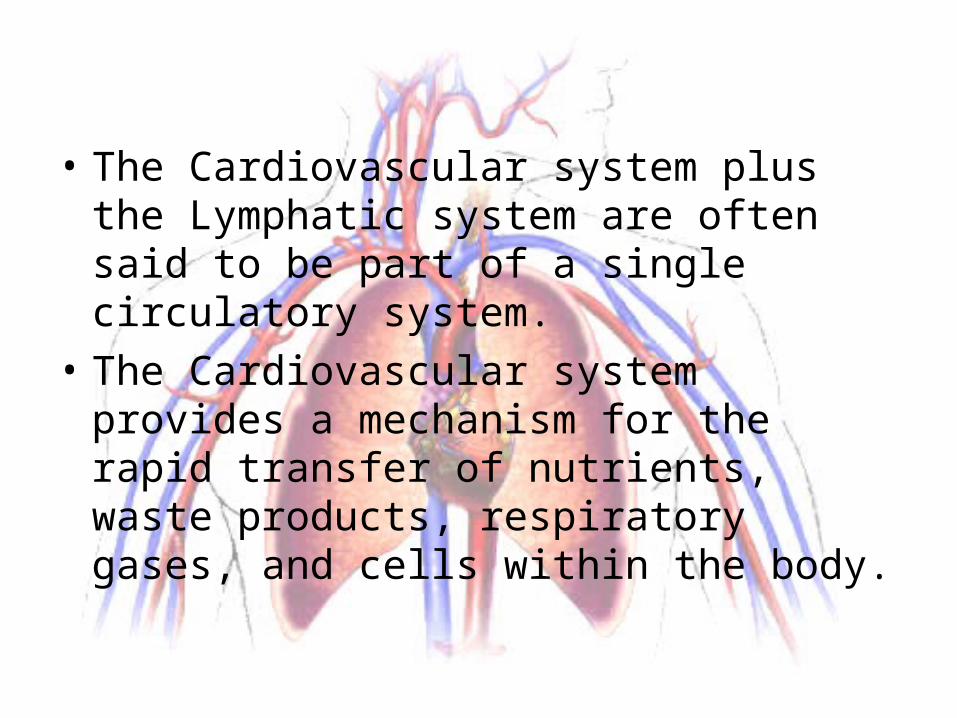
• The Cardiovascular system plus the Lymphatic system are often said to be part of a single circulatory system.
• The Cardiovascular system provides a mechanism for the rapid transfer of nutrients, waste products, respiratory gases, and cells within the body.
BLOBLOOD
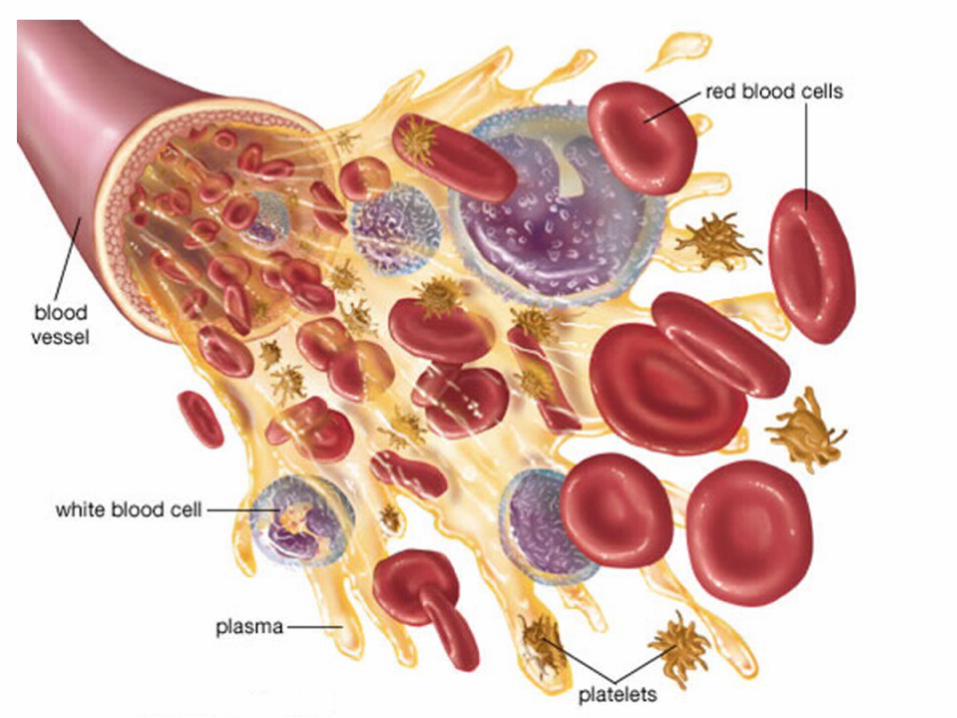
• Blood is a type of specialized connective tissue.
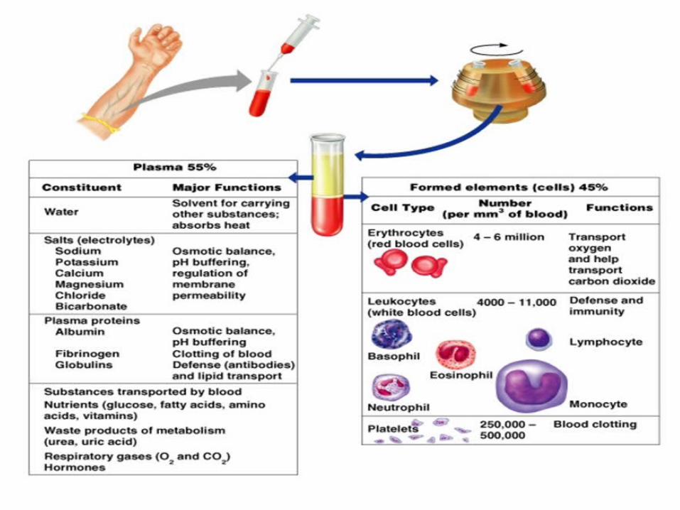
• Whole Blood is composed of Plasma (46-63%) and Formed Element (37-54%)-red blood cells, white blood cells, and platelets.
• Temperature--roughly 100.4 F
PLAPLASMA• makes up 46% to 63% of whole blood• 92% of plasma is water• the plasma proteins are produced in the liver• The primary plasma proteins are: albumins,
globulins, and fibrinogen
PRIMARY PLASMA PROTEINS
• ALBUMINS• Are important for transport of fatty acids, thyroid
hormones, some steroid hormones, and other substances
• GLOBULINS• most important globulins are antibodies(protect
against foreign proteins and pathogens), and transport proteins (bind ions and other components.)
• FIBROGEN• these are insoluble strands of fibrin• they aid in blood clots
FORMED ELEMENTS
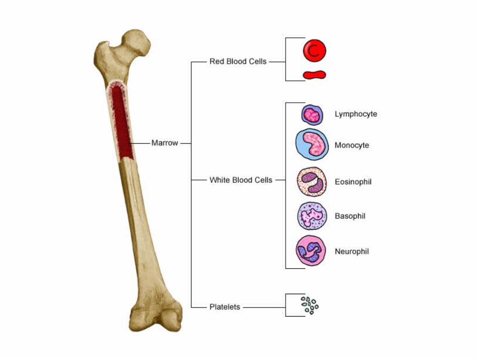
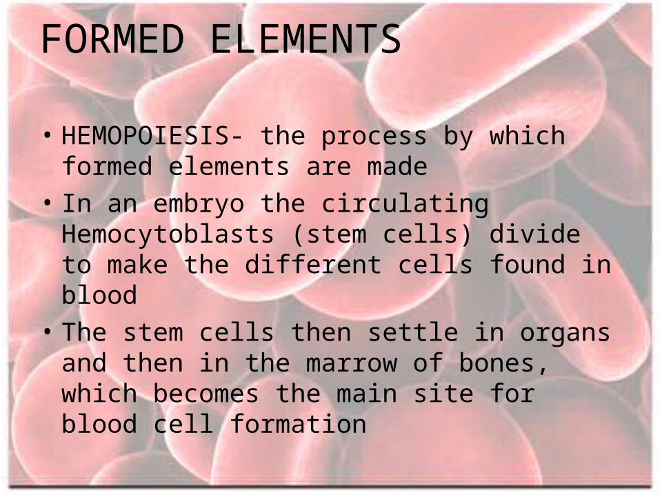
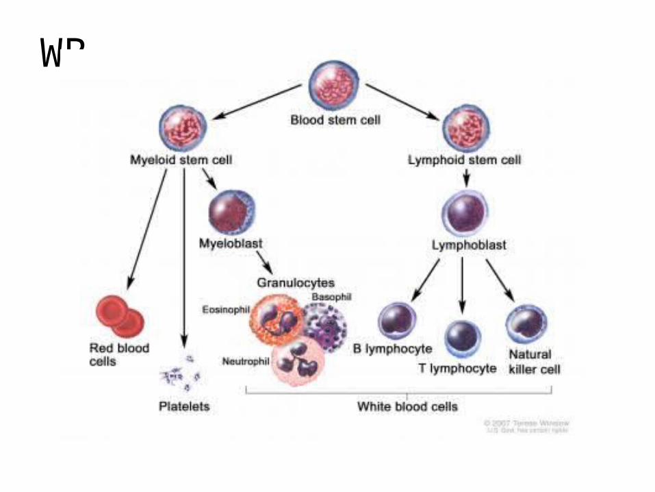
• HEMOPOIESIS- the process by which formed elements are made
• In an embryo the circulating Hemocytoblasts (stem cells) divide to make the different cells found in blood
• The stem cells then settle in organs and then in the marrow of bones, which becomes the main site for blood cell formation
RED BLOOD CELLS (RBCs)
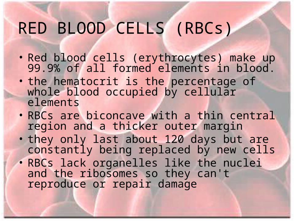
• Red blood cells (erythrocytes) make up 99.9% of all formed elements in blood.
• the hematocrit is the percentage of whole blood occupied by cellular elements
• RBCs are biconcave with a thin central region and a thicker outer margin
• they only last about 120 days but are constantly being replaced by new cells
• RBCs lack organelles like the nuclei and the ribosomes so they can't reproduce or repair damage
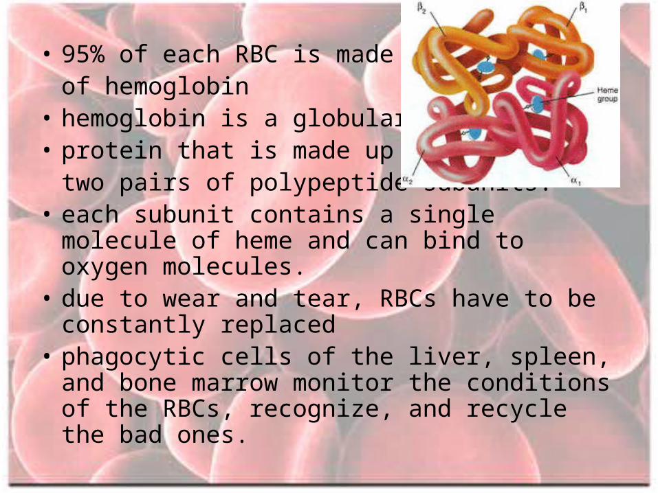
• 95% of each RBC is made upof hemoglobin
• hemoglobin is a globular • protein that is made up of
two pairs of polypeptide subunits.• each subunit contains a single molecule of heme
and can bind to oxygen molecules.• due to wear and tear, RBCs have to be constantly
replaced• phagocytic cells of the liver, spleen, and bone
marrow monitor the conditions of the RBCs, recognize, and recycle the bad ones.
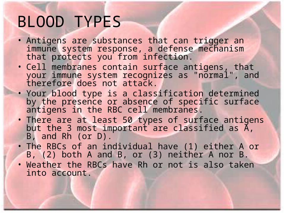
BLOOD TYPES• Antigens are substances that can trigger an immune system
response, a defense mechanism that protects you from infection.
• Cell membranes contain surface antigens, that your immune system recognizes as "normal", and therefore does not attack.
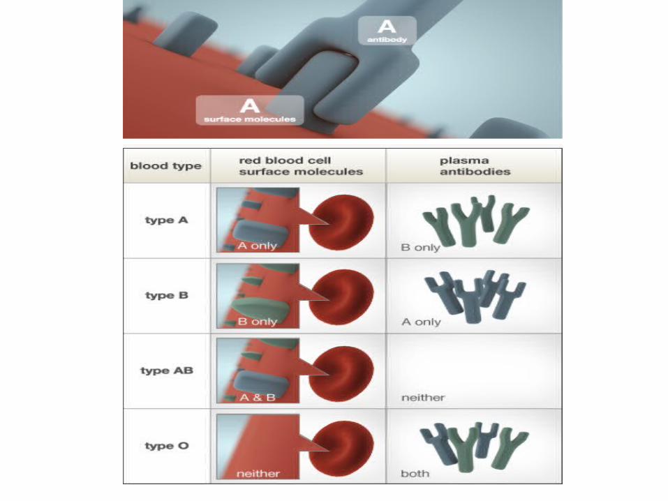
• Your blood type is a classification determined by the presence or absence of specific surface antigens in the RBC cell membranes.
• There are at least 50 types of surface antigens but the 3 most important are classified as A, B, and Rh (or D).
• The RBCs of an individual have (1) either A or B, (2) both A and B, or (3) neither A nor B.
• Weather the RBCs have Rh or not is also taken into account.
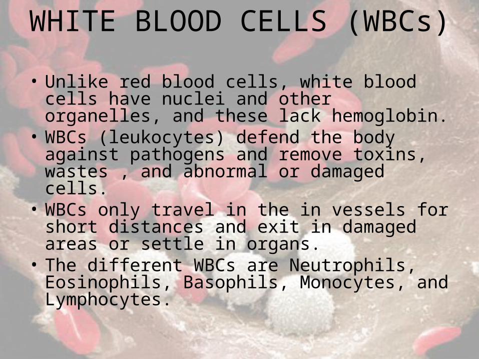
WHITE BLOOD CELLS (WBCs)
• Unlike red blood cells, white blood cells have nuclei and other organelles, and these lack hemoglobin.
• WBCs (leukocytes) defend the body against pathogens and remove toxins, wastes , and abnormal or damaged cells.
• WBCs only travel in the in vessels for short distances and exit in damaged areas or settle in organs.
• The different WBCs are Neutrophils, Eosinophils, Basophils, Monocytes, and Lymphocytes.
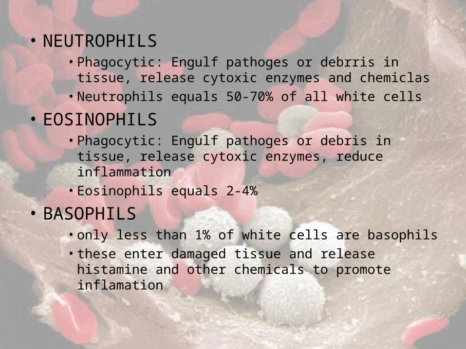
• NEUTROPHILS • Phagocytic: Engulf pathoges or debrris in tissue, release
cytoxic enzymes and chemiclas• Neutrophils equals 50-70% of all white cells
• EOSINOPHILS• Phagocytic: Engulf pathoges or debris in tissue, release
cytoxic enzymes, reduce inflammation• Eosinophils equals 2-4%
• BASOPHILS• only less than 1% of white cells are basophils• these enter damaged tissue and release histamine and
other chemicals to promote inflamation
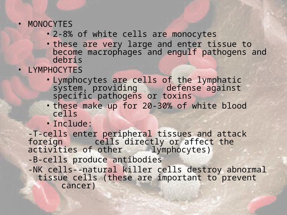
• MONOCYTES• 2-8% of white cells are monocytes• these are very large and enter tissue to become macrophages
and engulf pathogens and debris• LYMPHOCYTES
• Lymphocytes are cells of the lymphatic system, providing defense against specific pathogens or toxins
• these make up for 20-30% of white blood cells• Include:
-T-cells enter peripheral tissues and attack foreign cells directly or affect the activities of other lymphocytes)-B-cells produce antibodies-NK cells--natural killer cells destroy abnormal tissue cells (these are important to prevent
cancer)
WB
PLATELETS
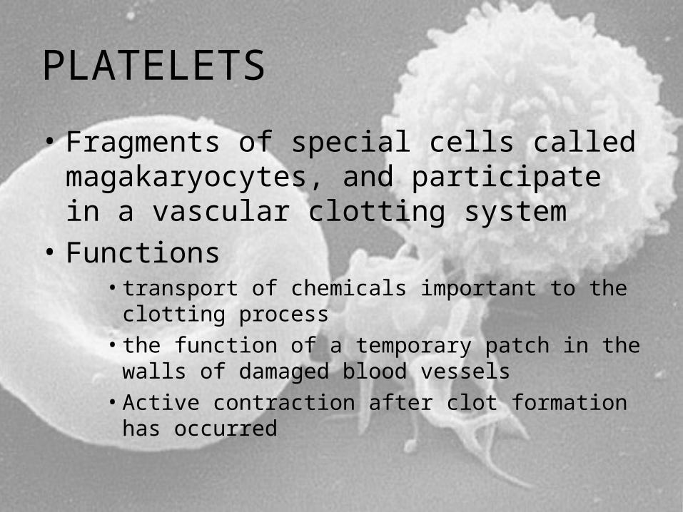
• Fragments of special cells called magakaryocytes, and participate in a vascular clotting system
• Functions• transport of chemicals important to the clotting
process• the function of a temporary patch in the walls of
damaged blood vessels• Active contraction after clot formation has occurred
HOMEOSTASIS• Homeostasis prevents the loss of blood through the walls
of damaged vessels• establishes a frame work for tissue repair• Consists of 3 phases
• The Vascular Phase--a period of local blood vessels constriction, or vascular spasms, at the injury site.
• The Platelet Phase--follows as platelets are activated, aggregate at the site, and adhere to the damaged surfaces.
• The Coagulation Phase--occurs as factors released by platelets and endothelial cells interact with clotting factors to form a blood clot.
• Clot Retraction--during clot retraction, platelets contract and pull the torn edges of the damaged vessels closer together.
• Fibrinolysis--during fibrinolysis, the clot gradually dissolves through the action of plasmin, the activated form of circulating plasminogen.
THE HEART
• The blood in our body has to keep on flowing to provide the cells of the body with the oxygen needed to and remove waste while transporting white cells to their intended targets.
• All functions of the circulatory system depend on the Heart to pump the blood through the body.
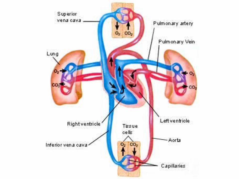
• The blood flows through a network of blood vessels that can be separated into two different sections-
• Pulmonary Circuit--carries blood to and from the gas exchange surfaces of the lungs.
• Systemic Circuit--transports blood to and from the rest of the body.
-The Arteries carry blood away from the heart
-The Veins return blood to the heart-Capillaries are the thin walled,
narrow- diameter vessels that connect the smallest arteries and veins and allow the exchange of O2 and CO2
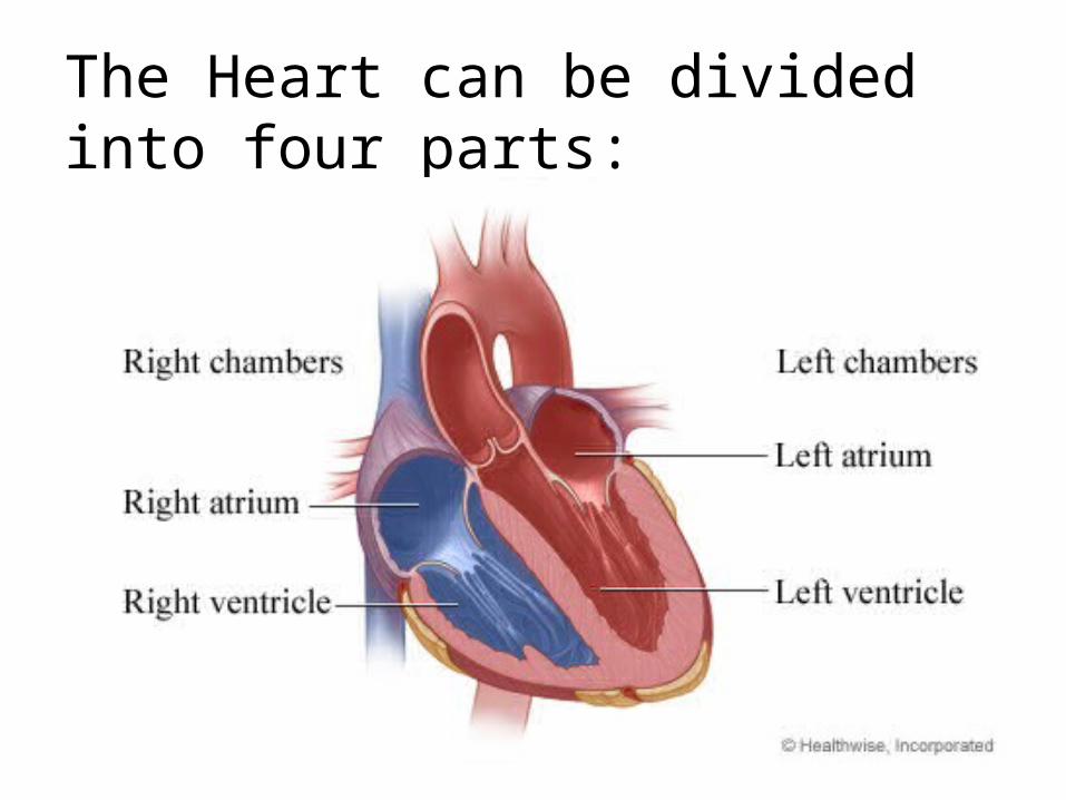
The Heart can be divided into four parts:
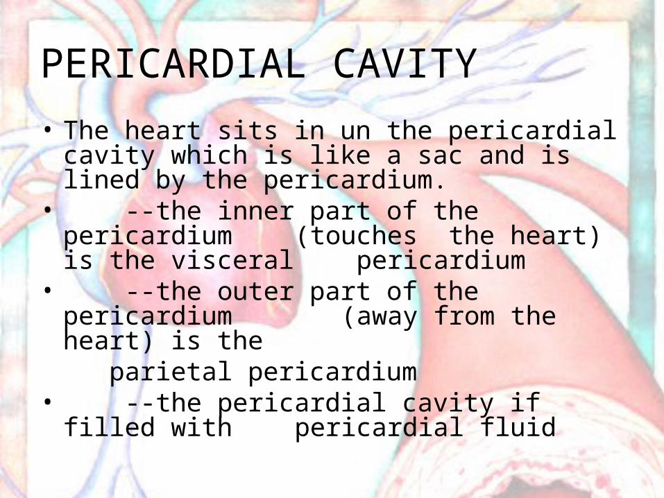
PERICARDIAL CAVITY• The heart sits in un the pericardial cavity which is
like a sac and is lined by the pericardium.• --the inner part of the pericardium
(touches the heart) is the visceral pericardium
• --the outer part of the pericardium (away from the heart) is the
parietal pericardium• --the pericardial cavity if filled with
pericardial fluid
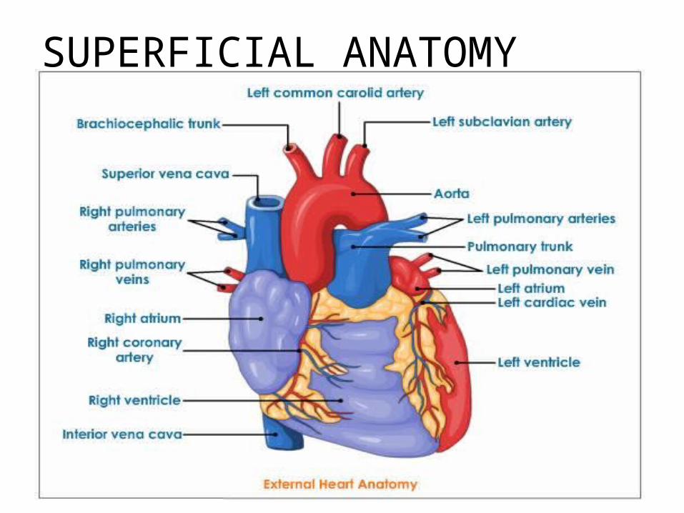
SUPERFICIAL ANATOMY
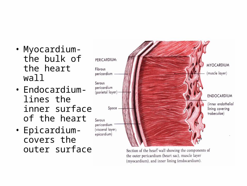
• Myocardium-the bulk of the heart wall
• Endocardium-lines the inner surface of the heart
• Epicardium-covers the outer surface
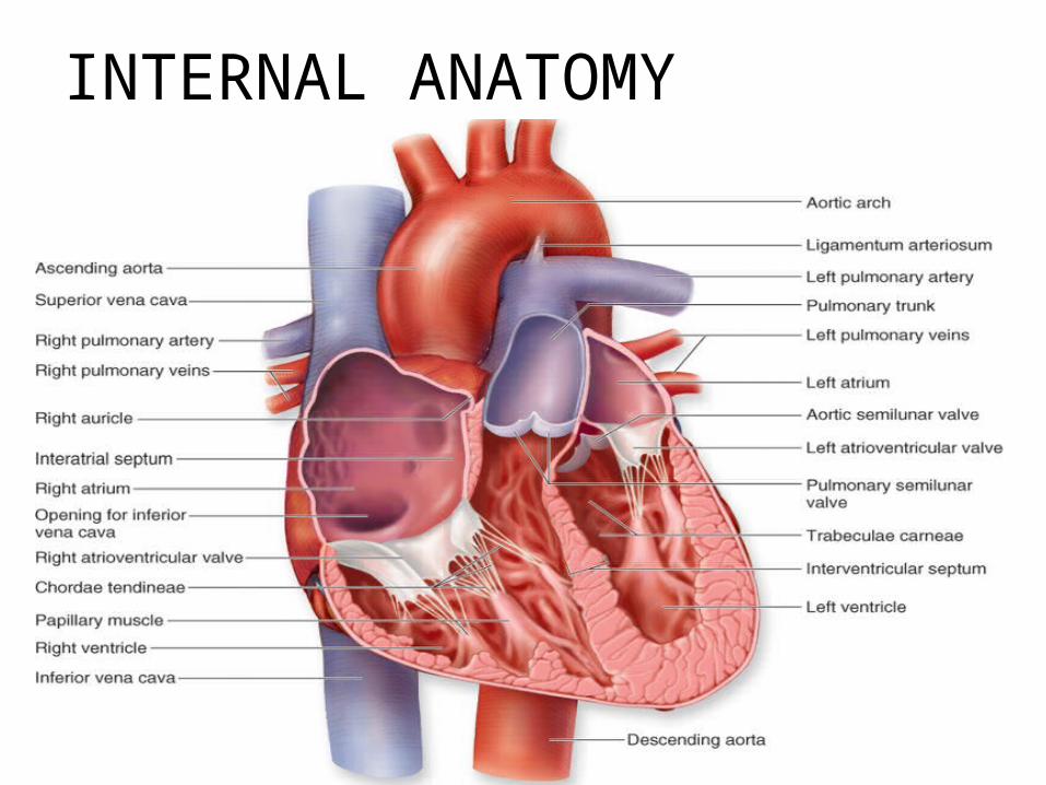
INTERNAL ANATOMY
THE BLOOD SUPPLY TO THER HEART• The heart muscles also need oxygen to function
properly, so there are arteries and veins running around the outside of the heart
• The Coronary Circulation supplies oxygen and nutrients needed by the cardiac muscle cells.
THE CORONARY ARTERIES-take oxygen rich blood from the aorta and take it to the heart’s muscles.
THE CARDIAC VEINS-connect with the coronary arteries and take the blood back to the heart.
THE HEART BEAT
• A heart beat is the coordinated contraction of the whole heart to make blood flow in the right direction at the proper time.
• the contractile cells in the heart are much faster than regular skeletal muscles.
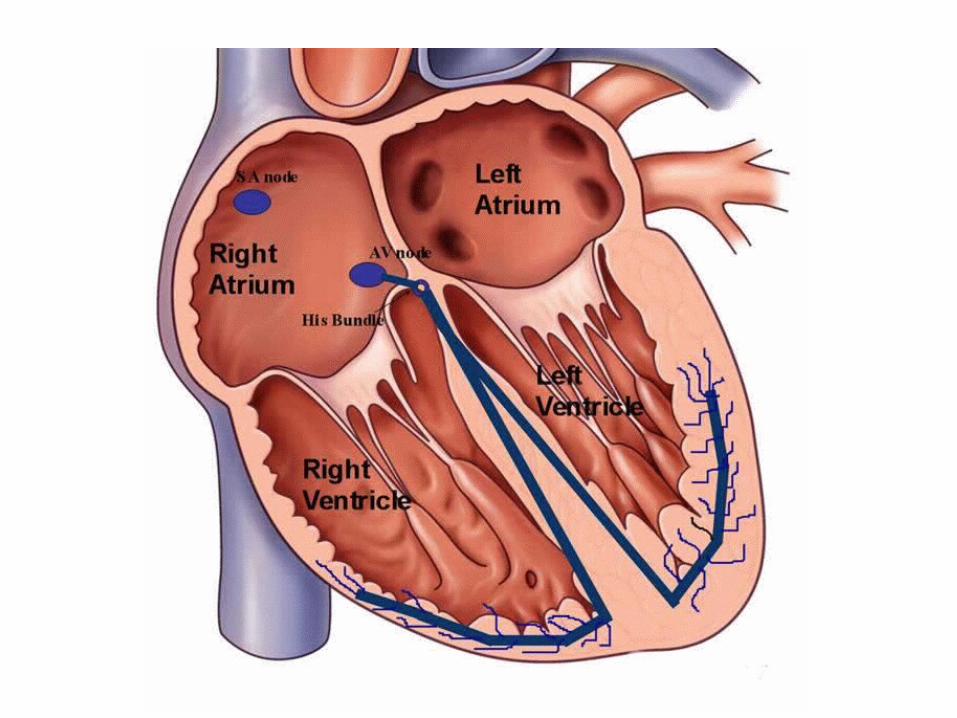
• THE CONDUCTING SYSTEM-the conducting system includes the
Senatorial (SA) Node, Antrioventricular (AV) Node, and Conducting cells.
THE HEART BEAT
• The senatorial (SA) Node (pacemaker)- located at the wall of the right atrium.
• an action potential appears at the SA Node• this action potential then spreads through the
Conducting cells around the Right Atrium and reaches the Left Atrium• the left and right Atrium begin to contract• the left and right Atrioventricular (AV) Valve close
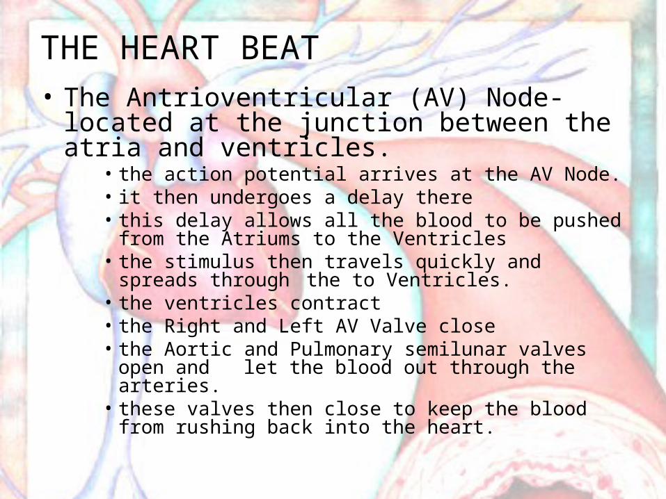
THE HEART BEAT• The Antrioventricular (AV) Node-located at the
junction between the atria and ventricles.• the action potential arrives at the AV Node.• it then undergoes a delay there• this delay allows all the blood to be pushed from the
Atriums to the Ventricles• the stimulus then travels quickly and spreads through the
to Ventricles.• the ventricles contract • the Right and Left AV Valve close• the Aortic and Pulmonary semilunar valves open and let
the blood out through the arteries.• these valves then close to keep the blood from rushing back
into the heart.
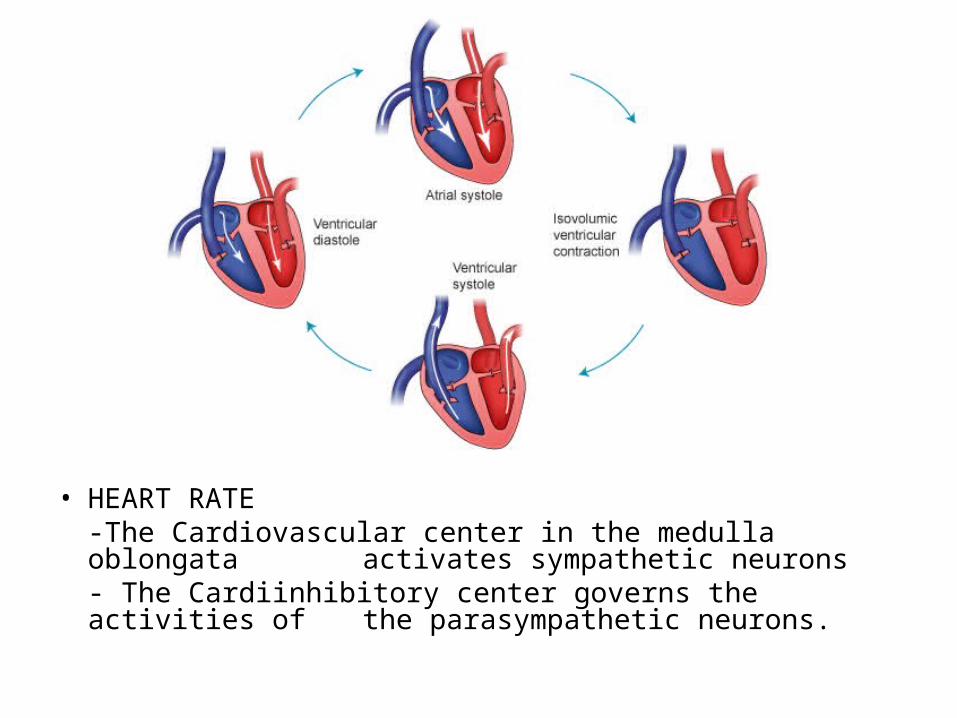
• HEART RATE-The Cardiovascular center in the medulla oblongata activates sympathetic neurons- The Cardiinhibitory center governs the activities of the parasympathetic neurons.

CARDIOVASCULAR REGULATION• Homeostasis mechanisms ensure that tissue
perfusion (blood flow) delivers adequate oxygen and nutrients.
• Local Factors (Autoregulation) change the pattern of blood flow within capillary beds in response to chemical changes in interstitial fluids.
• Central Mechanisms (Neural) respond to changes in arterial pressure or blood gas levels. Ex. Epinephrine and Norepinephrine create the “Fight or Flight” effect.
• The Endocrine Factors that help homeostasis are hormones, Which can assist in short term adjustments (change in cardiac output and peripheral resistance) and long term adjustments (changes in blood volume that affect cardiac output and gas transport).
Patterns of the Cardiovascular System• Exercise- by exercising often, you can train your heart a
bigger output and get improved Cardiovascular performance -athletes have larger stroke volumes, slower resting heart rates, and larger cardiac reserves than do nonathletes.
• Response to Hemorrhaging (excessive blood loss)-blood loss lower blood volume and venous return and decrease cardiac output.-peripheral vasoconstriction (vessel constriction)-hormones are released that promote fluid
retention and more erythrocytes (RBCs) are made.• Shock is acute circulatory crisis marked by hypotension and
inadequate peripheral blood flow
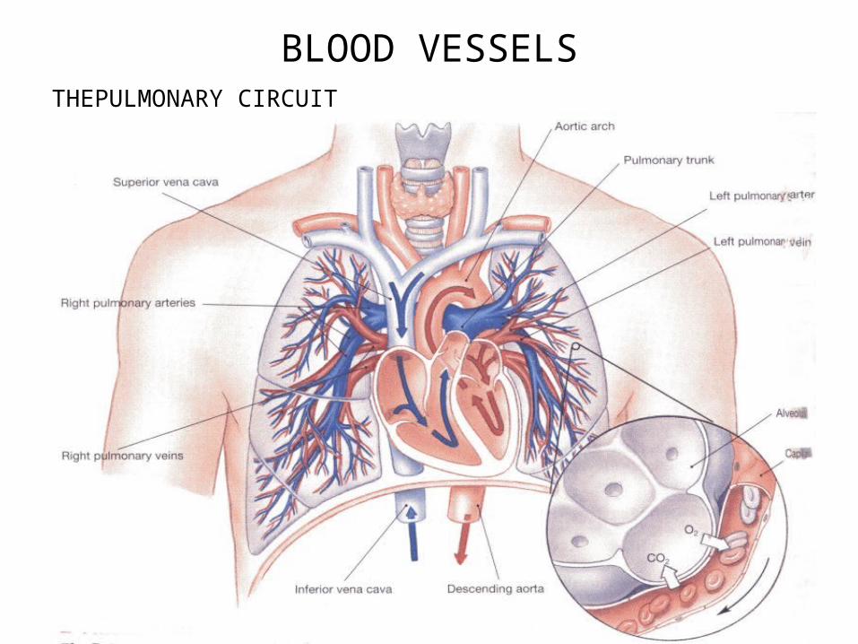
BLOOD VESSELSTHEPULMONARY CIRCUIT
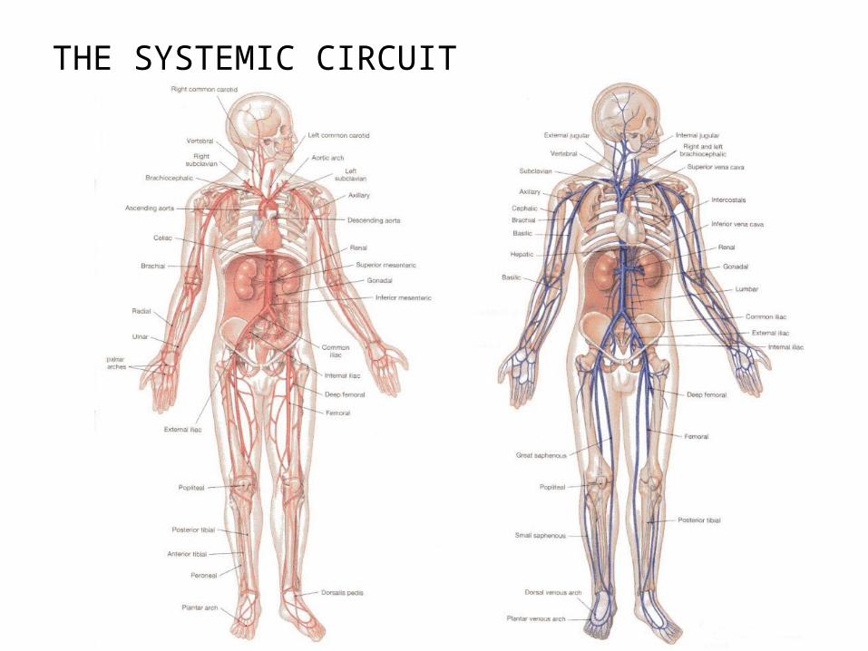
THE SYSTEMIC CIRCUIT
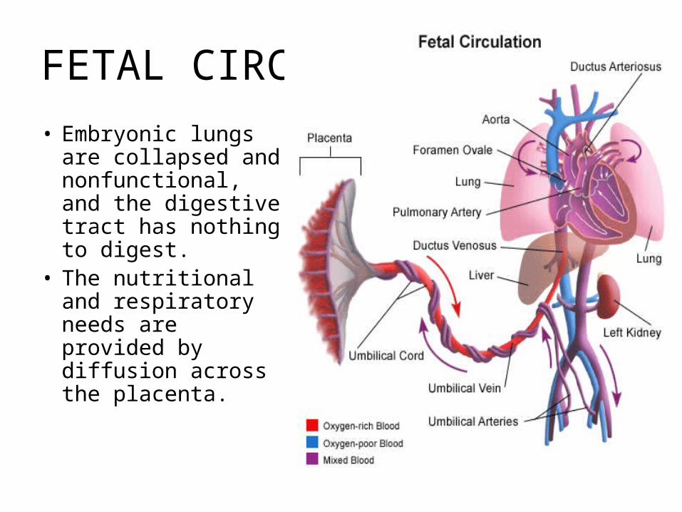
FETAL CIRCUIT
• Embryonic lungs are collapsed and nonfunctional, and the digestive tract has nothing to digest.
• The nutritional and respiratory needs are provided by diffusion across the placenta.
• AGING AND THE CARDIOVASCULAR• Age-related Changes in Blood-decrease in hematocrit,
blood clotting, pooling of blood • The Aging Heart-does not work as efficiently, and can’t
repair as well• Aging Blood Vessels-vessels can rupture more easily
causing blood loss
• INTEGRATION WITH OTHER SYSTEMS• The cardiovascular system is anatomically and
functionally linked to all other systems.