Embed Size (px)
Citation preview
Human Body:Cardiovascular System
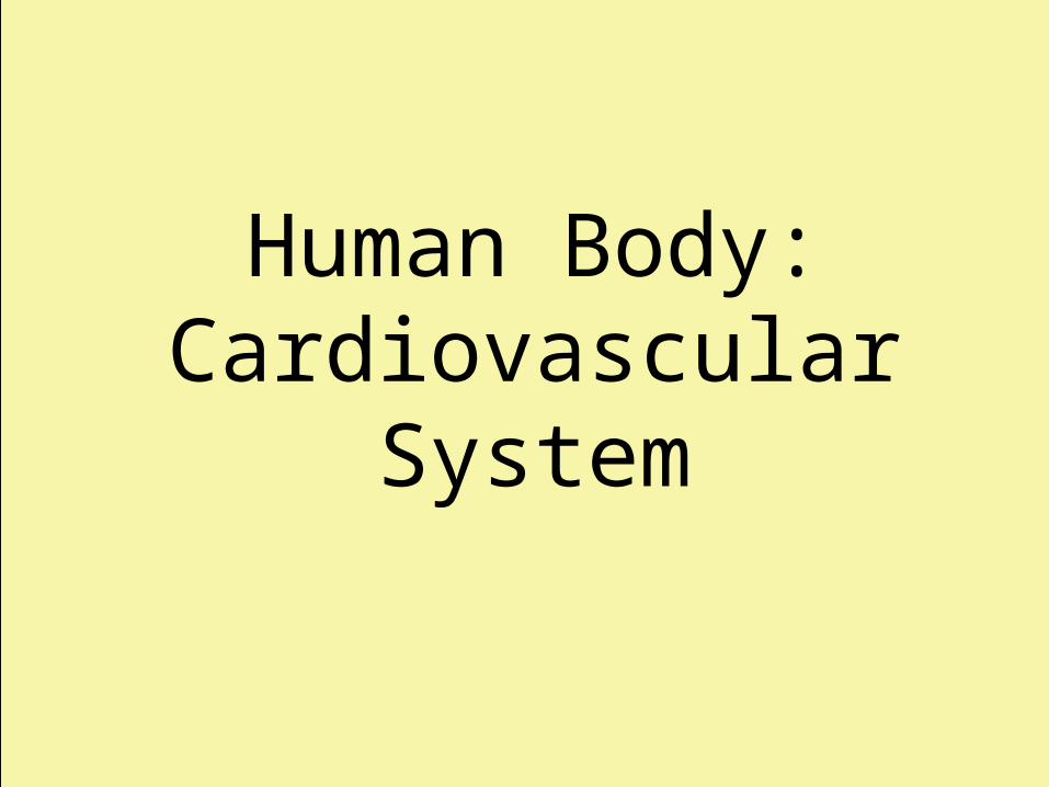
Closed vs. OpenCirculatory System
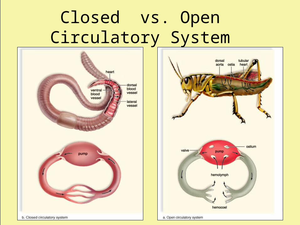
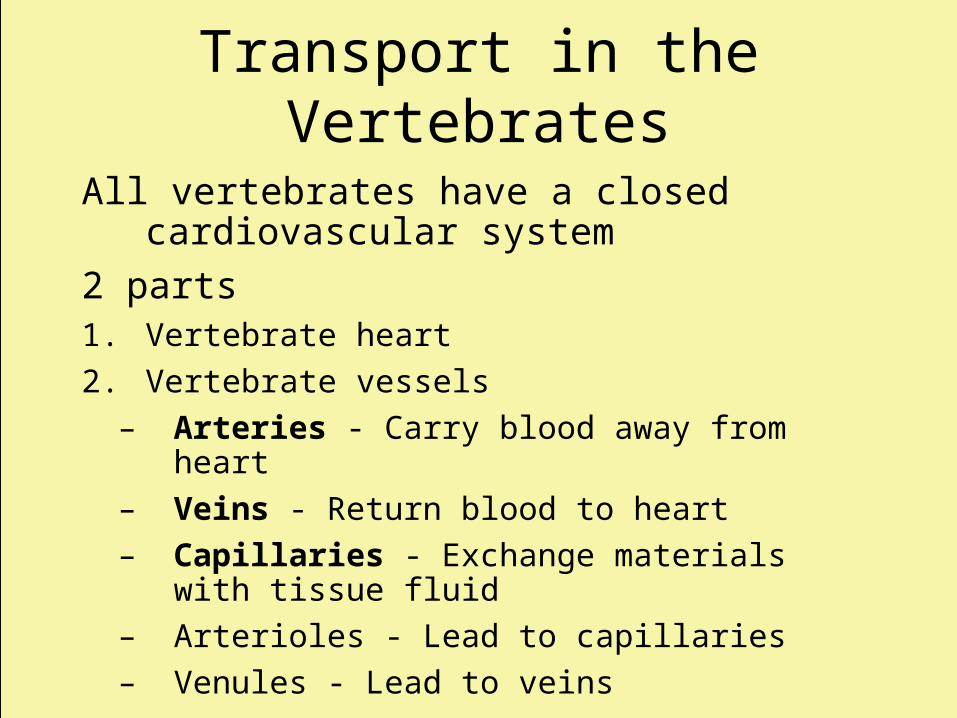
Transport in the Vertebrates
All vertebrates have a closed cardiovascular system
2 parts:1. Vertebrate heart
– Atrial chamber(s) of heart receive blood from general circulation
– Ventricle chamber(s) of heart pump blood out through blood vessels
Transport in the Vertebrates
All vertebrates have a closed cardiovascular system
2 parts1. Vertebrate heart
2. Vertebrate vessels
– Arteries - Carry blood away from heart
– Veins - Return blood to heart
– Capillaries - Exchange materials with tissue fluid
– Arterioles - Lead to capillaries
– Venules - Lead to veins
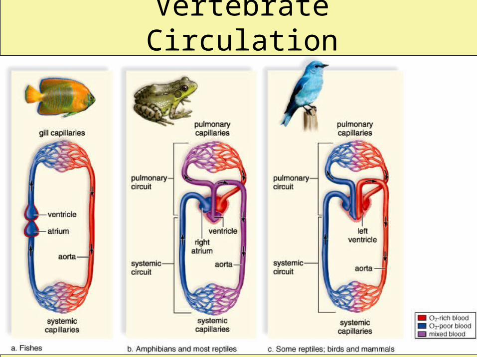
Vertebrate Circulation
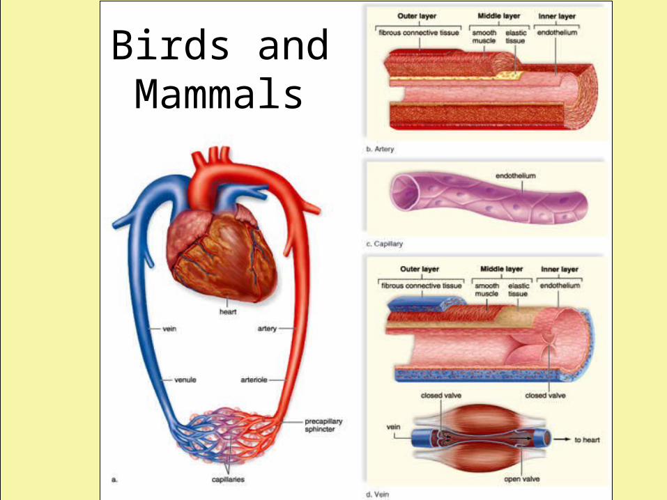
Birds and Mammals
Human Circulation
• Human Heart
– Fist-sized
– Cone-shaped
– Very muscular organ (special cardiac fibers)
– Lies within a fluid-filled sac (the pericardium)
Human Heart
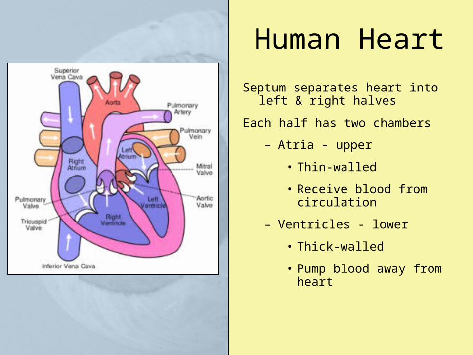
Septum separates heart into left & right halves
Each half has two chambers
– Atria - upper
• Thin-walled
• Receive blood from circulation
– Ventricles - lower
• Thick-walled
• Pump blood away from heart
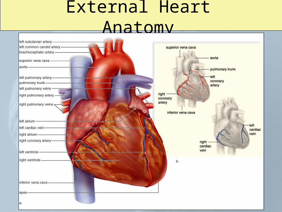
External Heart Anatomy
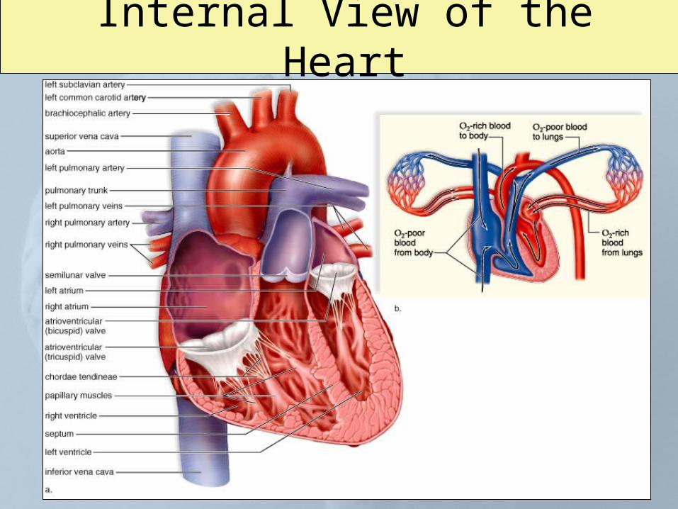
Internal View of the Heart
Human Heart:Valves
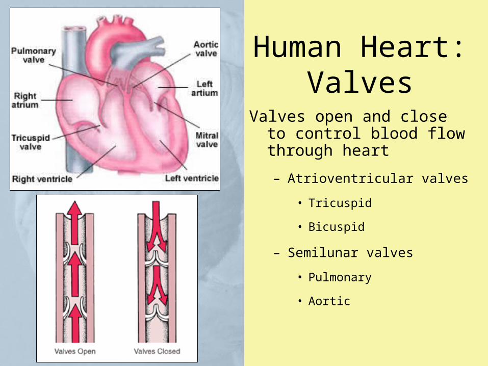
Valves open and close to control blood flow through heart
– Atrioventricular valves
• Tricuspid
• Bicuspid
– Semilunar valves
• Pulmonary
• Aortic
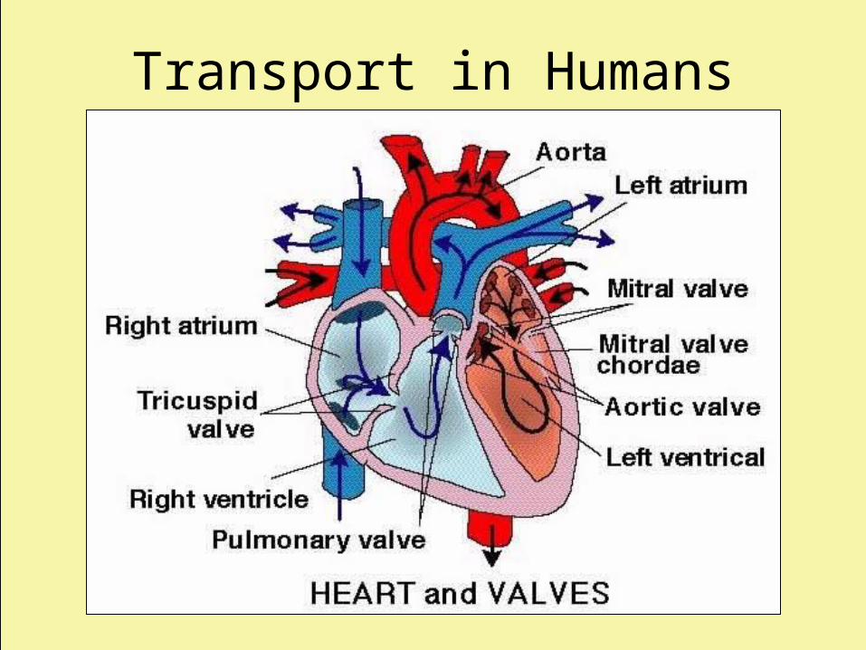
Transport in Humans
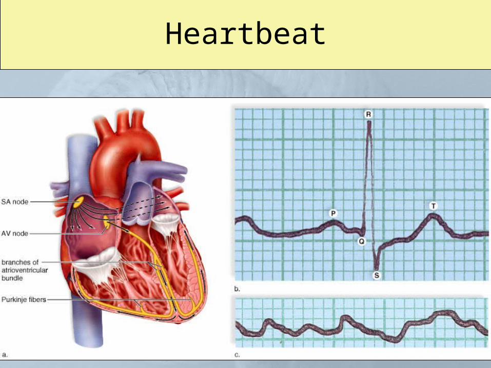
Heartbeat
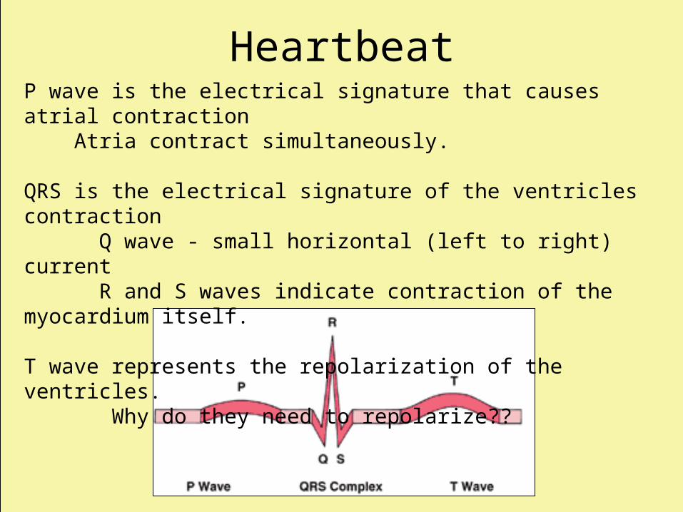
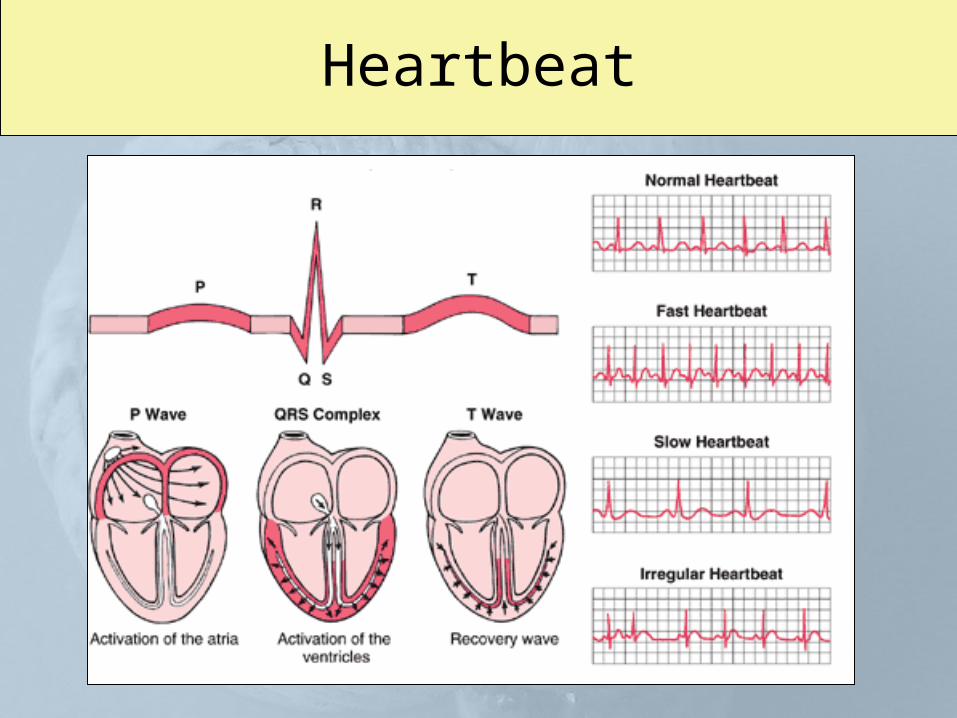
HeartbeatP wave is the electrical signature that causes atrial contraction Atria contract simultaneously.
QRS is the electrical signature of the ventricles contraction Q wave - small horizontal (left to right) current R and S waves indicate contraction of the myocardium itself.
T wave represents the repolarization of the ventricles. Why do they need to repolarize??
Heartbeat
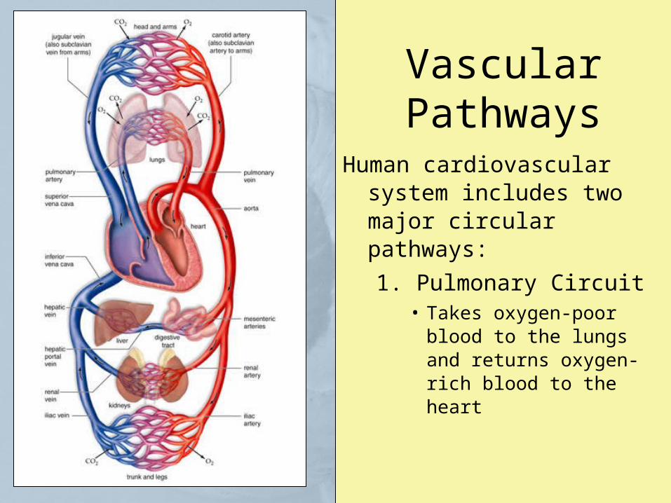
Vascular Pathways
Human cardiovascular system includes two major circular pathways:
1. Pulmonary Circuit• Takes oxygen-poor blood to
the lungs and returns oxygen-rich blood to the heart
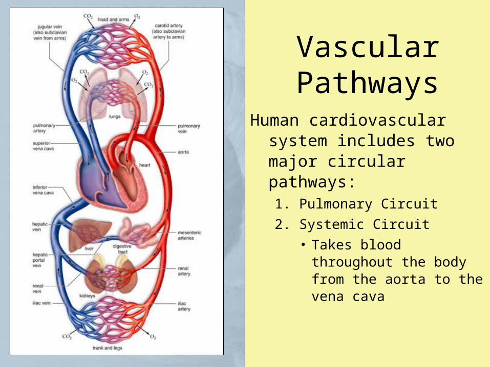
Vascular Pathways
Human cardiovascular system includes two major circular pathways:1. Pulmonary Circuit
2. Systemic Circuit
• Takes blood throughout the body from the aorta to the vena cava
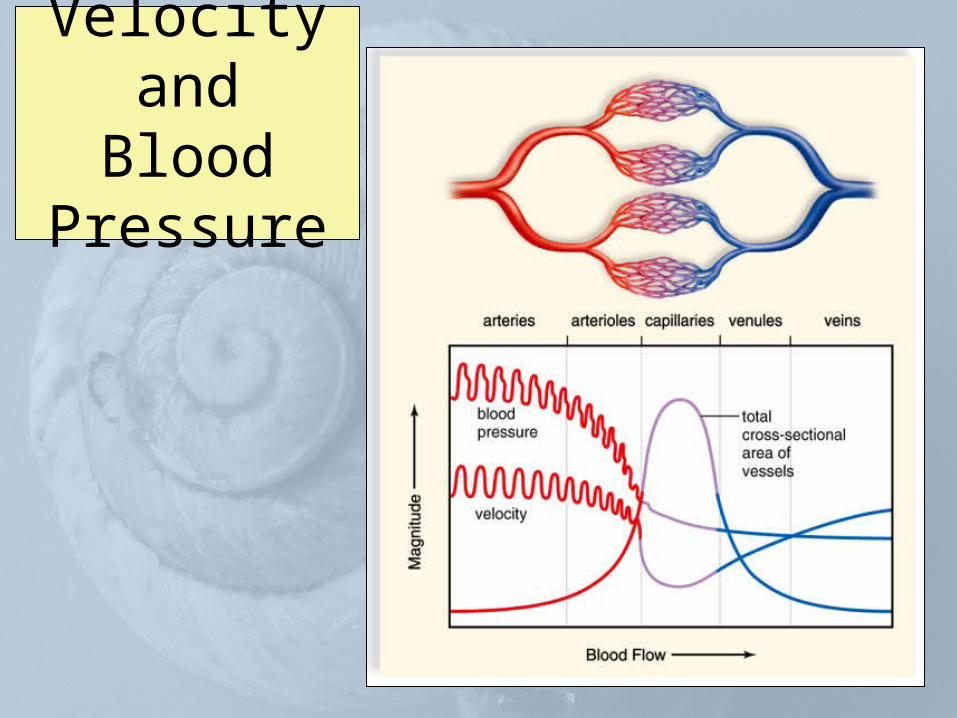
Velocity and Blood Pressure
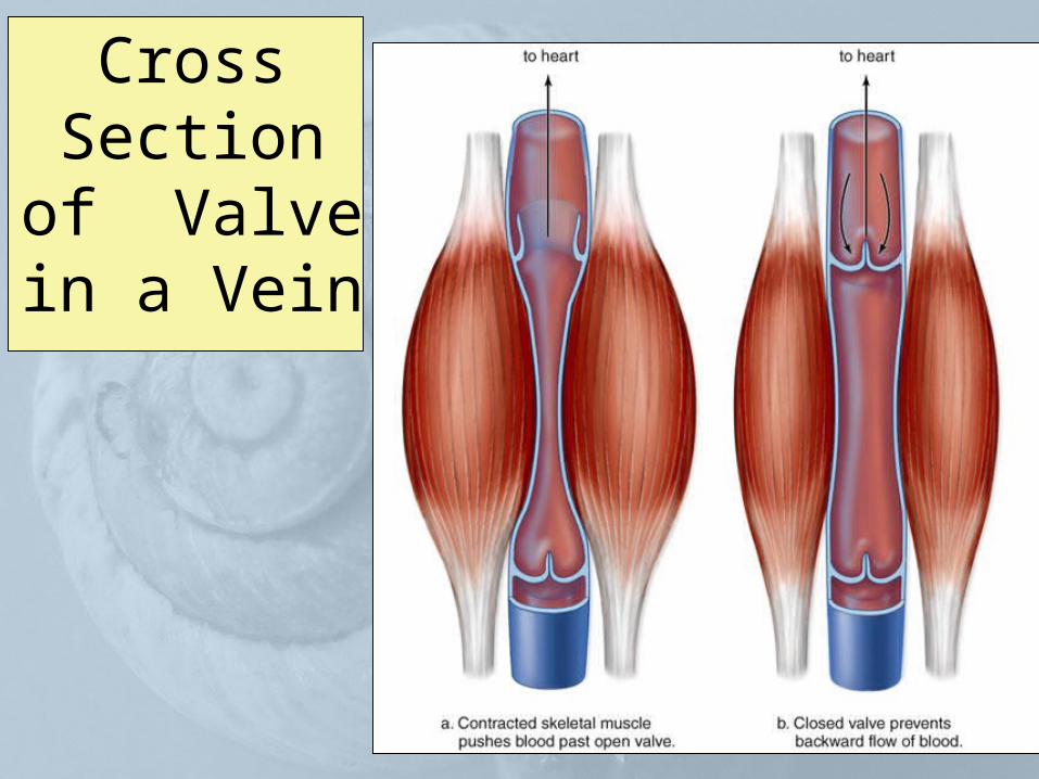
Cross Section of Valve in a
Vein
Blood Pressure
• The beat of the heart = pressure that keeps blood moving in the arteries
• Skeletal muscle contraction pushes blood in the veins toward the heart
Blood Pressure
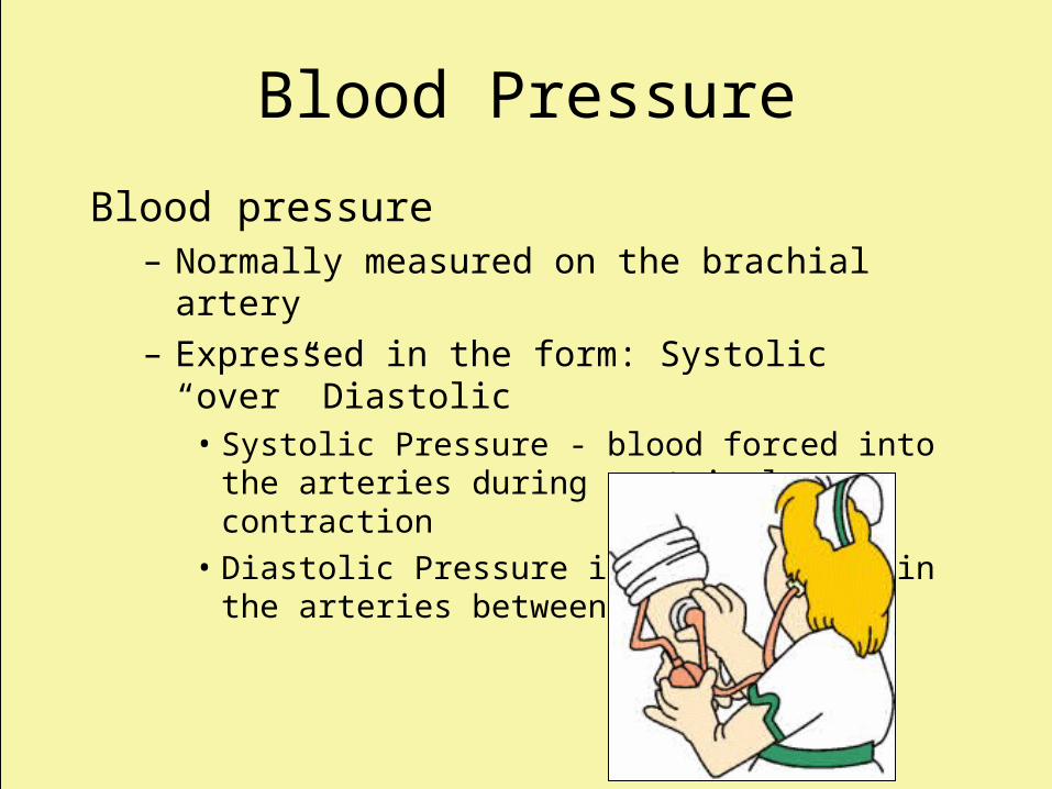
Blood pressure– Normally measured on the brachial artery– Expressed in the form: Systolic “over” Diastolic
• Systolic Pressure - blood forced into the arteries during ventricular contraction
• Diastolic Pressure is the pressure in the arteries between contractions
Cardiovascular Disorders
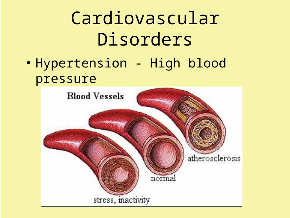
• Hypertension - High blood pressure
Cardiovascular Disorders
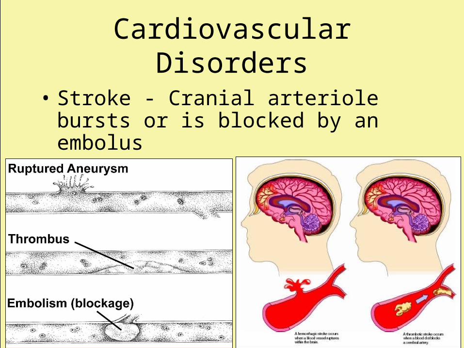
• Stroke - Cranial arteriole bursts or is blocked by an embolus
Cardiovascular Disorders
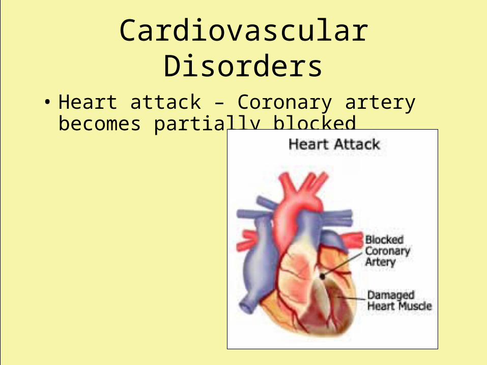
• Heart attack – Coronary artery becomes partially blocked
Cardiovascular Disorders
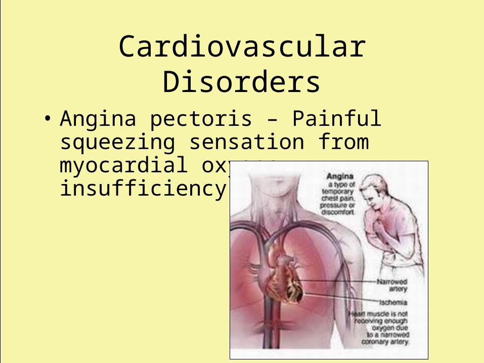
• Angina pectoris – Painful squeezing sensation from myocardial oxygen insufficiency
Blood Functions
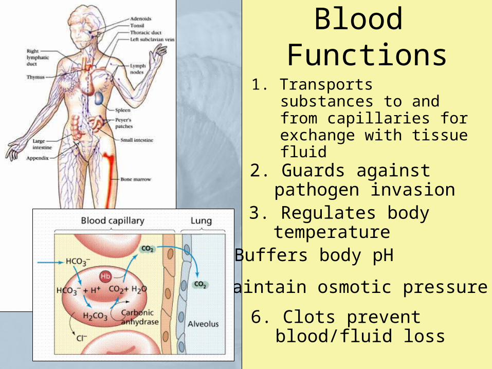
1. Transports substances to and from capillaries for exchange with tissue fluid
2. Guards against pathogen invasion
3. Regulates body temperature
4. Buffers body pH
5. Maintain osmotic pressure
6. Clots prevent blood/fluid loss
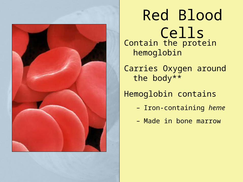
Red Blood CellsContain the protein hemoglobin
Carries Oxygen around the body**
Hemoglobin contains
– Iron-containing heme
– Made in bone marrow
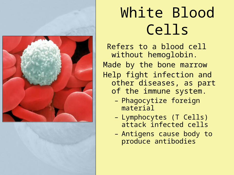
White Blood Cells
Refers to a blood cell without hemoglobin.
Made by the bone marrow Help fight infection and other
diseases, as part of the immune system.– Phagocytize foreign material– Lymphocytes (T Cells) attack
infected cells– Antigens cause body to produce
antibodies
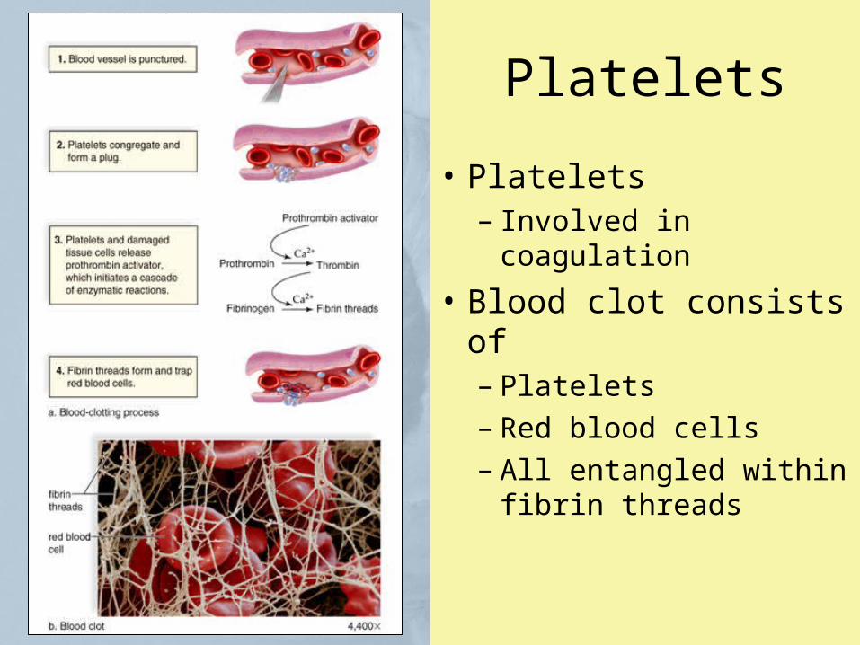
Platelets
• Platelets– Involved in coagulation
• Blood clot consists of– Platelets– Red blood cells– All entangled within
fibrin threads
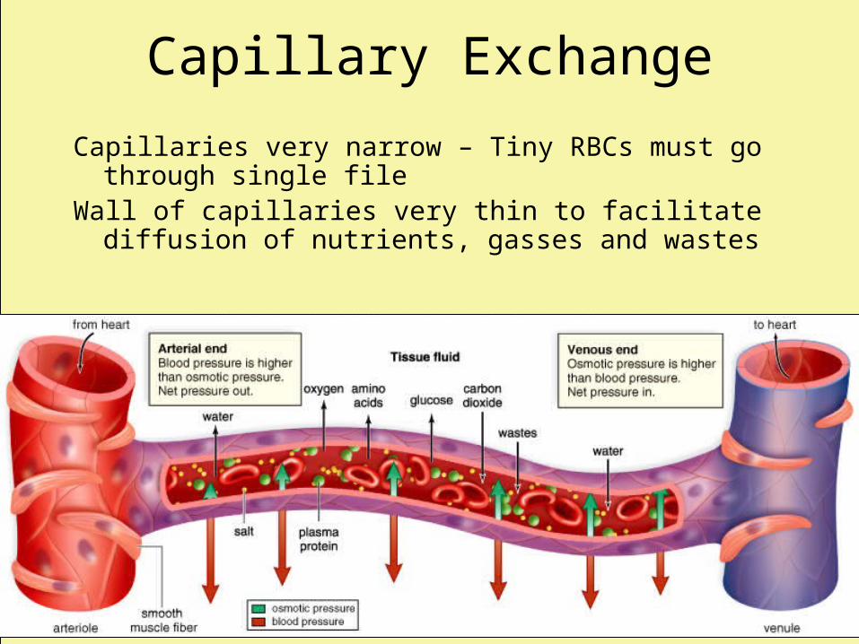
Capillary Exchange
Capillaries very narrow – Tiny RBCs must go through single file
Wall of capillaries very thin to facilitate diffusion of nutrients, gasses and wastes
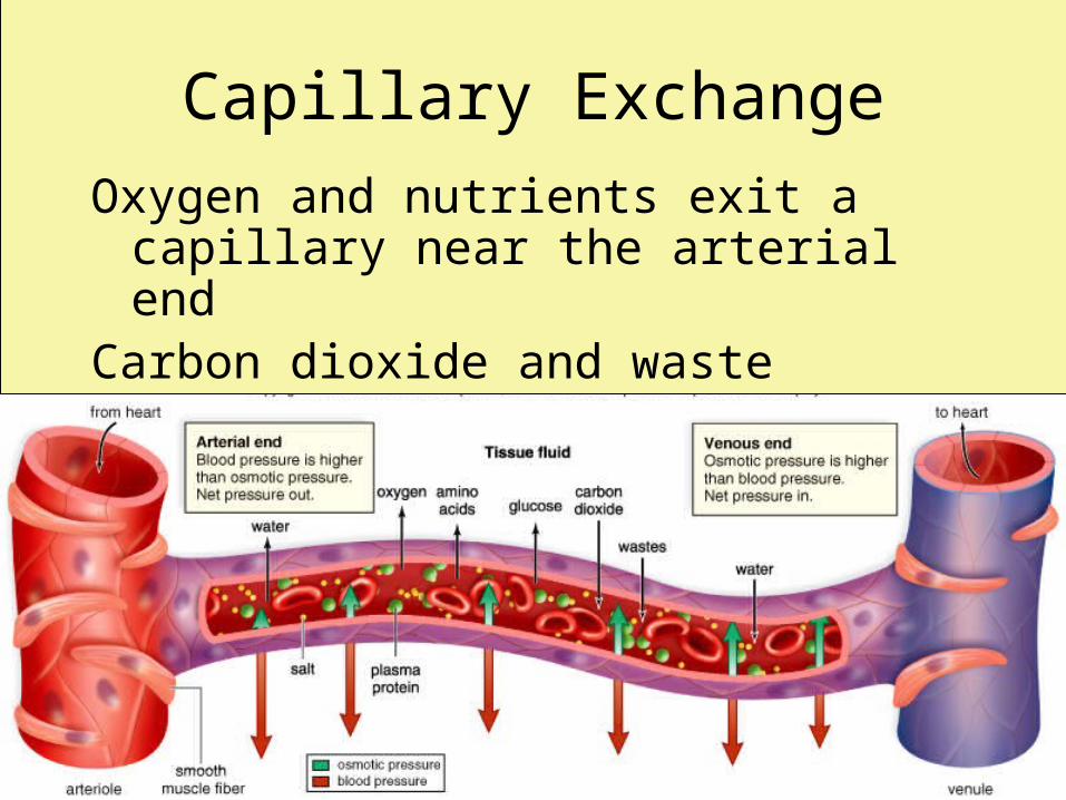
Capillary Exchange
Oxygen and nutrients exit a capillary near the arterial end
Carbon dioxide and waste molecules enter a capillary near the venous end
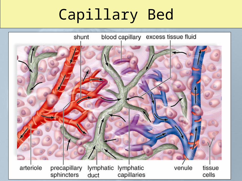
Capillary Bed
Human Body: Respiratory System
Gas Exchange
Respiration– The events associated with gas exchange
between the cells and the external environment
Gas Exchange SurfacesNeeds for effective, gas-exchange:
– Regions must be
• Moist
• Thin
• Relatively large (Surface Area/Vol - vascularization)
Delivery to cells is promoted by respiratory pigments (like hemoglobin)
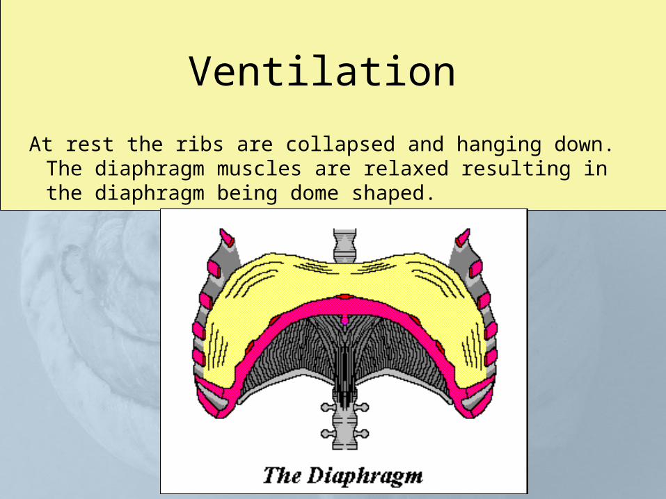
Ventilation
At rest the ribs are collapsed and hanging down. The diaphragm muscles are relaxed resulting in the diaphragm being dome shaped.
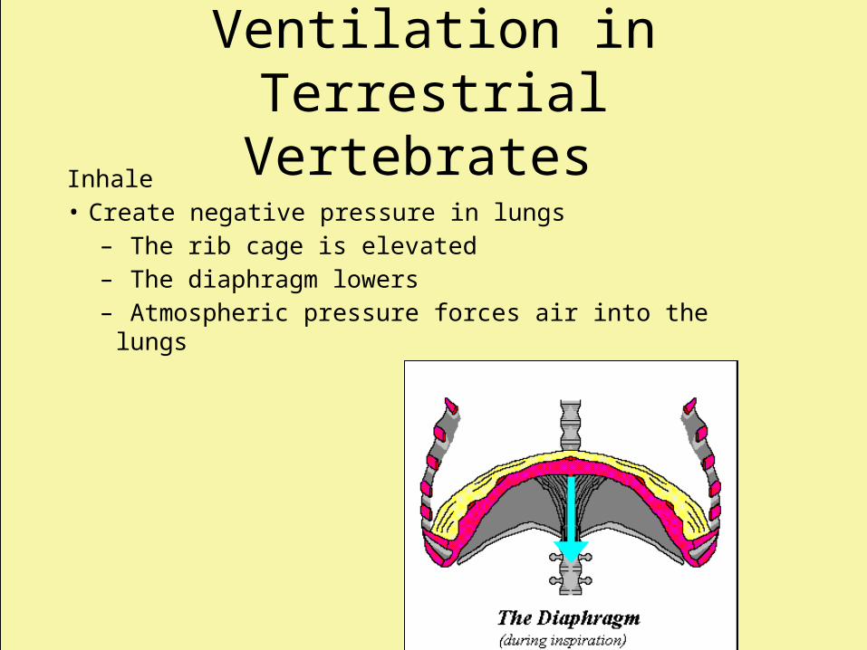
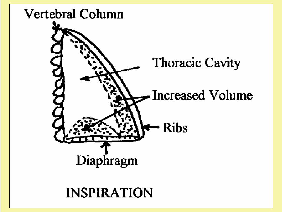
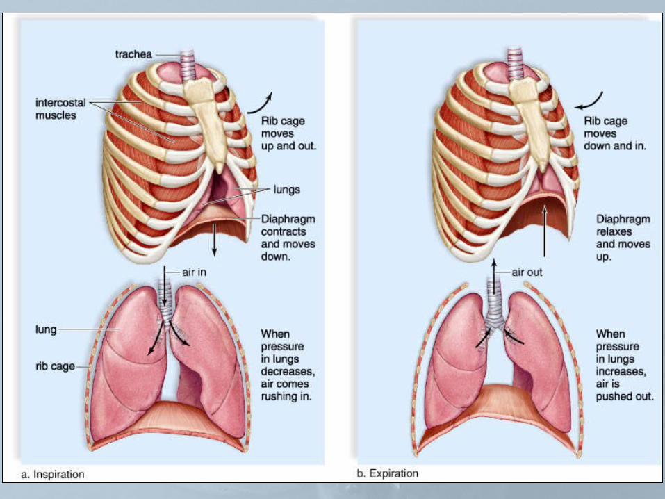
Ventilation inTerrestrial Vertebrates
Inhale• Create negative pressure in lungs
– The rib cage is elevated – The diaphragm lowers– Atmospheric pressure forces air into the lungs
Human Respiratory System
• As air moves through upper respiratory system– It is filtered to free it of debris– Warmed, and– Humidified
• When air reaches lungs– It is at body temperature, and– Its humidity is 100%
Upper Respiratory System
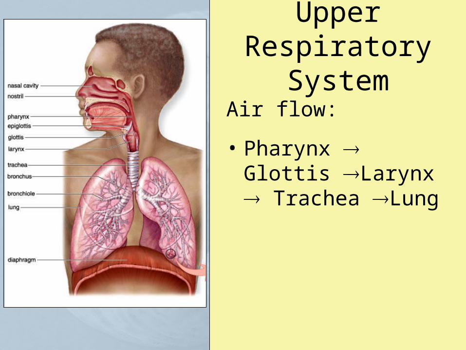
Air flow:
• Pharynx Glottis Larynx Trachea Lung
Upper Respiratory System
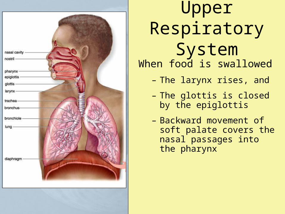
When food is swallowed
– The larynx rises, and
– The glottis is closed by the epiglottis
– Backward movement of soft palate covers the nasal passages into the pharynx
Lower Respiratory System
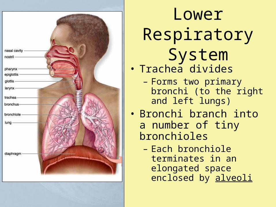
• Trachea divides– Forms two primary bronchi
(to the right and left lungs)
• Bronchi branch into a number of tiny bronchioles– Each bronchiole terminates
in an elongated space enclosed by alveoli
Gas Exchange and Transport
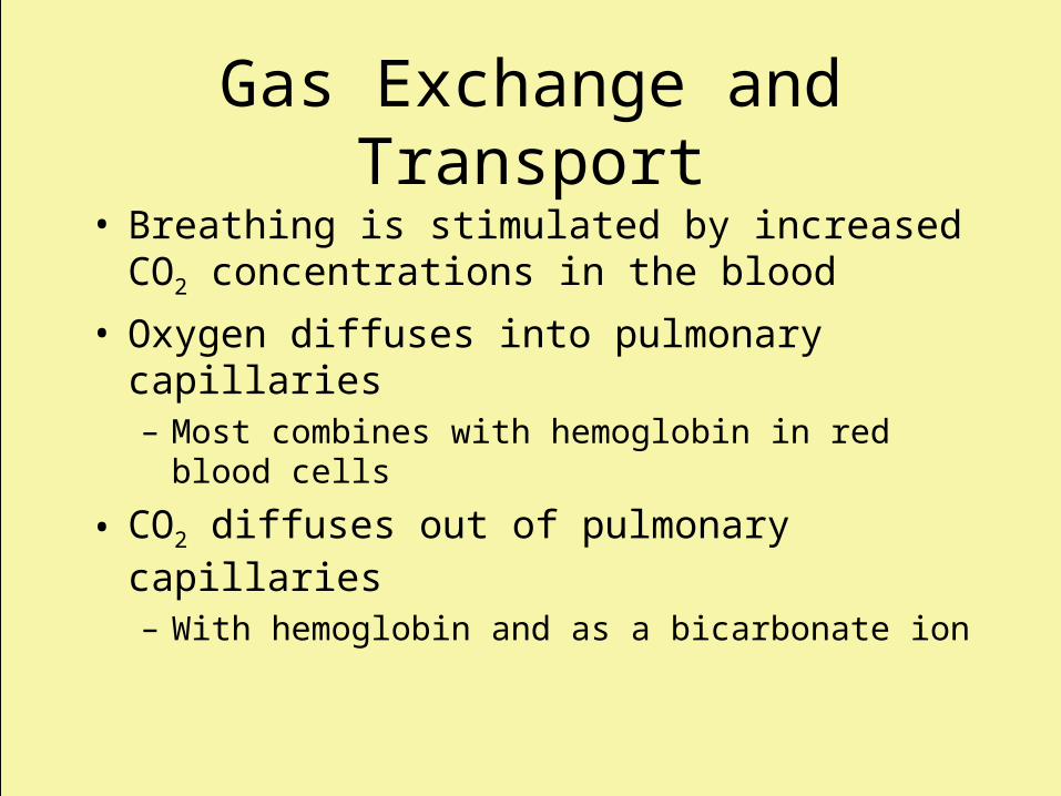
• Breathing is stimulated by increased CO2 concentrations in the blood
• Oxygen diffuses into pulmonary capillaries– Most combines with hemoglobin in red blood cells
• CO2 diffuses out of pulmonary capillaries– With hemoglobin and as a bicarbonate ion
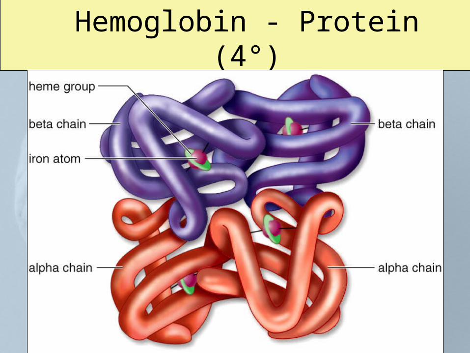
Hemoglobin - Protein (4°)
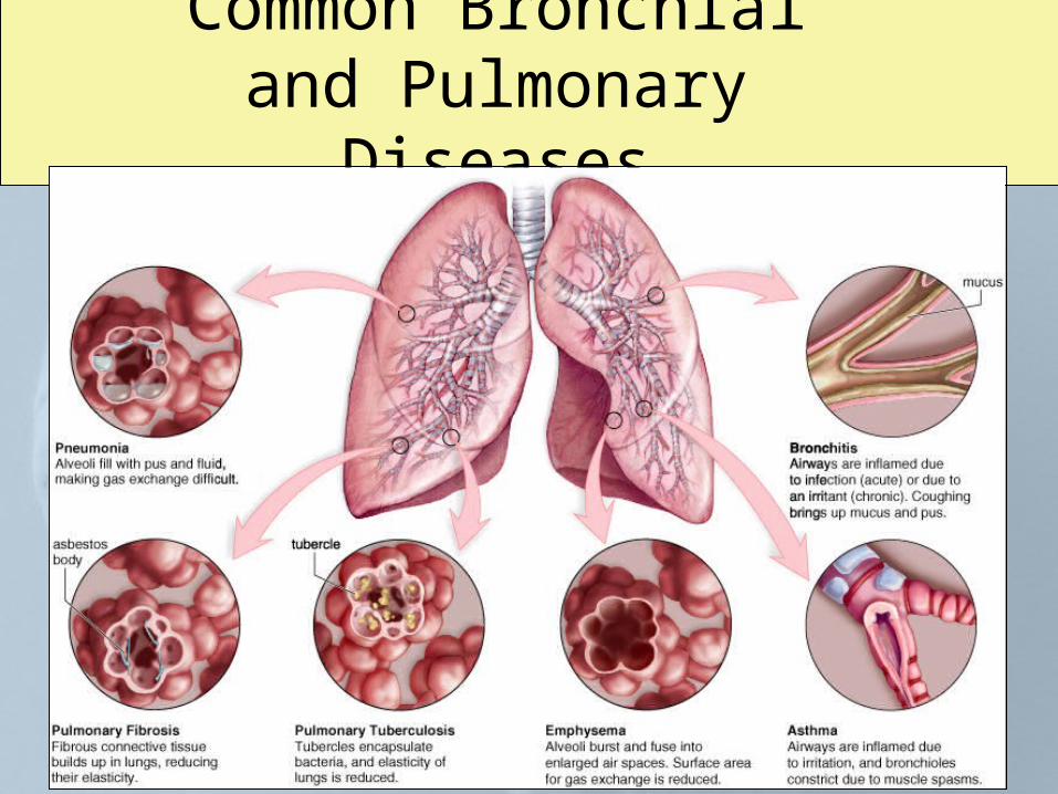
Common Bronchial and Pulmonary Diseases
Respiration and Health
Upper Respiratory Tract Infections
– Strep Throat
(Streptococcus pyogenes)
– Sinusitis - sinuse infection
– Tonsillitis - tonsil infection
– Laryngitis - larynx infection
Respiration and Health
Lower Respiratory Tract Infections– Acute bronchitis
• Infection of primary and secondary bronchi
– Pneumonia• Viral or bacterial infection of the lungs where
bronchi and alveoli fill with fluid
– Pulmonary tuberculosis• Caused by tubercle bacillus
Disorders
• Chronic bronchitis– Airways inflamed and filled with mucus
• Emphysema– Alveoli burst and refuse -- this damage
causes a decrease in surface area available for gas exchange
Disorders
• Asthma– Airways are unusually sensitive to specific
irritants• When exposed to the irritants, the smooth
muscles in the bronchioles undergo spasms
• Lung Cancer– Begins with thickening and callusing of the
cells lining the airways