Embed Size (px)
Citation preview
DIVISIONS OR COMPONENTS OF THE CIRCULATORY SYSTEM
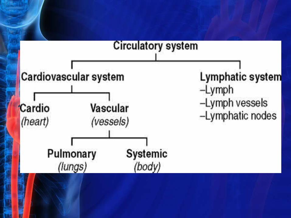
The circulatory system consists of the cardiovascular and lymphatic components. The cardiovascular portion includes the heart, blood, and vessels that transport the blood.
The lymphatic element of the circulatory system is composed of a clear, watery fluid called lymph, along with lymphatic vessels and lymphatic nodes. The cardiovascular and lymphatic components differ in their function and method of transporting their respective fluids within the vessels.
The cardiovascular, or blood circulatory, division may be divided further into the cardio (circulation within the heart) and vascular (blood vessel) components.
The heart is the major organ of the cardiovascular system; it functions as a pump to maintain circulation of blood throughout the body. The vascular component is composed of a network of blood vessels that carry blood from the heart to body tissues and back to the heart again.
Functions of the cardiovascular system include the following:
1.Transportation of oxygen, nutrients, hormones, and chemicals necessary for normal body activity
2.Removal of waste products through the kidneys and lungs
3.Maintenance of body temperature and water and electrolyte balance. These functions are performed by the following blood components: red blood cells, white blood cells, and platelets suspended in plasma.
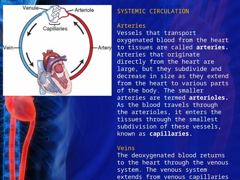
SYSTEMIC CIRCULATION
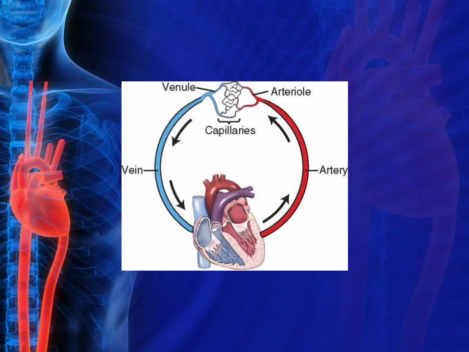
ArteriesVessels that transport oxygenated blood from the heart to tissues are called arteries. Arteries that originate directly from the heart are large, but they subdivide and decrease in size as they extend from the heart to various parts of the body. The smaller arteries are termed arterioles. As the blood travels through the arterioles, it enters the tissues through the smallest subdivision of these vessels, known as capillaries.
VeinsThe deoxygenated blood returns to the heart through the venous system. The venous system extends from venous capillaries to venules to veins, increasing in size as it nears the heart.
General Systemic Circulation
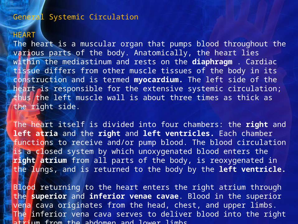
HEARTThe heart is a muscular organ that pumps blood throughout the various parts of the body. Anatomically, the heart lies within the mediastinum and rests on the diaphragm . Cardiac tissue differs from other muscle tissues of the body in its construction and is termed myocardium. The left side of the heart is responsible for the extensive systemic circulation; thus the left muscle wall is about three times as thick as the right side.
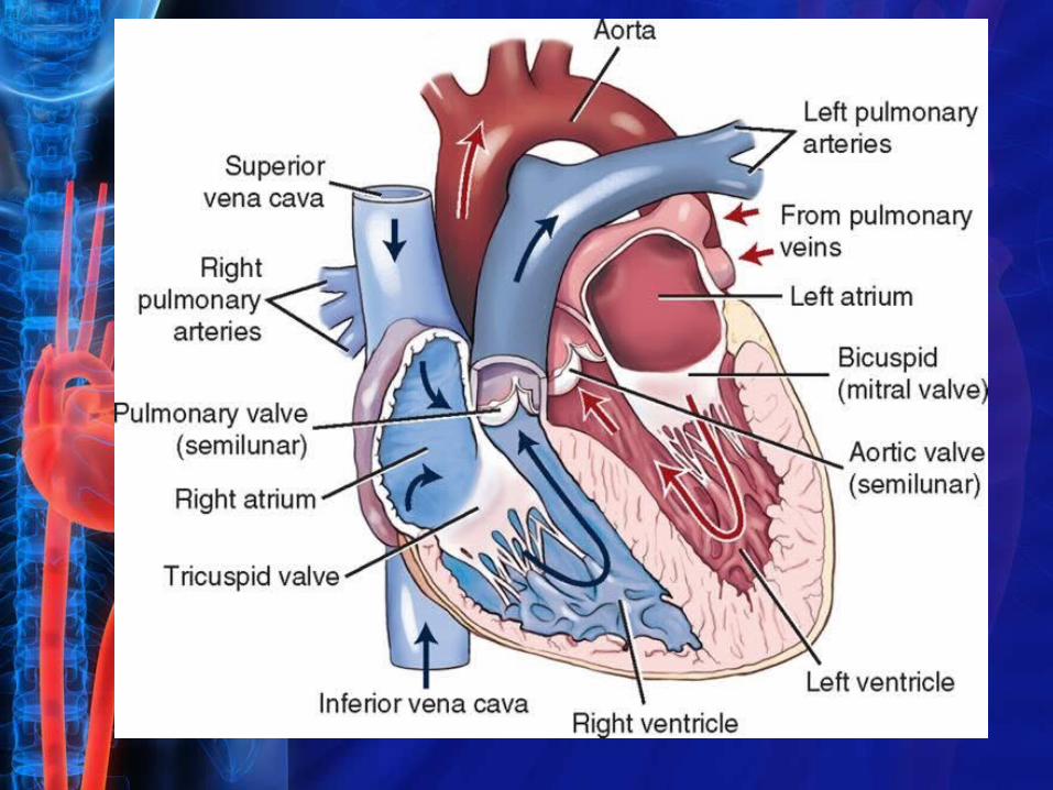
The heart itself is divided into four chambers: the right and left atria and the right and left ventricles. Each chamber functions to receive and/or pump blood. The blood circulation is a closed system by which unoxygenated blood enters the right atrium from all parts of the body, is reoxygenated in the lungs, and is returned to the body by the left ventricle.
Blood returning to the heart enters the right atrium through the superior and inferior venae cavae. Blood in the superior vena cava originates from the head, chest, and upper limbs. The inferior vena cava serves to deliver blood into the right atrium from the abdomen and lower limbs.
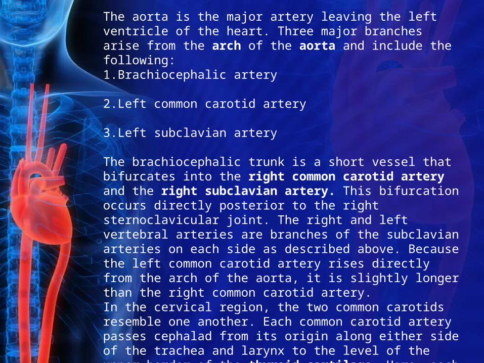
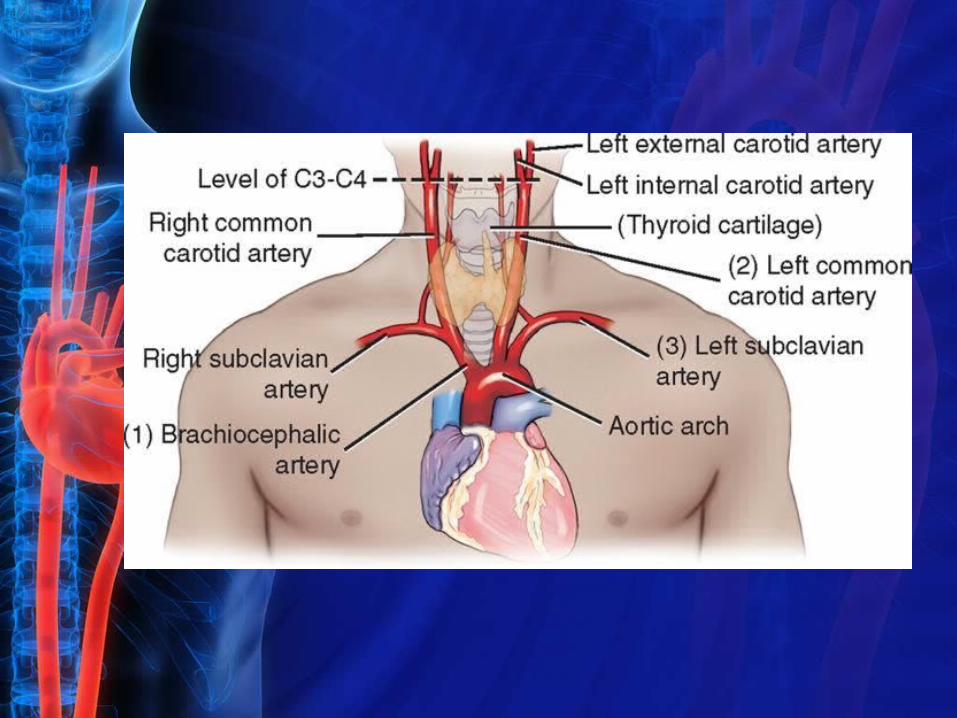
The aorta is the major artery leaving the left ventricle of the heart. Three major branches arise from the arch of the aorta and include the following:1.Brachiocephalic artery
2.Left common carotid artery
3.Left subclavian artery
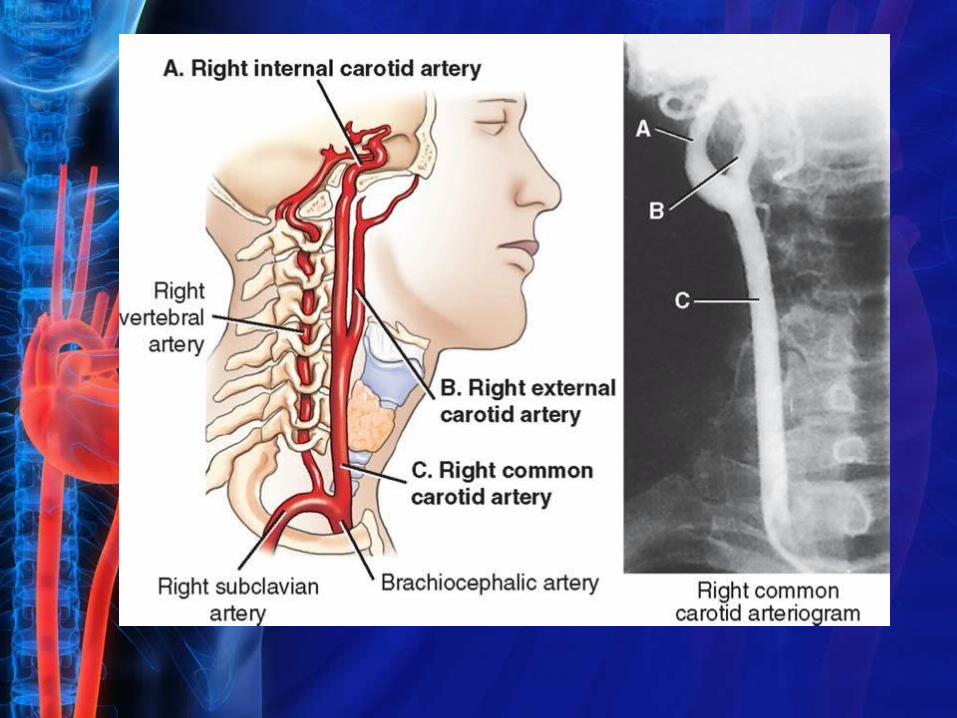
The brachiocephalic trunk is a short vessel that bifurcates into the right common carotid artery and the right subclavian artery. This bifurcation occurs directly posterior to the right sternoclavicular joint. The right and left vertebral arteries are branches of the subclavian arteries on each side as described above. Because the left common carotid artery rises directly from the arch of the aorta, it is slightly longer than the right common carotid artery.In the cervical region, the two common carotids resemble one another. Each common carotid artery passes cephalad from its origin along either side of the trachea and larynx to the level of the upper border of the thyroid cartilage. Here, each common carotid artery divides into external and internal carotid arteries. The site of bifurcation for each common carotid is at the level of the fourth cervical vertebra.
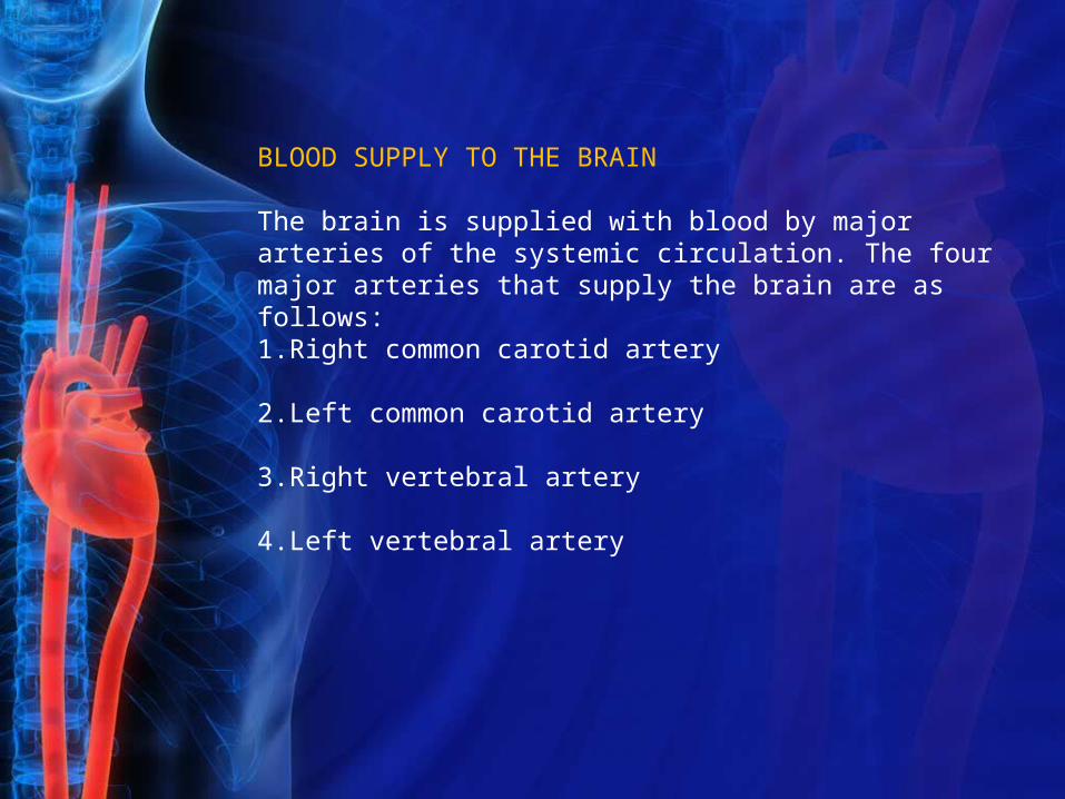
BLOOD SUPPLY TO THE BRAIN
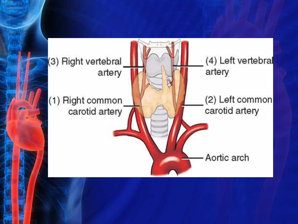
The brain is supplied with blood by major arteries of the systemic circulation. The four major arteries that supply the brain are as follows:1.Right common carotid artery
2.Left common carotid artery
3.Right vertebral artery
4.Left vertebral artery
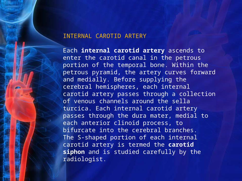
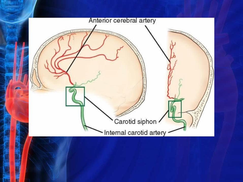
INTERNAL CAROTID ARTERY
Each internal carotid artery ascends to enter the carotid canal in the petrous portion of the temporal bone. Within the petrous pyramid, the artery curves forward and medially. Before supplying the cerebral hemispheres, each internal carotid artery passes through a collection of venous channels around the sella turcica. Each internal carotid artery passes through the dura mater, medial to each anterior clinoid process, to bifurcate into the cerebral branches.The S-shaped portion of each internal carotid artery is termed the carotid siphon and is studied carefully by the radiologist.
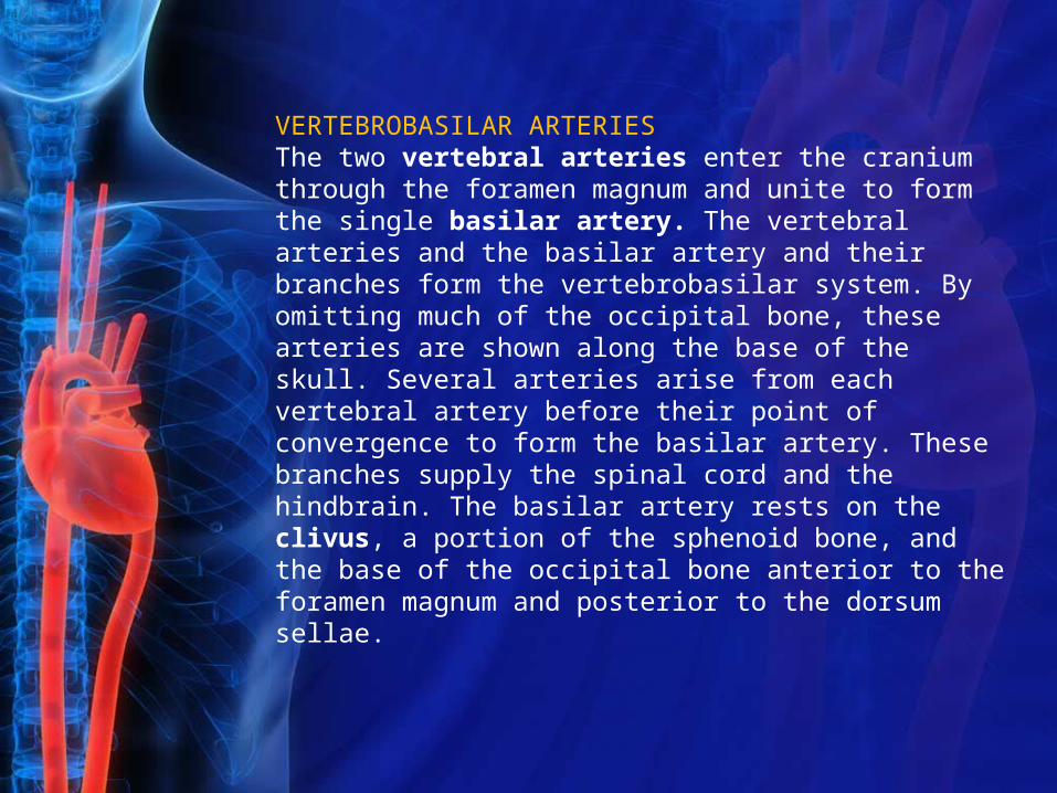
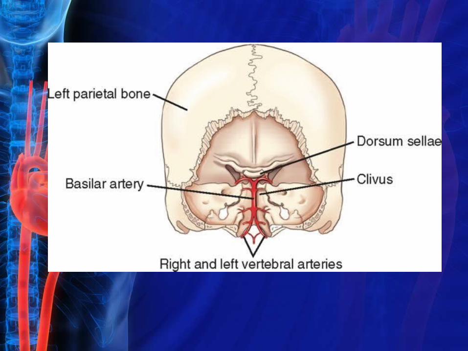
VERTEBROBASILAR ARTERIESThe two vertebral arteries enter the cranium through the foramen magnum and unite to form the single basilar artery. The vertebral arteries and the basilar artery and their branches form the vertebrobasilar system. By omitting much of the occipital bone, these arteries are shown along the base of the skull. Several arteries arise from each vertebral artery before their point of convergence to form the basilar artery. These branches supply the spinal cord and the hindbrain. The basilar artery rests on the clivus, a portion of the sphenoid bone, and the base of the occipital bone anterior to the foramen magnum and posterior to the dorsum sellae.
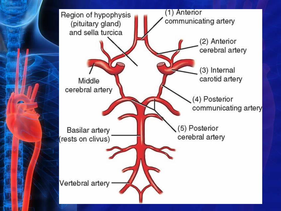
ARTERIAL CIRCLE (CIRCLE OF WILLIS)
The blood to the brain is supplied by the internal carotid and vertebral arteries. The posterior brain circulation communicates with the anterior circulation along the base of the brain in the arterial circle or circle of Willis The five arteries or branches that make up the arterial circle are (1) the anterior communicating artery, (2) the anterior cerebral arteries, (3) branches of the internal carotid arteries, (4) the posterior communicating artery, and (5) the posterior cerebral arteries.
Cerebral VeinsGREAT VEINS OF THE NECK
The three pairs of major veins that drain the head, face, and neck region include the following:
1.Right and left internal jugular veins
2.Right and left external jugular veins
3.Right and left vertebral veins
Each internal jugular vein drains the meninges and brain. In addition, many smaller veins join each internal jugular vein as it passes caudad to eventually become the brachiocephalic vein on each side. The right and left brachiocephalic veins join to form the superior vena cava, which returns blood to the right atrium of the heart.
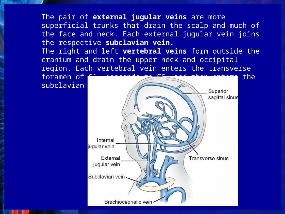
The pair of external jugular veins are more superficial trunks that drain the scalp and much of the face and neck. Each external jugular vein joins the respective subclavian vein.The right and left vertebral veins form outside the cranium and drain the upper neck and occipital region. Each vertebral vein enters the transverse foramen of C1, descends to C6, and then enters the subclavian vein.
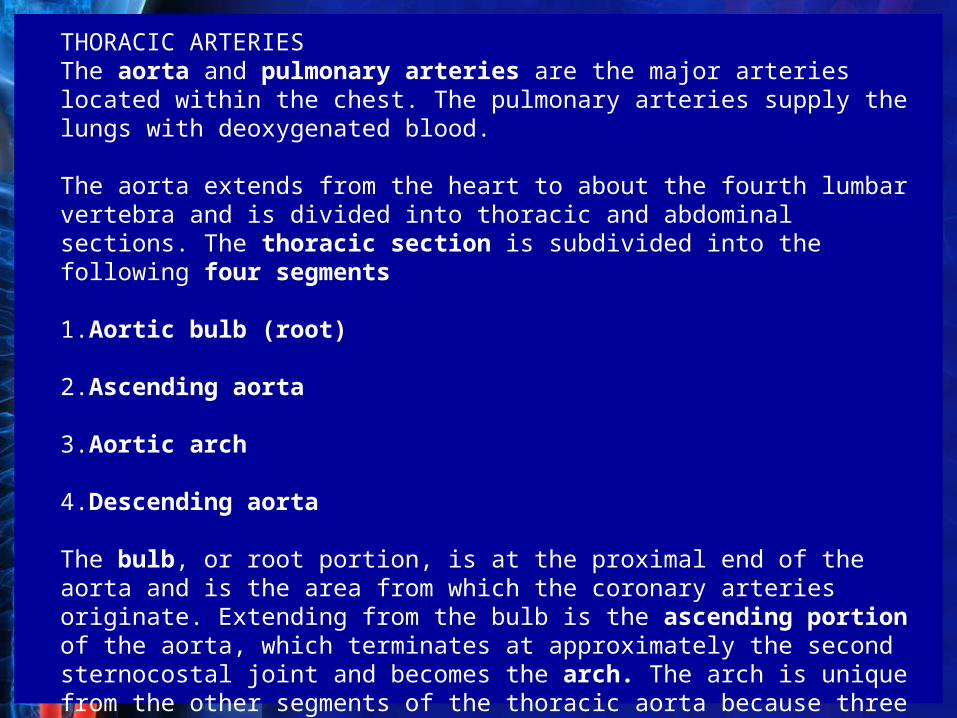
THORACIC ARTERIESThe aorta and pulmonary arteries are the major arteries located within the chest. The pulmonary arteries supply the lungs with deoxygenated blood.
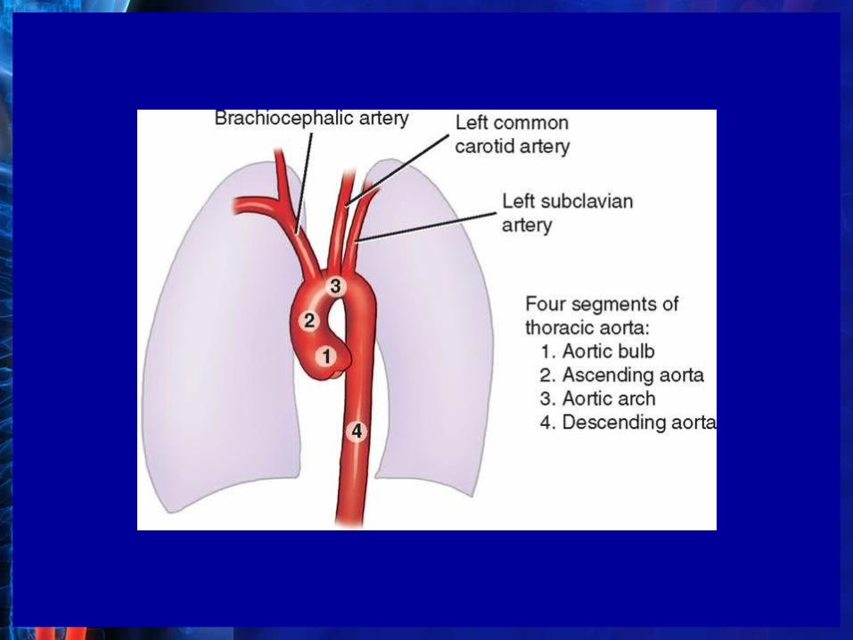
The aorta extends from the heart to about the fourth lumbar vertebra and is divided into thoracic and abdominal sections. The thoracic section is subdivided into the following four segments
1.Aortic bulb (root)
2.Ascending aorta
3.Aortic arch
4.Descending aorta
The bulb, or root portion, is at the proximal end of the aorta and is the area from which the coronary arteries originate. Extending from the bulb is the ascending portion of the aorta, which terminates at approximately the second sternocostal joint and becomes the arch. The arch is unique from the other segments of the thoracic aorta because three arterial branches arise from it: the brachiocephalic artery, the left common carotid artery, and the left subclavian artery.
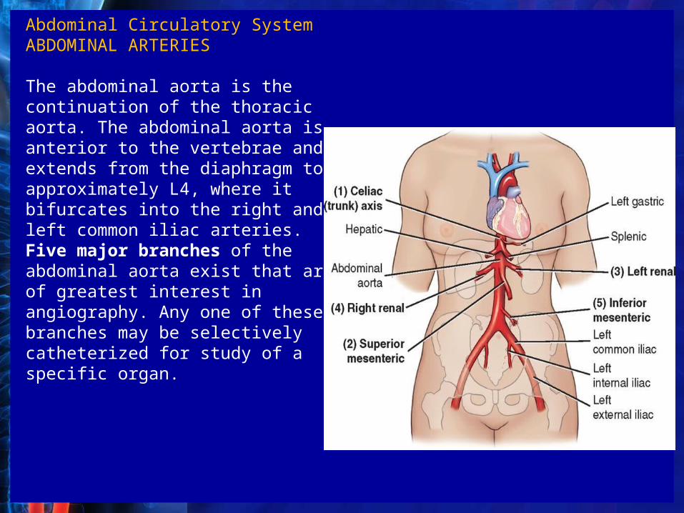
Abdominal Circulatory SystemABDOMINAL ARTERIES
The abdominal aorta is the continuation of the thoracic aorta. The abdominal aorta is anterior to the vertebrae and extends from the diaphragm to approximately L4, where it bifurcates into the right and left common iliac arteries. Five major branches of the abdominal aorta exist that are of greatest interest in angiography. Any one of these branches may be selectively catheterized for study of a specific organ.
The trunk of the celiac artery arises from the anterior aspect of the aorta just below the diaphragm and about 1.5 cm above the origin of the superior mesenteric artery. Organs supplied with blood by the three large branches of the celiac trunk are the liver, spleen, and stomach.
The superior mesenteric artery supplies blood to the pancreas, most of the small intestine, and portions of the right side of the large intestine (cecum, ascending, and about one half of the transverse colon). It originates from the anterior surface of the aorta at the level of the first lumbar vertebra about 1.5 centimeters below the celiac artery.The inferior mesenteric artery originates from the aorta at about the third lumbar vertebra (3 or 4 cm above the level of the bifurcation of the common iliac arteries). Blood is supplied to portions of the large intestine (left half of transverse colon, descending colon, sigmoid colon, and most of the rectum) by the inferior mesenteric artery.The right and left renal arteries supplying blood to the kidneys originate on each side of the aorta just below the superior mesenteric artery at the level of the disk between the first and second lumbar vertebrae
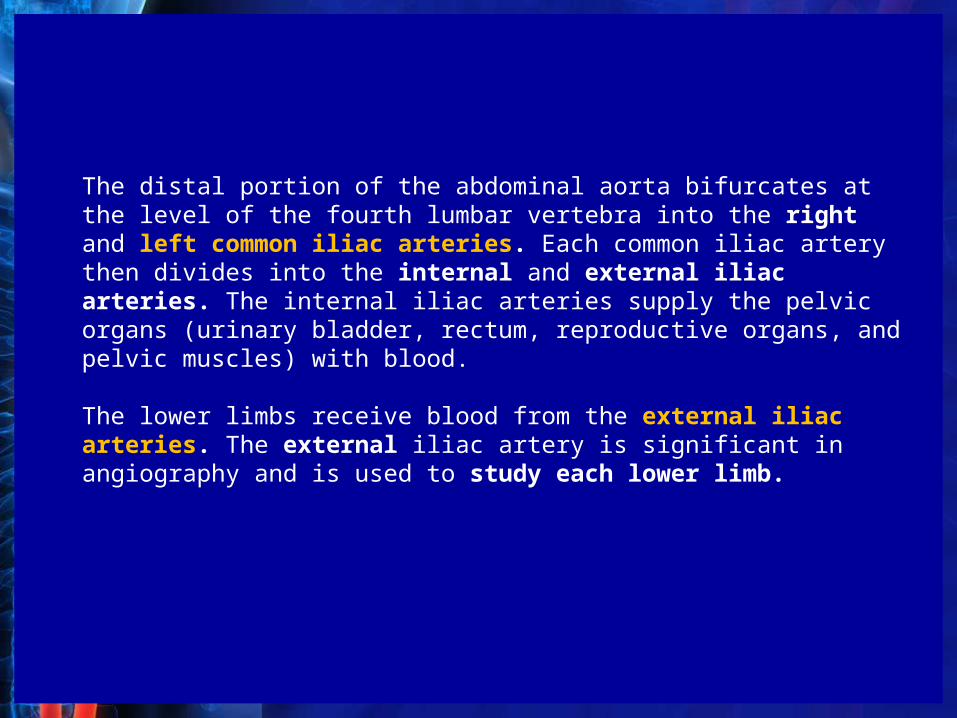
The distal portion of the abdominal aorta bifurcates at the level of the fourth lumbar vertebra into the right and left common iliac arteries. Each common iliac artery then divides into the internal and external iliac arteries. The internal iliac arteries supply the pelvic organs (urinary bladder, rectum, reproductive organs, and pelvic muscles) with blood.
The lower limbs receive blood from the external iliac arteries. The external iliac artery is significant in angiography and is used to study each lower limb.
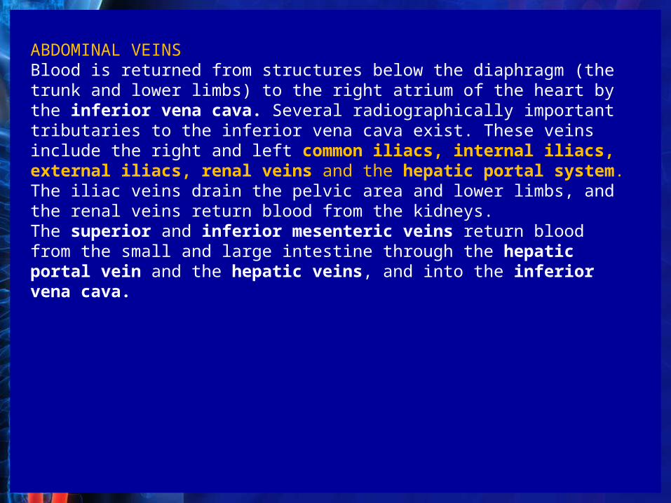
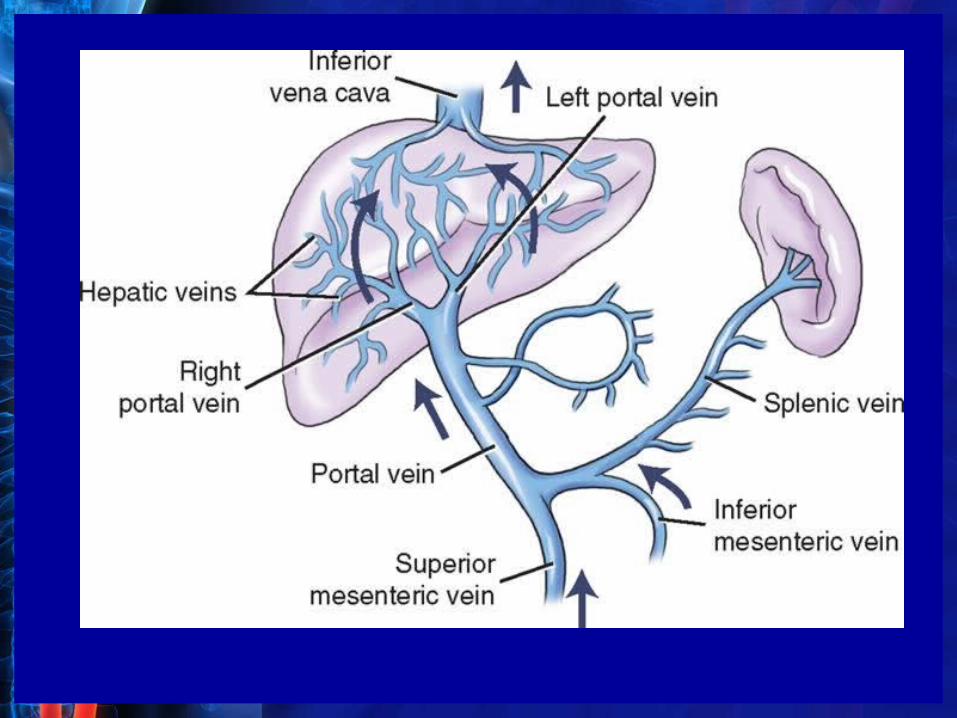
ABDOMINAL VEINSBlood is returned from structures below the diaphragm (the trunk and lower limbs) to the right atrium of the heart by the inferior vena cava. Several radiographically important tributaries to the inferior vena cava exist. These veins include the right and left common iliacs, internal iliacs, external iliacs, renal veins and the hepatic portal system. The iliac veins drain the pelvic area and lower limbs, and the renal veins return blood from the kidneys.The superior and inferior mesenteric veins return blood from the small and large intestine through the hepatic portal vein and the hepatic veins, and into the inferior vena cava.
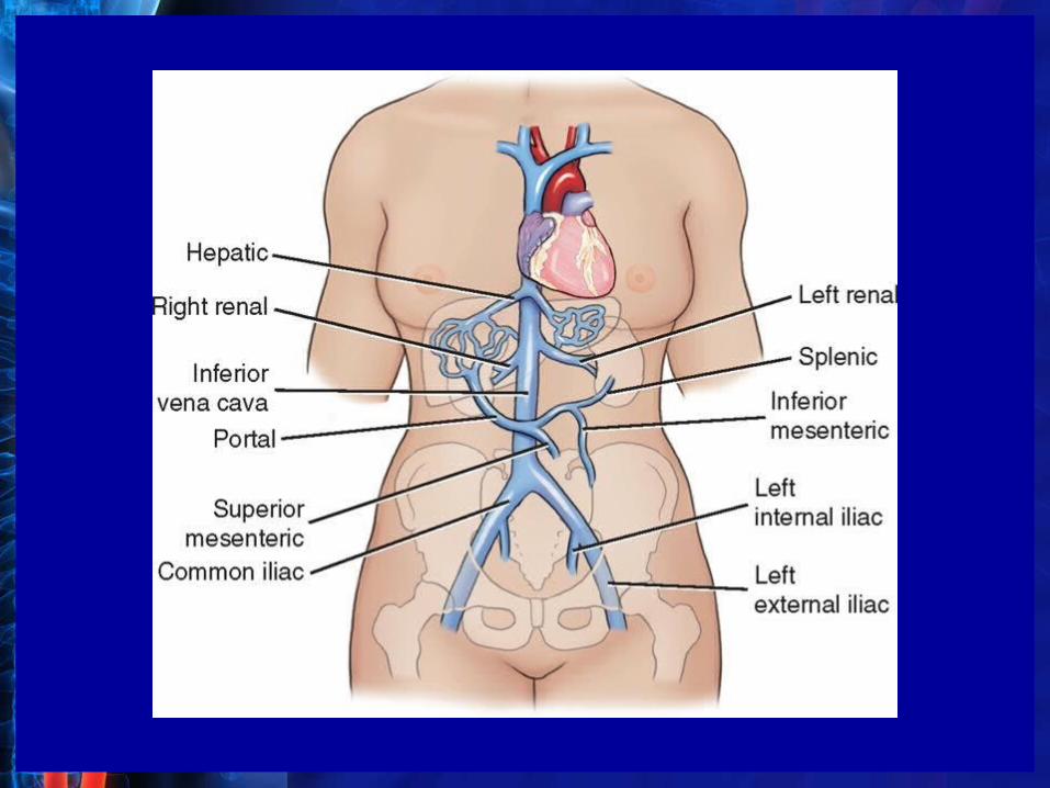
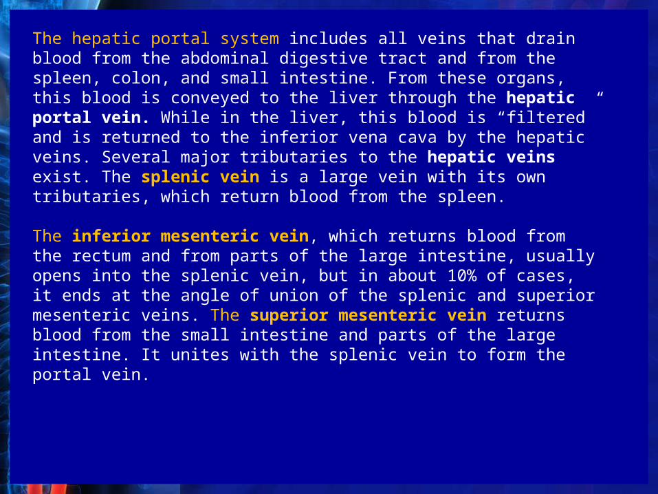
The hepatic portal system includes all veins that drain blood from the abdominal digestive tract and from the spleen, colon, and small intestine. From these organs, this blood is conveyed to the liver through the hepatic portal vein. While in the liver, this blood is “filtered” and is returned to the inferior vena cava by the hepatic veins. Several major tributaries to the hepatic veins exist. The splenic vein is a large vein with its own tributaries, which return blood from the spleen.
The inferior mesenteric vein, which returns blood from the rectum and from parts of the large intestine, usually opens into the splenic vein, but in about 10% of cases, it ends at the angle of union of the splenic and superior mesenteric veins. The superior mesenteric vein returns blood from the small intestine and parts of the large intestine. It unites with the splenic vein to form the portal vein.
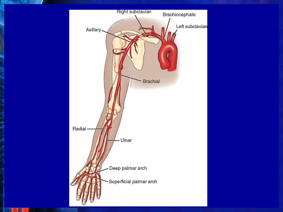
UPPER LIMB ARTERIESThe arterial circulation of the upper limb is generally considered to begin at the subclavian artery. The origin of the subclavian artery differs from the right side to the left side. On the right side, the subclavian arises from the brachiocephalic artery, whereas the left subclavian originates directly from the aortic arch.The subclavian continues to become the axillary artery, which gives rise to the brachial artery. The brachial artery bifurcates into the ulnar and radial arteries at approximately the level of the neck of the radius. The radial and ulnar arteries continue to branch until they join together to form two palmar arches (deep and superficial). Branches of these arches supply the hand and fingers with blood.
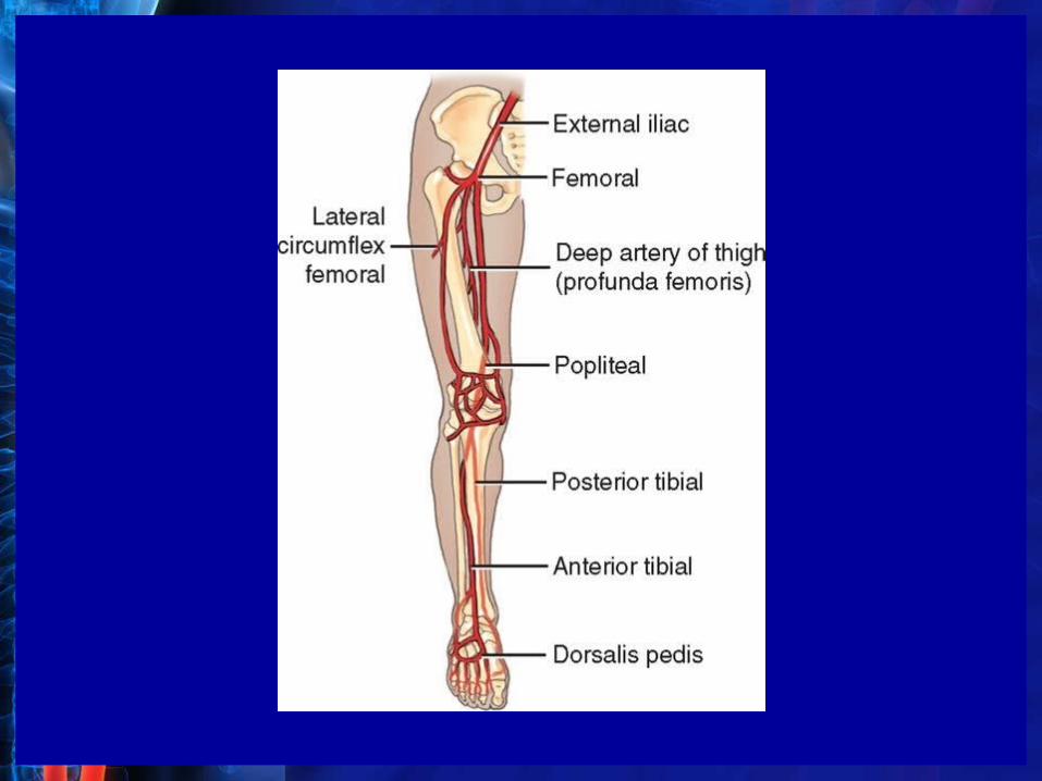
LOWER LIMB ARTERIESThe arterial circulation of the lower limb begins at the external iliac artery and ends at the veins of the foot . The first artery to enter the lower limb is the common femoral artery. The common femoral artery divides into the femoral and deep femoral arteries. The femoral artery extends down the leg and becomes the popliteal artery at the level of the knee. Major branches of the popliteal are the anterior tibial and posterior tibial arteries.The anterior tibial artery continues as the dorsalis pedis artery, with branches to the ankle and foot. The posterior tibial artery supplies the calf and plantar surface of the foot.
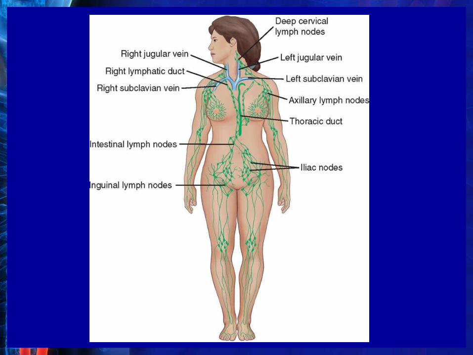
Lymphatic SystemLYMPH DRAINAGE
The lymphatic system serves to drain interstitial fluid (fluid in the spaces between the cells) and return it to the venous system. The fluid from the left side of the body, the lower limbs, pelvis, and abdomen enters the venous system by the thoracic duct (largest lymph vessel in the body), which drains into the left subclavian vein near its junction with the left jugular vein.The upper right side of the body, upper limb, and head and neck region drain lymph fluid into the venous system at the junction of the right jugular and right subclavian veins by the right lymph duct.
FUNCTIONSFunctions of the lymphatic portion of the circulatory system are as follows:1.Fights disease by producing lymphocytes and macrophages2.Returns proteins and other substances to the blood3.Filters the lymph in the lymph nodes4.Transfers fats from the intestine to the thoracic duct and hence to the blood