Embed Size (px)
Citation preview
Discoveries in recent years continue to unveil the role of chlamydia in human
pathology. This genus of intracellular bacteria, long linked to infectious disease
in humans and animals, includes three known pathogens responsible for diverse
disease syndromes: C. pneumoniae*, responsible for respiratory diseases
including bronchitis, pharyngitis, and pneumonia; C. trachomatis, the sexually
transmitted disease commonly associated with chlamydia; and C. psittaci*,
responsible for psittacosis, a zoonotic respiratory disease transmitted from birds
that is rare in humans.
Chlamydia do not grow in routine bacterial cultures for respiratory pathogens.
Micro-immunofluorescence (MIF) testing is an important diagnostic tool to help
identify patients infected with chlamydia.
Focus MIF Advantages
•TestforC. pneumoniae,
C. trachomatis, and
C. psittaci
•Time-saverforhigher
volume testing
•Commonsampledilutions
for IgG and IgA testing for
efficiency in panel testing
•Pre-absorptionstepfor
IgM, which removes
interfering IgG
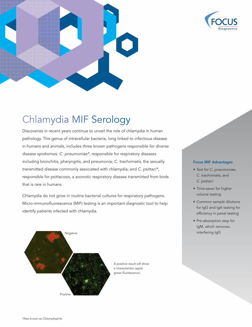
A positive result will show a characteristic apple green fluorescence.
Negative
*Also known as Chlamydophila
Positive
Chlamydia MIF Serology
Chlamydia MIF Serology
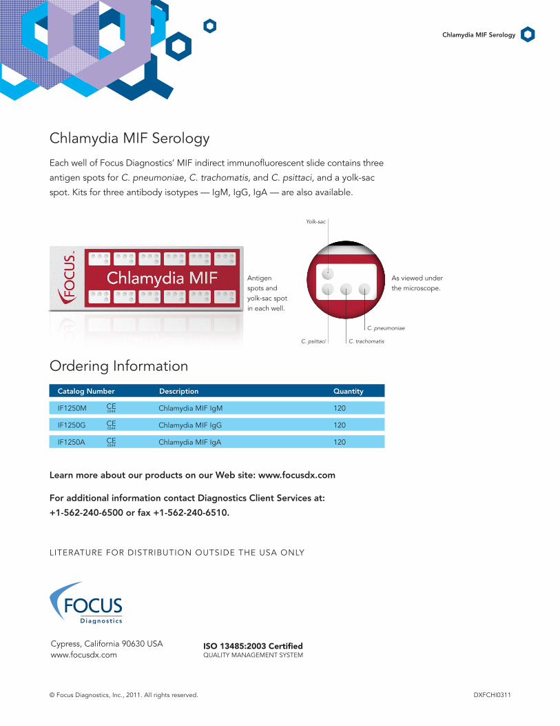
Each well of Focus Diagnostics’ MIF indirect immunofluorescent slide contains three
antigen spots for C. pneumoniae, C. trachomatis, and C. psittaci, and a yolk-sac
spot. Kits for three antibody isotypes — IgM, IgG, IgA — are also available.
Ordering Information
Catalog Number Description Quantity
IF1250M Chlamydia MIF IgM 120
IF1250G Chlamydia MIF IgG 120
IF1250A Chlamydia MIF IgA 120
Learn more about our products on our Web site: www.focusdx.com
For additional information contact Diagnostics Client Services at:
+1-562-240-6500 or fax +1-562-240-6510.
Chlamydia MIF Serology
Cypress, California 90630 USA www.focusdx.com
© Focus Diagnostics, Inc., 2011. All rights reserved. DXFCHI0311
ISO 13485:2003 Certified QUALITY MANAGEMENT SYSTEM
LITERATURE FOR DISTRIBUTION OUTSIDE THE USA ONLY
Antigen spots and yolk-sac spot in each well.
Yolk-sac
C. psittaci C. trachomatis
C. pneumoniae
As viewed under the microscope.
CE 0344
CE 0344
CE 0344