Embed Size (px)
Citation preview
Arch. Dis. Childh., 1967, 42, 62.
Chemical Studies in Gargoylism*J0RGEN CLAUSEN, HOLGER V. DYGGVE, JOHANNES C. MELCHIOR,
and HANS OLAV CHRISTENSEN LOUFrom the Neurochemical Institute, Danish Multiple Sclerosis Society, the University Clinic of Paediatrics, Rigshospitalet,
Copenhagen, and the State Institution for Mentally Retarded. 'Andersvaenge', Slagelse, Denmark
Recent studies of the chemical structure ofglycolipids (Klenk and Gielen, 1961, 1963; Sven-nerholm and Raal, 1961; Kuhn, 1961; Wolfe andLowden, 1964) have contributed to our knowledgeof the metabolism of different glycolipids. Thus, alow concentration of 3-glucosidase in human brainand other organs will give rise to a blocked degrada-tion of gangliosides, with accumulation of gluco-cerebrosides, the disease associated with thisenzymatic defect having previously been namedGaucher's disease (Gaucher, 1882; Philippart andMenkes, 1964; Brady, Kanfer, and Shapiro, 1965a, b;Svennerholm, 1966).
Again, the gluco-cerebrosides in brain may betransformed by means of specific enzymes togalacto-cerebrosides. This ceramide, as well asgalacto-gluco-ceramide, may be sulphated by meansof active sulphate (Svennerholm, 1966), to give riseto sulphatides which in their turn are degraded bythe action of sulphatases (Mehl and Jatzkewitz,1963, 1964), so that absence of the enzyme sulpha-tase, as occurs in metachromatic leucodystrophy,results in accumulation of the sulphatides (Jatzke-witz, 1960; Sourander and Svennerholm, 1962;Hagberg, Souranderand Svennerholm, 1962; Austin,McAfee, Armstrong, O'Rourke, Shearer, andBachhawat, 1964).
Finally, the initial splitting of N-acetyl-neuramin-ic acid (NANA) may be impaired, as is the case inTay-Sachs' disease (Klenk, Liedtke, and Gielen,1963), when mainly mono-sialo-gangliosides accum-ulate.Accumulation of carbohydrate derivatives occurs
not only in the lipidoses but also in the mucopoly-saccharidoses (Melchior, Clausen, and Dyggve,1965). Thus, in Hurler's syndrome (gargoylism)there are accumulations of chondroitin sulphate Band heparitin sulphate (Brante, 1952; Bishton,
Received April 7, 1966.* These investigations have been supported by Grant No. NB
05216-02 Public Service Grant, National Institutes of Health,Education and Welfare, Bethesda, U.S.A., and in part from theResearch Funds for Mental Retardation, Copenhagen, Denmark.
Norman, and Tingey, 1956), and in Morquio-Ullrich's disease chondroitin sulphate B andkeratosulphate and/or hyaluronic acid-like substan-ces are stored and excreted in increased amounts(Zellweger, Ponseti, Pedrini, Stamler, and vonNoorden, 1961; Clausen, Dyggve, and Melchior,1963).Because of the similarity between acid mucopoly-
saccharides (AMP) and polysaccharide moieties ofglycolipids, pathological changes in the synthesis orbreakdown of one or more of the carbohydrateconstituents may cause changes in the other.Following on our previous studies (Dyggve,Melchior, and Clausen, 1962; Clausen et al., 1963),this communication gives some data concerningthese two groups of carbohydrate-rich substances intwo cases of Hurler's syndrome. These datasuggest a metabolic defect common to the katabolismof both AMP and glycolipids in this disease.
Case ReportsCase 1. A boy, born September 1949, died aged 141
years. His case history has been published in detail byClausen et al. (1963). There are no known similardiseases in the family. Following a normal infancy hedeteriorated gradually into a typical case of gargoylism.He died from pneumonia and heart disease. Repeatedslit-lamp examinations revealed no corneal opacities.No abnormal inclusions were found in his lymphocytes.EEGs on several occasions were moderately abnormal,with an increased amount of slow activity. Pneumo-encephalography at the age of 5 years showed a sym-metrical dilatation of the lateral ventricles, with an Evansratio of 0 * 35. Radiological examination showedthickening of the skull bones and typical changes of thespine and the hands. The spinal fluid protein was44 mg./100 ml., with no cells.
Necropsy (Dr. P. Christoffersen). The boy's appear-ance was typical of gargoylism with coarse facial featuresand extremities and a short neck. The cranium wasenlarged with thickened bones measuring maximally 15mm.The heart was diffusely enlarged, weighing 240 g.
62
copyright. on 4 June 2018 by guest. P
rotected byhttp://adc.bm
j.com/
Arch D
is Child: first published as 10.1136/adc.42.221.62 on 1 F
ebruary 1967. Dow
nloaded from

Chemical Studies in GargoylismThe myocardium was hypertrophic, the thickness of theleft ventricle being 14 mm. The aortic and mitral valveswere thickened but without vegetations. The coronaryvessels were normal. Distally, in the aorta small yellowsub-intimal plaques were found. The liver (1540 g.)was enlarged and measured 17 x 17 x 7 cm.: the surfaceand the cut sections were yellow. The spleen (290 g.)was also enlarged measuring 14 x 8 x 6 cm. The lungsshowed bronchopneumonia.
Histologically, there was ballooning of the liver cells,with eccentrically placed nuclei. There was a strongpositive reaction to Sudan-staining, but only a slightreaction to PAS. In the myocardium there wereinfiltrations of lymphocytes, with oedema, and in theaorta there was also some lymphocyte infiltration. Theskin showed oedema of the corium where some PAS-positive substance was seen. A lymph node revealedballooned cells with PAS-positive substance.The brain (Dr. Erna Christensen) showed some
atrophy, with an increased amount of spinal fluid; theweight after removal of the right occipital lobe and afterfixation was 920 g. The gyri were small and the sulcienlarged. The basal arteries were normal. On frontalsection the ventricular system, including the 3rd ventricle,was seen to be much dilated. The white matter appear-ed underdeveloped and showed a great number oflacunae of up to 3 mm. Histologically, there weresevere changes throughout the whole central nervoussystem. The layers of the cortex were poorly markedand the ganglion cells were ballooned, with a strongPAS-positive reaction and deposits of sudanophilicsubstance intracellularly. The lacunae contained co-agulated fluid and were surrounded by perivascularproliferation of connective tissue, and macrophages.The myelination of the white matter was normal. Theependyma was lacking in several areas and the remainingshowed some granular ependymitis. Throughout thebrain the Purkinje cells were swollen and containedsudanophilic and PAS-positive material; these changeswere less pronounced in the distal parts of the medullaand in the spinal cord.No metachromasia was found by staining with
toluidine blue and cresyl violet. The Bielschowskystaining for axis cylinders showed swollen neurites. Theleptomeninges were thickened over large parts of thebrain, with oedema and moderate amounts of lympho-cytes, and macrophages containing PAS-positive andsudanophilic substances. Histological diagnosis wasgargoylism.
Case 2. A boy, born December 1958, birthweight3900 g. (for further details see Case 3 of Steiness, 1961),died when 5* years old. Two older sibs are healthy.Pregnancy and delivery were normal. At the age of6 months the possibility of gargoylism was raised and thediagnosis was established at the age of 9 months, atwhich time an abnormal excretion of acid mucopoly-saccharides was found. His mental and physicaldevelopment was increasingly retarded, requiring admis-sion to an institution for the mentally retarded where hedied.
Necropsy (Dr. H. Wolthers). The facial appearancewas typical of gargoylism with thick lips, a square cranialappearance, and a small body. The lungs showed anincreased amount of mucopurulent secretion in thebronchi. The heart measured 7 x 7 cm., and thethickness of both ventricles was increased. The walls ofthe aorta were thickened and somewhat fibrotic, resemb-ling the arteriosclerotic changes in older people. Theliver measured 25 x 15 x 5 cm., the spleen 9 x 6 x 3cm.The cranium showed thin bones. The subarachnoid
fluid was increased. The vessels on the lower surface ofthe brain were normal. On section a fairly pronounceddilatation of the ventricular system was found, especiallyinvolving the lateral ventricles, and there was someswelling of the brain tissue. No histological examina-tion was performed.The diagnosis from the necropsy was gargoylism.During the illness urinary specimens drawn from
24-hour collections were used for qualitative andquantitative estimations of AMP. Necropsy specimenswere taken 12 and 16 hours, respectively, after death,from the following organs: brain, liver, spleen, andkidney. These specimens were analysed for theircontent of polar lipids and AMP.
MethodsThe AMPs present in urine were screened by paper
electrophoresis of urine concentrated 100 times (Clausenand Rosenkast, 1962; Asboe-Hansen and Clausen, 1964;Dyggve et al., 1962; Clausen et al., 1963). Theelectrophoresis was performed in 0-2 M LiCl (pH 2-8)and the pattern was stained for AMPs with Alcian blue(Foster and Pearce, 1961). The total content ofAMP inurine was estimated by content of hexosamine and uronicacid bound to AMP. AMP and some uromucoid wereprecipitated with cetyl-pyridium bromide (CPB),followed by an estimation of hexosamine, uronic acid,and fucose in the precipitate. Because of the knownfucose/hexosamine ratio ofuromucoid, the hexosamine ofAMP can be estimated (Clausen and Asboe-Hansen,1966). The content ofAMP was thus expressed as mg.hexosamine and uronic acid excreted per 24 hours.The AMP in the necropsy specimens was similarly
determined by the total amount of hexosamine anduronic acid present per mg. extractable protein. How-ever, the hexosamine value thus expressed also covers theCPB-precipitable gangliosides and glycoproteins ofvarying composition. The fucose/hexosamine ratio wastherefore unknown, and the fucose content of the CPBprecipitate (mg./mg. protein) was only used as anindicator for the total CPB-precipitable glycoprotein.The polar lipids ofthe necropsy specimens were extractedwith chloroform : methanol (2 : 1) and separated intoindividual fractions by means of thin-layer chromato-graphy (Clausen, Christensen Lou, and Andersen, 1965).The distribution of the sialic acid-free polar lipids wasevaluated by photometric scanning of the chromato-plates. The patterns of polar lipids were developedquantitatively by spraying with ammonium-molybdateperchloric acid (Christensen Lou, Clausen, and Bierring,
63copyright.
on 4 June 2018 by guest. Protected by
http://adc.bmj.com
/A
rch Dis C
hild: first published as 10.1136/adc.42.221.62 on 1 February 1967. D
ownloaded from

Clausen, Dyggve, Melchior, and Christensen Lou1965; Clausen, 1966). The values obtained were
correlated with those of normal necropsy specimens fromadults.The identification of the glycolipids was performed by
infrared analysis of the fractions isolated from quantita-tive thin-layer chromatography (Clausen, 1966). Theglycolipids were further characterized by their resistanceto a 24-hour treatment (370 C.) with 0 5 N KOH. Theabnormal glycolipids visualized on thin-layer chroma-tography (see below) may be isolated by preparativechromatography (Christensen Lou et al., 1965). How-ever, for analytical purposes, the abnormal glycolipidfraction found in liver, spleen, and kidney was isolated bycolumn chromatography on silicic acid (O'Brien andSampson, 1965). This revealed the abnormal glycolipidfraction to be eluted in pure state in Case 1 by chloroform+ methanol (19 + 1) and in Case 2 by chloroform +methanol + H2O (4 + 1 + 0 5% H.O). The glyco-lipid fraction was hydrolysed with 4N HCI. Theliberated carbohydrates were identified qualitatively bypaper-chromatography, using 5% aqueous solutions ofglucose galactose, glucosamine, galactosamine, lactose,and maltose as standard markers (Schultze, Schmidt-berger, and Haupt, 1958). After 16 hours of developing,the papers were stained by spraying with aniliniumphthalate (Schultze et al., 1958). Quantitatively thehexose/lhexosamine ratio was determined by quantita-tive determination of hexose by the orcinol test andhexosamines by the Ehrlich's reaction (Clausen andAsboe-Hansen, 1966).The fatty acid composition of the isolated glycolipid
fraction (10 mg.) was evaluated by gas-chromatographyof the methyl esters. The fatty acids of the glycolipidwere saponified with 2 ml. 1 N KOH in 96% ethanol for30 minutes as described by Rathbone (1965). The fattyacids were liberated by addition of 1 - 5 ml. 2 N HCI +5 ml. H2O. In a separating funnel the fatty acids weretransferred to 10 ml. ethyl ether and washed three timeswith 10 ml. 0- 1 N HCI. The ether phase was driedabove CaCl2 for 16 hours and concentrated to drynessbelow N2. The fatty acids were afterwards methylatedwith 6 ml. super-dry methanol saturated with dry HCIgas. 250 [LI. dry benzene was added as catalyst.Methylation was performed for 30 minutes at 800 C. insealed bottles. The methyl esters were extracted andwashed as described above. Finally the esters wereconcentrated by evaporation of the ether below a streamof N2. Gas-chromatography was performed on 0 2 V.I.methyl esters in a Perkin-Elmer flame-ionization gas-chromatograph F-i 1-(Clausen, 1966, in preparation)(column dimensions 2 m. x 1 mm., stationary phase 8%1-4 butanediol succinate on Chromosorb W (mesh80-100)).Chromatograms were analysed by triangulation and the
proportions of individual fatty acids were expressed as a
percentage of the total. The fatty acids were identifiedby log plotting and by means of standard markers(Hormel Institute, Minnesota, U.S.A.). The sialic-richglycolipids (gangliosides) were separated by means ofthin-layer chromatography, using chloroform: methanol+ aqueous NH, (25%) (70 + 30 + 5) as an ascending
system. The pattern was developed with resorcinol-HCl-reagent for glycolipids as described by Svenner-holm (1963). The gangliosides were developed with aviolet colour, other glycolipids with a brownish colour.
ResultsInvestigations of urine specimens. In Case 2
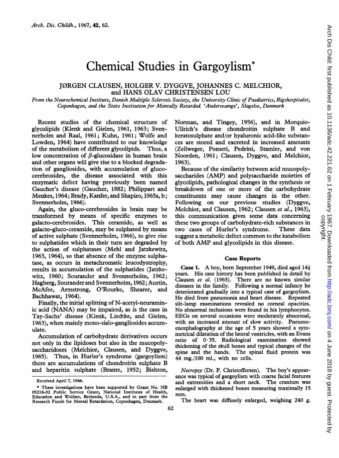
paper-electrophoresis of urine concentrated 100times revealed one fraction of AMP with themobility of chondroitin sulphate (Fig. 1). Quanti-tative estimation of AMP in a 24-hour specimen ofurine revealed a 20-fold increase in the amount ofAMP-bound hexosamine and of AMP-bounduronic acid (Table I). In Case 1 also, as previouslyreported by Dyggve et al. (1962) and by Clausenet al. (1963), a fraction with the mobility ofchondroitin sulphate was found, which, however,extended towards the heparin fraction. No quanti-tative estimation was performed in this case.
TABLE IAcid Mucopolysaccharide Excretion in Urine in
Normal Subjects and in Case 2
Acid Mucopoly- Acid Mucopoly-Age (yr.) saccharide saccharide
Hexosamine Glucuronic Acid(mg./24 hr.) (mg./24 hr.)
Normal subjects0-5 1-7( 09) 2-1 (05)5-20 6-3( 1-0) 49(± 05)Over 20 .35(09) 39 (±04)Mean of all groups of age 3*9 ( 05) 3 7 ( 03)
Case 2 .149-0 87 *9
Figures in parentheses are standard deviations of the mean.
Investigations of necropsy samples. InCase 2 electrophoresis gave a fraction with themobility of chondroitin sulphate in the heart, kidney,liver, and brain. Total estimation of fucose,hexosamine, and uronic acid in these organs revealedonly a slight increase in the glycoproteins (fucosecontent), but a 20- to 230-fold increase in hexosa-mine, and an 85- to 455-fold increase in uronic acid(Table II).
TABLE IICase 2: Acid Mucopolysaccharides in Necropsy
SpecimensFucose Hexosamine Uronic Acid(gg./mg. (ttg./mg. (gg./mg.protein) protein) protein)
Heart .. .. 157 25*00 13*20Kidney .. .. 082 19*00 11*90Normal kidney .. 0-14 0-16 0-14Liver .. 3-82 99*50 73*10Normal liver .0. 29 083 038Brain .. 1.117 12*00 6-82Normal brain .. 79 053 015
64copyright.
on 4 June 2018 by guest. Protected by
http://adc.bmj.com
/A
rch Dis C
hild: first published as 10.1136/adc.42.221.62 on 1 February 1967. D
ownloaded from
Chemical Studies in Gargoylism 65
hnonaroltin Case HeparinSulphate
FIG. 1.-Case 2. Paper electrophoresis of urine concentrated 100 times. The electrophoretic patterns are correlated tothe markers of chondroitin sulphate and heparin. A fraction with the mobility of chondroitin sulphate is seen. (For
experimental details, see the text.)
Quantitative thin-layer chromatography revealeda relative predominance of a glycolipid fraction inliver and kidney cortex, but not in the other organs
investigated (Table III).The Rf value, 0 * 55, was found to be slower than
that of normal galacto-cerebrosides (Rf 0 * 77). Theglycolipid fraction was resistant to hydrolysis by0 * 5 N KOH. In the brain necropsy specimen, an
increase in the phosphatidyl-ethanolamine fraction
was found, which may be explained by a partial over-lapping of the abnormal glycolipid fraction and theethanolamine-phosphatides, because this fraction inthe brain chromatogram extended more towards thefront of the developing fluid.
Paper chromatography of the abnormal fractionisolated from liver tissue revealed only glucose andgalactosamine (five experiments), but no galactose.However, traces of a fraction with an Rf value as
TABLE IIICase 2: Distribution of Phosphatides and Sialic Acid-free Glycolipids in Necropsy Specimens
Sf11 + Ps Sfi Le Pe Ce
Kidney cortex from patient 13*4 ± 3*6 23 5 ± 3 *4 10*4 ± 7*3 52*8 ± 9*2*Kidney cortex from normal subject.. .. 193 ± 13 27 2 ± 1*2 16*1 i 2-5 37*2 ± 3-2t
Brain, white matter from patient .. .. 14-5 ± 17 3-6 03 16 9 ± 2 4 49-1 ± 2-1 15-9 ± 3-2Brain, white matter from normal subject .. 125 ± 05 9*9 ± 01 14-7 ± 07 24-9 ± 06 38*6 ± 09
Liver from patient .13*3 ± 71 370± 7*3 18*1 ± 10 6 41*5 + 9*1Liver fromnormal subject 20*2+2*5 33*0 ± 2*5 22*8+3*7 22*3+3*2
* This fraction has a retention factor Rf 0 55. t This fraction has a retention factor Rf 0 77.Sf1l (C-18 sphingomyelins): polar lipid fractions demonstrated by means of infrared analysis to contain sphingomyelin (C-18). However,
this fraction also covers the serine-phosphatide band (Ps).Sfi (C-24 sphingomyelins): a fraction with a retention factor as C-24 sphingomyelins.Le, a fraction with retention factor as lecithin.Pe, a fraction with retention factor as ethanolamine-phosphatides.Ce, a fraction with retention factors as cerebrosides and other glycolipids (traces of aldehyde-derivatives of split products of unsaturated
fatty acids may adere to these fractions (L. Svennerholm 1965, personal communication)).All fractions mentioned above have been isolated by infrared analysis and identified as described by Clausen (1966).The values of the necropsy specimens are given + SD. The normal values are based upon studies of 10 different macroscopically normal
necropsy specimens, and are given + SD of the means.
copyright. on 4 June 2018 by guest. P
rotected byhttp://adc.bm
j.com/
Arch D
is Child: first published as 10.1136/adc.42.221.62 on 1 F
ebruary 1967. Dow
nloaded from

Clausen, Dyggve, Melchior, and Christensen LouTABLE IV
Chemical Data of Glycolipid Fraction (Rf value 0 55)Isolated From Necropsy Specimens in Two Cases of
Hurler's Syndrome
Hexose/ Paper ChromatographyCase HexosamineNo. Ratio Glucose Lactose Galacto-
samine*
1 3*4/1 + Traces +2 3 0/1 + Traces +
* Other carbohydrates tested (galactose, maltose, glucosamine)were absent.
lactose was found. The hexose/hexosamine ratiowas found to 3 0 (W/W) (Table IV).
In Case 1 paper electrophoretic studies of thebrain tissue revealed the presence of the AMPfraction found in urine. Quantitative estimationsshowed only a normal level of glycoproteins (fucosecontent), but a 13-fold increase in the hexosamineand a 33-fold increase in the uronic acid of the wholebrain matter (Table V).
Quantitative thin-layer chromatography of liverextract revealed, as in Case 2, an increase of theglycolipids with Rf value of 0 55, slower than that ofnormal galacto-cerebrosides (Rf 0 *77). In addition,the spleen revealed a high content of this component(Table VI).The fatty acid composition of the abnormal
TABLE VCase 1: Acid Mucopolysaccharides in Necropsy
Specimens
Fucose Hexosamine Uronic Acid(tig./mg. (tg-!mg. (tig-/mg.protein) protein) protein)
0-67 7-11 4-960.79 0.53 0.15
glycolipids is seen in Table VII. Only traces ofpolyenoic acids of 22-C atoms were found and no
C-24 derivatives could be detected. Staining ofsialic components (gangliosides) in thin-layerchromatography revealed an increase in mono- anddi-sialo-gangliosides (fraction GM 1' 2 and 3, GD lasSvennerholm, 1963) in both Cases 1 and 2 (Fig. 2and 3). No changes were found in other fractions,including the sulphatides which were normal (0 23to 0 17 parts of the cerebroside fraction).
DiscussionThe two cases of gargoylism were clinically
typical of this disorder, with thick lips, prominenteyebrows and flat nose, and the large head and smallbody. Case 1 had a hearing loss and Case 2 hadcorneal opacities; both had hepatosplenomegaly.Their development was characteristic, with a fairlynormal infancy and later deterioration, leading to
TABLE VICase 1: Distribution of Phosphatides and Sialic Acid-free Glycolipids in Necropsy Specimens
SfL11 + Ps Sf_L Le Pe Ce
Kidney cortex from patient 14*9 ± 3*6 200± 3*4 11*2 ± 7*3 53 *9 ± 992Kidney cortex from normal subject .. 19*3 ± 1*3 27 *2 + 1*2 16*1 ± 2*5 37*2 ± 3*2Spleen from patient .17*6 ± 73 25 *4 ± 6*8 22 *8 ± 3*1 34*2 ± 10*9Spleen from normal subject 23*6 ± 2*6 31*3 ± 2*4 220 ± 1*1 22*7 ± 3*8
Brain, white matterfrom patient .. 20*8 ± 17 63±03 160 ± 2*4 50*7 ± 2*1 6*3 ± 3*2Brain, white matter from normal subject .. 125 ± 05 9*9 ± 01 14-7 ± 07 24-9 ± 06 38 *6 ± 09
For key see Table III.
TABLE VII
Fatty Acid Composition (%) of Abnormal Glycolipid Isolated from Liver Tissue from Two Cases of Hurler'sSyndrome
* Unidentified.
66copyright.
on 4 June 2018 by guest. Protected by
http://adc.bmj.com
/A
rch Dis C
hild: first published as 10.1136/adc.42.221.62 on 1 February 1967. D
ownloaded from

Chemical Studies in Gargoylism
GM3
GM2
GMi
GDIa
GDlb
GM
GD and GT
FIG. 2.-Case 1. Thin-layer chromatogram ofglycolipids(gangliosides) from cortex of the brain correlated with thatof normal cortex (adult). The polar lipids are developedwith resorcinol (Svennerholm, 1963). The fraction namedGM refers to the abnormal gangliosides of Case 1. (Thechromatograms were developed for 30 minutes withchloroform + methanol + 10% aqueous NH3 (60 + 35 +8).) GM refers to mono-sialo-ganglioside fractions, GD todi-sialo-ganglioside, and GT to tri-sialo-gangliosides (for
technical details, see text).
idiocy. Radiologically there were typical bonechanges, particularly of the spine.Necropsy confirmed the diagnosis. In both cases
there was hepatosplenomegaly, and abnormalities ofthe aorta, and naked-eye appearances of the brainwere typical. In Case 1, where histological exami-nation was performed, the findings were character-istic of a storage disease and the histochemicalstudies supported the presence of gangliosides.The latter finding is in agreement with those ofJervis (1950) and Benda (1952), who were the first tosuggest a metabolic disorder. The deposits were
6
Case L Case IFIG. 3.-Thin-layer chromatograms of gangliosidesextracted from the grey matter of the brain from Cases1 and 2. GD and GM refer to the different gangliosides.(See Svennerholm (1963) and Fig. 2; the chromatograms
were developedfor 11 hours.)Mono- and di-sialo-gangliosides are found. No tri-sialo-
gangliosides can be seen.
proved to be a ganglioside, first by Brante (1952) andlater by others (Norman, Urich, Tingey, andGoodbody, 1959; O'Brien, Stem, Landing, O'Brien,and Donnell, 1965).The chemical studies revealed an accumulation
ofAMP in all organs investigated, while in additionthe urine from Case 2 revealed a large increase inAMP. The paper electrophoretic studies ofconcentrated urine revealed a fraction with themobility of chondroitin sulphate in both cases.Previous infrared studies revealed this fraction to be
67
copyright. on 4 June 2018 by guest. P
rotected byhttp://adc.bm
j.com/
Arch D
is Child: first published as 10.1136/adc.42.221.62 on 1 F
ebruary 1967. Dow
nloaded from
Clausen, Dyggve, Melchior, and Christensen Louidentical with chondroitin sulphate B. Althoughthe presence of a small amount of heparitin sulphatecannot be excluded (Bishton et al., 1956), the amountof this component was too low to allow isolation byalcohol precipitation before the infrared analysis(Dyggve et al., 1962; Clausen et al., 1963). Ourresults thus seem in agreement with those of Brante(1952). The slightly increased fucose value mayindicate a slight increase in the level of glycoproteinsin one of these cases of gargoylism.The quantitative thin-layer chromatography
revealed a predominance of the glycolipid fraction,containing both galactosamine and glucose. Be-cause the glycolipids are split products of thedegradation of gangliosides (see above), and becausemono- and di-sialo-gangliosides were found to beincreased in the brain matter of these two cases ofgargoylism, a common enzymatic defect in thedegradation of carbohydrate moieties of AMP,glycolipids, and glycoproteins may explain thefindings.
Recent studies (Klenk and Gielen, 1961, 1963;Kuhn, 1961; Wolfe and Lowden, 1964; Svenner-holm and Raal, 1961; Svennerholm, 1964, 1966)have shown the gangliosides to be composed oflipamide-glucose-galactose-galactosamine-galactose-(sialic acid)3, with the probable attachment of onesialic acid residue to the glucose molecule.
So, at the moment it cannot be decided whetherthe glycolipid containing only glucose and galacto-samine represents a new type of lipid, presentnormally merely in traces, or whether it representsan abnormal glycolipid only to be found in Hurler'ssyndrome. However, it is of interest that Booth,Goodwin, and Cumings (1966) have recentlydescribed a nearly hexosamine-free sialic- andhexose-containing glycolipid in necropsy specimensfrom a case of Hurler's syndrome. The presentstudies showed accumulations of both chondroitinsulphate and of a glycolipid, both containinggalactosamine, and this may be explained on thebasis of a common metabolic defect in the followingway.
Chondroitin sulphate B (see survey by Gibian,1959) and gangliosides may only be degraded toliberate the galactosamine, when hexosaminidase(,3-galactosaminidase) is present (Walker, 1961).Because the glycoproteins also may contain galacto-samine (Schultze et al., 1958), it seems reasonable toexplain the findings in gargoylism by a lack of aP-galactosaminidase-like enzyme, leading to accum-ulation of glycolipids, chondroitin sulphate, and theirprecursors. This may also explain, on the basis of'stasis' in the pathway, the increased level of mono-and di-sialo-gangliosides in the brain matter. This
explanation also seems in agreement with otherstudies (to be published later), that in Farber'sdisease a storage of both a glycolipid and AMPoccurs. Most degradative enzymes, as for in-stance hexosaminidases, are localized to thelysosomes intracellularly (de Duve, 1963). Becauseboth mucopolysaccharidoses and lipidoses may beexplained by lack of degradative enzymes and,because, as demonstrated in the present communica-tion, these diseases may be interrelated concerningthe chemical abnormalities, their pathogenesis maybe related to abnormalities in the lysosomes.When compared with the fatty acid composition
of normal cerebroside (O'Brien and Rouser, 1964),the present abnormal glycolipid seems to be lackinghigher polyenoic acids (C-24 derivatives) and onlythe glycolipid from Case 1 contained minor amountsof C-22 derivatives.The present pathological findings are in agreement
with the histological studies of Craig, Clarke, andBanker (1959), Norman et al. (1959), and Landing,Silverman, Craig, Jacoby, Lahey, and Chadwick(1964). These authors demonstrated foaming histio-cytosis of viscera as found in Tay-Sachs' disease,while in our two cases the increase in the glycolipidswas found in the spleen and liver, containing reticulo-endothelial cells. The similarity of the findings inTay-Sachs' disease may be explained by the com-promised degradation of the gangliosides. A familywith one child with gargoylism and another withTay-Sachs' disease has been described by Shanklinand Salam (1963), and similarity between ganglio-sides from a gargoyle brain and from a Tay-Sachs'brain has been reported by Taghavy, Salsman, andLedeen (1964).The finding of an increased sulphatase activity in
cases of gargoylism (Austin et al., 1964) may be dueto increased sulphation of galacto-AMP stored inthis disease.
Summary
Two cases of gargoylism were investigatedchemically. Necropsy specimens revealed accumu-lation in liver, kidney, spleen, and brain of acidmucopolysaccharides. In the liver and spleen, andin the kidney cortex, there was an accumulation ofmono-, di-sialo-gangliosides and glycolipids.These findings may be explained by a deficiency
of hexosaminidase (/-galactosaminidase) activity.
Our thanks are due to Dr. Annalise Dupont forpermission to publish one of the cases of gargoylism, andto Drs. H. Wolthers, P. Christoffersen, and ErnaChristensen, for the necropsies.
68
copyright. on 4 June 2018 by guest. P
rotected byhttp://adc.bm
j.com/
Arch D
is Child: first published as 10.1136/adc.42.221.62 on 1 F
ebruary 1967. Dow
nloaded from

Chemical Studies in Garffovlism 69REFERENCES
Asboe-Hansen, G., and Clausen, J. (1964). Mastocytosis (urticariapigmentosa) with urinary excretion of hyaluronic acid andchondroitin sulfuric acid. Changes induced by polymyxin B.Amer. J3. Med., 36, 144.
Austin, J., McAfee, D., Armstrong, D., O'Rourke, M., Shearer, L.,and Bachhawat, B. (1964). Abnormal sulphatase activities in twohuman diseases (metachromatic leucodystrophy and gargoylism).Biochem. J., 93, 15c.
Benda, C. E. (1952). Developmental Disorders of Mentation andCerebral Palsies. Grune & Stratton, New York.
Bishton, R. L., Norman, R. M., and Tingey, A. (1956). Thepathology and chemistry of a case of gargoylism. J3. clin. Path.,9, 305.
Booth, D. A., Goodwin, H., and Cumings, J. N. (1966). Abnormalgangliosides in Tay-Sachs disease, Niemann-Pick's disease andgargoylism. J. Lipid Res., 7, 337.
Brady, R. O., Kanfer, J., and Shapiro, D. (1965a). The metabolismof glucocerebrosides. I. Purification and properties of aglucocerebroside-cleaving enzyme from spleen tissue. J7. biol.Chem., 240, 39.
-, -, and- (1965b). The metabolism of glucocerebrosid-es. II. Evidence of an enzymatic deficiency in Gaucher'sdisease. Biochem. biophys. Res. Commun., 18, 221.
Brante, G. (1952). Gargoylism. A mucopolysaccharidosis.Scand. J. clin. Lab. Invest., 4, 43.
Christensen Lou, H. O., Clausen, J., and Bierring, F. (1965). Phos-pholipids and glycolipids of tumours in the central nervoussystem. J. Neurochem., 12, 619.
Clausen, J. (1966). Polar lipids and structural proteins of humanbrain. Properties and pathological changes. In A NA TOAdvanced Study Institute on the Molecular Basis of some Aspectsof Mental Activity, ed. 0. Walaas. Academic Press, London.In the press.
-, and Asboe-Hansen, G. (1966). Urinary acid mucopolysaccha-ridosis in mastocytosis. A new technique for quantitativeestimation. Clin. chim. Acta, 13, 475.
-, Christensen Lou, H. O., and Andersen, H. (1965). Phospho-lipid and glycolipid patterns of infant and foetal brain. J.Neurochem., 12, 599.
--, Dyggve, H. V., and Melchior, J. C. (1963). Mucopolysac-charidosis. Arch. Dis. Childh., 38, 364.
-, and Rosenkast, P. (1962). Isolation of acid mucopolysaccha-rides of human brain. J. Neurochem., 9, 393.
Craig, J. M., Clarke, J. T., and Banker, B. Q. (1959). A metabolicneurovisceral disorder with the accumulation of an unidentifiedsubstance. A variant of Hurler's syndrome. A.M.A. J. Dis.Child., 98, 577.
de Duve, C. (1963). The lysosome concept. In Ciba FoundationSymposium on Lysosomes, ed. A. V. S. de Reuck and M. P.Cameron, p. 1. Little Brown, Boston, Massachusetts;Churchill, London.
Dyggve, H. V., Melchior, J. C., and Clausen, J. (1962). Morquio-Ullrich's disease. Arch. Dis. Childh., 37, 525.
Foster, T. S., and Pearce, R. H. (1961). Zone electrophoresis ofacid mucopolysaccharides. Canad. J. Biochem., 39, 1771.
Gaucher, P. C. E. (1882). De l epithelioma primitif de la rate:hypertrophie idiopathique de la rate sans leucemie. Thesis,Paris.
Gibian, H. (1959). Mucopolysaccharide und Mucopolysaccharidasen.Franz Deuticke, Vienna.
Hagberg, B., Sourander, P., and Svennerholm, L. (1962). Sulfatidelipidosis in childhood. A.M.A. Jr. Dis. Child., 104, 644.
Jatzkewitz, H. (1960). Cerebron-und Kerasin-schwefelsaureesterals Speichersubstanzen bei der Leukodystrophie Typ Scholz(metachromatische Form der diffusen Sklerose). Hoppe-Seylers Z. physiol. Chem., 320, 134.
Jervis, G. A. (1950). Gargoylism (lipochondrodystrophy). Arch.Neurol. Psychiat. (Chic.), 63, 681.
Klenk, E., and Gielen, W. (1961). Uber die Gehirnganglioside.Hoppe-Seylers Z. physiol. Chem., 323, 126.
-, and - (1963). Uber ein zweites hexosaminhaltigesGangliosid aus Menschengehirn. ibid., 330, 218.
-, Liedtke, U., and Gielen, W. (1963). Das Gangliosid desGehirns bei der infantilen amaurotischen Idiotie vom Typ Tay-Sachs. ibid., 334, 186.
Kuhn, R. (1961). Brain gangliosides. Angew. Chem., 72, 186.Landing, B. H., Silverman, F. N., Craig, J. M., Jacoby, M. D.,
Lahey, M. E., and Chadwick, D. L. (1964). Familial neuro-visceral lipidosis. A.M.A. J. Dis. Child., 108, 503.
Mehl, E., and Jatzkewitz, H. (1963). (Yber ein Cerebrosid-schwefel-saure-ester spaltendes Enzym aus Schweineniere. Hoppe-Seylers Z. physiol. Chem., 331, 292.
-, and- (1964). Eine Cerebrosidsulfatase aus Schweinen-iere. ibid., 339, 260.
Melchior, J. C., Clausen, J., and Dyggve, H. V. (1965). Themucopolysaccharidoses. Clin. Pediat. (Philad.), 4, 468.
Norman, R. M., Urich, H., Tingey, A. H., and Goodbody, R. A.(1959). Tay-Sachs' disease with visceral involvement and itsrelationship to Niemann-Pick's disease. J. Path. Bact., 78, 409.
O'Brien, J. S., and Rouser, G. (1964). The fatty acid compositionof brain sphingolipids: sphingomyelin, ceramide, cerebrosideand cerebroside sulphate. J. Lipid Res., 5, 339.
-, and Sampson, E. L. (1965). Lipid composition of the normalhuman brain: gray matter, white matter and myelin. ibid., 6,537.
-, Stern, M.B., Landing, B. H., O'Brien, J. K., and Donnell, G.N. (1965). Generalized gangliosidosis. Amer. J. Dis. Child.,109, 338.
Philippart, M., and Menkes, J. (1964). Isolation and characteriza-tion of the main splenic glycolipids in Gaucher's disease.Evidence for the site of metabolic block. Biochem. biophys. Res.Commun., 15, 551.
Rathbone, L. (1965). The effect of diet on the fatty acid composi-tions of serum, brain, brain mitochondria and myelin in the rat.Biochem. J., 97, 620.
Schultze, H. E., Schmidtberger, R., and Haupt, H. (1958). Unter-suchungen uber die gebundenen Kohlenhydrate in isoliertenPlasmaproteiden. Biochem. Z., 329, 490.
Shanklin, W. M., and Salam, M. (1963). A comparison of thehistochemistry of the cerebral cortex from siblings withgargoylism and Tay-Sachs' disease. Acta neuroveg. (Wien),25, 297.
Sourander, P., and Svennerholm, L. (1962). Sulphatide lipidosis inthe adult with the clinical picture of progressive organic demen-tia with epileptic seizures. Acta neuropath. (Berl.), 1, 384.
Steiness, I. (1961). Acid mucopolysaccharides in urine in gargoy-lism. Pediatrics, 27, 112.
Svennerholm, L., (1963). Chromatographic separation of humanbrain gangliosides. J. Neurochem., 10, 613.
- (1964). The gangliosides. J. Lipid Res., 5, 145.(1966). Lipid changes in mental and neurological disorders.In A NA TO Advanced Study Institute on The Molecular Basis ofSome Aspects of Mental Activity, ed. 0. Walaas. AcademicPress, London. In the press.
-, and Raal, A. (1961). Composition of brain gangliosides.Biochem. biophys. Acta (Amst.), 53, 422.
Taghavy, A., Salsman, K., and Ledeen, R. (1964). An abnormalganglioside pattern from a gargoyle brain. Fed. Proc., 23, 128.
Walker, P. G. (1961). The enzymatic degradation of mucopoly-saccharides. Biochem. Soc. Sympos., 20, 109.
Wolfe, L. S., and Lowden, J. A. (1964). Studies on brain ganglio-sides. I. The isolation and composition of a trisialoganglio-side. Canad. J. Biochem., 42, 1041.
Zellweger, H., Ponseti, I. V., Pedrini, V., Stamler, F. S., and vonNoorden, G. K. (1961). Morquio-Ullrich's disease. J.Pediat., 59, 549.
copyright. on 4 June 2018 by guest. P
rotected byhttp://adc.bm
j.com/
Arch D
is Child: first published as 10.1136/adc.42.221.62 on 1 F
ebruary 1967. Dow
nloaded from