Embed Size (px)
Citation preview
RESEARCH Open Access
Characterization of viroplasm formation duringthe early stages of rotavirus infectionJosé J Carreño-Torres, Michelle Gutiérrez, Carlos F Arias, Susana López, Pavel Isa*
Abstract
Background: During rotavirus replication cycle, electron-dense cytoplasmic inclusions named viroplasms areformed, and two non-structural proteins, NSP2 and NSP5, have been shown to localize in these membrane-freestructures. In these inclusions, replication of dsRNA and packaging of pre-virion particles occur. Despite theimportance of viroplasms in the replication cycle of rotavirus, the information regarding their formation, and thepossible sites of their nucleation during the early stages of infection is scarce. Here, we analyzed the formation ofviroplasms after infection of MA104 cells with the rotavirus strain RRV, using different multiplicities of infection(MOI), and different times post-infection. The possibility that viroplasms formation is nucleated by the entering viralparticles was investigated using fluorescently labeled purified rotavirus particles.
Results: The immunofluorescent detection of viroplasms, using antibodies specific to NSP2 showed that both thenumber and size of viroplasms increased during infection, and depend on the MOI used. Small-size viroplasmspredominated independently of the MOI or time post-infection, although at MOI’s of 2.5 and 10 the proportion oflarger viroplasms increased. Purified RRV particles were successfully labeled with the Cy5 mono reactive dye,without decrease in virus infectivity, and the labeled viruses were clearly observed by confocal microscope. PAGEgel analysis showed that most viral proteins were labeled; including the intermediate capsid protein VP6. Only 2out of 117 Cy5-labeled virus particles colocalized with newly formed viroplasms at 4 hours post-infection.
Conclusions: The results presented in this work suggest that during rotavirus infection the number and size ofviroplasm increases in an MOI-dependent manner. The Cy5 in vitro labeled virus particles were not found tocolocalize with newly formed viroplasms, suggesting that they are not involved in viroplasm nucleation.
BackgroundRotaviruses are the major cause of severe diarrhea inchildren and young animals worldwide. As a membersof the family Reoviridae, they have a genome of 11 seg-ments of double-stranded RNA (dsRNA) enclosed inthree protein layers, forming infectious triple-layeredparticles (TLP) [1]. During, or just after entering thecell’s cytoplasm, the outer capsid, composed of VP4 andVP7, is released, yielding transcriptionally active double-layered particles (DLP). The produced viral transcriptsdirect the synthesis of viral proteins and serve as tem-plates for the synthesis of negative-RNA strands to formthe genomic dsRNA. During the replication cycle ofrotavirus electron-dense cytoplasmic inclusions, namedviroplasms, are formed [2]. Such cytoplasmic inclusions
are observed during infection with a number of animalviruses [3], including reoviruses, as other members ofthe Reoviridae family [4].In rotaviruses two non-structural proteins, NSP2 and
NSP5, have been shown to be sufficient to form mem-brane-free cytoplasmic inclusions, which are known asviroplasms-like structures [5]. In vivo immunofluores-cence visualization of viroplasms shows they are hetero-geneous in size [6,7]. It is in these structures where thesynthesis of dsRNA and its packaging into pre-virioncore particles take place [8]. Besides NSP2 and NSP5,other viral proteins accumulate in viroplasms - namelyVP1, VP2, VP3, VP6, and NSP6 [7,9-11]. The key roleof NSP2 and NSP5 proteins in the formation of viro-plasms has been demonstrated by knocking-down theirexpression by RNA interference, which results in theinhibition of viroplasm formation, genome replication,virion assembly, and a general decrease of viral protein
* Correspondence: [email protected] de Genética del Desarrollo y Fisiología Molecular, Instituto deBiotecnología, Universidad Nacional Autónoma de México
Carreño-Torres et al. Virology Journal 2010, 7:350http://www.virologyj.com/content/7/1/350
© 2010 Carreño-Torres et al; licensee BioMed Central Ltd. This is an Open Access article distributed under the terms of the CreativeCommons Attribution License (http://creativecommons.org/licenses/by/2.0), which permits unrestricted use, distribution, andreproduction in any medium, provided the original work is properly cited.
synthesis [7,8,12]. Viroplasm formation has been studiedusing electron or fluorescence microscopy [6,13-15],however, despite their importance in the replicationcycle of rotavirus, little is know about their dynamics offormation. The observation that bromouridine-labeledRNA localizes to viroplasms suggested that the viraltranscripts are synthesized within viroplasms, which ledto the hypothesis that the entering viral particles couldserve as points of nucleation for the formation of viro-plasms [8]. In this work, the dynamics of viroplasm for-mation in MA104 cells infected with rotavirus strainRRV was studied as a function of time and multiplicityof infection (MOI). Using fluorescently labeled purifiedrotavirus particles; we showed that the incoming TLPsdo not seem to be involved in the formation ofviroplasms.
Materials and methodsCells, viruses, antibodies, and fluorophoresMA104 cells were cultured in Advanced Dulbecco’sModified Eagle’s Medium (DMEM) supplemented with3% fetal calf serum (FBS). The rhesus rotavirus strainRRV, obtained from H.B. Greenberg (Stanford Univer-sity, Stanford CA), was propagated in MA104 cells.The rabbit polyclonal serum to NSP2 protein has beendescribed previously [16]. Horseradish peroxidase-conjugated goat anti-rabbit polyclonal antibody wasfrom Perkin Elmer Life Sciences (Boston, MA), Alexa488 and 568 -conjugated goat anti-rabbit polyclonalantibodies, FluoSpheres carboxylate-modified micro-spheres, 0.1 μm, yellow-green fluorescent (505/515),were from Molecular Probes (Eugene, OR), and Cy™5Mono-Reactive Dye pack was from Amersham, GEHealthcare, UK.
Identification, quantitation and size analysis of viroplasmsMA104 cells grown in 10 mm coverslips were infectedwith rotavirus strain RRV at different MOI’s for 1 hourat 4°C. After washing unbound virus, the cells wereincubated at 37°C for different times post-infection. Thecells were fixed with 2% paraformaldehyde, and permea-bilized with 0.5% Triton X-100 in PBS containing 1%bovine serum albumin, as described previously [17].Cells were then incubated with rabbit polyclonal sera toNSP2 protein, followed by staining with goat anti-rabbitIgG coupled to Alexa-488 or 568. The images wereacquired using a Zeiss Axioskop 2 Mot Plus microscopeand analyzed by Image Pro Plus 5.0.2.9 and AdobePhotoshop 7.0. All images were acquired with a 60×objective, with a real time CCD Camera in 256 greyscales, and the size of the images was 1392 × 1040 pix-els, with 8 bits. The estimation of viroplasm size wasdone using the Analyze particle function of Image J1.32j program (Wayne Rasband, NIH, USA).
Immunodetection of rotavirus NSP2 proteinMA104 cells grown in 24-well plates were infected withrotavirus strain RRV at different MOI’s for 1 hour at4oC. After washing unbound virus, the cells were incu-bated at 37oC for different times post-infection. At theindicated time points, the cells were washed twice withPBS and lysed with Laemmli sample buffer. Proteinswere separated by 10% SDS-PAGE and transferred tonitrocellulose membranes (Millipore, Bedford, MA).Membranes were blocked with 5% non-fat dried milk inPBS, and incubated at 4oC with primary anti NSP2 poly-clonal antibody in PBS with 0.1% milk, followed byincubation with secondary, horseradish peroxidase-con-jugated antibodies. The peroxidase activity was revealedusing the Western Lightning™ChemiluminiscenceReagent Plus (Pelkin Elmer Life Sciences). The imagesobtained were scanned and the band densities analyzedusing Image pro software.
Conjugation of virus with fluorophore and colocalizationof labeled viruses with viroplasmsTo label virus with fluorophores, RRV virions were puri-fied by cesium chloride gradient centrifugation asdescribed previously [18]. The purified TLP’s of simianstrain RRV were washed twice with 10 mM Hepes pH8.2, 5 mM CaCl2, 140 mM NaCl, and labeled with Cy5mono reactive dye (0.1, 0.5, 1, 2.5, and 5 nmol of fluoro-phore for 1 μg of purified virus) at room temperaturefor 1 hour with gentle agitation. The reaction wasstopped by addition of Tris-HCl pH 8.8 to a final con-centration of 50 mM. Labeled viruses were separatedfrom unbound fluorophore by gel filtration on a G25sepharose column. As control, the purified TLP’s ofstrain RRV were processed in identical way withoutaddition of fluorophore. Viral titres were determined bya standard immunoperoxydase assay as described pre-viously [19]. DLP’s were prepared by EDTA treatmentof labeled TLP’s. To determine which viral proteinswere conjugated with fluorophore, labeled and non-labeled TLP’s and DLP’s were resolved in PAGE gel,analyzed on Typhoon-Trio (Amersham Biosciences) andstained by silver nitrate. Labeled particles were com-pared with FluoSpheres [carboxylate-modified micro-spheres, 0.1 μm, yellow-green fluorescent (505/515)]using confocal microscope LSM-510 Zeiss, mounted oninverted microscope Zeiss Axiovert 200 M, with AIMsoftware, using objective Plan-neofluor 100×/1.30 OilPh3 (Carl Zeiss). To detect green staining, excitationlaser Argon 2 488 nm was used with emission filter BP500-530 nm, and for far red staining laser Helio-Neon633 nm was used with emission filter BP650-670 nm.To colocalize labeled TLP’s with viroplasms, MA104cells grown on coverslips were infected with Cy5-labeled RRV TLP’s (MOI of 2) for 1 hour at 37°C.
Carreño-Torres et al. Virology Journal 2010, 7:350http://www.virologyj.com/content/7/1/350
Page 2 of 11
After washing unbound virus, the infection was left toproceed for 4 hours, and then the cells were fixed andthe viroplasms were detected as described above using arabbit polyclonal antibody specific for rotavirus NSP2protein, and a goat anti rabbit IgG coupled to Alexa488. Images were acquired using a confocal microscopeas described above, as stacks of 10 images 800 nm thick,with resolution of 1024 × 1024 pixels, and processed bynearest neighbor deconvolution using AIM software.Acquired images were processed by Image J 1.32j andAdobe Photoshop 7.0. Analyzing corresponding indivi-dual images ensured localization of all Cy-5 labeled viralparticles inside cytoplasm.
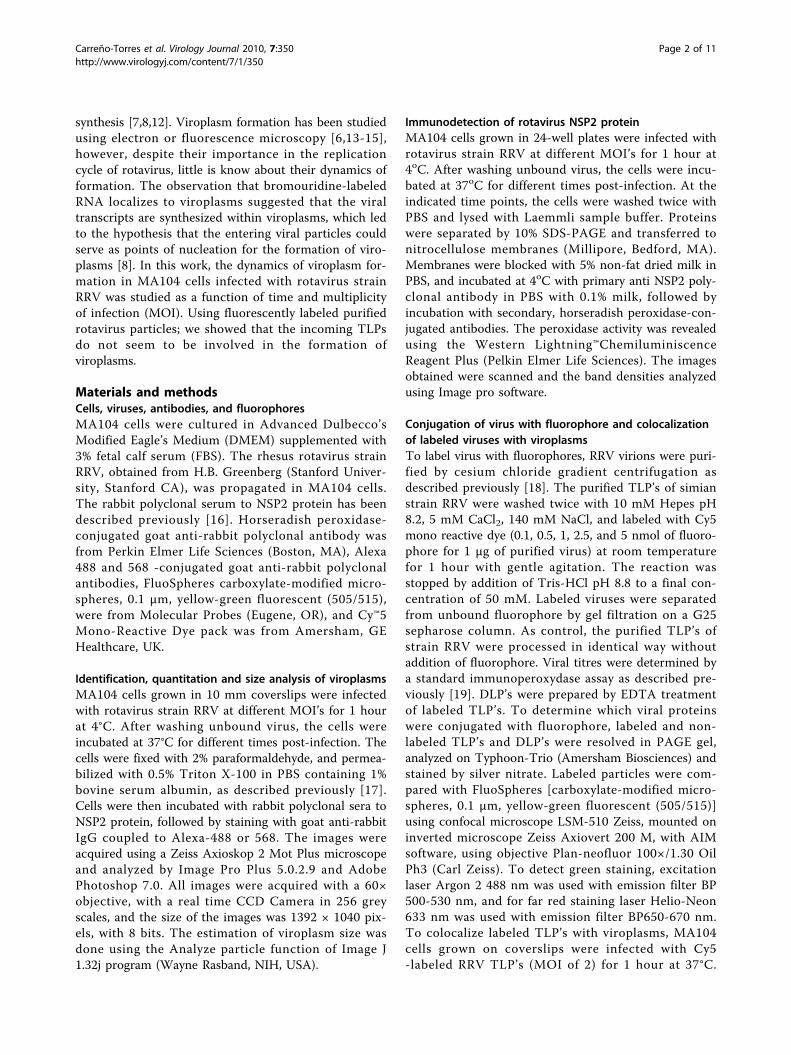
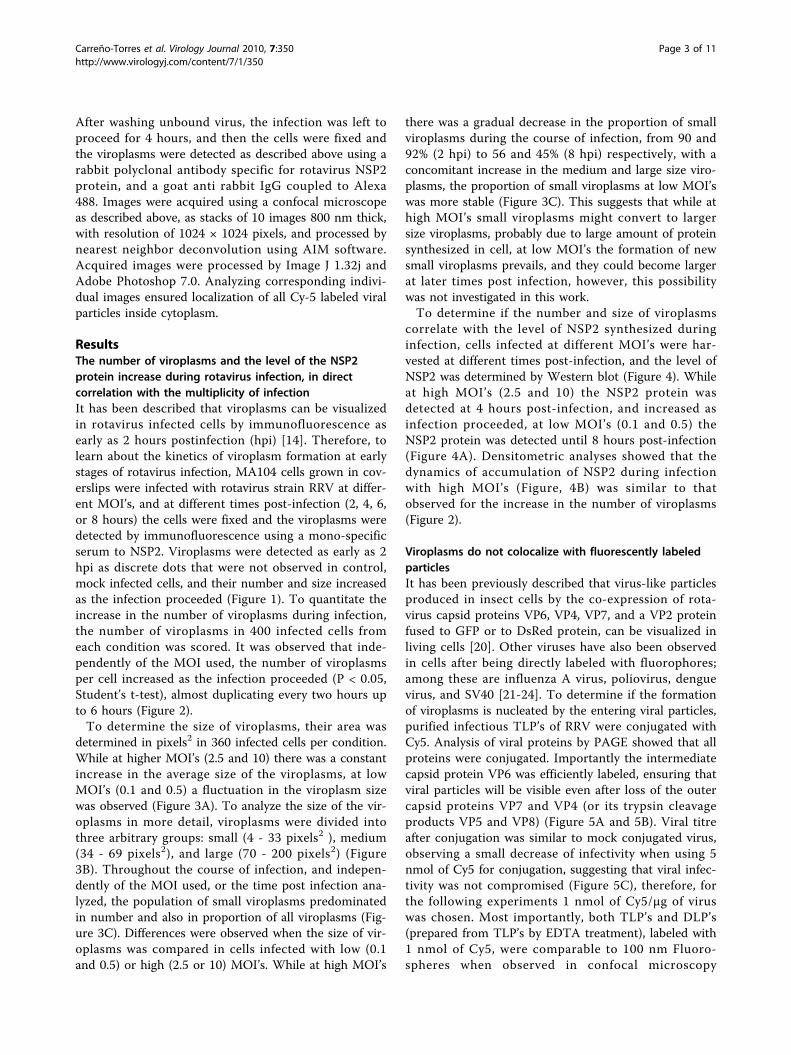
ResultsThe number of viroplasms and the level of the NSP2protein increase during rotavirus infection, in directcorrelation with the multiplicity of infectionIt has been described that viroplasms can be visualizedin rotavirus infected cells by immunofluorescence asearly as 2 hours postinfection (hpi) [14]. Therefore, tolearn about the kinetics of viroplasm formation at earlystages of rotavirus infection, MA104 cells grown in cov-erslips were infected with rotavirus strain RRV at differ-ent MOI’s, and at different times post-infection (2, 4, 6,or 8 hours) the cells were fixed and the viroplasms weredetected by immunofluorescence using a mono-specificserum to NSP2. Viroplasms were detected as early as 2hpi as discrete dots that were not observed in control,mock infected cells, and their number and size increasedas the infection proceeded (Figure 1). To quantitate theincrease in the number of viroplasms during infection,the number of viroplasms in 400 infected cells fromeach condition was scored. It was observed that inde-pendently of the MOI used, the number of viroplasmsper cell increased as the infection proceeded (P < 0.05,Student’s t-test), almost duplicating every two hours upto 6 hours (Figure 2).To determine the size of viroplasms, their area was
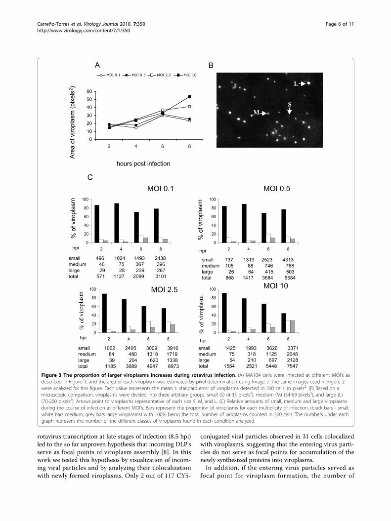
determined in pixels2 in 360 infected cells per condition.While at higher MOI’s (2.5 and 10) there was a constantincrease in the average size of the viroplasms, at lowMOI’s (0.1 and 0.5) a fluctuation in the viroplasm sizewas observed (Figure 3A). To analyze the size of the vir-oplasms in more detail, viroplasms were divided intothree arbitrary groups: small (4 - 33 pixels2 ), medium(34 - 69 pixels2), and large (70 - 200 pixels2) (Figure3B). Throughout the course of infection, and indepen-dently of the MOI used, or the time post infection ana-lyzed, the population of small viroplasms predominatedin number and also in proportion of all viroplasms (Fig-ure 3C). Differences were observed when the size of vir-oplasms was compared in cells infected with low (0.1and 0.5) or high (2.5 or 10) MOI’s. While at high MOI’s
there was a gradual decrease in the proportion of smallviroplasms during the course of infection, from 90 and92% (2 hpi) to 56 and 45% (8 hpi) respectively, with aconcomitant increase in the medium and large size viro-plasms, the proportion of small viroplasms at low MOI’swas more stable (Figure 3C). This suggests that while athigh MOI’s small viroplasms might convert to largersize viroplasms, probably due to large amount of proteinsynthesized in cell, at low MOI’s the formation of newsmall viroplasms prevails, and they could become largerat later times post infection, however, this possibilitywas not investigated in this work.To determine if the number and size of viroplasms
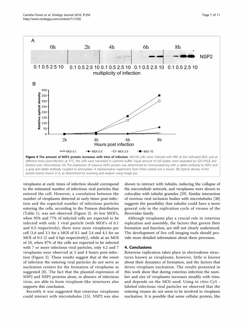
correlate with the level of NSP2 synthesized duringinfection, cells infected at different MOI’s were har-vested at different times post-infection, and the level ofNSP2 was determined by Western blot (Figure 4). Whileat high MOI’s (2.5 and 10) the NSP2 protein wasdetected at 4 hours post-infection, and increased asinfection proceeded, at low MOI’s (0.1 and 0.5) theNSP2 protein was detected until 8 hours post-infection(Figure 4A). Densitometric analyses showed that thedynamics of accumulation of NSP2 during infectionwith high MOI’s (Figure, 4B) was similar to thatobserved for the increase in the number of viroplasms(Figure 2).
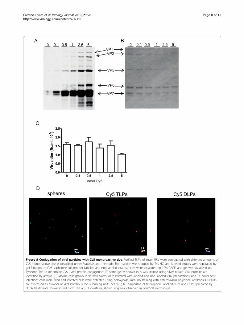
Viroplasms do not colocalize with fluorescently labeledparticlesIt has been previously described that virus-like particlesproduced in insect cells by the co-expression of rota-virus capsid proteins VP6, VP4, VP7, and a VP2 proteinfused to GFP or to DsRed protein, can be visualized inliving cells [20]. Other viruses have also been observedin cells after being directly labeled with fluorophores;among these are influenza A virus, poliovirus, denguevirus, and SV40 [21-24]. To determine if the formationof viroplasms is nucleated by the entering viral particles,purified infectious TLP’s of RRV were conjugated withCy5. Analysis of viral proteins by PAGE showed that allproteins were conjugated. Importantly the intermediatecapsid protein VP6 was efficiently labeled, ensuring thatviral particles will be visible even after loss of the outercapsid proteins VP7 and VP4 (or its trypsin cleavageproducts VP5 and VP8) (Figure 5A and 5B). Viral titreafter conjugation was similar to mock conjugated virus,observing a small decrease of infectivity when using 5nmol of Cy5 for conjugation, suggesting that viral infec-tivity was not compromised (Figure 5C), therefore, forthe following experiments 1 nmol of Cy5/μg of viruswas chosen. Most importantly, both TLP’s and DLP’s(prepared from TLP’s by EDTA treatment), labeled with1 nmol of Cy5, were comparable to 100 nm Fluoro-spheres when observed in confocal microscopy
Carreño-Torres et al. Virology Journal 2010, 7:350http://www.virologyj.com/content/7/1/350
Page 3 of 11
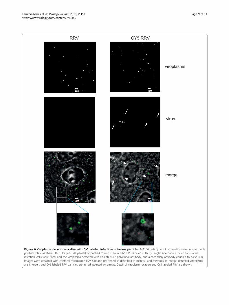
(Figure 5D). Since it was possible to visualize the fluor-escently labeled viral particles, we used them to observetheir intracellular distribution with respect to the newlyformed viroplasms. To do this, MA104 cells grown incoverslips were infected with Cy5-conjugated RRVTLP’s at an MOI of 2, and 4 hpi the cells were fixed,the viroplasms were immunostained using a polyclonal
sera to NSP2, and images were acquired using confocalmicroscopy, as described under material and methods.Fluorescently labeled viral particles were observed dis-tributed in the cytoplasm as discrete spots (Figure 6).The number of labeled viral particles, viroplasms, andtheir co-localization was counted independently by twopersons in 31 cells. In these, 117 labeled virus particles
Time post infection
2 hours 4 hours 6 hours 8 hours
Mock infected cells
MOI
0.1
0.5
2.5
10
Figure 1 Detection of viroplasms in cells infected at different MOI’s, and at distinct times post infection. MA104 cells were infected withRRV at the indicated MOI, and at different times post infection at 37°C, the cells were fixed and immunostained with a rabbit antibody to NSP2and a goat anti-rabbit antibody coupled to Alexa-488 or Alexa-568. Images were acquired using Zeiss Axioskop 2 Mot Plus microscope andImage Pro Plus 5.0.2.9 program. Mock-infected cells are shown as control.
Carreño-Torres et al. Virology Journal 2010, 7:350http://www.virologyj.com/content/7/1/350
Page 4 of 11
and 467 viroplasms were observed, however, only 2 ofthe viral particles observed colocalized with viroplasms,while the rest appeared independent of each other inthe cell cytoplasm.
DiscussionThe formation of viroplasms has been previously studiedusing electron and fluorescence microscopy, however,those studies have focused only on late (4 to 24 hpi)stages of infection [6,13,15]. Only Eichwald et al [14]have studied earlier stages of viroplasm formation, andin their work, following the expression of an NSP2 pro-tein fused to EGFP in rotavirus SA-11 infected cells,they observed that the total number of viroplasmsdecreased with time, with a concomitant increase intheir size, starting at 6 hpi. This observation was inter-preted as fusion events between smaller viroplasms.Similar results were reported by Cabral-Romero andPadilla-Noriega [15] using the strain SA-11 in BSC1cells, although at even later (10 hpi) stages of infection.Comparing the formation of viroplasms between SA-11and OSU rotavirus strains, Campagna et al. [6] observedthat the viroplasms formed in OSU infected cells didnot increase in size as readily as those formed duringinfection with SA-11. In this work, after infection withrotavirus strain RRV, using different MOI’s, an increasein the number of viroplasms and in the amount of theNSP2 protein was observed. The size of viroplasms wasobserved to increase when higher MOI’s were used.There are several possibilities to explain the discrepan-
cies reported. First, the decrease in the number of
viroplasms was observed only during infection withstrain SA-11 [14,15], but not with strains OSU [12], andRRV (this work). It is known that some viral functions(receptor specificity, plaque formation, extraintestinalspread, IRF3 degradation, etc) may vary among differentrotavirus strains [25-28] what opens the possibility thatthere could also be strain-specific differences for viro-plasm formation. In fact, an impaired phosphorylationof NSP5 affected differently the morphogenesis of viro-plasms in cells infected with either SA-11 or OSU rota-virus strains [6]. The differences observed between ourstudies and those of other groups could also arise fromthe different methodologies used to detect viroplasms.While in our case the newly synthesized rotavirus pro-teins were immunodetected and analyzed in 400 cells, inthe study by Eichwald et al. [14] the identification of vir-oplasms was based on the detection of NSP2-EGFP orNSP5-EGFP fusion proteins in 20 cells. It is possiblethat the large amount of recombinant fusion proteinsthat accumulated in the cytoplasm of transfected cellsbefore rotavirus infection could change the kinetics ofviroplasm formation, since upon rotavirus infection arapid redistribution of the EGFP - proteins wasobserved. It was not possible to compare the exact num-ber of viroplasms obtained in that study, since the MOIthat was used to infect the transfected MA104 cells wasnot mentioned.In this work, studying the kinetics of viroplasm forma-
tion during the infection of strain RRV, we observed anincrease in both the number and size of viroplasms withtime and this increment was dependent on the MOIused. At high MOI’s (2.5 and 10) the increase correlatedwith the amount of NSP2 protein detected at a giventime point, while at lower MOI’s (0.1 and 0.5), the smal-ler increase in NSP2 protein correlated with a less vari-able viroplasm size. It is possible that when a criticalconcentration of NSP2 and NSP5 is reached, and asother viral proteins accumulate, viroplasms start toform, first as small entities, and then becoming larger atlater stages of the replication cycle. Although, it is notpossible to determine if the increase in size is caused byfusion of smaller viroplasms or by addition of newlyproduced rotavirus proteins to small viroplasms, ourobservations are more consistent with the idea that newsmall viroplasms are generated constantly during thereplication cycle, since even at later stages of infection alarge proportion of small viroplasms was observed. Itremains to be determined if the small viroplasms, pre-sumably generated by the aggregation of NSP2 andNSP5 require an additional priming signal, or if it isonly the concentration of free NSP2 and NSP5 whatdictates the formation of a new viroplasms.The mechanism of viroplasms formation and its protein
content is unknown. The fact that viroplasms are sites for
0
5
10
15
20
25
2 4 6 8
hours post infection
num
ber
of
viropla
sm
s p
er
cell
MOI 0.1 MOI 0.5 MOI 2.5 MOI 10
Figure 2 The number of viroplasms per cell increases withtime of infection. MA104 cells were infected at different MOI’s asdescribed in Figure 1, and viroplasms were detected byimmunofluorescent staining of NSP2. Viroplasms were counted in400 infected cells in each condition. Each value is expressed asmean ± standard error. The increase in the number of viroplasmsduring the infection at each MOI, and the differences in the numberof viroplasms between different MOI at each time point werestatistically significant (P < 0.05, student T-test).
Carreño-Torres et al. Virology Journal 2010, 7:350http://www.virologyj.com/content/7/1/350
Page 5 of 11
rotavirus transcription at late stages of infection (8.5 hpi)led to the so far unproven hypothesis that incoming DLP’sserve as focal points of viroplasm assembly [8]. In thiswork we tested this hypothesis by visualization of incom-ing viral particles and by analyzing their colocalizationwith newly formed viroplasms. Only 2 out of 117 CY5-
conjugated viral particles observed in 31 cells colocalizedwith viroplasms, suggesting that the entering virus parti-cles do not serve as focal points for accumulation of thenewly synthesized proteins into viroplasms.In addition, if the entering virus particles served as
focal point for viroplasm formation, the number of
% o
f vi
ropl
asm
C
hpi
small 496 1024 1493 2438
medium 46 75 367 396
large 29 28 239 267
total 571 1127 2099 3101
small 1425 1993 3626 3371
medium 75 318 1125 2048
large 54 210 697 2128
total 1554 2521 5448 7547
% o
f viropla
sm
small 737 1319 2523 4313
medium 105 88 746 768
large 26 64 415 503
total 868 1417 3684 5584
% o
f viropla
sm
% o
f vi
ropl
asm
small 1062 2405 3009 3916
medium 84 480 1318 1719
large 39 204 620 1338
total 1185 3089 4947 6973
hpi
hpihpi
0
20
40
60
80
100
2 4 6 8
0
20
40
60
80
100
2 4 6 8
0
20
40
60
80
100
2 4 6 8
0
20
40
60
80
100
2 4 6 8
MOI 10MOI 2.5
MOI 0.1 MOI 0.5
AA
rea o
f viropla
sm
(pix
els
2)
0
10
20
30
40
50
60
2 4 6 8
MOI 0.1 MOI 0.5 MOI 2.5 MOI 10
hours post infection
L
MS
B
Figure 3 The proportion of larger viroplasms increases during rotavirus infection. (A) MA104 cells were infected at different MOI’s asdescribed in Figure 1, and the area of each viroplasm was estimated by pixel determination using Image J. The same images used in Figure 2were analyzed for this figure. Each value represents the mean ± standard error of viroplasms detected in 360 cells, in pixels2. (B) Based on amicroscopic comparison, viroplasms were divided into three arbitrary groups, small (S) (4-33 pixels2), medium (M) (34-69 pixels2), and large (L)(70-200 pixels2). Arrows point to viroplasms representative of each size S, M, and L. (C) Relative amounts of small, medium and large viroplasmsduring the course of infection at different MOI’s. Bars represent the proportion of viroplasms for each multiplicity of infection, (black bars - small;white bars medium; grey bars large viroplasms) with 100% being the total number of viroplasms counted in 360 cells. The numbers under eachgraph represent the number of the different classes of viroplasms found in each condition analyzed.
Carreño-Torres et al. Virology Journal 2010, 7:350http://www.virologyj.com/content/7/1/350
Page 6 of 11
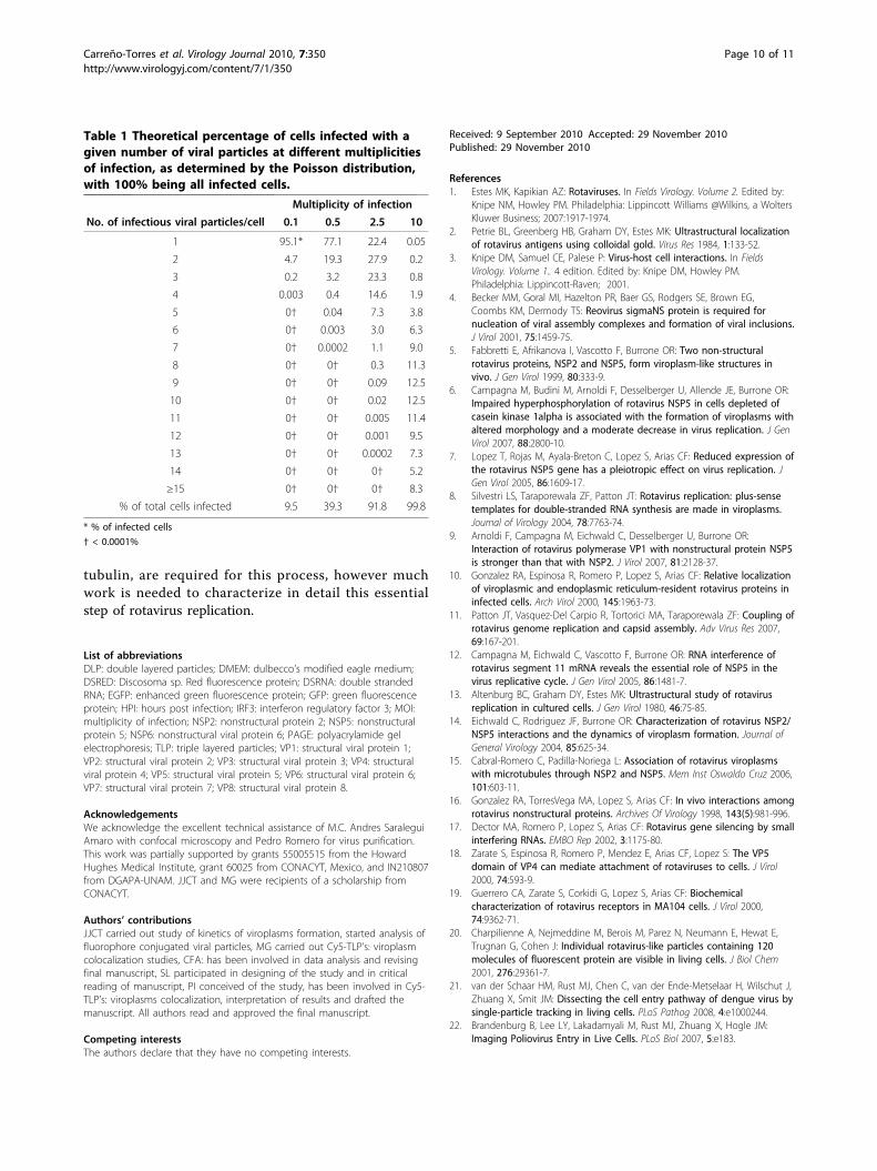
viroplasms at early times of infection should correspondto the estimated number of infectious viral particles thatentered the cell. However, a correlation between thenumber of viroplasms detected at early times post-infec-tion and the expected number of infectious particlesentering the cells, according to the Poisson distribution(Table 1), was not observed (Figure 2). At low MOI’s,when 95% and 77% of infected cells are expected to beinfected with only 1 viral particle (with MOI’s of 0.1and 0.5 respectively), there were more viroplasms percell [1.6 and 3.1 for a MOI of 0.1 and 2.4 and 4.1 for anMOI of 0.5 (2 and 4 hpi respectively)], while at an MOIof 10, when 87% of the cells are expected to be infectedwith 7 or more infectious viral particles, only 4.2 and 7viroplasms were observed at 2 and 4 hours post-infec-tion (Figure 2). These results suggest that at the onsetof infection the entering viral particles do not serve asnucleation centers for the formation of viroplasms assuggested [8]. The fact that the plasmid expression ofNSP2 and NSP5 proteins alone, in absence of infectiousvirus, are able to form viroplasm-like structures alsosupports this conclusion.Recently it was suggested that rotavirus viroplasms
could interact with microtubules [15]. NSP2 was also
shown to interact with tubulin, inducing the collapse ofthe microtubule network, and viroplasms were shown tocolocalize with tubulin granules [29]. Similar interactionof reovirus viral inclusion bodies with microtubules [30]suggests the possibility that tubulin could have a moregeneral role in the replication cycle of viruses of theReoviridae family.Although viroplasms play a crucial role in rotavirus
replication and assembly, the factors that govern theirformation and function, are still not clearly understood.The development of live cell imaging tools should pro-vide more detailed information about these processes.
4. ConclusionsRotavirus replication takes place in electrodense struc-tures known as viroplasms, however, little is knownabout their dynamics of formation, and the factors thatdrives viroplasm nucleation. The results presented inthis work show that during rotavirus infection the num-ber and size of viroplasms increases steadily with time,and depends on the MOI used. Using in vitro Cy5 -labeled infectious viral particles we observed that theentering viruses do not seem to be involved in viroplasmnucleation. It is possible that some cellular protein, like
0
50
100
150
200
250
300
350
400
2h 4h 6h 8hHours post infection
Rela
tive o
pti
cal d
en
sit
y
MOI 0.1 MOI 0.5 MOI 2.5 MOI 10
A
B
0.1 0.5 2.5 100.1 0.5 2.5 10 0.1 0.5 2.5 10 0.1 0.5 2.5 100.1 0.5 2.5 10
0h 2h 4h 6h 8h
NSP2
multiplicity of infection
Figure 4 The amount of NSP2 protein increases with time of infection. MA104 cells were infected with RRV at the indicated MOI, and atdifferent times post-infection at 37°C, the cells were harvested in Laemmli buffer. Equal amount of cell lysates were separated by SDS-PAGE andblotted onto nitrocellulose. (A) The expression of rotavirus NSP2 protein was determined by immunostaining with a rabbit antibody to NSP2 anda goat anti-rabbit antibody coupled to peroxydase. A representative experiment from three carried out is shown. (B) Optical density of theprotein bands shown in A, as determined by scanning and analysis using Image pro.
Carreño-Torres et al. Virology Journal 2010, 7:350http://www.virologyj.com/content/7/1/350
Page 7 of 11
A B
Figure 5 Conjugation of viral particles with Cy5 monoreactive dye. Purified TLP’s of strain RRV were conjugated with different amounts ofCy5 monoreactive dye as described under Materials and methods. The reaction was stopped by Tris-HCl and labeled viruses were separated bygel filtration on G25 sepharose column. (A) Labeled and non-labeled viral particles were separated on 10% PAGE, and gel was visualized onTyphoon Trio to determine Cy5 - viral protein conjugation. (B) Same gel as shown in A was stained using silver nitrate. Viral proteins areidentified by arrows. (C) MA104 cells grown in 96 well plates were infected with labeled and non labeled viral preparations, and 14 hours postinfections cells were fixed and infected cells were detected using peroxydase immuno staining with anti-rotavirus polyclonal antibodies. Resultsare expressed as number of viral infectious focus forming units per ml. (D) Comparison of fluorophore labelled TLP’s and DLP’s (prepared byEDTA treatment), shown in red, with 100 nm Fluorosferes, shown in green, observed in confocal microscope.
Carreño-Torres et al. Virology Journal 2010, 7:350http://www.virologyj.com/content/7/1/350
Page 8 of 11
viroplasms
virus
CY5 RRVRRV
merge
Figure 6 Viroplasms do not colocalize with Cy5 labeled infectious rotavirus particles. MA104 cells grown in coverslips were infected withpurified rotavirus strain RRV TLPs (left side panels) or purified rotavirus strain RRV TLP’s labeled with Cy5 (right side panels). Four hours afterinfection, cells were fixed, and the viroplasms detected with an anti-NSP2 polyclonal antibody, and a secondary antibody coupled to Alexa-488.Images were obtained with confocal microscope LSM 510 and processed as described in material and methods. In merge, detected viroplasmsare in green, and Cy5 labeled RRV particles are in red, pointed by arrows. Detail of viroplasm location and Cy5 labeled RRV are shown.
Carreño-Torres et al. Virology Journal 2010, 7:350http://www.virologyj.com/content/7/1/350
Page 9 of 11
tubulin, are required for this process, however muchwork is needed to characterize in detail this essentialstep of rotavirus replication.
List of abbreviationsDLP: double layered particles; DMEM: dulbecco’s modified eagle medium;DSRED: Discosoma sp. Red fluorescence protein; DSRNA: double strandedRNA; EGFP: enhanced green fluorescence protein; GFP: green fluorescenceprotein; HPI: hours post infection; IRF3: interferon regulatory factor 3; MOI:multiplicity of infection; NSP2: nonstructural protein 2; NSP5: nonstructuralprotein 5; NSP6: nonstructural viral protein 6; PAGE: polyacrylamide gelelectrophoresis; TLP: triple layered particles; VP1: structural viral protein 1;VP2: structural viral protein 2; VP3: structural viral protein 3; VP4: structuralviral protein 4; VP5: structural viral protein 5; VP6: structural viral protein 6;VP7: structural viral protein 7; VP8: structural viral protein 8.
AcknowledgementsWe acknowledge the excellent technical assistance of M.C. Andres SaraleguiAmaro with confocal microscopy and Pedro Romero for virus purification.This work was partially supported by grants 55005515 from the HowardHughes Medical Institute, grant 60025 from CONACYT, Mexico, and IN210807from DGAPA-UNAM. JJCT and MG were recipients of a scholarship fromCONACYT.
Authors’ contributionsJJCT carried out study of kinetics of viroplasms formation, started analysis offluorophore conjugated viral particles, MG carried out Cy5-TLP’s: viroplasmcolocalization studies, CFA: has been involved in data analysis and revisingfinal manuscript, SL participated in designing of the study and in criticalreading of manuscript, PI conceived of the study, has been involved in Cy5-TLP’s: viroplasms colocalization, interpretation of results and drafted themanuscript. All authors read and approved the final manuscript.
Competing interestsThe authors declare that they have no competing interests.
Received: 9 September 2010 Accepted: 29 November 2010Published: 29 November 2010
References1. Estes MK, Kapikian AZ: Rotaviruses. In Fields Virology. Volume 2. Edited by:
Knipe NM, Howley PM. Philadelphia: Lippincott Williams @Wilkins, a WoltersKluwer Business; 2007:1917-1974.
2. Petrie BL, Greenberg HB, Graham DY, Estes MK: Ultrastructural localizationof rotavirus antigens using colloidal gold. Virus Res 1984, 1:133-52.
3. Knipe DM, Samuel CE, Palese P: Virus-host cell interactions. In FieldsVirology. Volume 1.. 4 edition. Edited by: Knipe DM, Howley PM.Philadelphia: Lippincott-Raven; 2001.
4. Becker MM, Goral MI, Hazelton PR, Baer GS, Rodgers SE, Brown EG,Coombs KM, Dermody TS: Reovirus sigmaNS protein is required fornucleation of viral assembly complexes and formation of viral inclusions.J Virol 2001, 75:1459-75.
5. Fabbretti E, Afrikanova I, Vascotto F, Burrone OR: Two non-structuralrotavirus proteins, NSP2 and NSP5, form viroplasm-like structures invivo. J Gen Virol 1999, 80:333-9.
6. Campagna M, Budini M, Arnoldi F, Desselberger U, Allende JE, Burrone OR:Impaired hyperphosphorylation of rotavirus NSP5 in cells depleted ofcasein kinase 1alpha is associated with the formation of viroplasms withaltered morphology and a moderate decrease in virus replication. J GenVirol 2007, 88:2800-10.
7. Lopez T, Rojas M, Ayala-Breton C, Lopez S, Arias CF: Reduced expression ofthe rotavirus NSP5 gene has a pleiotropic effect on virus replication. JGen Virol 2005, 86:1609-17.
8. Silvestri LS, Taraporewala ZF, Patton JT: Rotavirus replication: plus-sensetemplates for double-stranded RNA synthesis are made in viroplasms.Journal of Virology 2004, 78:7763-74.
9. Arnoldi F, Campagna M, Eichwald C, Desselberger U, Burrone OR:Interaction of rotavirus polymerase VP1 with nonstructural protein NSP5is stronger than that with NSP2. J Virol 2007, 81:2128-37.
10. Gonzalez RA, Espinosa R, Romero P, Lopez S, Arias CF: Relative localizationof viroplasmic and endoplasmic reticulum-resident rotavirus proteins ininfected cells. Arch Virol 2000, 145:1963-73.
11. Patton JT, Vasquez-Del Carpio R, Tortorici MA, Taraporewala ZF: Coupling ofrotavirus genome replication and capsid assembly. Adv Virus Res 2007,69:167-201.
12. Campagna M, Eichwald C, Vascotto F, Burrone OR: RNA interference ofrotavirus segment 11 mRNA reveals the essential role of NSP5 in thevirus replicative cycle. J Gen Virol 2005, 86:1481-7.
13. Altenburg BC, Graham DY, Estes MK: Ultrastructural study of rotavirusreplication in cultured cells. J Gen Virol 1980, 46:75-85.
14. Eichwald C, Rodriguez JF, Burrone OR: Characterization of rotavirus NSP2/NSP5 interactions and the dynamics of viroplasm formation. Journal ofGeneral Virology 2004, 85:625-34.
15. Cabral-Romero C, Padilla-Noriega L: Association of rotavirus viroplasmswith microtubules through NSP2 and NSP5. Mem Inst Oswaldo Cruz 2006,101:603-11.
16. Gonzalez RA, TorresVega MA, Lopez S, Arias CF: In vivo interactions amongrotavirus nonstructural proteins. Archives Of Virology 1998, 143(5):981-996.
17. Dector MA, Romero P, Lopez S, Arias CF: Rotavirus gene silencing by smallinterfering RNAs. EMBO Rep 2002, 3:1175-80.
18. Zarate S, Espinosa R, Romero P, Mendez E, Arias CF, Lopez S: The VP5domain of VP4 can mediate attachment of rotaviruses to cells. J Virol2000, 74:593-9.
19. Guerrero CA, Zarate S, Corkidi G, Lopez S, Arias CF: Biochemicalcharacterization of rotavirus receptors in MA104 cells. J Virol 2000,74:9362-71.
20. Charpilienne A, Nejmeddine M, Berois M, Parez N, Neumann E, Hewat E,Trugnan G, Cohen J: Individual rotavirus-like particles containing 120molecules of fluorescent protein are visible in living cells. J Biol Chem2001, 276:29361-7.
21. van der Schaar HM, Rust MJ, Chen C, van der Ende-Metselaar H, Wilschut J,Zhuang X, Smit JM: Dissecting the cell entry pathway of dengue virus bysingle-particle tracking in living cells. PLoS Pathog 2008, 4:e1000244.
22. Brandenburg B, Lee LY, Lakadamyali M, Rust MJ, Zhuang X, Hogle JM:Imaging Poliovirus Entry in Live Cells. PLoS Biol 2007, 5:e183.
Table 1 Theoretical percentage of cells infected with agiven number of viral particles at different multiplicitiesof infection, as determined by the Poisson distribution,with 100% being all infected cells.
Multiplicity of infection
No. of infectious viral particles/cell 0.1 0.5 2.5 10
1 95.1* 77.1 22.4 0.05
2 4.7 19.3 27.9 0.2
3 0.2 3.2 23.3 0.8
4 0.003 0.4 14.6 1.9
5 0† 0.04 7.3 3.8
6 0† 0.003 3.0 6.3
7 0† 0.0002 1.1 9.0
8 0† 0† 0.3 11.3
9 0† 0† 0.09 12.5
10 0† 0† 0.02 12.5
11 0† 0† 0.005 11.4
12 0† 0† 0.001 9.5
13 0† 0† 0.0002 7.3
14 0† 0† 0† 5.2
≥15 0† 0† 0† 8.3
% of total cells infected 9.5 39.3 91.8 99.8
* % of infected cells
† < 0.0001%
Carreño-Torres et al. Virology Journal 2010, 7:350http://www.virologyj.com/content/7/1/350
Page 10 of 11
23. Pelkmans L, Kartenbeck J, Helenius A: Caveolar endocytosis of simian virus40 reveals a new two-step vesicular-transport pathway to the ER. NatCell Biol 2001, 3:473-83.
24. Rust MJ, Lakadamyali M, Zhang F, Zhuang X: Assembly of endocyticmachinery around individual influenza viruses during viral entry. NatStruct Mol Biol 2004, 11:567-73.
25. Mossel EC, Ramig RF: Rotavirus genome segment 7 (NSP3) is adeterminant of extraintestinal spread in the neonatal mouse. J Virol2002, 76:6502-9.
26. Offit PA, Blavat G, Greenberg HB, Clark HF: Molecular basis of rotavirusvirulence: role of gene segment 4. J Virol 1986, 57:46-9.
27. Haddow J, Clark B, Ni Y, Desselberger U: Biological function of therotavirus protein VP4: observations on porcine isolates from China. MedMicrobiol Immunol 1989, 178:163-76.
28. Sen A, Feng N, Ettayebi K, Hardy ME, Greenberg HB: IRF3 inhibition byrotavirus NSP1 is host cell and virus strain dependent but independentof NSP1 proteasomal degradation. J Virol 2009, 83:10322-35.
29. Martin D, Duarte M, Lepault J, Poncet D: Sequestration of free tubulinmolecules by the viral protein NSP2 induces microtubuledepolymerization during rotavirus infection. J Virol 84:2522-32.
30. Parker JS, Broering TJ, Kim J, Higgins DE, Nibert ML: Reovirus core proteinmu2 determines the filamentous morphology of viral inclusion bodiesby interacting with and stabilizing microtubules. J Virol 2002, 76:4483-96.
doi:10.1186/1743-422X-7-350Cite this article as: Carreño-Torres et al.: Characterization of viroplasmformation during the early stages of rotavirus infection. Virology Journal2010 7:350.
Submit your next manuscript to BioMed Centraland take full advantage of:
• Convenient online submission
• Thorough peer review
• No space constraints or color figure charges
• Immediate publication on acceptance
• Inclusion in PubMed, CAS, Scopus and Google Scholar
• Research which is freely available for redistribution
Submit your manuscript at www.biomedcentral.com/submit
Carreño-Torres et al. Virology Journal 2010, 7:350http://www.virologyj.com/content/7/1/350
Page 11 of 11