Embed Size (px)
Citation preview

459© The Society of Nematologists 2018.
JOURNAL OF NEMATOLOGYIssue 4 | Vol. 50Article | DOI: 10.21307/jofnem-2018-042
Characterization of Juvenile Stages of Bursaphelenchus crenati Rhm, 1956 (Nematoda: Aphelenchoidoidea)
AbstractIn this study juvenile stages of Bursaphelenchus crenati (Sexdentati group) were distinguished based mainly on the genital primordium structure. Dauer stage juveniles were sampled under elytra of Hylesinus crenatus in galleries in bark of the wilted Fraxinus excelsior in the Voronezh region of Russia and were multiplied in laboratory cultures on the fungus Botrytis cinerea. Individual development included five stages (J1 – J4 and adults) separated by molts. The first molt J1 to J2 occurred inside the egg and the J2 was at the hatching stage. Sex of juvenile stages can be identified using the morphology and size of the genital primordia and a body size of nematodes. Sex of juveniles may be identified from the J3 stage by the presence of the cloacal primordium in male juvenile and orientation of the germinal zone of the genital primordium. A tabular key to developmental stages of B. crenati is given. The body grows during molts and within each stage. The body increases rapidly after J3 stage combined with the most active cellular differentiation and elongation of the genital primordium. The dauer juveniles collected from elytra of vector beetles were similar to the third juvenile stage of the propagative generation by genital primordium structure and body size.
Key wordsBursaphelenchus crenati, Life cycle, Dauer stage, Postembryogenesis, Diagnostics, Saproxylic nematodes, Fraxinus, Hylesinus, Russia, Ash wilt.
The nematode species Bursaphelenchus crenati Rühm, 1956 belonging to Sexdentati group was origi-nally described from the galleries of Hylesinus crenati Fabricius in bark of ash Fraxinus excelsior L. in Bavaria, Germany and later was re – described by Gu et al. (2017) using the populations from Poland. This species was recently detected in several localities of Russia and Belarus (Ryss et al., unpublished). Trans-missive dauer juveniles of B. crenati were found in senior larvae, pupae, and female imagoes of the bee-tle species. From these dauer juveniles, adult nema-todes were grown in cultures of the spore – less strain of the fungus Botrytis cinerea on the 2% potato dex-trose agar (PDA). All records revealed associations of B. crenati with wilt symptoms of the ash Fraxinus
excelsior. In the same beetle vector, the fungi belong-ing to the family Ophiostomataceae were extracted and cultured in 2% PDA and 2% MEA (malt agar). Nutrition relations between the nematodes and fungi were established, it has been shown that the nema-tode population grown in slow rate (5 wk) comparing to that culture with Botrytis cinerea (7 d at room tem-perature). To study a functional role of different juve-nile stages of B. crenati in the adaptations to vector and plant and fungus host, as well as in survival of winter seasonal stress, the identification of the life cy-cle stages would be necessary.
Special attention was given to observe the genital primordium, because according to previous studies this organ is most useful in identification of stage and
Alexander Y. Ryss and Kristina S. Polyanina
Zoological Institute, Russian Academy of Sciences, St. Petersburg 199034, Russia.
E-mail: [email protected].
This paper was edited by Zafar Ahmad Handoo.
Received for publication June 23, 2018.
460
Characterization of Juvenile Stages of Bursaphelenchus crenati Rhm, 1956 (Nematoda: Aphelenchoidoidea)
sex of juveniles (Hirschmann, 1962, 1971; Hirschmann and Triantaphyllou, 1968; Ryss, 1981, 1988; Ryss and Chernetskaya, 2009, 2010; Ryss and Polyanina, 2017). The purpose of this research is to characterize the juvenile stages of the species.
Materials and methods
Nematode samples
The bark and wood samples with the beetle bark tun-nels of Hylesinus crenatus Fabricius, in the inner bark layer and sapwood in wilted ash tree, Fraxinus excel-sior L. were collected by A.V. Petrov in the forestry plantation “Tellermanovskoye Opytnoye Lesnichest-vo ILAN RAS,” Tellermanovsky settlement, Gribano-vsky District, Voronezh Oblast, Russia, June 2017, in a location with the GPS. 51°21¢46.8¢N 42°02¢40.3¢E. Samples were sent to Zoological Institute RAS. In the laboratory, the nematode Bursaphelenchus crenati dauers were collected from beetle imagoes, where they formed semi – dry spot – like assemblages (“nematangia”) on the inner surface of elytra.
Nematode culturing
Nematodes were multiplied from dauers in the sterile laboratory cultures on the lawn of spore – less strain of the fungus Botrytis cinerea, inoculated in the 2% potato dextrose agar media in petri dish (Ryss, 2015). Over 2 wk after 20 nematode specimen inoculation, nematodes occupied all the petri dish space (diam. 6 cm), devastating the fungus lawn, showing ac-tive oviposition and population growth. To maintain the nematode isolate, approximately 100 nematode specimens were transferred to the fresh culture of B. cinerea, multiplying population occupied all the petri dish space in 5 to 7 d at 20 to 22°C.
Morphological study
To study the morphology, nematodes were washed out from petri dish with 1 ml of distilled water and fixed in hot TAF as described by Ryss (2017b). Over 2 d after fixation, nematodes were stained with ace-tic orcein or methylene blue. A modification of the staining technique (Ryss, 1988; Ryss and Chernet-skaya, 2009; Ryss and Polyanina, 2017) was used
Figure 1: Bursaphelenchus crenati. Development in egg. A. Cleavage; B. Gastrula; C,D. First stage juvenile (J1) within egg shell, at different focal planes; E,F,G. Second stage juvenile (J2) molting with shed cuticle, at different focal planes; H. Hatching J2, preparing to leave the egg shell. Scale 20 µm.

461
JOURNAL OF NEMATOLOGY
to stain nematodes with acetic orcein. A 1.5 ml Ep-pendorf tube with nematodes in TAF was placed in upright position for 10 min to settle nematodes down, and the upper layer of fixative was removed with a syringe up to the two fold level of the nematode sus-pension height (usually 100 µL). Then 200 µL of 70% acetic acid was added into the tube. After shaking the tube, two drops of saturated acetic orcein solu-tion in 65% acetic acid were added, which is usually used for chromosome staining. The mixture was kept overnight for staining procedure and then a drop of staining suspension was placed into the glycerin in a cavity slide. The stained nematodes picked out by needle and transferred to 5 µL drop of glycerin.
To stain with methylene blue, the nematodes were fixed in TAF and processed to glycerin according to Ryss (2017a). Then, nematodes were picked out and transferred from glycerin suspension into a small 1 µL drop of glycerin on a glass slide, and a 5 µL drop of saturated methylene blue water solution was added from above. The drop was placed for 12 hr in open air at room temperature for water evaporation and staining. Then nematodes were transferred in another small glycerin drop on glass slide, heated softly and quickly for stain contrasting and differentiation.
Stained nematodes were picked out with needle under a stereomicroscope and placed in a minute drop of glycerin on a microscopic slide. The drop was covered with 20 × 20 mm coverslip. The drop was spread under coverslip to press nematodes to obtain flattened worm bodies for detailed study of nematode genital primordia structures. After drop spreading, coverslip was sealed with nail polish along borders. All stained nematodes in slides were studied, measured, and photographed under the automated microscope system Leica DM5000 B with differential interference contrast (DIC) and with the Leica DFC320 (R2) digital camera with Leica DFC Twain Software for PC and Leica IM50 Image Manager for PC. All measurements were made using the software ImageJ 1.48 v (Nation-al Institute of Health, USA, http://imagej.nih.gov/ij).
Measurements and values used in morphometry of the individual development stages
Measures: L: body length; BW: body width; pharynx length till the pharyngo – intestinal valve; pharynx length till the end of the pharyngeal gland lobe; tail length; tail width at anus (ABW); GP: genital primordium length;
Figure 2: Bursaphelenchus crenati. Methylene blue staining. Junior juveniles. A,B. Second stage juvenile (J2): A. Genital primordium; B. Tail; C,D. Genital primordium of J3 female juveniles (arrows: anterior germinal zone); E,F. Tails of female J3; G, H. Genital primordium of J3 male juveniles (arrows: posterior germinal zone); I,J. Tails of male J3 (arrows: cloaca primordium). Scale 20 μm.

462
Characterization of Juvenile Stages of Bursaphelenchus crenati Rhm, 1956 (Nematoda: Aphelenchoidoidea)
GW: genital primordium width; spicule length along arc; hyaline part of tail in male juveniles (future bursal flap of male). Ratios: a: body length divided to body width; b: body length divided to pharynx length till the pharyn-go – intestinal valve; b’: body length divided to pharynx length till the end of pharyngeal glands; c: body length divided to tail length; c’: tail length divided to tail width; V or (V): distance from anterior body end to vulva, or to the center of the genital primordium, divided to the body length, in %; GP/GW: genital primordium length divided to its width; GP/L: genital primordium length divided to body length, in %.
Because the bodies were flattened, the re – calcu-lation of the body and tail diameter was used: the flat-tened width was considered as a half of the circle (π R or pi R), thus the diameter (2 R) was calculated as the flattened body width (BW) × (2/pi) = 0.64 × BW.
Results
The stained nematodes and eggs were studied. The first molt took place inside the egg shell; thus dur-ing hatching the second stage juvenile (J2) broke the egg – shell and came out (Fig. 1E,F,G).
Other stages may be classified as J2, J3, J4, and adult males and females, separated by three molts. Molts were detected by cuticle separation from the body at body extremities, the head and tail tips. The sex of J3 and J4 may be recognized, whereas it cannot be determined in J2. The stage and sex of the juvenile may be determined in the genital primordium structure
Figure 3: Bursaphelenchus crenati. Methylene blue staining. Genital primordium and tails of the fourth stage juveniles and adults. A. Genital primordium of the fourth stage female juvenile (J4); B,C. Molting female juvenile J4 – adult; B. Genital primordium; C. Tail; D. Genital system of adult female; E, F. Fourth stage male juvenile; E. Genital primordium; F. Tail and cloacal primordium; G. Genital system of adult male. Scale 50 µm.
Figure 4: Position of genital primordia in bodies of male juveniles and adults. Scale 50 µm.

463
JOURNAL OF NEMATOLOGY
and its position in the body. The body size and the ratio between genital primordium length and the body are useful to identify the stage. Genital primordia in every stage and sex are described and illustrated below.
Second stage juveniles (J2) (Figs. 2A,B, 4,5; Table 1)
The genital primordium consists of four cells: two large germinal cells in the central part and two small somatic nuclei at extremities (Fig. 2A). Primordium is in the middle of the intestine part of the body (Fig. 4).
Differences from other stages
J2 differs from senior stages by the smallest number of cells in primordium as well as in the body size less than 200 µm vs body length of 300 µm or longer in juveniles of senior stages.
Third stage juveniles
Third stage male juveniles (Figs. 2,4; Table 1)
In the genital primordium, 3 to 5 germinal cells in the posterior part and 12 somatic nuclei in anterior part (Fig. 2G,H) are present. In addition, a single somatic apical nucleus at every extremity of the primordium is found. The primordium is situated in the middle of the intes-tine part of the body. Cloaca primordium is present as a dense agglomeration of somatic nuclei around the rec-tum (Fig. 2I,J).
Third stage female juvenile (Figs. 2C,D, 5; Table 1)
In the genital primordium, 2 large germinal cells in the anterior part and 12 somatic nuclei in the posterior primordium part are present together with a single somatic apical nucleus on every extremity of the pri-mordium (Fig. 2C,D). A circle of six somatic nuclei in-terrupt the ventral hypodermal cord chain of somatic nuclei. It is a primordium of vulva.
Differences of the third stage juveniles
Differences of male juvenile of the third stage from fe-male juvenile are manifested by the presence of cloa-ca primordium and the position of germinal cells in the posterior part of the primordium. In female juveniles, germinal cells are located in the anterior part of the primordium. Third stage juveniles differ from J2 in the number of cells of the primordium (more than 10 vs 4 in J2). From J4 juveniles, the third stage juveniles dif-fer by the length of primordium occupying 6% of the body length or less vs 11% or more in J4 and adults.
Fourth stage juveniles
Fourth stage male juveniles (Figs. 3G, 4; Table 1)
Cloaca primordium is massive, with numerous so-matic nuclei around rectum and a transparent cavity anterior to rectum with rudimentary spicules. The genital primordium is distinctly divided into an anterior germinal part of 30 to 60 large cells and a
Figure 5: Position of genital primordia in bodies of female juveniles and adults. Scale 50 µm.
464
Characterization of Juvenile Stages of Bursaphelenchus crenati Rhm, 1956 (Nematoda: Aphelenchoidoidea)
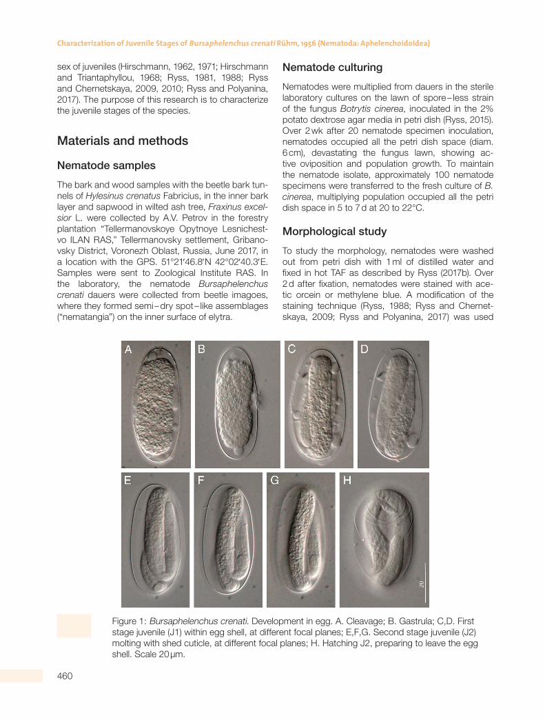
Table 1. Measurements (in µm) and ratios of the development stages of Bursaphelenchus crenati.
Stage/Character
J2 J3 male J3 female J4 male J4 female Male Female
n 20 20 20 15 18 15 15
L 166 ± 22 367 ± 21 375 ± 26 455 ± 21 489 ± 14 698 ± 69 843 ± 94
(150 – 181) (338 – 400) (339 – 402) (377 – 532) (479 – 508) (607 – 804) (750 – 1082)
Body width 7 ± 1 10 ± 2 11 ± 2 14 ± 4 12 ± 0.2 17 ± 3 16 ± 3
(6 – 8) (6 – 12) (8 – 13) (12 – 17) (10 – 15) (14 – 216) (13 – 20)
Pharynx 68 ± 4 111 ± 5 115 ± 6 106 ± 0.7 125 ± 12 122 ± 12 136 ± 22
(65 – 70) (104 – 118) (109 – 125) (105 – 106) (111 – 140) (105 – 144) (108 – 178)
P haryngeal gland
30 ± 5(27 – 34)
50 ± 5(44 – 58)
52 ± 5(45 – 60)
49 ± 6(45 – 53)
50 ± 6(42 – 55)
50 ± 6(41 – 64)
59 ± 14(40 – 85)
Stylet 7 ± 1 8 ± 1 8 ± 0.8 10 ± 1 10 ± 1 10 ± 0.1 11 ± 1
(6 – 8) (6 – 9) (7 – 9) (9 – 11) (9 – 11) (8 – 12) (10 – 13)
L ip region width
4.0 ± 0.3(3.5 – 4.5)
5 ± 0.2(4 – 5)
5 ± 0.4(4 – 5)
5 ± 0.2(5 – 5.5)
5.8 ± 0.2(5.5 – 6.0)
7.0 ± 0.6(7 – 8)
7.0 ± 0.5(6.0 – 7.5)
L ip region height
2.3 ± 0.5(2.0 – 2.5)
3 ± 0.3(2 – 3)
2 ± 0.2(2 – 3)
3 ± 0.2(3 – 3.5)
2.5 ± 0.1(2.4 – 2.7) –
4 ± 0.5(3 – 5)
3.6 ± 0.3(3 – 4)
M edian bulb from Anterior
30 ± 2(29 – 31)
48 ± 2(45 – 52)
48 ± 1.4(46 – 50)
40 ± 3(38 – 43)
56 ± 6(51 – 64)
58 ± 5(51 – 71)
60 ± 9(51 – 73)
M edian bulb length
9.4 ± 1(9 – 10)
12 ± 1(10 – 14)
12 ± 1.3(10 – 14)
12 ± 0.3(11.5 – 12)
12.7 ± 0.7(11.5 – 13.0)
15 ± 2(13 – 18)
18 ± 4(14 – 23)
M edian bulb width
5.7 ± 0.3(5.5 – 6.0)
9 ± 2(6 – 11)
9 ± 1.2(7 – 10)
9 ± 2(7 – 10)
10.3 ± 1.3(10 – 12)
12 ± 1(11 – 14)
12 ± 1(10 – 13)
Excretory pore from anterior
35 ± 3(33 – 37)
53 ± 2.1(50 – 57)
54.0 ± 3.9(49 – 61)
46 ± 4(44 – 49)
61.3 ± 0.9(60 – 62)
63 ± 2(60 – 77)
70 ± 11(59 – 90)
Tail 9 ± 3 22 ± 3 25 ± 2 23 ± 3 27.5 ± 1.4 29 ± 3 31 ± 5
(7 – 11) (19 – 28) (21 – 28) (21 – 25) (26 – 29) (26 – 35) (20 – 37)
A nal body width
4 ± 1(4 – 5)
9 ± 1.1(7 – 10)
9 ± 2(6 – 11)
8 ± 1(7 – 9)
10 ± 2(9 – 13)
17 ± 2(12 – 19)
11 ± 2(8 – 14)
G enital primordium 7 ± 1 20 ± 5 15 ± 2 78 ± 25 132 ± 40 472 ± 68 526 ± 90
length (GPL) (6 – 8) (14 – 28) (12 – 17) (60 – 95) (77 – 160) (355 – 581) (413 – 630)
G enital primordium 2.7 ± 0.1 5 ± 0.4 6 ± 1 5 ± 1 8 ± 2 14 ± 3 16 ± 3
Width (GPW) (2.5 – 3.0) (4 – 5) (4 – 7) (4 – 6) (6 – 10) (11 – 14) (12 – 21)
Spicule length along arc
– – – – – 17 ± 1(16 – 19)
–
a 23 ± 2(22 – 24)
40 ± 8(32 – 58)
36 ± 4(32 – 41)
31.6 ± 1.2(31 – 33)
40 ± 7(33 – 49)
40 ± 5(34 – 50)
53 ± 8(41 – 60)
b 4.5 ± 0.4 6.1 ± 0.3 6.0 ± 0.3 8.0 – 1.2 6.6 ± 0.6 9.7 ± 1.2 11.1 – 2.0
(4.2 – 4.8) (5.6 – 6.5) (5.6 – 6.3) (7.1 – 8.9) (5.7 – 7.0) (7.7 – 11.3) (8.8 – 14.1)
b’ 2.5 ± 0.5 3.3 ± 0.2 3.3 ± 0.2 4.3 ± 1.1 3.9 ± 0.4 5.8 ± 0.6 6.3 ± 1.0
(2.1 – 2.8) (3.1 – 3.6) (3.0 – 3.6) (3.6 – 5.1) 3.4 – 4.3) (4.9 – 6.6) (4.6 – 7.6)
c 19.0 ± 4.3 16.6 ± 2.1 15.2 ± 0.5 20.0 ± 2.4 17.9 ± 1.5 24.3 ± 2.9 27.7 ± 5.5
(15.9 – 22.1) (12.4 – 20.5) (14.6 – 16.2) (18.4 – 21.7) (16.8 – 19.9) (20.4 – 30.5) (22 – 37)
c’ 2.1 ± 0.4 2.7 ± 0.4 2.8 ± 0.4 2.8 ± 0.1 2.8 ± 0.4 1.8 ± 0.2 2.9 ± 0.3
(1.8 – 2.4) (2.3 – 3.4) (2.4 – 3.4) (2.7 – 2.9) (2.3 – 3.1) (1.4 – 2.2) (2.4 – 3.4)
V % or (V): genital primordium center from anterior, to body length, in %
65 ± 2(63 – 66)
61 ± 2(56 – 64)
66 ± 2(63 – 68)
58 ± 12(49 – 66)
75 ± 2(72 – 77)
– 75 ± 2(73 – 77)
GPL/GPW 2.6 ± 0.4 4.3 ± 1.0 2.4 ± 0.2 17 ± 10 16.8 ± 3.0 34 ± 7 34 ± 8
(2.4 – 2.9) (3.0 – 5.7) (2.2 – 2.8) (10 – 24) (13.6 – 20.6) (26 – 49) (24 – 46)
GPL/L, % 4.3 ± 0.04 5.4 ± 1.1 4 ± 0.3 18 ± 10 27 ± 8 67 ± 4 62 ± 6
(4.26 – 4.32) (4.2 – 7.1) (3 – 4) (11 – 25) (16 – 33) (58 – 72) (55 – 71)
L ip region length to its Width
1.8 ± 0.3(1.6 – 2.0)
1.9 ± 0.2(1.6 – 2.3)
1.9 ± 0.1(1.8 – 2.1)
1.7 ± 0.1(1.7 – 1.8)
2.3 ± 0.1(2.2 – 2.5)
2.0 ± 0.3(1.6 – 2.5)
1.9 ± 0.2(1.6 – 2.2)
M edian bulb length to Its width
1.6 ± 0.1(1.6 – 1.7)
1.4 ± 0.3(1.0 – 1.9)
1.4 ± 0.1(1.2 – 1.6)
1.4 ± 0.3(1.2 – 1.6)
1.3 ± 0.2(1.0 – 1.5)
1.3 ± 0.1(1.1 ± 1.5)
1.4 ± 0.2(1.2 – 1.8)
All values are given as mean ± SD (minimum-maximum).

465
JOURNAL OF NEMATOLOGY
somatic part of two rows of somatic nuclei with 15 to 16 nuclei in every row. The somatic part is not divided into sections (Fig. 3G). Genital primordium occupies 11 to 25% of the body length (Fig. 4). Tail tip with nar-rowly conical 6 (4 – 9) µm hyaline zone is curved ven-trally, corresponding to bursal flap of adult male.
Fourth stage female juvenile (Figs. 3,5; Table 1). Genital primordium is divided into the anterior ger-minal part, consisting of 20 to 30 large cells and the posterior somatic part consisting of more than 60 so-matic nuclei. On the ventral side of body wall in the middle of somatic primordium part, the lens – like in-vagination surrounded by massive structure attached to the ventral body wall, is distinct. It is the vulva primordium. Genital primordium occupies 16 to 33% of the body length (Fig. 5).
Differences of the fourth stage juveniles
Differences between male juveniles of the fourth stage from female juveniles are manifested by the presence of cloaca primordium. In female juveniles, the lens – like transparent vulva primordium at the lev-
el of uterus primordium part is present. In J4 male juveniles, the vulva primordium is absent. The fourth stage juveniles differ from J3 in the larger genital pri-mordium (11% of body length or more vs 6% or less in J3) and in numerous cells of the primordium: more than 50 in J4 vs 20 or less in J3. From adult nema-todes, J4s differ in the absence of copulative organs vs spicules or vulva is present in adult nematodes.
Adults (Figs. 3,4,5; Table 1)
Staining did not reveal any new morphological features in addition to detailed description (Gu et al., 2017).
Molting individuals
Molting individuals are characterized by the separa-tion of molting cuticle at extremities: head and tail tip (Figs. 3C, 6B,D,E,H,J). The genital primordium structure of molting specimens is intermediate between struc-tures of primordia described above for the juveniles of J2 to J4 stages.
b 4.5 ± 0.4 6.1 ± 0.3 6.0 ± 0.3 8.0 – 1.2 6.6 ± 0.6 9.7 ± 1.2 11.1 – 2.0
(4.2 – 4.8) (5.6 – 6.5) (5.6 – 6.3) (7.1 – 8.9) (5.7 – 7.0) (7.7 – 11.3) (8.8 – 14.1)
b’ 2.5 ± 0.5 3.3 ± 0.2 3.3 ± 0.2 4.3 ± 1.1 3.9 ± 0.4 5.8 ± 0.6 6.3 ± 1.0
(2.1 – 2.8) (3.1 – 3.6) (3.0 – 3.6) (3.6 – 5.1) 3.4 – 4.3) (4.9 – 6.6) (4.6 – 7.6)
c 19.0 ± 4.3 16.6 ± 2.1 15.2 ± 0.5 20.0 ± 2.4 17.9 ± 1.5 24.3 ± 2.9 27.7 ± 5.5
(15.9 – 22.1) (12.4 – 20.5) (14.6 – 16.2) (18.4 – 21.7) (16.8 – 19.9) (20.4 – 30.5) (22 – 37)
c’ 2.1 ± 0.4 2.7 ± 0.4 2.8 ± 0.4 2.8 ± 0.1 2.8 ± 0.4 1.8 ± 0.2 2.9 ± 0.3
(1.8 – 2.4) (2.3 – 3.4) (2.4 – 3.4) (2.7 – 2.9) (2.3 – 3.1) (1.4 – 2.2) (2.4 – 3.4)
V % or (V): genital primordium center from anterior, to body length, in %
65 ± 2(63 – 66)
61 ± 2(56 – 64)
66 ± 2(63 – 68)
58 ± 12(49 – 66)
75 ± 2(72 – 77)
– 75 ± 2(73 – 77)
GPL/GPW 2.6 ± 0.4 4.3 ± 1.0 2.4 ± 0.2 17 ± 10 16.8 ± 3.0 34 ± 7 34 ± 8
(2.4 – 2.9) (3.0 – 5.7) (2.2 – 2.8) (10 – 24) (13.6 – 20.6) (26 – 49) (24 – 46)
GPL/L, % 4.3 ± 0.04 5.4 ± 1.1 4 ± 0.3 18 ± 10 27 ± 8 67 ± 4 62 ± 6
(4.26 – 4.32) (4.2 – 7.1) (3 – 4) (11 – 25) (16 – 33) (58 – 72) (55 – 71)
L ip region length to its Width
1.8 ± 0.3(1.6 – 2.0)
1.9 ± 0.2(1.6 – 2.3)
1.9 ± 0.1(1.8 – 2.1)
1.7 ± 0.1(1.7 – 1.8)
2.3 ± 0.1(2.2 – 2.5)
2.0 ± 0.3(1.6 – 2.5)
1.9 ± 0.2(1.6 – 2.2)
M edian bulb length to Its width
1.6 ± 0.1(1.6 – 1.7)
1.4 ± 0.3(1.0 – 1.9)
1.4 ± 0.1(1.2 – 1.6)
1.4 ± 0.3(1.2 – 1.6)
1.3 ± 0.2(1.0 – 1.5)
1.3 ± 0.1(1.1 ± 1.5)
1.4 ± 0.2(1.2 – 1.8)
All values are given as mean ± SD (minimum-maximum).
466
Characterization of Juvenile Stages of Bursaphelenchus crenati Rhm, 1956 (Nematoda: Aphelenchoidoidea)
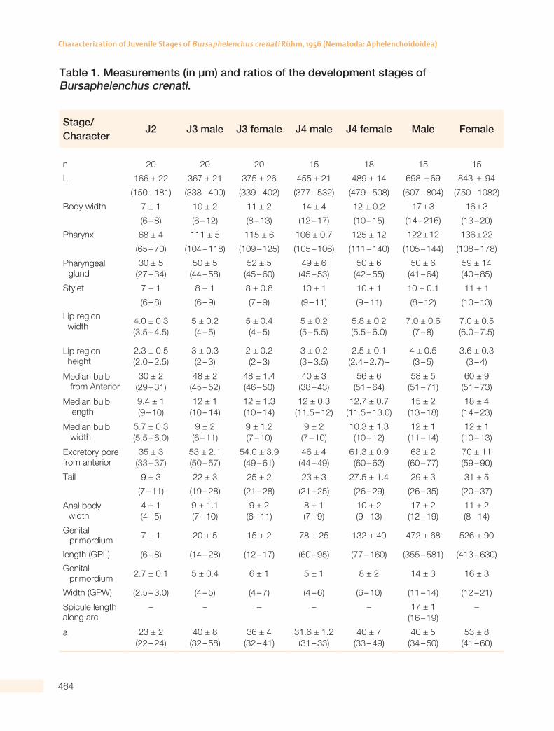
Molt from J2 to J3 (Fig. 6A,B; Table 2)
In the center of primordium, 3 to 4 mitoses of somatic nuclei are distinct between germinal cells (Fig. 6A).
Molt from J3 to J4, male juvenile (Figs. 4, 6C – H; Table 2)
The genital primordium is initially straight, then transforming to loop – shaped (Fig. 6F,G), with the anterior apex and both somatic and germinal parts reversed posteriorly; the somatic part is set off by constriction from germinal part and its tip is curved hook – like (Fig. 6G). The cloaca primordium is en-larged with a transparent inner cavity anterior to the rectum (Fig. 6H). This molting phase is an in-dication of the orientation change of the somatic part, which in male juvenile J3 is located anteriorly
Figure 6: Bursaphelenchus crenati. Molting juveniles (shedding cuticle in tail). A,B. Molting male juvenile from J2 to J3; A. Genital primordium (gp); B. Tail and cloacal primordium (cp); C – H. Molting male juvenile from J3 to J4; C. Genital primordium of molting J3 – J4 (aac – anterior apical cell, sz – somatic zone, gerz – germinal zone); D, E, H. Tail and cloacal primordium (cp) of J3 – J4 male juvenile; F. Genital primordium of molting male J3 – J4 juvenile at “loop stage”; G. Drawing of F with explanation of the reversion of the somatic zone (sz) with anterior apical cell (aac) flexed to the germinal zone (gerz); gc – germinal cells; pac – posterior apical cell, sn – somatic nuclei, vc – ventral cord nuclei; I,J – molting female J3 – J4 juvenile; I. Genital primordium with anterior germinal zone (gerz); J. Tail. Scale 50 µm.
and in J4 is shifted posteriorly, thus pulling a germi-nal part to the anterior direction (Fig. 6F,G).
Molt from J3 to J4, female juvenile (Figs. 5,6I,J; Table 2)
The somatic part of the genital primordium in its pos-terior part possesses more than 20 somatic nuclei, the germinal part in the anterior part of the primordi-um with 5 to 10 large germinal cells.
Molt from J4 to male (Fig. 4; Table 2)
The somatic part of the genital primordium reaches in some specimens the massive cloaca primordium, in which inner cavity transparent spicules outlines are distinct. The bursal flap is not formed, the tail is nar-rowly conical with 9 µm long hyaline zone.
Table 2. Measurements (in µm) and ratios of the development stages of Bursaphelenchus crenati.
Stage/Character
J2®J3 male
J2®J3 female
J3®J4 male
J3®J4 female
J4®male J4®femaleDauer J3D
n 16 5 10 20 5 17 20
L 347 ± 7 323 ± 86 370 ± 3 377 ± 10 596 ± 91 544 ± 69 413 ± 20
(338 – 354) (181 – 410) (366 – 374) (364 – 398) (532 – 660) (433 – 614) (392 – 463)
Body width 9 ± 1 10 ± 2 11 ± 2 12 ± 1 16 ± 0.3 13 ± 3 8 ± 1
(8 – 10) (8 – 14) (9 – 13) (11 – 13) (14 – 17) (9 – 17) (6 – 9)
Pharynx 107 ± 4 103 ± 21 110 ± 5 110 ± 5 112 ± 9 117 ± 6 119 ± 9
(104 – 112) (65 – 116) (105 – 115) (102 – 117) (105 – 118) (109 – 123) (101 – 128)
P haryngeal gland
49 ± 5(44 – 56)
51 ± 1(49 – 53)
49 ± 4(44 – 55)
48 ± 4(41 – 54)
46 ± 1(45 – 47)
48 ± 6(40 – 55)
43 ± 7(35 – 55)
Stylet 7 ± 1.2 8 ± 1 7 ± 1 9 ± 0.7 9 ± 0.4 9 ± 1.4 9.4 ± 0.4
(6 – 9) (6 – 9) (6 – 8) (8 – 10) (9 – 10) (8 – 11) (9 – 10)
L ip region width
5.0 ± 0.3(4 – 5)
5 ± 0.6(4 – 6)
5.6 ± 0.5(4.8 – 6.0)
5 ± 0.6(4 – 6)
5 ± 0.4(5 – 6)
5.4 ± 0.4(5 – 6)
4.8 ± 1.0(4 – 7)
L ip region height
3 ± 0.3(2 – 3)
2.4 ± 0.2(2 – 3)
2.4 ± 0.1(2.3 – 2.5)
3 ± 0.3(2 – 3)
3 ± 1(2 – 3) –
2.9 ± 0.4(2 – 3)
2.9 ± 0.3(2.5 – 4)
M edian bulb from anterior
46 ± 1(45 – 47)
45 ± 8(31 – 50)
47 ± 0.1(46 – 48)
47 ± 3(40 – 50)
46 ± 12(38 – 54)
53 ± 3(51 – 58)
49 ± 2(45 – 51)
M edian bulb length
11 ± 1.2(10 – 13)
11 ± 0.9(10 – 12)
12 ± 1(11 – 13)
12 ± 1(10 – 13)
13 ± 1(12 – 14)
13 ± 0.2(12.5 – 13.5)
11 ± 2(9 – 15)
M edian bulb width
9 ± 1.1(8 – 10)
8 ± 1.5(6 – 10)
10 ± 1(9 – 11)
10 ± 1(7 – 10)
9.9 ± 0.2(9.5 – 10.0)
10 ± 1(9 – 11)
8 ± 2(6 – 10)
E xcretory pore from anterior
51 ± 1(50 – 52)
50 ± 8(37 – 56)
53 ± 2(51 – 54)
54 ± 4(45 – 59)
52 ± 12(44 – 61)
58 ± 3(54 – 62)
40 ± 3(37 – 44)
Tail 22 ± 4 21 ± 6 22 ± 1 26 ± 1.5 28 ± 5 28 ± 3 37 ± 3
(20 – 28) (11 – 27) (21 – 24) (24 – 28) (25 – 32) (23 – 31) (30 – 41)
A nal body width
8 ± 0.4(7 – 8)
8 ± 3(5 – 12)
11 ± 2(8 – 13)
11 ± 1.1(10 – 13)
11 ± 3(9 – 13)
10.1 ± 0.9(9 – 11)
8 ± 1(5 – 9)
G enital primordium
18 ± 4 10 ± 3 20 ± 2 18 ± 5 158 ± 138 197 ± 105 14.8 ± 2.1
Length (GPL) (14 – 23) (6 – 14) (17 – 22) (13 – 28) (60 – 255) (122 – 369) (11 – 18)
G enital primordium Width (GPW)
5 ± 1(4 – 5)
5 ± 1(3 – 7)
4.8 ± 05(4.0 – 5.3)
5 ± 1(3 – 6)
10 ± 5(6 – 13)
9 ± 3(7 – 14)
3.9 ± 0.4(3 – 5)

467
JOURNAL OF NEMATOLOGY
Table 2. Measurements (in µm) and ratios of the development stages of Bursaphelenchus crenati.
Stage/Character
J2®J3 male
J2®J3 female
J3®J4 male
J3®J4 female
J4®male J4®femaleDauer J3D
n 16 5 10 20 5 17 20
L 347 ± 7 323 ± 86 370 ± 3 377 ± 10 596 ± 91 544 ± 69 413 ± 20
(338 – 354) (181 – 410) (366 – 374) (364 – 398) (532 – 660) (433 – 614) (392 – 463)
Body width 9 ± 1 10 ± 2 11 ± 2 12 ± 1 16 ± 0.3 13 ± 3 8 ± 1
(8 – 10) (8 – 14) (9 – 13) (11 – 13) (14 – 17) (9 – 17) (6 – 9)
Pharynx 107 ± 4 103 ± 21 110 ± 5 110 ± 5 112 ± 9 117 ± 6 119 ± 9
(104 – 112) (65 – 116) (105 – 115) (102 – 117) (105 – 118) (109 – 123) (101 – 128)
P haryngeal gland
49 ± 5(44 – 56)
51 ± 1(49 – 53)
49 ± 4(44 – 55)
48 ± 4(41 – 54)
46 ± 1(45 – 47)
48 ± 6(40 – 55)
43 ± 7(35 – 55)
Stylet 7 ± 1.2 8 ± 1 7 ± 1 9 ± 0.7 9 ± 0.4 9 ± 1.4 9.4 ± 0.4
(6 – 9) (6 – 9) (6 – 8) (8 – 10) (9 – 10) (8 – 11) (9 – 10)
L ip region width
5.0 ± 0.3(4 – 5)
5 ± 0.6(4 – 6)
5.6 ± 0.5(4.8 – 6.0)
5 ± 0.6(4 – 6)
5 ± 0.4(5 – 6)
5.4 ± 0.4(5 – 6)
4.8 ± 1.0(4 – 7)
L ip region height
3 ± 0.3(2 – 3)
2.4 ± 0.2(2 – 3)
2.4 ± 0.1(2.3 – 2.5)
3 ± 0.3(2 – 3)
3 ± 1(2 – 3) –
2.9 ± 0.4(2 – 3)
2.9 ± 0.3(2.5 – 4)
M edian bulb from anterior
46 ± 1(45 – 47)
45 ± 8(31 – 50)
47 ± 0.1(46 – 48)
47 ± 3(40 – 50)
46 ± 12(38 – 54)
53 ± 3(51 – 58)
49 ± 2(45 – 51)
M edian bulb length
11 ± 1.2(10 – 13)
11 ± 0.9(10 – 12)
12 ± 1(11 – 13)
12 ± 1(10 – 13)
13 ± 1(12 – 14)
13 ± 0.2(12.5 – 13.5)
11 ± 2(9 – 15)
M edian bulb width
9 ± 1.1(8 – 10)
8 ± 1.5(6 – 10)
10 ± 1(9 – 11)
10 ± 1(7 – 10)
9.9 ± 0.2(9.5 – 10.0)
10 ± 1(9 – 11)
8 ± 2(6 – 10)
E xcretory pore from anterior
51 ± 1(50 – 52)
50 ± 8(37 – 56)
53 ± 2(51 – 54)
54 ± 4(45 – 59)
52 ± 12(44 – 61)
58 ± 3(54 – 62)
40 ± 3(37 – 44)
Tail 22 ± 4 21 ± 6 22 ± 1 26 ± 1.5 28 ± 5 28 ± 3 37 ± 3
(20 – 28) (11 – 27) (21 – 24) (24 – 28) (25 – 32) (23 – 31) (30 – 41)
A nal body width
8 ± 0.4(7 – 8)
8 ± 3(5 – 12)
11 ± 2(8 – 13)
11 ± 1.1(10 – 13)
11 ± 3(9 – 13)
10.1 ± 0.9(9 – 11)
8 ± 1(5 – 9)
G enital primordium
18 ± 4 10 ± 3 20 ± 2 18 ± 5 158 ± 138 197 ± 105 14.8 ± 2.1
Length (GPL) (14 – 23) (6 – 14) (17 – 22) (13 – 28) (60 – 255) (122 – 369) (11 – 18)
G enital primordium Width (GPW)
5 ± 1(4 – 5)
5 ± 1(3 – 7)
4.8 ± 05(4.0 – 5.3)
5 ± 1(3 – 6)
10 ± 5(6 – 13)
9 ± 3(7 – 14)
3.9 ± 0.4(3 – 5)

468
Characterization of Juvenile Stages of Bursaphelenchus crenati Rhm, 1956 (Nematoda: Aphelenchoidoidea)
Molt from J4 to female (Fig. 5; Table 2)
Vulva primordium is a massive cellular agglomeration with an inner transversal slit, but without opening out-side. All sections of the genital system are distinct: the ovary of 30 to 40 germinal cells, the oviduct, empty spermatheca, crustaformeria, and the uterus with in-ner empty cavity (Fig. 5).Tabular key to juvenile stages of Bursaphelenchus crenati (Key is given in Table 3).
Morphometric ratios of juvenile development
Individual growth is illustrated in diagrams (Fig. 7) with two main parameters: the body length and the
ratio between the genital primordium length and the body length. The elongation of the body is faster af-ter the third stage (Fig. 7A). At the same period, the maximum elongation and differentiation of the geni-tal primordium takes place in both male and female development lines (Fig. 7B). Juvenile body increas-es between molts and within developmental stages, which is evident in the size ranges of every stage (Tables 1,2).
Dauer juveniles (Figs. 4,5,8,9; Table 2)
The dauer juveniles have the same genital primordi-um structure and size as J3. The differences are: the slender and straight body and straight long tail vs curved ventrally tail in the J3 of the propagative gen-
Spicule length along arc
– – – – – – –
a 40 ± 5 34 ± 8 34.8 ± 5.2 32 ± 2 39 ± 12 41.5 ± 4.6 54 ± 8.3
(34 – 46) (22 – 41) (28.4 – 41.8) (29 – 35) (31 – 48) (36.8 – 48.9) (44.6 – 69.6)
b 6.0 ± 0.3 5.6 ± 0.6 6.1 ± 0.2 6.1 – 0.2 9.1 ± 0.3 7.8 ± 0.6 5.5 ± 0.4
(5.7 – 6.4) (4.8 – 6.4) (5.9 – 6.3) (5.8 – 6.5) (8.9 – 9.2) (6.7 – 8.3) (5.1 – 6.4)
b’ 3.2 ± 0.1 3.1 ± 0.3 3.4 ± 0.2 3.4 ± 0.2 5.3 ± 0.4 4.6 ± 0.5 3.5 ± 0.3
(3.1 – 3.3) (2.8 – 3.6) (3.2 – 3.6) (3.1 – 3.7) (5.1 – 5.6) (4.0 – 5.2) (3.0 – 4.1)
c 16.0 ± 2.5 15.6 ± 0.6 16.6 ± 0.9 14.8 ± 0.8 21.1 ± 0.9 19.7 ± 1.7 11.2 ± 0.9
(12.4 – 17.9) (14.7 – 16.2) (15.1 – 17.3) (13.8 – 1.58) (20.5 – 21.7) (18.0 – 22.3) (9.9 – 13.2)
c’ 2.8 ± 0.4 2.8 ± 0.5 2.1 ± 0.3 2.3 ± 0.2 2.6 ± 0.1 2.8 ± 0.3 4.7 ± 0.5
(2.4 – 3.4) (2.2 – 3.4) (1.7 – 2.5) (2.0 – 2.8) (2.6 – 2.7) (2.3 – 3.2) (4.3 – 5.5)
V % or (V): genital primordium center from anterior, to body length, in %
61 ± 2(59 – 64)
64 ± 3(59 – 68)
62 ± 1(60 – 64)
66 ± 2(63 – 68)
52 ± 3(49 – 54)
73.2 ± 1.6(70.5 – 74.3)
62 ± 5(56 – 72)
GPL/GPW 4.0 ± 1.1 2.3 ± 0.6 4.2 ± 0.2 3.9 ± 1 15 ± 6 20 ± 4 3.8 ± 0.5
(3.0 – 5.4) (1.5 – 2.9) (3.9 – 4.5) (2.7 – 6.1) (10 – 19) (16 – 27) (3 – 4.5)
GPL/L, % 5.2 ± 1.2 3.3 ± 1.0 5.5 ± 0.6 5 ± 1 25 ± 19 35.3 ± 15.2 4 ± 1
(4.2 – 6.6) (1.8 – 4.3) (4.5 – 5.9) (4 – 7) (11 – 39) (24.3 – 60.0) (3 – 5)
L ip region length to its width
1.8 ± 0.3(1.6 – 2.3)
2.0 ± 0.3(1.6 – 2.4)
2.3 ± 0.2(2.0 – 2.6)
1.9 ± 0.2(1.6 – 2.4)
2.1 ± 0.6(1.7 – 2.5)
1.9 ± 0.1(1.8 – 2.0)
1.6 ± 0.3(1.4 – 2.4)
M edian bulb length to its width
1.4 ± 0.2(1.1 – 1.6)
1.4 ± 0.2(1.3 – 1.7)
1.2 ± 0.1(1.2 – 1.3)
1.2 ± 0.1(1.0 – 1.4)
1.3 ± 0.2(1.2 – 1.4)
1.3 ± 0.1(1.2 ± 1.4)
1.4 ± 0.2(1.1 – 1.7)
All values are given as mean±SD (minimum-maximum).
469
JOURNAL OF NEMATOLOGY
eration. Lateral field with two closely located bands of equal width (three incisures). Dauers differ in hem-ispherical continuous head with hyaline inner cap vs set off head and well developed cephalic circu-lar framework. Stylet is very thin capillary tube, co-nus and basal thickenings not distinct. Median bulb elongated, its length twice as its width, central valve is weakly developed. Pharyngo – intestinal junction is one half of the median bulb length posterior to
median bulb, surrounded by nerve ring with ex-cretory pore at the posterior third of median bulb. Pharyngeal glands form very narrow band, gland nuclei indistinct.
In 3 hr after pouring beetle elytra with “nematan-gia” in water or 0.9% NaCl solution, the J3D juve-niles started to molt to J4 juvenile with long genital primordium and well developed stylet and median bulb (Fig. 9).
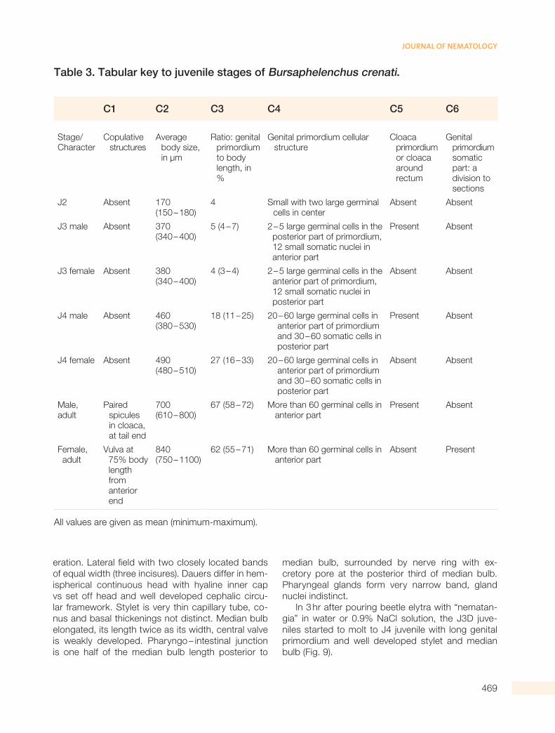
Table 3. Tabular key to juvenile stages of Bursaphelenchus crenati.
C1 C2 C3 C4 C5 C6
Stage/Character
C opulative structures
A verage body size, in µm
R atio: genital primordium to body length, in %
G enital primordium cellular structure
C loaca primordium or cloaca around rectum
G enital primordium somatic part: a division to sections
J2 Absent 170 (150 – 180)
4 S mall with two large germinal cells in center
Absent Absent
J3 male Absent 370 (340 – 400)
5 (4 – 7) 2 – 5 large germinal cells in the posterior part of primordium, 12 small somatic nuclei in anterior part
Present Absent
J3 female Absent 380 (340 – 400)
4 (3 – 4) 2 – 5 large germinal cells in the anterior part of primordium, 12 small somatic nuclei in posterior part
Absent Absent
J4 male Absent 460 (380 – 530)
18 (11 – 25) 20 – 60 large germinal cells in anterior part of primordium and 30 – 60 somatic cells in posterior part
Present Absent
J4 female Absent 490 (480 – 510)
27 (16 – 33) 20 – 60 large germinal cells in anterior part of primordium and 30 – 60 somatic cells in posterior part
Absent Absent
Male, adult
P aired spicules in cloaca, at tail end
700 (610 – 800)
67 (58 – 72) M ore than 60 germinal cells in anterior part
Present Absent
F emale, adult
V ulva at 75% body length from anterior end
840 (750 – 1100)
62 (55 – 71) M ore than 60 germinal cells in anterior part
Absent Present
All values are given as mean (minimum-maximum).

470
Characterization of Juvenile Stages of Bursaphelenchus crenati Rhm, 1956 (Nematoda: Aphelenchoidoidea)
Discussion
The embryonic development with hatching of J2 af-ter the first molt within egg shell is the ovoviviparity. The sex of juvenile is distinctly observed from the third stage. It is an indication of the possible phys-iological sex determination at the second stage of juvenile (Ryss and Polyanina, 2017). The fast-est growth and genital primordium differentiation takes place after the J3 to J4 molt. Earlier study of the development of Bursaphelenchus mucronatus kolymensis (Ryss and Chernetskaya, 2009) and B.
ulmophilus (Ryss and Polyanina, 2017) gave similar results: the first molt occurring in the egg shell and four stages outside of egg are divided by molts: J2 to J4 and adult worms. The only difference is the dauer juvenile stage. In B. mucronatus it is the fourth stage juvenile (Ryss, 2008), but in B. ulmophilus and B. crenati dauers are J3D stage. Leaving the beetle surface, the B. crenati dauers start to molt to the fourth stage juvenile with elongated and differenti-ated genital primordium, just in 0.9% NaCl solution or in water. The same molting just after reviving in water was detected for dauers J3D of B. ulmophilus
Figure 8: Dauer juveniles. Methylene blue staining. A. Male juvenile J3D; B. Female juvenile J3D; C. Genital primordium of male dauer juvenile; D. Tail of male dauer juvenile. Scale 50 µm for A,B and 20 µm for C,D.
Figure 7: Diagrams of juvenile growth. A. Body length; B. Ratio of genital primordium length to body length (GP/L, %). Standard deviation (STD) levels are marked for mean values.

471
JOURNAL OF NEMATOLOGY
Figure 9: Dauer juveniles molted to J4D after 3 hr in 0.9% NaCl solution. A. Male juvenile; B. Female juvenile.
(Ryss et al., 2015). Difference between the Bursaph-elenchus clades in the dauer juvenile stages (J3D or J4D) is the important character for the genus Bursa-phelenchus phylogeny and intrageneric taxonomic diagnostics (Ryss and Subbotin, 2017). Dauers play the important role in the deciduous woody plant wilt spread, as the infective juveniles vectored by insects (Polyanina et al., 2016). The stage of dauers in nem-atode life cycles and their adaptations to insect vec-tors is of special interest.
Acknowledgments
The authors are grateful to the State Academ-ic Programs FSR: AAAA – A17 – 117030310322 – 3, AAAA – A17 – 117080110040 – 3 and the grant RFBR 17 – 04 – 00360a. The authors are grateful to the for-est entomologist Dr. Alexander V. Petrov (ILAN RAS)
for wood and bark samples with tunnels of the bark beetle Hylesinus crenatus Fabricius.
ReferencesGu, J., Tomalak, M., He, J., and Fang, Y. 2017. Bur-
saphelenchus crenati Rühm, 1956 (Tylenchina: Aphel-enchoididae), a nematode associated with the Greater ash bark beetle, Hylesinus crenatus Fabricius, in dy-ing ash, Fraxinus excelsior L., in Europe. Nematology 19:413–26.
Hirschmann, H. 1962. Life cycle of the Ditylen-chus triformis (Nematoda: Tylenchida) with emphasis on post – embryonic development. Proceedings of the Helminthological Society Washington 29(1):30–43.
Hirschmann, H. 1971. Comparative morphology and anatomy, in Zuckerman, B. M., Mai, W. F., and Ro-hde, R. A. (Eds), Plant parasitic nematodes Vol. 1, Aca-demic Press, New York, NY: 9–63.
Hirschmann, H., and Triantaphyllou, A. C. 1968. Mode of reproduction and development of the repro-ductive system of Helicotylenchus dihystera. Nemato-logica 13(4):558–74.
Polyanina, K. S., Popovichev, B. G., and Ryss, A. Y. 2016. Elm wood nematodes as a threat to the ur-ban woody plantations in the Russian North – West: the laboratory tests of woody plant host range and pathogenicity, in Musolin, D. L., and Selikhovkin, A. V. (Eds), The Kataev memorial readings – IX. dendro-biotic invertebrates and fungi and their role in forest ecosystems. Proceedings of the International Confer-ence, November 23–25, Saint Petersburg State For-est Technical University, Saint Petersburg: 90–3, (in Russian).
Ryss, A., Polyanina, K. S., Popovichev, B. G., and Subbotin, S. A. 2015. Description of Bursaphelen-chus ulmophilus sp.n. (Nematoda: Parasitaphelenchi-nae) associated with Dutch elm disease of Ulmus gla-bra Huds. in the Russian North West. Nematology 17 (6):685–703.
Ryss, A. Y., and Polyanina, K. S. 2017. Diagnostics of the stages of post – embryonic development in Bur-saphelenchus ulmophilus (Nematoda: Aphelenchoidi-dae). Parazitologiya 51(6):466–80.
Ryss, A. Y., and Subbotin, S. A. 2017. Coevolution of wood – inhabiting nematodes of the genus Bursap-helenchus Fuchs, 1937 with their insect vectors and plant hosts. Zhurnal Obshei Biologii 78(3):13–42, (in Russian).
Ryss, A. Y. 1981. Morphogenesis of the female genital system in superfamily Tylenchoidea (Nemato-da). Parazitologiya 15:533–42, (in Russian).
Ryss, A. Y. 1988. Root parasitic nematodes of the family Pratylenchidae (Tylenchida) of the world fauna, Nauka Publishers, Leningrad, 368, (in Russian).
472
Characterization of Juvenile Stages of Bursaphelenchus crenati Rhm, 1956 (Nematoda: Aphelenchoidoidea)
Ryss, A. Y. 2008. Entomophilic juveniles of the nematode Bursaphelenchus mucronatus (Nematoda, Aphelenchida, Parasitaphelenchidae) obtained from the cerambycid vector Monochamus urussovi (Coleop-tera, Cerambycidae) under experimental conditions. Entomological Review 88(1):108–14.
Ryss, A. Y. 2015. The most simple techniques for detection and laboratory cultivation of woody plant wilt nematodes. Izvestia Sankt – Peterburgskoj Lesotehnic-eskoj Akademii 211:287–95, (in Russian).
Ryss, A. Y. 2017a. A simple express technique to process nematodes for collection slide mounts. Jour-nal of Nematology 49:27–32.
Ryss, A. Y. 2017b. The simplest ‘field’ methods for extraction of nematodes from plants, wood, insects and soil, with additional description how to keep ex-tracted nematodes alive for a long time. Parazitologiya 15(1):57–67.
Ryss, A. Y., and Chernetskaya, A. Y. 2009. Life – cy-cle of Bursaphelenchus mucronatus Mamiya et Enda, 1979 (Nematoda: Aphelenchida)]. Parazitologiya. 43 3:206–24, (in Russian).
Ryss, A. Y., and Chernetskaya, A. Y. 2010. Life – cy-cle of Paraphelenchus myceliophtorus Goodey, 1958 (Nematoda: Aphelenchida). Parazitologiya 44:105–27, (in Russian).


![The larval and juvenile stages of the Plecostei. Suisan Gakki Ho 3(2). [Translation 98]](https://img.pdfslide.us/doc/110x75/568bdbdf1a28ab2034b020aa/the-larval-and-juvenile-stages-of-the-plecostei-suisan-gakki-ho-32-translation.jpg)