Embed Size (px)
Citation preview
Characterization of the MSMEG_2631 Gene (mmp) Encoding aMultidrug and Toxic Compound Extrusion (MATE) Family Protein inMycobacterium smegmatis and Exploration of Its Polyspecific NatureUsing Biolog Phenotype MicroArray
Mukti Nath Mishra, Lacy Daniels
Department of Pharmaceutical Sciences, Texas A&M Health Science Center Irma Lerma Rangel College of Pharmacy, Kingsville, Texas, USA
In Mycobacterium, multidrug efflux pumps can be associated with intrinsic drug resistance. Comparison of putative mycobacte-rial transport genes revealed a single annotated open reading frame (ORF) for a multidrug and toxic compound extrusion(MATE) family efflux pump in all sequenced mycobacteria except Mycobacterium leprae. Since MATE efflux pumps function asmultidrug efflux pumps by conferring resistance to structurally diverse antibiotics and DNA-damaging chemicals, we studiedthis gene (MSMEG_2631) in M. smegmatis mc2155 and determined that it encodes a MATE efflux system that contributes to in-trinsic resistance of Mycobacterium. We propose that the MSMEG_2631 gene be named mmp, for mycobacterial MATE protein.Biolog Phenotype MicroArray data indicated that mmp deletion increased susceptibility for phleomycin, bleomycin, capreomy-cin, amikacin, kanamycin, cetylpyridinium chloride, and several sulfa drugs. MSMEG_2619 (efpA) and MSMEG_3563 mask theeffect of mmp deletion due to overlapping efflux capabilities. We present evidence that mmp is a part of an MSMEG_2626-2628-2629-2630-2631 operon regulated by a strong constitutive promoter, initiated from a single transcription start site. All together,our results show that M. smegmatis constitutively encodes an Na�-dependent MATE multidrug efflux pump from mmp in anoperon with putative genes encoding proteins for apparently unrelated functions.
Tuberculosis (TB) is still one of the biggest killers; it is estimatedthat Mycobacterium tuberculosis causes 8.8 million new cases
of TB and kills 1.4 million people per year (1). Factors contribut-ing to therapeutic complications are latency in an asymptomatichost and later reactivation to cause active disease (2, 3), intrinsicantibiotic resistance (4), and acquired drug resistance (5). Multi-drug-resistant (MDR) strains further complicate therapy. Al-though changes responsible for acquired resistance are often un-derstood (6–10), the basis of MDR is not yet fully defined (11, 12).
Bacteria have several antibiotic resistance strategies: (i) pro-duce enzymes to inactivate antibiotics, (ii) alter targets to reduceantibiotic affinity, and (iii) reduce drug accumulation by de-creased permeability or increased efflux (4). These strategies maybe an intrinsic feature of an organism or acquired via mutations oracquisition of exogenous genes (13, 14). Low mycobacterial cellwall permeability due to its lipid-rich composition, limiting drugentry, is one reason for intrinsic resistance (15, 16), but Mycobac-terium also has several multidrug efflux pumps (17) that enhancedrug resistance by drug extrusion; however, their roles in intrinsicresistance are not well explored.
Multidrug efflux pumps extrude a wide range of structurallyunrelated compounds and belong to five families: (i) ATP bindingcassette (ABC), (ii) major facilitator superfamily (MFS), (iii) smallmultidrug resistance (SMR), (iv) resistance-nodulation-division(RND), and (v) multidrug and toxic compound extrusion(MATE). ABC family members are ATP-dependent primarytransporters, but the other families are secondary transportersthat use an electrochemical transmembrane gradient. MATE pro-teins (18) have a 12-membrane helix topology and use either H�
or Na� gradients to drive efflux (19). In plants, MATE transport-ers secrete secondary metabolites to defend against microbialpathogens (20, 21), and in the liver and kidney they extrude xeno-
biotic cations (22, 23). Bacterial MATE transporters function asxenobiotic efflux pumps and can confer resistance to structurallydiverse drugs, including antibiotics and DNA-damaging agents(24, 25).
MATE systems from only a few eukaryotes (26) and pro-karyotes (19) have been functionally characterized. The role formost reported prokaryotic MATE transporters is not well defined,since most have been studied by heterologous expression (19). Toinvestigate the role of a MATE transporter in Mycobacterium in-trinsic resistance, we made an M. smegmatis mutant lacking thisprotein and characterized it using Phenotype MicroArray (PM).The present study reveals the polyspecific and Na�-dependentnature of the M. smegmatis MATE transporter and transcriptionalcoupling of its gene with 4 upstream genes. By determining thetranscription start site, we have also identified its promoter andexamined its activity.
MATERIALS AND METHODSBacterial strains, growth conditions, plasmids, and chemicals. Table 1describes the strains and plasmids used. Mycobacteria were cultivated at37°C in Difco 7H9 broth supplemented with 0.2% (vol/vol) glycerol and0.05% (wt/vol) Tween 80 or on Difco 7H10 agar plates supplemented with0.2% (vol/vol) glycerol. Escherichia coli was grown at 37°C (or 30°C whenusing strain BW25113/pKD46) in Luria-Bertani broth. Plasmids were
Received 12 September 2012 Accepted 2 January 2013
Published ahead of print 4 January 2013
Address correspondence to Lacy Daniels, [email protected].
Copyright © 2013, American Society for Microbiology. All Rights Reserved.
doi:10.1128/JB.01724-12
1610 jb.asm.org Journal of Bacteriology p. 1610–1621 April 2013 Volume 195 Number 7
on October 29, 2018 by guest
http://jb.asm.org/
Dow
nloaded from
maintained in E. coli DH5� with 100 �g ampicillin (Amp), 75 �g kana-mycin (Km), or 25 �g gentamicin (Gen) per ml.
Bioinformatic analysis. Mycobacterial transporter families werecompared using TransportDB (http://www.membranetransport.org/)(32). Protein BLAST analysis was performed via the National Center forBiotechnology Information (NCBI) (http://www.ncbi.nlm.nih.gov/BLAST). MATE homologs were retrieved from GenBank (http://www.ncbi.nlm.nih.gov/GenBank/). ClustalW2 (http://www.ebi.ac.uk/Tools/msa/clustalw2/) and MEGA (33) were used for multiple sequence align-ment and phylogenetic trees. Chromosomal regions were retrieved fromNCBI (http://www.ncbi.nlm.nih.gov/genome/) and J. Craig Venter Insti-tute-Comprehensive Microbial Resource (JCVI-CMR; http://cmr.jcvi.org/tigr-scripts/CMR). Sequence Manipulation Suite (http://www.ualberta.ca/) (34) and Vector NTI Advance (Invitrogen) were used formolecular weight estimation, genomic map construction, and primer de-sign.
Mutant construction. M. smegmatis mutants were constructed usinghomologous recombination as follows. The mmp (MSMEG_2631) locuswas PCR amplified using 2629:F:XbaI/2633:R:BglII primers to produce a4,030-bp fragment, which was cloned in pJQ200 (28) to generate pLD183,which was transferred to E. coli25113/pKD46 (to use the �-Red recombi-nase system [27]) and grown at 30°C in the presence of 1 mM arabinose tomake electrocompetent cells. The Km resistance gene was PCR amplifiedfrom pKD4 (27) using primers 2631:H1/2631:H2, designed to produce a
40-bp homologous region for mmp at both ends. PCR product was puri-fied and electroporated into 25113/pKD46 having pLD183. Transfor-mants were selected with Km (75 �g/ml) at 37°C on LB plates. Plasmidswere isolated and examined for Kmr cassette insertion in mmp and desig-nated pLD187. pLD187 was mobilized in M. smegmatis mc2155, andtransformants were selected with Km (50 �g mg�1) and 2% sucrose on LBplates. Disruption was confirmed by PCR, and confirmed mutants weredesignated LD1158. Unmarked mmp deletion mutants were developedusing the FLP/FLP recognition target (FLP/FRT) system. A variant ofpMN234 (29) that has a Genr cassette (S. Jaques and L. Daniels, unpub-lished data) in the place of the Kmr cassette in pMN234 was mobilized inLD1158, and transformants were selected with gentamicin (20 �g ml�1)on LB plates. Transformants were kept with gentamicin for 2 to 3 revivalsand transferred to plain LB plates, and Kmr cassette deletion was con-firmed by Km sensitivity and PCR assays. Mutants with confirmed dele-tion of MSMEG_2631 (�MSMEG2631) were designated LD1184. Thesame procedure was used to develop all unmarked deletion mutants.
Construction of complementation plasmids. M. smegmatis (mmp)and Mycobacterium bovis BCG (BCG_2856c) were PCR amplified using2631:F:NdeI/2631:R:MluI and dinF:F:NdeI/dinF:R:MluI primers, respec-tively. Products digested with NdeI/MluI were ligated with digestedpTB21 (intermediate mycobacterial promoter) or pUV15 (strong myco-bacterial promoter) to make two expression plasmids for each open read-ing frame (ORF). Recombinant plasmids were purified, confirmed, and
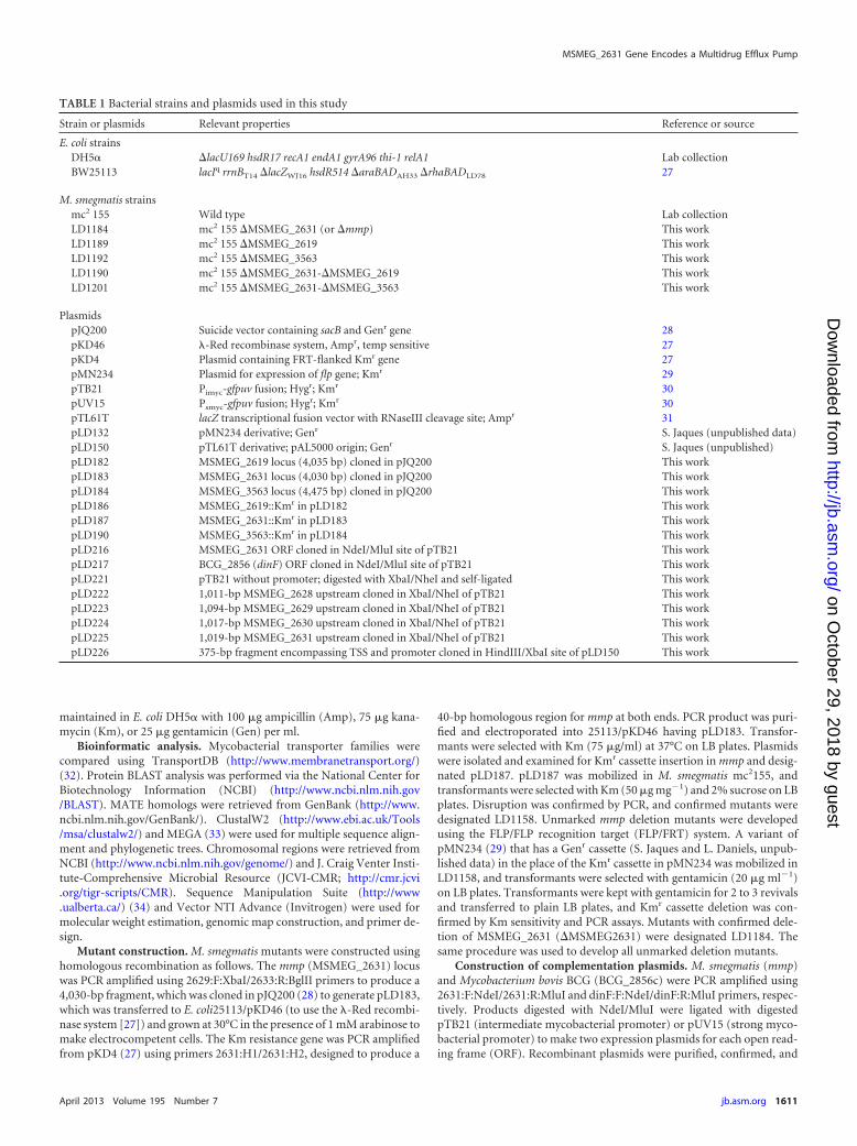
TABLE 1 Bacterial strains and plasmids used in this study
Strain or plasmids Relevant properties Reference or source
E. coli strainsDH5� �lacU169 hsdR17 recA1 endA1 gyrA96 thi-1 relA1 Lab collectionBW25113 lacIq rrnBT14 �lacZWJ16 hsdR514 �araBADAH33 �rhaBADLD78 27
M. smegmatis strainsmc2 155 Wild type Lab collectionLD1184 mc2 155 �MSMEG_2631 (or �mmp) This workLD1189 mc2 155 �MSMEG_2619 This workLD1192 mc2 155 �MSMEG_3563 This workLD1190 mc2 155 �MSMEG_2631-�MSMEG_2619 This workLD1201 mc2 155 �MSMEG_2631-�MSMEG_3563 This work
PlasmidspJQ200 Suicide vector containing sacB and Genr gene 28pKD46 �-Red recombinase system, Ampr, temp sensitive 27pKD4 Plasmid containing FRT-flanked Kmr gene 27pMN234 Plasmid for expression of flp gene; Kmr 29pTB21 Pimyc-gfpuv fusion; Hygr; Kmr 30pUV15 Psmyc-gfpuv fusion; Hygr; Kmr 30pTL61T lacZ transcriptional fusion vector with RNaseIII cleavage site; Ampr 31pLD132 pMN234 derivative; Genr S. Jaques (unpublished data)pLD150 pTL61T derivative; pAL5000 origin; Genr S. Jaques (unpublished)pLD182 MSMEG_2619 locus (4,035 bp) cloned in pJQ200 This workpLD183 MSMEG_2631 locus (4,030 bp) cloned in pJQ200 This workpLD184 MSMEG_3563 locus (4,475 bp) cloned in pJQ200 This workpLD186 MSMEG_2619::Kmr in pLD182 This workpLD187 MSMEG_2631::Kmr in pLD183 This workpLD190 MSMEG_3563::Kmr in pLD184 This workpLD216 MSMEG_2631 ORF cloned in NdeI/MluI site of pTB21 This workpLD217 BCG_2856 (dinF) ORF cloned in NdeI/MluI site of pTB21 This workpLD221 pTB21 without promoter; digested with XbaI/NheI and self-ligated This workpLD222 1,011-bp MSMEG_2628 upstream cloned in XbaI/NheI of pTB21 This workpLD223 1,094-bp MSMEG_2629 upstream cloned in XbaI/NheI of pTB21 This workpLD224 1,017-bp MSMEG_2630 upstream cloned in XbaI/NheI of pTB21 This workpLD225 1,019-bp MSMEG_2631 upstream cloned in XbaI/NheI of pTB21 This workpLD226 375-bp fragment encompassing TSS and promoter cloned in HindIII/XbaI site of pLD150 This work
MSMEG_2631 Gene Encodes a Multidrug Efflux Pump
April 2013 Volume 195 Number 7 jb.asm.org 1611
on October 29, 2018 by guest
http://jb.asm.org/
Dow
nloaded from
electroporated into M. smegmatis. Transformants were selected with 100�g ml�1 of hygromycin on LB plates.
PM tests. Phenotype MicroArray (PM; Biolog, Hayward CA) antimi-crobial susceptibility tests for M. smegmatis were performed with modifi-cations of earlier methods (35; S. Jaques III and L. Daniels, presented atthe SCASM Conference, Austin, TX, 2008; M. Rahman, S. Jaques III, andL. Daniels, presented at the SC-ASM Conference, Austin, TX, 2008).Briefly, cells were streaked onto 7H10 agar plates supplemented with 0.2%(vol/vol) glycerol and grown 3 to 4 days at 37°C. Growth was removedfrom 7H10 plates using a cotton swab dipped in 2% Tween 80, and cellswere suspended in GN/GP-IF-0a (Biolog inoculating fluid) to give 85%transmittance (Biolog turbidimeter, 20-mm diameter tube). Cell suspen-sions were diluted 60-fold in 120 ml of 1� IF-10b (Biolog) supplementedwith 1� Biolog Redox Dye Mix H to make inoculum (100 �l/well) for 10plates (PM-11 to -20), which measure sensitivity to antibiotics and otherinhibitors. PM plates were sealed with plastic film (Axygen Inc., UnionCity, CA), incubated at 37°C in the OmniLog instrument (Biolog, Hay-ward, CA) for 168 h, and monitored for color change in the wells. Datawere analyzed with OmniLog-PM software, release OL_PM_109M of 14January 2002.
MIC determination. MICs were determined in 7H9 medium supple-mented with Tween 80 and oleic acid-albumin-dextrose-catalase(OADC) with different inhibitor concentrations. Growth was monitoredvisually after 3 to 4 days of growth at 37°C. MIC determination was re-peated five times, and only results reproduced more than three times arelisted.
EtBr accumulation and efflux assay. Ethidium bromide (EtBr) accu-mulation and efflux assay was performed as described previously (4, 36).M. smegmatis was grown at 37°C in 7H9 medium to an optical density at600 nm (OD600 nm) of ca. 0.7 to 0.8, and cells were harvested, washed twicewith 0.1 M potassium phosphate (pH 7.2), resuspended in the same bufferat an OD600 nm of 1.2, and shaken for 90 min at 37°C to starve for energy.EtBr accumulation was monitored in the presence of 50 mM NaCl, 0.2%glycerol, or NaCl plus glycerol with a NOVOstar microplate reader (BMGLabtechnologies, Inc., Durham, NC). For efflux assay, cells were loadedwith EtBr by incubating cells in potassium phosphate supplemented with5 �M ethidium bromide for 1 h at 37°C. EtBr efflux from loaded cells wasmonitored using the same instrument. In both assays, the final EtBr con-centration was 5 �M, and excitation and emission wavelengths were 520nm and 590 nm, respectively.
RNA extraction and RT-PCR. Total RNA was isolated from 7H9-grown, mid-exponential-phase M. smegmatis (OD600 nm, ca. 0.8 to 0.9)using the RNeasy Protect Bacteria Mini Kit (Qiagen). On-column DNasedigestion was performed using RNase-Free DNase (Qiagen). Reversetranscriptase PCR (RT-PCR) was carried out with 0.3 to 0.5 �g RNA usinga one-step RT-PCR kit (Qiagen). Cycling conditions were 52°C for 30min, 95°C for 15 min, 30 cycles of 95°C for 30 s, 56 to 60°C (according tothe primer) for 30 s, and 72°C for 1 min, followed by incubation at 72°Cfor 10 min. For two-step RT-PCR, RNA samples were reverse transcribedusing the AffinityScript Multiple Temperature cDNA synthesis kit (Agi-lent Technologies) with template cDNA with PCR primer sets.
5= RACE. The mmp transcription start site (TSS) was determined us-ing the 3=/5= rapid amplification of cDNA ends (3=/5=RACE) kit, 2ndGeneration (Roche), according to the manufacturer’s instructions.Briefly, mmp transcripts were reverse transcribed from total RNA intocDNA using 2631-SP1:R (Table 2). cDNA was purified using a High PurePCR kit (Roche) and 3=-poly(dA) tailed and then used as the template intwo PCRs designed to amplify the 5= end of mmp and MSMEG_2628 usingoligo(dT)-anchor/2631-SP2:R and oligo(dT)-anchor/2628-SP1:R prim-ers, respectively. The oligo(dT)-anchor primer was provided in the kit toanneal to the poly(dA) tail, and 2631-SP2:R (Table 2) was complementaryto a region upstream of the 2631-SP1:R binding site. First PCR productswere separately used as the template in second PCRs using anchor/2631-SP3:R and anchor/2628-SP2:R primer sets. Anchor primer was providedin the kit to anneal at a region generated by oligo(dT)-anchor primer at
the 3= end of cDNA, and 2631-SP3:R and 2628-SP2:R (Table 2) werecomplementary to the region upstream of 2631-SP2:R and 2628-SP1:Rbinding sites, respectively. Product was ligated into pGEM-T Easy (Pro-mega), and several clones were sequenced.
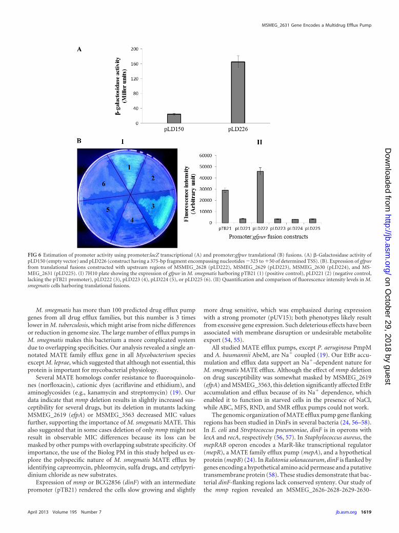
Promoter:reporter fusion construction and promoter activity de-termination. A 375-bp fragment (�325 to �50 of the TSS) was amplifiedusing Pr:F1:HindIII/Pr:R1:XbaI primers and cloned in pLD150 (apTL61T derivative [31] of pAL5000 origin; Genr [Jaques and Daniels,unpublished]) to construct a promoter:lacZ transcriptional fusion, desig-nated pLD226. pLD150 and pLD226 were mobilized in M. smegmatis. The�-galactosidase assay (37) was performed with M. smegmatis strains har-boring pLD150 or pLD226. Cells were grown in 7H9 to mid-exponentialphase and then grown for two more hours after supplementing with EtBr(2 �g ml�1), norfloxacin (5 �g/ml), kanamycin (2 �g ml�1), or sulfachlo-ropyridazine (400 �g ml�1). Untreated cultures were used as controls.Assay cell density was equalized by diluting with fresh medium or mediumsupplemented with drugs.
Upstream regions of MSMEG_2628, MSMEG_2629, and MSMEG_2630 and mmp (approximately 1,000 bp) were amplified using primers2628US:F:XbaI/2628:R:NheI (1,011 bp), 2629US:F:XbaI/2629:R1:NheI(1,094 bp), 2630US:F:XbaI/2630:R1:NheI (1,017 bp), and 2631US:F:XbaI/2631:R1:NheI (1,019 bp), respectively. Amplified fragments werecloned at the XbaI/NheI site of pTB21 to construct promoter:gfpuv trans-lational fusions by replacing the pTB21 promoter. Constructs with theupstream region of MSMEG_2628, MSMEG_2629, and MSMEG_2630 andmmp were designated pLD222, pLD223, pLD224, and pLD225, respec-tively. XbaI/NheI-digested pTB21 was self-ligated to construct a promot-erless plasmid, designated pLD221, as a negative control; pTB21 was thepositive control. GFPuv fluorescence was measured with a NOVOstar
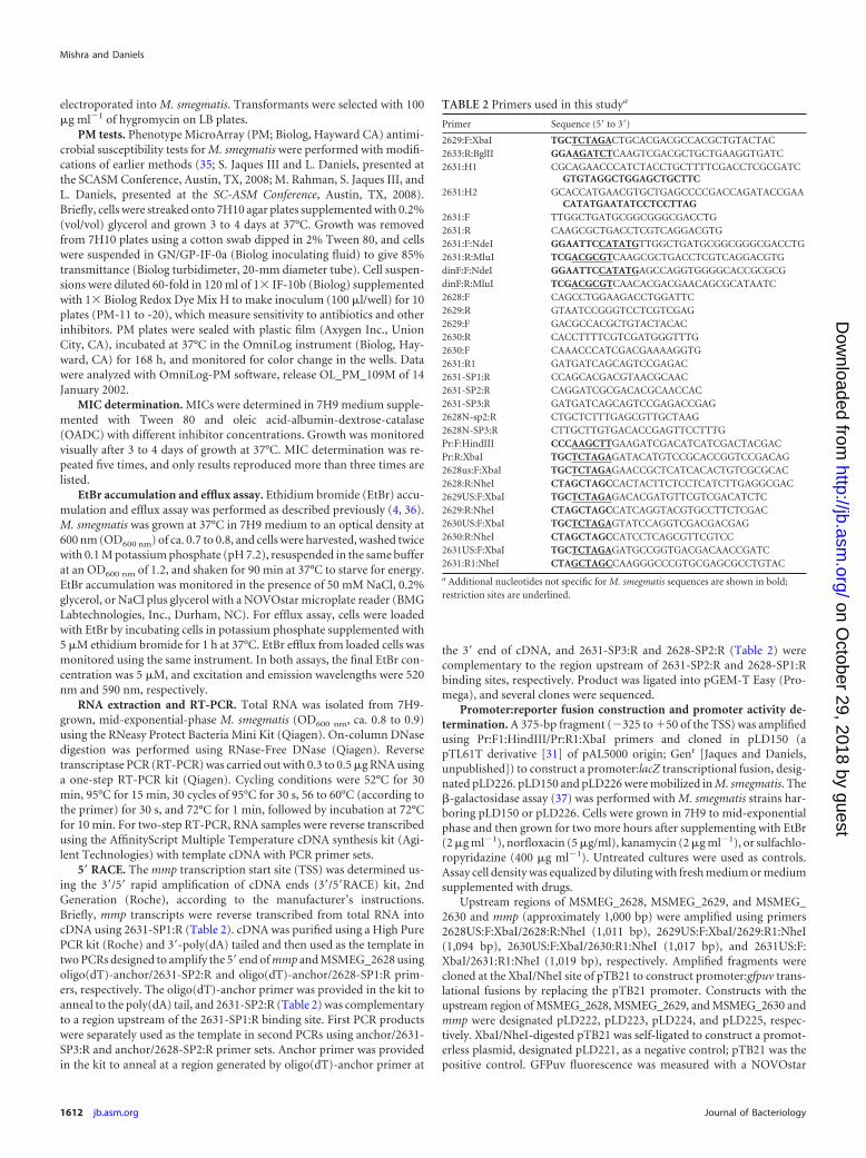
TABLE 2 Primers used in this studya
Primer Sequence (5= to 3=)2629:F:XbaI TGCTCTAGACTGCACGACGCCACGCTGTACTAC2633:R:BglII GGAAGATCTCAAGTCGACGCTGCTGAAGGTGATC2631:H1 CGCAGAACCCATCTACCTGCTTTTCGACCTCGCGATC
GTGTAGGCTGGAGCTGCTTC2631:H2 GCACCATGAACGTGCTGAGCCCCGACCAGATACCGAA
CATATGAATATCCTCCTTAG2631:F TTGGCTGATGCGGCGGGCGACCTG2631:R CAAGCGCTGACCTCGTCAGGACGTG2631:F:NdeI GGAATTCCATATGTTGGCTGATGCGGCGGGCGACCTG2631:R:MluI TCGACGCGTCAAGCGCTGACCTCGTCAGGACGTGdinF:F:NdeI GGAATTCCATATGAGCCAGGTGGGGCACCGCGCGdinF:R:MluI TCGACGCGTCAACACGACGAACAGCGCATAATC2628:F CAGCCTGGAAGACCTGGATTC2629:R GTAATCCGGGTCCTCGTCGAG2629:F GACGCCACGCTGTACTACAC2630:R CACCTTTTCGTCGATGGGTTTG2630:F CAAACCCATCGACGAAAAGGTG2631:R1 GATGATCAGCAGTCCGAGAC2631-SP1:R CCAGCACGACGTAACGCAAC2631-SP2:R CAGGATCGCGACACGCAACCAC2631-SP3:R GATGATCAGCAGTCCGAGACCGAG2628N-sp2:R CTGCTCTTTGAGCGTTGCTAAG2628N-SP3:R CTTGCTTGTGACACCGAGTTCCTTTGPr:F:HindIII CCCAAGCTTGAAGATCGACATCATCGACTACGACPr:R:XbaI TGCTCTAGAGATACATGTCCGCACCGGTCCGACAG2628us:F:XbaI TGCTCTAGAGAACCGCTCATCACACTGTCGCGCAC2628:R:NheI CTAGCTAGCCACTACTTCTCCTCATCTTGAGGCGAC2629US:F:XbaI TGCTCTAGAGACACGATGTTCGTCGACATCTC2629:R:NheI CTAGCTAGCCATCAGGTACGTGCCTTCTCGAC2630US:F:XbaI TGCTCTAGAGTATCCAGGTCGACGACGAG2630:R:NheI CTAGCTAGCCATCCTCAGCGTTCGTCC2631US:F:XbaI TGCTCTAGAGATGCCGGTGACGACAACCGATC2631:R1:NheI CTAGCTAGCCAAGGGCCCGTGCGAGCGCCTGTAC
a Additional nucleotides not specific for M. smegmatis sequences are shown in bold;restriction sites are underlined.
Mishra and Daniels
1612 jb.asm.org Journal of Bacteriology
on October 29, 2018 by guest
http://jb.asm.org/
Dow
nloaded from
microplate reader, using excitation and emission wavelengths of 395 nmand 508 nm, respectively.
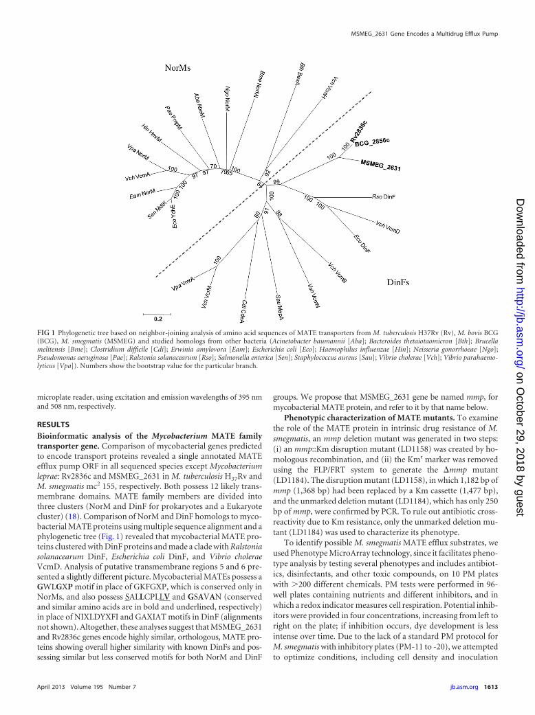
RESULTSBioinformatic analysis of the Mycobacterium MATE familytransporter gene. Comparison of mycobacterial genes predictedto encode transport proteins revealed a single annotated MATEefflux pump ORF in all sequenced species except Mycobacteriumleprae: Rv2836c and MSMEG_2631 in M. tuberculosis H37Rv andM. smegmatis mc2 155, respectively. Both possess 12 likely trans-membrane domains. MATE family members are divided intothree clusters (NorM and DinF for prokaryotes and a Eukaryotecluster) (18). Comparison of NorM and DinF homologs to myco-bacterial MATE proteins using multiple sequence alignment and aphylogenetic tree (Fig. 1) revealed that mycobacterial MATE pro-teins clustered with DinF proteins and made a clade with Ralstoniasolanacearum DinF, Escherichia coli DinF, and Vibrio choleraeVcmD. Analysis of putative transmembrane regions 5 and 6 pre-sented a slightly different picture. Mycobacterial MATEs possess aGWLGXP motif in place of GKFGXP, which is conserved only inNorMs, and also possess SALLCPLLV and GSAVAN (conservedand similar amino acids are in bold and underlined, respectively)in place of NIXLDYXFI and GAXIAT motifs in DinF (alignmentsnot shown). Altogether, these analyses suggest that MSMEG_2631and Rv2836c genes encode highly similar, orthologous, MATE pro-teins showing overall higher similarity with known DinFs and pos-sessing similar but less conserved motifs for both NorM and DinF
groups. We propose that MSMEG_2631 gene be named mmp, formycobacterial MATE protein, and refer to it by that name below.
Phenotypic characterization of MATE mutants. To examinethe role of the MATE protein in intrinsic drug resistance of M.smegmatis, an mmp deletion mutant was generated in two steps:(i) an mmp::Km disruption mutant (LD1158) was created by ho-mologous recombination, and (ii) the Kmr marker was removedusing the FLP/FRT system to generate the �mmp mutant(LD1184). The disruption mutant (LD1158), in which 1,182 bp ofmmp (1,368 bp) had been replaced by a Km cassette (1,477 bp),and the unmarked deletion mutant (LD1184), which has only 250bp of mmp, were confirmed by PCR. To rule out antibiotic cross-reactivity due to Km resistance, only the unmarked deletion mu-tant (LD1184) was used to characterize its phenotype.
To identify possible M. smegmatis MATE efflux substrates, weused Phenotype MicroArray technology, since it facilitates pheno-type analysis by testing several phenotypes and includes antibiot-ics, disinfectants, and other toxic compounds, on 10 PM plateswith 200 different chemicals. PM tests were performed in 96-well plates containing nutrients and different inhibitors, and inwhich a redox indicator measures cell respiration. Potential inhib-itors were provided in four concentrations, increasing from left toright on the plate; if inhibition occurs, dye development is lessintense over time. Due to the lack of a standard PM protocol forM. smegmatis with inhibitory plates (PM-11 to -20), we attemptedto optimize conditions, including cell density and inoculation
FIG 1 Phylogenetic tree based on neighbor-joining analysis of amino acid sequences of MATE transporters from M. tuberculosis H37Rv (Rv), M. bovis BCG(BCG), M. smegmatis (MSMEG) and studied homologs from other bacteria (Acinetobacter baumannii [Aba]; Bacteroides thetaiotaomicron [Bth]; Brucellamelitensis [Bme]; Clostridium difficile [Cdi]; Erwinia amylovora [Eam]; Escherichia coli [Eco]; Haemophilus influenzae [Hin]; Neisseria gonorrhoeae [Ngo];Pseudomonas aeruginosa [Pae]; Ralstonia solanacearum [Rso]; Salmonella enterica [Sen]; Staphylococcus aureus [Sau]; Vibrio cholerae [Vch]; Vibrio parahaemo-lyticus [Vpa]). Numbers show the bootstrap value for the particular branch.
MSMEG_2631 Gene Encodes a Multidrug Efflux Pump
April 2013 Volume 195 Number 7 jb.asm.org 1613
on October 29, 2018 by guest
http://jb.asm.org/
Dow
nloaded from
fluid modifications, to provide reproducible susceptibility pheno-types. We tested PM-11 to -20 data reproducibility with replicatesfor wild-type cells with IF-10b or 7H9 plus 5% OADC and differ-ent cell suspension dilutions. Data with 60� dilution of 85% T cellsuspension in IF-10b were fairly reproducible (data not shown).Data with 7H9 plus 5% OADC were also reproducible, but growthwas slower than preferred.
M. smegmatis has more than 100 putative drug efflux genesbelonging to different families. If some have overlapping substratespecificity, this may mask the effect of deleting one gene, resultingin little or no susceptibility difference. With this in mind, we cre-
ated two other unmarked deletion mutants, �MSMEG_2619(efpA) (LD1189) and �MSMEG_3563 (LD1192), lacking genesfor reported drug efflux pumps (4), as positive controls to exam-ine with PM plates. PM data suggested susceptibility differencesfor each of the three mutants in comparison to the wild type.Although differences were small in the majority of wells, suggest-ing enhanced mutant sensitivity, they were consistent. Drugs andchemicals identified as likely substrates by PM data for LD1189and LD1192 include most chemicals reported earlier (4) (data notshown), which indicates PM reliability for drug susceptibility test-ing in M. smegmatis.
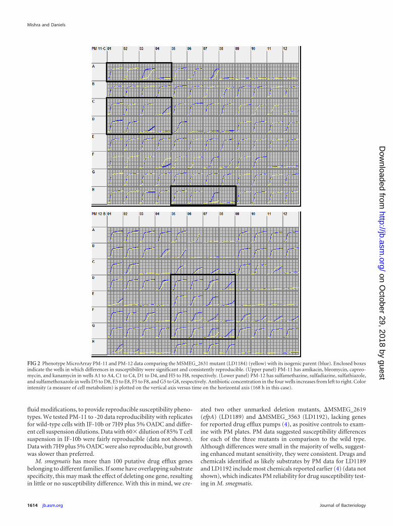
FIG 2 Phenotype MicroArray PM-11 and PM-12 data comparing the MSMEG_2631 mutant (LD1184) (yellow) with its isogenic parent (blue). Enclosed boxesindicate the wells in which differences in susceptibility were significant and consistently reproducible. (Upper panel) PM-11 has amikacin, bleomycin, capreo-mycin, and kanamycin in wells A1 to A4, C1 to C4, D1 to D4, and H5 to H8, respectively. (Lower panel) PM-12 has sulfamethazine, sulfadiazine, sulfathiazole,and sulfamethoxazole in wells D5 to D8, E5 to E8, F5 to F8, and G5 to G8, respectively. Antibiotic concentration in the four wells increases from left to right. Colorintensity (a measure of cell metabolism) is plotted on the vertical axis versus time on the horizontal axis (168 h in this case).
Mishra and Daniels
1614 jb.asm.org Journal of Bacteriology
on October 29, 2018 by guest
http://jb.asm.org/
Dow
nloaded from
PM data for the wild type and LD1184 (Fig. 2) revealed that thismutant was more susceptible to several antibiotics and antiseptics,including capreomycin, amikacin, kanamycin, phleomycin, bleo-mycin, cetylpyridinium chloride, and all sulfa drugs on PM plates(sulfamethazine, sulfadiazine, sulfathiazole, sulfamethoxazole,sulfachloropyridazine, and sulfamonomethoxine). PM data re-vealed that (i) except for sulfa drugs, LD1189 was more suscep-tible to all compounds listed for LD1184 than LD1184 but notvice versa, and (ii) LD1184 and LD1192 were equally sensitiveto the sulfa drugs. This observation indicated overlapping genefunctions and prompted us to delete efpA or MSMEG_3563 inLD1184 to make double mutants LD1190 (�MSMEG_2631 and�MSMEG_2619) and LD1201 (�MSMEG_2631 and �MSMEG_3563), respectively, to examine the effect of mmp deletion in thepresence and absence of efpA or MSMEG_3563. In most cases,mmp deletion resulted in a slight decrease in MIC values; however,double mutants showed greater MIC decreases (Table 3). MICsfor LD1190 (lacking efpA and mmp) were always lower than thoseof LD1189 (lacking only efpA), indicating contributions by mmp.Additionally, in some cases (e.g., capreomycin) a significant MICdifference for the wild type and for LD1184 was not observed, butdeletion of this gene in the efpA-lacking strain (LD1189) resultedin further MIC decrease. Similarly, deletion of either mmp orMSMEG_3563 did not produce a significant difference in somecases, but deletion of both genes resulted in a greater MIC decreasein LD1201.
Finally, ethidium bromide and norfloxacin were selected forsusceptibility testing because both are NorM substrates and EtBr isa preferred DinF substrate (19). Deletion of mmp (mutantLD1184) rendered the strain more susceptible to EtBr (2-fold de-crease in MIC) and slightly susceptible for norfloxacin (Table 2),suggesting that EtBr is a good M. smegmatis MATE substrate andthat this protein provides low-level intrinsic resistance for struc-turally unrelated compounds.
Effect of mmp and BCG_2856c expression. Data indicatingmmp involvement in multidrug resistance prompted us to exam-ine complementation and the effect of its expression at higher
levels. We made expression constructs with intermediate (pTB21)and strong (pUV15) promoters to express mmp and M. bovis BCGdinF (BCG_2856c, identical to Rv2836c). Using pUV15 resultedin growth-defective transformants, with small colonies after aweek. Using pTB21 gave normal transformants, but the expres-sion constructs grew slowly, and wild-type strains bearing eitherpLD216 (pTB21carrying mmp) or pLD217 (pTB21/BCG carryingdinF) were slightly more sensitive to EtBr and other drugs thanstrains bearing empty vector (pTB21). Complemented mutantsbearing expression constructs grew slowly or were slightly moresensitive to drugs than mutants harboring empty vector. This sug-gests that higher expression of this gene renders cells growth de-fective, causing increased sensitivity.
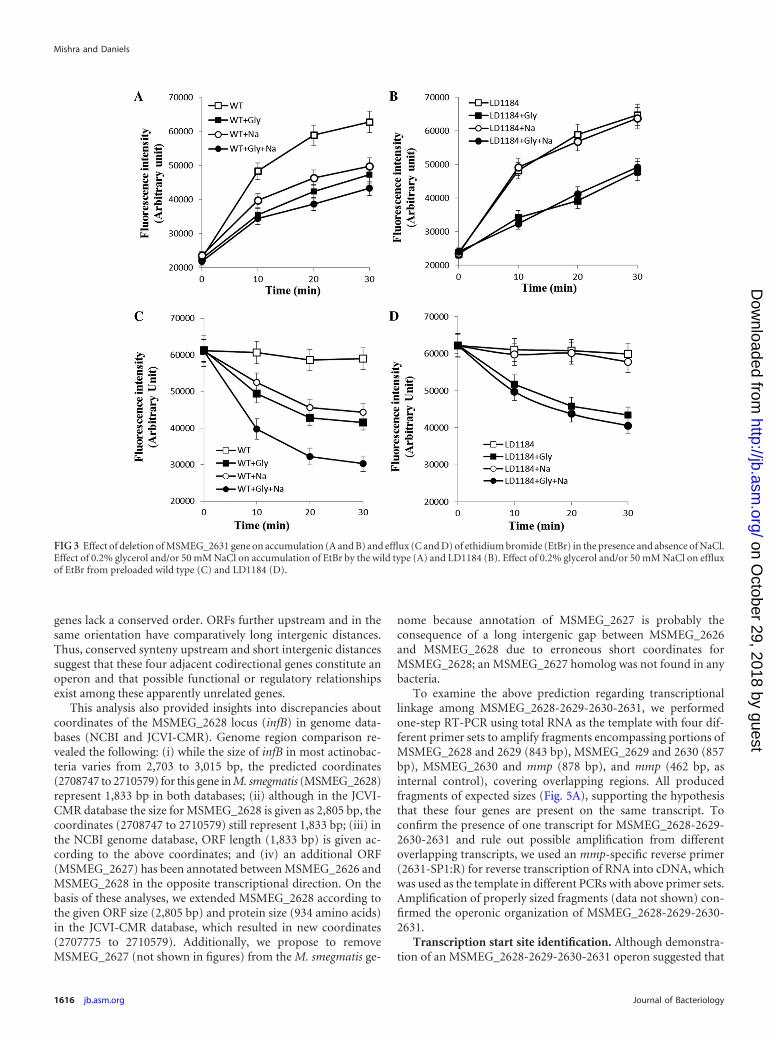
Effect of mmp deletion on ethidium bromide accumulationand efflux. Several MATE members are active efflux pumps, themajority of which (NorM [38], VmrA [39], VcmA [40], VcrM[41], YdhE [36], and HmrM [42]) are Na� dependent. However,Pseudomonas aeruginosa PmpM (43) and Acinetobacter bauman-nii AbeM (44) are H� coupled. Thus, we examined whether mmpencoded an active efflux pump and if so, whether it is coupled toNa� or H�. We performed EtBr accumulation and efflux assayswith the wild type and LD1184 in the presence and absence ofNa�, with the anticipation that (i) mmp deletion would affectaccumulation (positively) and efflux (negatively) if this gene en-coded an efflux pump and (ii) if mmp encoded an Na�-coupledMATE efflux pump, the effect of mmp deletion would be signifi-cant in the presence of Na� because other EtBr efflux pumps suchas LfrA and EfpA belonging to MFS (both are H� coupled) wouldnot contribute significantly without an energy source. Figure 3Ashows that wild-type cells accumulate 22%, 26%, and 34% lessEtBr in the presence of NaCl, glycerol, or NaCl plus glycerol, re-spectively, than control cells (with no NaCl or glycerol). WithLD1184, glycerol reduced EtBr accumulation at a rate similar(28%) to that of the wild type (Fig. 3B). However, NaCl did notsignificantly affect EtBr accumulation by LD1184. EtBr efflux datafrom preloaded cells (Fig. 3C and D) revealed that EtBr effluxfrom wild-type cells was accelerated by either NaCl or glycerol butin the case of LD1184 only glycerol, and not NaCl, acceleratedefflux. Additionally, efflux was maximum in the presence of NaClplus glycerol, but only from wild-type cells. These results supportthe hypothesis that the M. smegmatis MATE efflux protein is Na�
coupled and active.mmp organization and its transcriptional linkage with up-
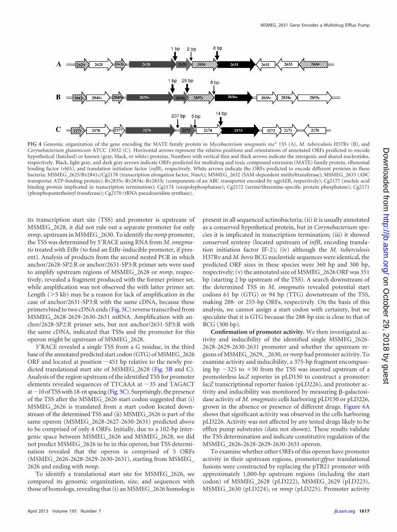
stream genes. To investigate mmp inducibility by EtBr and anti-biotics with promoter:reporter assays, we analyzed the 600 bp up-stream to identify a putative mycobacterial promoter consensussequence but found none. Further analysis revealed that mmp andthree other upstream ORFs, i.e., MSMEG_2630 (encoding a DHHfamily protein), MSMEG_2629 (rbfA, encoding ribosome bindingfactor A), and MSMEG_2628 (infB, encoding translation initia-tion factor IF-2), were present in the same orientation.MSMEG_2629 and 2630 have an intergenic distance of 2 nucleo-tides, while MSMEG_2628 and 2629 and MSMEG_2630 and mmpoverlap by 1 and 8 nucleotides, respectively. Since short intergenicdistance and phylogenetically conserved gene order are importantpredictors of operon structure for genes in the same orientation(45), we compared the mmp region with that of other genomes.This gene order was fairly well conserved in more than 13 actino-bacteria. Figure 4 shows that gene organization upstream of themycobacterial MATE gene is highly conserved, but downstream
TABLE 3 Antimicrobial susceptibility of M. smegmatis mc2 155mutantsa
M.smegmatisstrain Description
MIC (�g ml�1)
EtBr NOR CP KM PM CPC SCP SML SMT
mc2 155 Wild type 4 6 8 4 0.5 10 600 700 600LD1184 �2631 (MATE) 2 5 6 2 0.3 8 400 450 400LD1189 �2619 (efpA) 2 4 5 4 0.3 7 600 700 600LD1190 �2631-�2619 1.5 2 3 2 0.15 4 400 500 400LD1192 �3563 3 6 8 2 0.5 10 450 400 450LD1201 �2631-�3563 2 4 6 1 0.3 7 300 300 300
a MICs were determined by growth in 8 ml 7H9 –10% OADC– 0.05% Tween 80supplemented with different concentrations of either EtBr (ethidium bromide), NOR(norfloxacin), CP (capreomycin), PM (phleomycin), CPC (cetylpyridinium chloride),SCP (sulfachloropyridazine), SML (sulfamethoxazole), or SMT (sulfamethazine).Growth was monitored by visual inspection after 3 to 4 days. Experiments wererepeated at least five times to confirm the difference between wild-type and mutantstrains for their susceptibility to these antimicrobial agents. Final concentration ranges(�g/ml): EtBr, 0, 0.5, 1.0, 1.5, 2.0, 2.5, 3.0, 3.5, 4.5, 5.0, 5.5, 6.0. 6.5, 7.0, 7.5, 8.0; NOR,1, 2, 3, 4, 5, 6, 7, 8, 9, 10; CP, 1, 2, 3, 4, 5, 6, 7, 8, 9, 10; KM, 0, 0.5, 1.0, 1.5, 2.0, 2.5, 3.0,3.5, 4.5, 5.0, 5.5, 6.0; PM, 0, 0.05, 0.1, 0.15, 0.2, 0.25, 0.30, 0.35, 0.45, 0.5, 0.55, 0.6, 0.65,0.7, 0.75, 0.8; CPC, 0, 2, 4, 6, 8, 10, 12, 14, 16, 18, 20; all sulfa drugs (SCP, SML, andSMT), 0, 50, 100, 150, 200, 250, 300, 350, 400, 450, 500, 650, 700, 750, 800, 850, 900,950, 1,000.
MSMEG_2631 Gene Encodes a Multidrug Efflux Pump
April 2013 Volume 195 Number 7 jb.asm.org 1615
on October 29, 2018 by guest
http://jb.asm.org/
Dow
nloaded from
genes lack a conserved order. ORFs further upstream and in thesame orientation have comparatively long intergenic distances.Thus, conserved synteny upstream and short intergenic distancessuggest that these four adjacent codirectional genes constitute anoperon and that possible functional or regulatory relationshipsexist among these apparently unrelated genes.
This analysis also provided insights into discrepancies aboutcoordinates of the MSMEG_2628 locus (infB) in genome data-bases (NCBI and JCVI-CMR). Genome region comparison re-vealed the following: (i) while the size of infB in most actinobac-teria varies from 2,703 to 3,015 bp, the predicted coordinates(2708747 to 2710579) for this gene in M. smegmatis (MSMEG_2628)represent 1,833 bp in both databases; (ii) although in the JCVI-CMR database the size for MSMEG_2628 is given as 2,805 bp, thecoordinates (2708747 to 2710579) still represent 1,833 bp; (iii) inthe NCBI genome database, ORF length (1,833 bp) is given ac-cording to the above coordinates; and (iv) an additional ORF(MSMEG_2627) has been annotated between MSMEG_2626 andMSMEG_2628 in the opposite transcriptional direction. On thebasis of these analyses, we extended MSMEG_2628 according tothe given ORF size (2,805 bp) and protein size (934 amino acids)in the JCVI-CMR database, which resulted in new coordinates(2707775 to 2710579). Additionally, we propose to removeMSMEG_2627 (not shown in figures) from the M. smegmatis ge-
nome because annotation of MSMEG_2627 is probably theconsequence of a long intergenic gap between MSMEG_2626and MSMEG_2628 due to erroneous short coordinates forMSMEG_2628; an MSMEG_2627 homolog was not found in anybacteria.
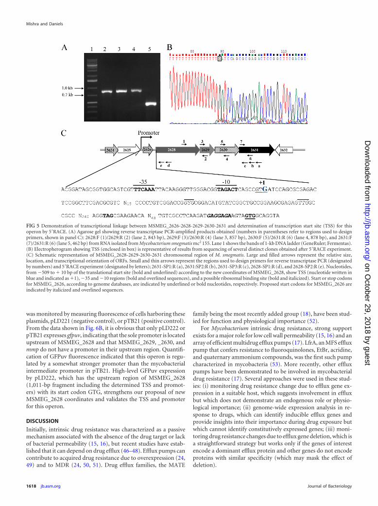
To examine the above prediction regarding transcriptionallinkage among MSMEG_2628-2629-2630-2631, we performedone-step RT-PCR using total RNA as the template with four dif-ferent primer sets to amplify fragments encompassing portions ofMSMEG_2628 and 2629 (843 bp), MSMEG_2629 and 2630 (857bp), MSMEG_2630 and mmp (878 bp), and mmp (462 bp, asinternal control), covering overlapping regions. All producedfragments of expected sizes (Fig. 5A), supporting the hypothesisthat these four genes are present on the same transcript. Toconfirm the presence of one transcript for MSMEG_2628-2629-2630-2631 and rule out possible amplification from differentoverlapping transcripts, we used an mmp-specific reverse primer(2631-SP1:R) for reverse transcription of RNA into cDNA, whichwas used as the template in different PCRs with above primer sets.Amplification of properly sized fragments (data not shown) con-firmed the operonic organization of MSMEG_2628-2629-2630-2631.
Transcription start site identification. Although demonstra-tion of an MSMEG_2628-2629-2630-2631 operon suggested that
FIG 3 Effect of deletion of MSMEG_2631 gene on accumulation (A and B) and efflux (C and D) of ethidium bromide (EtBr) in the presence and absence of NaCl.Effect of 0.2% glycerol and/or 50 mM NaCl on accumulation of EtBr by the wild type (A) and LD1184 (B). Effect of 0.2% glycerol and/or 50 mM NaCl on effluxof EtBr from preloaded wild type (C) and LD1184 (D).
Mishra and Daniels
1616 jb.asm.org Journal of Bacteriology
on October 29, 2018 by guest
http://jb.asm.org/
Dow
nloaded from
its transcription start site (TSS) and promoter is upstream ofMSMEG_2628, it did not rule out a separate promoter for onlymmp, upstream in MSMEG_2630. To identify the mmp promoter,the TSS was determined by 5=RACE using RNA from M. smegma-tis treated with EtBr (to find an EtBr-inducible promoter, if pres-ent). Analysis of products from the second nested PCR in whichanchor/2628-SP2:R or anchor/2631-SP3:R primer sets were usedto amplify upstream regions of MSMEG_2628 or mmp, respec-tively, revealed a fragment produced with the former primer set,while amplification was not observed the with latter primer set.Length (5 kb) may be a reason for lack of amplification in thecase of anchor/2631-SP3:R with the same cDNA, because theseprimers bind to two cDNA ends (Fig. 5C) reverse transcribed fromMSMEG_2628-2629-2630-2631 mRNA. Amplification with an-chor/2628-SP2:R primer sets, but not anchor/2631-SP3:R withthe same cDNA, indicated that TSSs and the promoter for thisoperon might be upstream of MSMEG_2628.
5=RACE revealed a single TSS from a G residue, in the thirdbase of the annotated predicted start codon (GTG) of MSMEG_2626ORF and located at position �451 bp relative to the newly pre-dicted translational start site of MSMEG_2628 (Fig. 5B and C).Analysis of the region upstream of the identified TSS for promoterelements revealed sequences of TTCAAA at �35 and TAGACTat�10 of TSS with 18-nt spacing (Fig. 5C). Surprisingly, the presenceof the TSS after the MSMEG_2626 start codon suggested that (i)MSMEG_2626 is translated from a start codon located down-stream of the determined TSS and (ii) MSMEG_2626 is part of thesame operon (MSMEG_2628-2627-2630-2631) predicted aboveto be comprised of only 4 ORFs. Initially, due to a 102-bp inter-genic space between MSMEG_2626 and MSMEG_2628, we didnot predict MSMEG_2626 to be in this operon, but TSS determi-nation revealed that the operon is comprised of 5 ORFs(MSMEG_2626-2628-2629-2630-2631), starting from MSMEG_2626 and ending with mmp.
To identify a translational start site for MSMEG_2626, wecompared its genomic organization, size, and sequences withthose of homologs, revealing that (i) an MSMEG_2626 homolog is
present in all sequenced actinobacteria; (ii) it is usually annotatedas a conserved hypothetical protein, but in Corynebacterium spe-cies it is implicated in transcription termination; (iii) it showedconserved synteny (located upstream of infB, encoding transla-tion initiation factor IF-2); (iv) although the M. tuberculosisH37Rv and M. bovis BCG nucleotide sequences were identical, thepredicted ORF sizes in these species were 360 bp and 300 bp,respectively; (v) the annotated size of MSMEG_2626 ORF was 351bp (starting 2 bp upstream of the TSS). A search downstream ofthe determined TSS in M. smegmatis revealed potential startcodons 61 bp (GTG) or 94 bp (TTG) downstream of the TSS,making 288- or 255-bp ORFs, respectively. On the basis of thisanalysis, we cannot assign a start codon with certainty, but wespeculate that it is GTG because the 288-bp size is close to that ofBCG (300 bp).
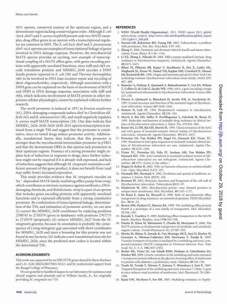
Confirmation of promoter activity. We then investigated ac-tivity and inducibility of the identified single MSMEG_2626-2628-2629-2630-2631 promoter and whether the upstream re-gions of MSMEG_2629, _2630, or mmp had promoter activity. Toexamine activity and inducibility, a 375-bp fragment encompass-ing bp �325 to �50 from the TSS was inserted upstream of apromoterless lacZ reporter in pLD150 to construct a promoter:lacZ transcriptional reporter fusion (pLD226), and promoter ac-tivity and inducibility was monitored by measuring �-galactosi-dase activity of M. smegmatis cells harboring pLD150 or pLD226,grown in the absence or presence of different drugs. Figure 6Ashows that significant activity was observed in the cells harboringpLD226. Activity was not affected by any tested drugs likely to beefflux pump substrates (data not shown). These results validatethe TSS determination and indicate constitutive regulation of theMSMEG_2626-2628-2629-2630-2631 operon.
To examine whether other ORFs of this operon have promoteractivity in their upstream regions, promoter:gfpuv translationalfusions were constructed by replacing the pTB21 promoter withapproximately 1,000-bp upstream regions (including the startcodon) of MSMEG_2628 (pLD222), MSMEG_2629 (pLD223),MSMEG_2630 (pLD224), or mmp (pLD225). Promoter activity
FIG 4 Genomic organization of the gene encoding the MATE family protein in Mycobacterium smegmatis mc2 155 (A), M. tuberculosis H37Rv (B), andCorynebacterium glutamicum ATCC 13032 (C). Horizontal arrows represent the relative positions and orientations of annotated ORFs predicted to encodehypothetical (hatched) or known (gray, black, or white) proteins. Numbers with vertical thin and thick arrows indicate the intergenic and shared nucleotides,respectively. Black, light gray, and dark gray arrows indicate ORFs predicted for multidrug and toxic compound extrusion (MATE) family protein, ribosomalbinding factor (rbfA), and translation initiation factor (infB), respectively. White arrows indicate the ORFs predicted to encode different proteins in thesebacteria: MSMEG_2625/Rv2841c/Cg2178 (transcription elongation factor, NusA); MSMEG_2632 (SAM-dependent methyltransferase); MSMEG_2633 (ABCtransporter ATP-binding protein); Rv2835c-Rv2834c-Rv2833c (components of an ABC transporter encoded by ugpAEB, respectively); Cg2177 (nucleic acidbinding protein implicated in transcription termination); Cg2174 (exopolyphosphatase); Cg2172 (serine/threonine-specific protein phosphatase); Cg2171(phosphopantetheinyl transferase); Cg2170 (tRNA pseudouridine synthase).
MSMEG_2631 Gene Encodes a Multidrug Efflux Pump
April 2013 Volume 195 Number 7 jb.asm.org 1617
on October 29, 2018 by guest
http://jb.asm.org/
Dow
nloaded from
was monitored by measuring fluorescence of cells harboring theseplasmids, pLD221 (negative control), or pTB21 (positive control).From the data shown in Fig. 6B, it is obvious that only pLD222 orpTB21 expresses gfpuv, indicating that the sole promoter is locatedupstream of MSMEG_2628 and that MSMEG_2629, _2630, andmmp do not have a promoter in their upstream region. Quantifi-cation of GFPuv fluorescence indicated that this operon is regu-lated by a somewhat stronger promoter than the mycobacterialintermediate promoter in pTB21. High-level GFPuv expressionby pLD222, which has the upstream region of MSMEG_2628(1,011-bp fragment including the determined TSS and promot-ers) with its start codon GTG, strengthens our proposal of newMSMEG_2628 coordinates and validates the TSS and promoterfor this operon.
DISCUSSION
Initially, intrinsic drug resistance was characterized as a passivemechanism associated with the absence of the drug target or lackof bacterial permeability (15, 16), but recent studies have estab-lished that it can depend on drug efflux (46–48). Efflux pumps cancontribute to acquired drug resistance due to overexpression (24,49) and to MDR (24, 50, 51). Drug efflux families, the MATE
family being the most recently added group (18), have been stud-ied for function and physiological importance (52).
For Mycobacterium intrinsic drug resistance, strong supportexists for a major role for low cell wall permeability (15, 16) and anarray of efficient multidrug efflux pumps (17). LfrA, an MFS effluxpump that confers resistance to fluoroquinolones, EtBr, acridine,and quaternary ammonium compounds, was the first such pumpcharacterized in mycobacteria (53). More recently, other effluxpumps have been demonstrated to be involved in mycobacterialdrug resistance (17). Several approaches were used in these stud-ies: (i) monitoring drug resistance change due to efflux gene ex-pression in a suitable host, which suggests involvement in effluxbut which does not demonstrate an endogenous role or physio-logical importance; (ii) genome-wide expression analysis in re-sponse to drugs, which can identify inducible efflux genes andprovide insights into their importance during drug exposure butwhich cannot identify constitutively expressed genes; (iii) moni-toring drug resistance changes due to efflux gene deletion, which isa straightforward strategy but works only if the genes of interestencode a dominant efflux protein and other genes do not encodeproteins with similar specificity (which may mask the effect ofdeletion).
FIG 5 Demonstration of transcriptional linkage between MSMEG_2626-2628-2629-2630-2631 and determination of transcription start site (TSS) for thisoperon by 5=RACE. (A) Agarose gel showing reverse transcriptase PCR-amplified products obtained (numbers in parentheses refer to regions used to designprimers, shown in panel C): 2628:F (1)/2629:R (2) (lane 2, 843 bp), 2629:F (3)/2630:R (4) (lane 3, 857 bp), 2630:F (5)/2631:R (6) (lane 4, 878 bp), and 2631:F(7)/2631:R (6) (lane 5, 462 bp) from RNA isolated from Mycobacterium smegmatis mc2 155. Lane 1 shows the bands of 1-kb DNA ladder (GeneRuler; Fermentas).(B) Electropherogram showing TSS (enclosed in box) is representative of results from sequencing of several distinct clones obtained after 5=RACE experiment.(C) Schematic representation of MSMEG_2628-2629-2630-2631 chromosomal region of M. smegmatis. Large and filled arrows represent the relative size,location, and transcriptional orientation of ORFs. Small and thin arrows represent the regions used to design primers for reverse transcriptase PCR (designatedby numbers) and 5=RACE experiment (designated by letters); 2631-SP1:R (a), 2631:SP2:R (b), 2631-SP3:R (c), 2628-SP1:R (d), and 2628-SP2:R (e). Nucleotides,from �509 to � 10 bp of the translational start site (bold and underlined) according to the new coordinates of MSMEG_2628, show TSS (nucleotide written inblue and indicated as �1), �35 and �10 regions (bold and overlined sequences), and a possible ribosomal binding site (bold and italicized). Start or stop codonsfor MSMEG_2626, according to genome databases, are indicated by underlined or bold nucleotides, respectively. Proposed start codons for MSMEG_2626 areindicated by italicized and overlined sequences.
Mishra and Daniels
1618 jb.asm.org Journal of Bacteriology
on October 29, 2018 by guest
http://jb.asm.org/
Dow
nloaded from
M. smegmatis has more than 100 predicted drug efflux pumpgenes from all drug efflux families, but this number is 3 timeslower in M. tuberculosis, which might arise from niche differencesor reduction in genome size. The large number of efflux pumps inM. smegmatis makes this bacterium a more complicated systemdue to overlapping specificities. Our analysis revealed a single an-notated MATE family efflux gene in all Mycobacterium speciesexcept M. leprae, which suggested that although not essential, thisprotein is important for mycobacterial physiology.
Several MATE homologs confer resistance to fluoroquinolo-nes (norfloxacin), cationic dyes (acriflavine and ethidium), andaminoglycosides (e.g., kanamycin and streptomycin) (19). Ourdata indicate that mmp deletion results in slightly increased sus-ceptibility for several drugs, but its deletion in mutants lackingMSMEG_2619 (efpA) or MSMEG_3563 decreased MIC valuesfurther, supporting the importance of M. smegmatis MATE. Thisalso suggested that in some cases deletion of only mmp might notresult in observable MIC differences because its loss can bemasked by other pumps with overlapping substrate specificity. Ofimportance, the use of the Biolog PM in this study helped us ex-plore the polyspecific nature of M. smegmatis MATE efflux byidentifying capreomycin, phleomycin, sulfa drugs, and cetylpyri-dinium chloride as new substrates.
Expression of mmp or BCG2856 (dinF) with an intermediatepromoter (pTB21) rendered the cells slow growing and slightly
more drug sensitive, which was emphasized during expressionwith a strong promoter (pUV15); both phenotypes likely resultfrom excessive gene expression. Such deleterious effects have beenassociated with membrane disruption or undesirable metaboliteexport (54, 55).
All studied MATE efflux pumps, except P. aeruginosa PmpMand A. baumannii AbeM, are Na� coupled (19). Our EtBr accu-mulation and efflux data support an Na�-dependent nature forM. smegmatis MATE efflux. Although the effect of mmp deletionon drug susceptibility was somewhat masked by MSMEG_2619(efpA) and MSMEG_3563, this deletion significantly affected EtBraccumulation and efflux because of its Na� dependence, whichenabled it to function in starved cells in the presence of NaCl,while ABC, MFS, RND, and SMR efflux pumps could not work.
The genomic organization of MATE efflux pump gene flankingregions has been studied in DinFs in several bacteria (24, 56–58).In E. coli and Streptococcus pneumoniae, dinF is in operons withlexA and recA, respectively (56, 57). In Staphylococcus aureus, themepRAB operon encodes a MarR-like transcriptional regulator(mepR), a MATE family efflux pump (mepA), and a hypotheticalprotein (mepB) (24). In Ralstonia solanacearum, dinF is flanked bygenes encoding a hypothetical amino acid permease and a putativetransmembrane protein (58). These studies demonstrate that bac-terial dinF-flanking regions lack conserved synteny. Our study ofthe mmp region revealed an MSMEG_2626-2628-2629-2630-
FIG 6 Estimation of promoter activity using promoter:lacZ transcriptional (A) and promoter:gfpuv translational (B) fusions. (A) �-Galactosidase activity ofpLD150 (empty vector) and pLD226 (construct having a 375-bp fragment encompassing nucleotides �325 to �50 of determined TSS). (B). Expression of gfpuvfrom translational fusions constructed with upstream regions of MSMEG_2628 (pLD222), MSMEG_2629 (pLD223), MSMEG_2630 (pLD224), and MS-MEG_2631 (pLD225). (I) 7H10 plate showing the expression of gfpuv in M. smegmatis harboring pTB21 (1) (positive control), pLD221 (2) (negative control,lacking the pTB21 promoter), pLD222 (3), pLD223 (4), pLD224 (5), or pLD225 (6). (II) Quantification and comparison of fluorescence intensity levels in M.smegmatis cells harboring translational fusions.
MSMEG_2631 Gene Encodes a Multidrug Efflux Pump
April 2013 Volume 195 Number 7 jb.asm.org 1619
on October 29, 2018 by guest
http://jb.asm.org/
Dow
nloaded from
2631 operon, conserved synteny of the upstream region, and adownstream region lacking conserved gene order. Although E. coli(lexA-dinF) and S. aureus mepRAB present only two MATE exam-ples, drug efflux genes in an operon with a transcriptional regula-tor are common in MFS. The E. coli lexA-dinF and S. pneumoniaedinF-recA operons are examples of transcriptional linkage of genesinvolved in DNA damage response. However, the mycobacterialMATE operon provides an exciting, new example of transcrip-tional coupling of a MATE efflux gene, with genes encoding pro-teins with apparently unrelated functions, since infB and rbfA en-code translation proteins and MSMEG_2630 encodes a DHHfamily protein reported in E. coli (59) and Thermus thermophilus(60) to be involved in DNA base excision-repair and recycling ofshort oligonucleotides, respectively. Although association with aDHH gene can be explained on the basis of involvement of MATEand DHH in DNA damage response, association with infB andrbfA, which indicates involvement of MATE protein in other im-portant cellular physiologies, cannot be explained without furtherresearch.
The norM promoter is induced at 18°C in Erwinia amylovora(61), DNA-damaging compounds induce dinF in Clostridium dif-ficile (62) and R. solanacearum (58), and mepR negatively regulatesS. aureus mepB MATE transcription (24). Our data indicate thatMSMEG_2626-2628-2629-2630-2631 operon transcription is ini-tiated from a single TSS and suggest that the promoter is consti-tutive, since no tested drugs induce promoter activity. Addition-ally, translational fusion data indicate that this promoter isstronger than the mycobacterial intermediate promoter in pTB21and that the downstream ORFs in this operon lack promoters intheir upstream regions. Presence of a strong promoter might ex-plain lack of induction in response to drugs, since further induc-tion might not be required if it is already well expressed, and lackof induction suggests that although M. smegmatis maintains a suf-ficient amount of this gene product, it does not benefit from (andmay suffer from) increased expression.
This study provides evidence that M. smegmatis encodes anNa�-dependent MATE family multidrug efflux pump from mmp,which contributes to intrinsic resistance against antibiotics, DNA-damaging chemicals, and disinfectants. mmp is a part of an operonthat includes genes encoding proteins with apparently unrelatedfunctions and is expressed efficiently from a strong constitutivepromoter. By confirmation of transcriptional linkage, determina-tion of the TSS, and estimation of promoter activity, we can now(i) correct the MSMEG_2628 coordinates by replacing positions2708747 to 2710579 (given in databases) with positions 2707775to 2710579 (proposed); (ii) remove MSMEG_2627 from the M.smegmatis genome, because its annotation is probably the conse-quence of a long intergenic gap associated with short coordinatesfor MSMEG_2628 and since a homolog for this protein was notfound in any bacteria; (iii) indicate a new translational start site forMSMEG_2626, since the predicted start codon is located withinthe determined TSS.
ACKNOWLEDGMENTS
This work was supported by an NIH STTR grant shared by Barry Bochnerand L.D. (GM 2R42GM073965-02A1) and by institutional support fromthe Rangel College of Pharmacy.
We are grateful to Sandford Jaques in our laboratory for assistance andshared reagents and plasmids and to William Jacobs, Jr., for originallyproviding M. smegmatis mc2155.
REFERENCES1. WHO (World Health Organization). 2011. WHO report 2011: global
tuberculosis control. http://www.who.int/tb/publications/global_report/2011/gtbr11_full.pdf.
2. Stewart GR, Robertson BD, Young DB. 2003. Tuberculosis: a problemwith persistence. Nat. Rev. Microbiol. 1:97–105.
3. Zhang Y. 2004. Persistent and dormant tubercle bacilli and latent tuber-culosis. Front. Biosci. 9:1136 –1156.
4. Li XZ, Zhang L, Nikaido H. 2004. Efflux pump-mediated intrinsic drugresistance in Mycobacterium smegmatis. Antimicrob. Agents Chemother.48:2415–2423.
5. Bifani PJ, Plikaytis BB, Kapur V, Stockbauer K, Pan X, Lutfey ML,Moghazeh SL, Eisner W, Daniel TM, Kaplan MH, Crawford JT, MusserJM, Kreiswirth BN. 1996. Origin and interstate spread of a New York Citymultidrug-resistant Mycobacterium tuberculosis clone family. JAMA 275:487– 489.
6. Banerjee A, Dubnau E, Quémard A, Balasubramian V, Um KS, WilsonT, Collins D, de Lisle G, Jacobs WR. 1994. inhA, a gene encoding a targetfor isoniazid and ethionamide in Mycobacterium tuberculosis. Science 263:227–230.
7. Dessen A, Quémard A, Blanchard JS, Jacobs WR, Jr, Sacchettini JC.1995. Crystal structure and function of the isoniazid target of Mycobacte-rium tuberculosis. Science 267:1638 –1641.
8. Honore N, Cole ST. 1994. Streptomycin resistance in mycobacteria.Antimicrob. Agents Chemother. 38:238 –242.
9. Morris S, Bai GH, Suffys P, Portillogomez L, Fairchok M, Rouse D.1995. Molecular mechanisms of multiple drug resistance in clinical iso-lates of Mycobacterium tuberculosis. J. Infect. Dis. 171:954 –960.
10. Rouse DA, Li ZM, Bai GH, Morris SL. 1995. Characterization of the katGand inhA genes of isoniazid-resistant clinical isolates of Mycobacteriumtuberculosis. Antimicrob. Agents Chemother. 39:2472–2477.
11. Pretorius GS, Van Helden PD, Sirgel FA, Eisenach KD, Victor TC.1995. Mutations in katG gene sequences in isoniazid-resistant clinical iso-lates of Mycobacterium tuberculosis are rare. Antimicrob. Agents Che-mother. 39:2276 –2281.
12. Victor TC, Pretorius GS, Felix JV, Jordaan AM, Van Helden PD,Eisenach KD. 1996. katG mutations in isoniazid-resistant strains of My-cobacterium tuberculosis are not infrequent. Antimicrob. Agents Che-mother. 40:1572. (Letter to the editor.)
13. Hogan D, Kolter R. 2002. Why are bacteria refractory to antimicrobials?Curr. Opin. Microbiol. 5:472– 477.
14. Normark BH, Normark S. 2002. Evolution and spread of antibiotic re-sistance. J. Intern. Med. 252:91–106.
15. Brennan PJ. 2003. Structure, function, and biogenesis of the cell wall ofMycobacterium tuberculosis. Tuberculosis 83:91–97.
16. Niederweis M. 2003. Mycobacterial porins—new channel proteins inunique outer membranes. Mol. Microbiol. 49:1167–1177.
17. De Rossi E, Ainsa JA, Riccardi G. 2006. Role of mycobacterial effluxtransporters in drug resistance: an unresolved question. FEMS Microbiol.Rev. 30:36 –52.
18. Brown MH, Paulsen IT, Skurray RA. 1999. The multidrug efflux proteinNorM is a prototype of a new family of transporters. Mol. Microbiol.31:394 –395.
19. Kuroda T, Tsuchiya T. 2009. Multidrug efflux transporters in the MATEfamily. Biochim. Biophys. Acta 1794:763–768.
20. Omote H, Hiasa M, Matsumoto T, Otsuka M, Moriyama Y. 2006. TheMATE proteins as fundamental transporters of metabolic and xenobioticorganic cations. Trends Pharmacol. Sci. 27:587–593.
21. Morita M, Shitan N, Sawada K, Van Montagu MCE, Inzé D, Rischer H,Goossens A, Oksman-Caldentey KM, Moriyama Y, Yazaki K. 2009.Vacuolar transport of nicotine is mediated by a multidrug and toxic com-pound extrusion (MATE) transporter in Nicotiana tabacum. Proc. Natl.Acad. Sci. U. S. A. 106:2447–2452.
22. Becker ML, Visser LE, van Schaik RHN, Hofman A, Uitterlinden AG,Stricker BH. 2009. Genetic variation in the multidrug and toxin extrusion1 transporter protein influences the glucose-lowering effect of metforminin patients with diabetes: a preliminary study. Diabetes 58:745–749.
23. Tsuda M, Terada T, Mizuno T, Katsura T, Shimakura J, Inui K. 2009.Targeted disruption of the multidrug and toxin extrusion 1 (Mate 1) genein mice reduces renal secretion of metformin. Mol. Pharmacol. 75:1280 –1286.
24. Kaatz GW, McAleese F, Seo SM. 2005. Multidrug resistance in Staphy-
Mishra and Daniels
1620 jb.asm.org Journal of Bacteriology
on October 29, 2018 by guest
http://jb.asm.org/
Dow
nloaded from
lococcus aureus due to overexpression of a novel multidrug and toxin ex-trusion (MATE) transport protein. Antimicrob. Agents Chemother. 49:1857–1864.
25. McAleese F, Petersen P, Ruzin A, Dunman PM, Murphy E, Projan SJ,Bradford PA. 2005. A novel MATE family efflux pump contributes to thereduced susceptibility of laboratory-derived Staphylococcus aureus mu-tants to tigecycline. Antimicrob. Agents Chemother. 49:1865–1871.
26. Moriyama Y, Hiasa M, Matsumoto T, Omote H. 2008. Multidrug andtoxic compound extrusion (MATE)-type proteins as anchor transportersfor the excretion of metabolic waste products and xenobiotics. Xenobi-otica 38:1107–1118.
27. Datsenko KA, Wanner BL. 2000. One-step inactivation of chromosomalgenes in Escherichia coli K-12 using PCR products. Proc. Natl. Acad. Sci.U. S. A. 97:6640 – 6645.
28. Quandt J, Hynes MF. 1993. Versatile suicide vectors which allowdirect selection for gene replacement in Gram-negative bacteria. Gene127:15–21.
29. Stephan J, Stemmer V, Niederweis M. 2004. Consecutive gene deletionsin Mycobacterium smegmatis using the yeast FLP recombinase. Gene 343:181–190.
30. Kapsa I, Ehrt S, Seeber S, Schnappinger D, Martin C, Riley LW,Niederweis M. 2001. Energy transfer between fluorescent proteins using aco-expression system in Mycobacterium smegmatis. Gene 278:115–124.
31. Linn T, Pierre RS. 1990. Improved vector system for constructing tran-scriptional fusions that ensures independent translation of lacZ. J. Bacte-riol. 172:1077–1084.
32. Ren Q, Chen K, and, Paulsen IT. 2007. TransportDB: a comprehensivedatabase resource for cytoplasmic membrane transport systems and outermembrane channels. Nucleic Acids Res. 35:D274 –D279.
33. Tamura K, Dudley J, Nei M, Kumar S. 2007. MEGA4: Molecular Evo-lutionary Genetics Analysis (MEGA) software version 4.0. Mol. Biol. Evol.24:1596 –1599.
34. Stothard P. 2000. The Sequence Manipulation Suite: JavaScript programsfor analyzing and formatting protein and DNA sequences. Biotechniques28:1102–1104.
35. Zhou L, Lei X-H, Bochner BR, Wanner BL. 2003. Phenotype MicroArrayanalysis of Escherichia coli K-12 mutants with deletions of all two-componentsystems. J. Bacteriol. 185:4956–4972.
36. Long F, Rouquette-Loughlin C, Shafer WM, Yu EW. 2008. Functionalcloning and characterization of the multidrug efflux pumps NorM fromNeisseria gonorrhoeae and YdhE from Escherichia coli. Antimicrob. AgentsChemother. 52:3052–3060.
37. Miller, JH. 1972. Experiments in molecular genetics, p 352–355. ColdSpring Harbor Laboratory, Cold Spring Harbor, NY.
38. Morita Y, Kataoka A, Shiota S, Mizushima T, Tsuchiya T. 2000. NorMof Vibrio parahaemolyticus is a Na�-driven multidrug efflux pump. J. Bac-teriol. 182:6694 – 6697.
39. Chen J, Morita Y, Huda MN, Kuroda T, Mizushima T, Tsuchiya T.2002. VmrA, a member of a novel class of Na�-coupled multidrug effluxpumps from Vibrio parahaemolyticus. J. Bacteriol. 184:572–576.
40. Huda MN, Morita Y, Kuroda T, Mizushima T, Tsuchiya T. 2001.Na�-driven multidrug efflux pump VcmA from Vibrio cholerae non-O1, anon-halophilic bacterium. FEMS Microbiol. Lett. 203:235–239.
41. Huda MN, Chen J, Morita Y, Kuroda T, Mizushima T, Tsuchiya T.2003. Gene cloning and characterization of VcrM, a Na�-coupled multi-drug efflux pump, from Vibrio cholerae non-O1. Microbiol. Immunol.47:419 – 427.
42. Xu XJ, Su XZ, Morita Y, Kuroda T, Mizushima T, Tsuchiya T. 2003.Molecular cloning and characterization of the HmrM multidrug effluxpump from Haemophilus influenza. Microbiol. Immunol. 47:937–943.
43. He GX, Kuroda T, Mima T, Morita Y, Mizushima T, Tsuchiya T. 2004.An H�-coupled multidrug efflux pump, PmpM, a member of the MATE
family of transporters, from Pseudomonas aeruginosa. J. Bacteriol. 186:262–265.
44. Su XZ, Chen J, Mizushima T, Kuroda T, Tsuchiya T. 2005. AbeM, anH�-coupled Acinetobacter baumannii multidrug efflux pump belongingto the MATE family of transporters. Antimicrob. Agents Chemother. 49:4362– 4364.
45. Bergman NH, Passalacqua KD, Hanna PC, Qin ZS. 2007. Operonprediction for sequenced bacterial genomes without experimental infor-mation. Appl. Environ. Microbiol. 73:846 – 854.
46. Li XZ, Livermore DM, Nikaido H. 1994. Role of efflux pump(s) inintrinsic resistance of Pseudomonas aeruginosa-resistance to tetracycline,chloramphenicol, and norfloxacin. Antimicrob. Agents Chemother. 38:1732–1741.
47. Nikaido H. 2001. Preventing drug access to targets: cell surface permea-bility barriers and active efflux in bacteria. Semin. Cell Dev. Biol. 12:215–223.
48. Ryan BM, Dougherty TJ, Beaulieu D, Chuang J, Dougherty BA, BarrettJF. 2001. Efflux in bacteria: what do we really know about it? Expert Opin.Investig. Drugs 10:1409 –1422.
49. Zgurskaya HI, Nikaido H. 2000. Multidrug resistance mechanisms: drugefflux across two membranes. Mol. Microbiol. 37:219 –225.
50. Li XZ, Nikaido H, Poole K. 1995. Role of mexA-mexB-oprM in antibioticefflux in Pseudomonas aeruginosa. Antimicrob. Agents Chemother. 39:1948 –1953.
51. Mata MT, Baquero F, Perez-Diaz JC. 2000. A multidrug efflux trans-porter in Listeria monocytogenes. FEMS Microbiol. Lett. 187:185–188.
52. Li XZ, Nikaido H. 2009. Efflux-mediated drug resistance in bacteria: anupdate. Drugs 69:1555–1623.
53. Takiff HE, Cimino M, Musso MC, Weisbrod T, Martinez R, DelgadoMB, Salazar L, Bloom BR, Jacobs WR, Jr. 1996. Efflux pump of theproton antiporter family confers low-level fluoroquinolone resistance inMycobacterium smegmatis. Proc. Natl. Acad. Sci. U. S. A. 93:362–366.
54. Eckert B, Beck CF. 1989. Overproduction of transposon Tn10-encodedtetracycline resistance protein results in cell death and loss of membranepotential. J. Bacteriol. 171:3557–3559.
55. Hickman RK, McMurry LM, Levy SB. 1990. Overproduction and puri-fication of the Tn10-specified inner membrane tetracycline resistance pro-tein Tet with fusions to �-galactosidase. Mol. Microbiol. 4:1241–1251.
56. Krueger JH, Elledge SJ, Walker GC. 1983. Isolation and characterizationof Tn5 insertion mutations in the lexA gene of Escherichia coli. J. Bacteriol.153:1368 –1378.
57. Mortier-Barriere I, De Saizieu A, Claverys JP, Martin B. 1998. Compe-tence-specific induction of recA is required for full recombination profi-ciency during transformation in Streptococcus pneumoniae. Mol. Micro-biol. 27:159 –170.
58. Brown DG, Swanson JK, Allen C. 2007. Two host-induced Ralstoniasolanacearum genes, acrA and dinF, encode multidrug efflux pumps andcontribute to bacterial wilt virulence. Appl. Environ. Microbiol. 73:2777–2786.
59. Dianov G, Sedgwick B, Daly G, Olsson M, Lovett S, Lindahl T. 1994.Release of 5=-terminal deoxyribose-phosphate residues from incised aba-sic sites in DNA by the Escherichia coli RecJ protein. Nucleic Acids Res.22:993–998.
60. Wakamatsu T, Kim K, Uemura Y, Nakagawa N, Kuramitsu S, Masui R.2011. Role of RecJ-like protein with 5=-3= exonuclease activity in oligo(de-oxy)nucleotide degradation. J. Biol. Chem. 286:2807–2816.
61. Burse A, Weingart H, Ullrich MS. 2004. NorM, an Erwinia amylovoramultidrug efflux pump involved in in vitro competition with other epi-phytic bacteria. Appl. Environ. Microbiol. 70:693–703.
62. Dridi L, Tankovic J, Petit JC. 2004. CdeA of Clostridium difficile, a newmultidrug efflux transporter of the MATE family. Microb. Drug Resist.10:191–196.
MSMEG_2631 Gene Encodes a Multidrug Efflux Pump
April 2013 Volume 195 Number 7 jb.asm.org 1621
on October 29, 2018 by guest
http://jb.asm.org/
Dow
nloaded from