Embed Size (px)
Citation preview

University of Nebraska - Lincoln University of Nebraska - Lincoln
DigitalCommons@University of Nebraska - Lincoln DigitalCommons@University of Nebraska - Lincoln
Dr. Heather Hallen-Adams Publications Gut Function Initiative
11-2007
Gene family encoding the major toxins of lethal Gene family encoding the major toxins of lethal Amanita Amanita
mushrooms mushrooms
Heather E. Hallen-Adams University of Nebraska-Lincoln, [email protected]
Hong Luo DOE Plant Research Lab, East Lansing, MI
John S. Scott-Craig DOE Plant Research Lab, East Lansing, MI
Jonathan D. Walton DOE Plant Research Lab, East Lansing, MI, [email protected]
Follow this and additional works at: https://digitalcommons.unl.edu/gfihallenadams
Part of the Medical Sciences Commons
Hallen-Adams, Heather E.; Luo, Hong; Scott-Craig, John S.; and Walton, Jonathan D., "Gene family encoding the major toxins of lethal Amanita mushrooms" (2007). Dr. Heather Hallen-Adams Publications. 1. https://digitalcommons.unl.edu/gfihallenadams/1
This Article is brought to you for free and open access by the Gut Function Initiative at DigitalCommons@University of Nebraska - Lincoln. It has been accepted for inclusion in Dr. Heather Hallen-Adams Publications by an authorized administrator of DigitalCommons@University of Nebraska - Lincoln.

Gene family encoding the major toxins of lethalAmanita mushroomsHeather E. Hallen*†, Hong Luo‡, John S. Scott-Craig‡, and Jonathan D. Walton*†‡
*Department of Plant Biology and ‡U.S. Department of Energy Plant Research Laboratory, Michigan State University, East Lansing, MI 48824
Edited by Joan Wennstrom Bennett, Rutgers, The State University of New Jersey, New Brunswick, NJ, and approved October 3, 2007 (received for reviewAugust 6, 2007)
Amatoxins, the lethal constituents of poisonous mushrooms in thegenus Amanita, are bicyclic octapeptides. Two genes in A.bisporigera, AMA1 and PHA1, directly encode �-amanitin, anamatoxin, and the related bicyclic heptapeptide phallacidin, aphallotoxin, indicating that these compounds are synthesizedon ribosomes and not by nonribosomal peptide synthetases.�-Amanitin and phallacidin are synthesized as proproteins of 35and 34 amino acids, respectively, from which they are predicted tobe cleaved by a prolyl oligopeptidase. AMA1 and PHA1 are presentin other toxic species of Amanita section Phalloidae but are absentfrom nontoxic species in other sections. The genomes of A.bisporigera and A. phalloides contain multiple sequences related toAMA1 and PHA1. The predicted protein products of this family ofgenes are characterized by a hypervariable ‘‘toxin’’ region capableof encoding a wide variety of peptides of 7–10 amino acids flankedby conserved sequences. Our results suggest that these fungi havea broad capacity to synthesize cyclic peptides on ribosomes.
amanitin � cyclic peptide � phalloidin � phallotoxin � amatoxin
Mushrooms in the genus Amanita section Phalloideae ac-count for �90% of all fatal mushroom poisonings (1). The
human LD50 for �-amanitin (Fig. 1A) is �0.1 mg/kg, and onemature destroying angel (A. bisporigera, A. virosa, A. suballiacea,and allied species) (Fig. 2A) or death cap (A. phalloides) (Fig.2B) can contain a fatal dose of 10–12 mg (2). Only thecarpophores (fruiting bodies) contain high concentrations of thetoxins. Like other ectomycorrhizal basidiomycetes, species ofAmanita grow slowly and do not form carpophores in culture (3).There are �900–1,000 species of Amanita, but most do notproduce amatoxins or phallotoxins, and some are edible (Fig.2C) (4, 5).
The mammalian toxicity of amatoxins is because of activecellular uptake followed by inhibition of RNA polymerase II(6–9). The typical symptoms of amatoxin poisoning are gastro-intestinal distress beginning 6–12 h after ingestion, a remissionphase lasting 12–24 h, and progressive loss of liver functionculminating in death within 3–5 days. One of the few effectivetreatments is liver transplantation (10).
In addition to amatoxins, several members of Amanita sectionPhalloideae produce bicyclic heptapeptides called phallotoxins(Fig. 1B). Although structurally related to amatoxins, phallo-toxins have a different mode of action, which is the stabilizationof F-actin (11). Phallotoxins are poisonous when administeredparenterally, but not orally because of poor absorption.
The biosynthetic origin of the Amanita toxins has been un-known. Because of the difficulty of working with Amanita fungiin culture, we took a genomic approach to identify genes involvedin the biosynthesis of the amatoxins and phallotoxins.
Results and DiscussionThe genome of A. bisporigera, an amatoxin- and phallotoxin-producing species native to North America (Fig. 2 A), wasshotgun-sequenced to approximately two times the coverage ofthe genome (�70 MB total based on the known size of otherhomobasidiomycetes) (12) by a combination of automated
Sanger sequencing and pyrosequencing (13). Because all knownfungal cyclic peptides are biosynthesized by nonribosomal pep-tide synthetases (NRPSs) (14, 15), the genome survey sequenceswere first queried with known bacterial and fungal NRPSs. Noevidence for any NRPS was found in A. bisporigera; the mostclosely related sequences were orthologs of aminoadipate re-ductase and acyl-CoA synthase, which are other members of theaminoacyl-adenylating superfamily (15).
We then searched the A. bisporigera genome for DNA encod-ing amanitins’ amino acid sequences. Simplifed to the unmod-ified 20 proteogenic amino acids (i.e., ignoring the hydroxyla-
Author contributions: H.E.H. and J.S.S.-C. designed research; H.E.H., H.L., and J.S.S.-C.performed research; H.E.H., H.L., J.S.S.-C., and J.D.W. analyzed data; and H.E.H. and J.D.W.wrote the paper.
The authors declare no conflict of interest.
This article is a PNAS Direct Submission.
Data deposition: The sequences reported in this paper have been deposited in the GenBankdatabase (accession nos. EU196139–EU196158).
†To whom correspondence may be addressed. E-mail: [email protected] or [email protected].
This article contains supporting information online at www.pnas.org/cgi/content/full/0707340104/DC1.
© 2007 by The National Academy of Sciences of the USA
A
B
Fig. 1. Structures of �-amanitin (A) and phallacidin (B). All of the amino acidshave the L configuration except hydroxyAsp in phallacidin (Thr in phalloidin).
www.pnas.org�cgi�doi�10.1073�pnas.0707340104 PNAS � November 27, 2007 � vol. 104 � no. 48 � 19097–19101
MIC
ROBI
OLO
GY

tions and Trp-Cys cross-bridge) (Fig. 1), the sequence of theamatoxins is a cyclic permutation of either IWGIGCNP (�- and�-amanitins) or IWGIGCDP (�- and �-amanitins). Nucleotidesequences that could encode the amino acid sequence of�-amanitin were found in the genome of A. bisporigera. Thissequence is not present in any protein or gene in the GenBankdatabase, therefore it is not likely to be present in A. bisporigeraby chance. Inverse PCR by using the restriction enzyme PvuIresulted in the isolation of a 2.5-kb fragment of flanking genomicDNA. An RNA blot probed with this DNA indicated that this
region of the genome is transcribed into an mRNA of �400 nt(data not shown). PCR primers based on the genomic sequencewere used to amplify a cDNA of �380 bp by 3� and 5� rapidamplification of cDNA ends (RACE). Comparison of thecloned, polyadenylated cDNA to the genomic sequence indi-cated that the gene, AMA1, has three introns with conventionalGT/AG intron borders. Two of the introns (53 and 59 nt inlength) are in the 3� untranslated region, and one intron (58 nt)interrupts the fourth from the last codon (Fig. 3A). The presenceof these features indicates that AMA1 constitutes a true tran-scribed and processed gene. Assuming that translation starts atthe first ATG downstream of the transcriptional start site, AMA1encodes a proprotein of 35 amino acids (Fig. 3A).
A genomic survey sequence of A. bisporigera also predicted thepeptide AWLVDCP, which matches phallacidin, one of themajor phallotoxins (Fig. 1B). Inverse PCR using PvuI and SacIwas used to isolate genomic fragments of 1.6 and 1.9 kb,respectively, covering the PHA1 gene. Two different classes ofsequences were found, which were identical in the region ofphallacidin but diverged �135 nt upstream. This finding indi-cates that A. bisporigera has at least two copies of the PHA1 gene,both of which could encode phallacidin. A cDNA for PHA1 wasisolated by using 3� and 5� RACE. Like AMA1, the cDNA forPHA1 also has three introns (57, 70, and 51 nt in length) inapproximately the same positions as the introns in AMA1. Theproprotein of PHA1 is 34 amino acids (Fig. 3B).
AMA1 and PHA1 and their translation products are similar inoverall size and sequence (Fig. 4). The translated regions upstreamof the toxin sequences have 28 of 30 nt in common (93%), theregions downstream have 40 of 48 nt in common (83%), butthe toxin regions have only 11 of 24 nt in common (46%). Thus, theproproteins of �-amanitin and phallacidin are composed of twodomains, a variable toxin region flanked by conserved regions(Fig. 4).
Many secondary metabolites are limited in their taxonomicdistribution, and most species of Amanita do not make amatoxinsor phallotoxins. To test whether the lack of toxin productionamong other species of Amanita were because of absence of theencoding genes, a blot of genomic DNA from 12 species ofAmanita was hybridized with AMA1 and PHA1. The speciesinclude four from section Phalloideae (this section contains all of
Fig. 2. Fungi of the genus Amanita. (A) A. bisporigera (Oakland County, MI).(B) A. phalloides (Alameda County, CA). (C) Nondeadly species of Amanita.Shown from left to right are three specimens of A. gemmata, one specimen ofA. muscaria, and two specimens of A. franchetii (Mendocino County, CA).
A
B
Fig. 3. Nucleotide sequences of cDNAs for AMA1 and PHA1. (A) AMA1. The sequence of �-amanitin is underlined. Carets indicate the positions of the threeintrons. (B) PHA1. The sequence of phallacidin is underlined. Carets indicate the positions of the three introns.
19098 � www.pnas.org�cgi�doi�10.1073�pnas.0707340104 Hallen et al.

the species that make amatoxins and phallotoxins), three fromsection Validae (the sister group to section Phalloideae), twofrom section Amanita, one from section Caesarea, and two fromsection Vaginatae (4, 5). All mushrooms were tested and con-firmed by HPLC for the expected presence or absence ofamatoxins and phallotoxins. All of the tested species thatsynthesize amatoxins and phallotoxins, but none of the nonpro-ducers, hybridize to AMA1 and PHA1 (Fig. 5). This finding isconsistent with AMA1 and PHA1 being responsible for amanitinand phallacidin biosynthesis and provides a molecular explana-tion for why Amanita species outside of section Phalloideae arenot deadly poisonous. (Some of the Amanita species that do notmake amatoxins or phallotoxins are edible, but others makedifferent toxic compounds.)
The complex hybridization patterns shown in Fig. 5 indicatethat AMA1 and PHA1 are members of gene families. Therefore,the conserved upstream and downstream amino acid sequencesof AMA1 and PHA1 were used as queries to search for additionalrelated sequences in the A. bisporigera genome. We thereby
found at least 13 new, related complete or almost completesequences (Fig. 6A) and another 10–15 sequences missing oneend or the other (data not shown). All of these new sequenceshave an upstream conserved consensus sequence MSDIN-TARLP (MSDIN, R, and P are invariant) and a downstreamconserved consensus sequence CVGDDV (the first D is invari-ant). The putative toxin regions, which start immediately down-stream of the invariant Pro residue and end after an invariant Proresidue, are hypervariable compared with the upstream anddownstream sequences. The hypervariable regions contain 7–10amino acids, and all 20 proteogenic amino acids are representedat least once.
To detect related genes in A. phalloides, which worldwideaccounts for the majority of fatal mushroom poisonings, degen-erate PCR primers were designed against the conserved up-stream and downstream sequences of AMA1 and PHA1. Thepredicted translations of four amplicons from A. phalloides andone from A. ocreata are shown in Fig. 6B. One of them(IWGIGCDP) matches the amino acid sequence of �-amanitin,
A
B
Fig. 4. Alignment of the cDNA nucleotide (A) and predicted amino acid sequences (B) of the coding regions of AMA1 and PHA1. The mature toxin sequencesare underlined.
Fig. 5. DNAblotsofdifferent speciesofAmanita. (A) ProbedwithAMA1 cDNA. (B) ProbedwithPHA1 cDNA. (C) Probedwithafragmentof the �-tubulingene isolatedfrom A. bisporigera (see SI Text). (D) Ethidium-stained gel showing relative lane loading. Markers are � phage DNA cut with BstEII. Species and provenances are asfollows: lane 1, A. aff. suballiacea (Ingham County, MI); lane 2, A. bisporigera (Ingham County); lane 3, A. phalloides (Alameda County, CA); lane 4, A. ocreata (SonomaCounty, CA); lane 5, A. novinupta (Sonoma County); lane 6, A. franchetii (Mendocino County, CA); lane 7, A. porphyria (Sonoma County); lane 8, a second isolate ofA. franchetii (Sonoma County); lane 9, A. muscaria (Monterey County, CA); lane 10, A. gemmata (Mendocino County); lane 11, A. hemibapha (Mendocino County);lane 12, A. velosa (Napa County, CA); and lane 13, Amanital section Vaginatae (Mendocino County). Mushrooms represent sections Phalloideae (1–4), Validae (5–8),Amanita (9 and 10), Caesareae (11), and Vaginatae (12 and 13). Four separate gels were run; the lanes are in the same order on each gel, and approximately the sameamount of DNA was loaded per lane. A and B are to the same scale, and C and D are to the same scale.
Hallen et al. PNAS � November 27, 2007 � vol. 104 � no. 48 � 19099
MIC
ROBI
OLO
GY
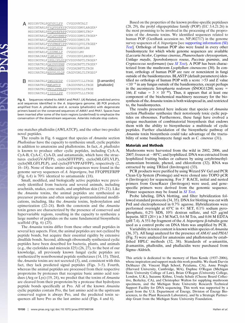
one matches phalloidin (AWLATCP), and the other two predictnovel peptides.
The results in Fig. 6 suggest that species of Amanita sectionPhalloideae have the capacity to synthesize small, cyclic peptidesin addition to amatoxins and phallotoxins. In fact, A. phalloidesis known to produce other cyclic peptides, including CyA-A,CyA-B, CyA-C, CyA-D, and antamanide, which have the struc-tures cyclo(GVAFFP), cyclo(SFFFPIP), cyclo(MLGFLVLP),cyclo(MLGFLPLP), and cyclo(FFVPPAFFPP), respectively (2,16–18). None of these amino acid sequences were found in thegenome survey sequences of A. bisporigera, but FFQPPEFRPP(Fig. 6A) is 70% identical to antamanide (18).
Small, modified, and biologically active peptides were previ-ously identified from bacteria and several animals, includingarachnids, snakes, cone snails, and amphibian skin (19–21). Likethe Amanita toxins, the animal peptides are synthesized asprecursor proteins and often undergo posttranslational modifi-cations, including, like the Amanita toxins, hydroxylation andepimerization (22–24). Both the conotoxin and the Amanitatoxin genes are characterized by the presence of conserved andhypervariable regions, resulting in the capacity to synthesize alarge number of peptides on the same fundamental biosyntheticscaffold (Fig. 6) (25).
The Amanita toxins differ from these other small peptides inseveral key aspects. First, the animal peptides are not cyclized bypeptide bonds, but acquire their essential rigidity by extensivedisulfide bonds. Second, although ribosomally synthesized cyclicpeptides have been described for bacteria, plants, and animals(e.g., the cyclotides and microcin J25) (26, 27), to the best of ourknowledge, all previously known fungal cyclic peptides aresynthesized by nonribosomal peptide synthetases (14, 15). Third,the Amanita toxins are not secreted (3), and, consistent with thisfact, they lack predicted signal peptides (Figs. 3–5). Fourth,whereas the animal peptides are processed from their respectiveproproteins by proteases that recognize basic amino acid resi-dues (Arg or Lys) (19, 24), we predict that the toxins of Amanitaare cleaved from their proproteins by a protease that hydrolyzespeptide bonds specifically at Pro. All of the known Amanitacyclic peptides contain Pro, the last amino acid in the upstreamconserved region is always Pro, and the predicted toxin se-quences all have Pro as the last amino acid (Figs. 4 and 6).
Based on the properties of the known proline-specific peptidases(28, 29), the prolyl oligopeptidase family (POP) (EC 3.4.21.26) isthe most promising to be involved in the processing of the propro-teins of the Amanita toxins. We identified sequences related tohuman POP (GenBank accession no. NP�002717) in the genomesurvey sequences of A. bisporigera [see supporting information (SI)Text]. Orthologs of human POP also were found in every otherbasidiomycete for which whole genome sequences are available(Laccaria bicolor, Coprinus cinereus, Phanerochaete chrysosporium,Ustilago maydis, Sporobolomyces roseus, Puccinia graminis, andCryptococcus neoformans) (see SI Text). A POP has been charac-terized from the mushroom Lyophyllum cinerascens (30). In con-trast, orthologs of human POP are rare or nonexistent in fungioutside of the basidiomycetes. BLASTP (default parameters) iden-tified no orthologs of human POP with a score �53 and E value�10�6 in any fungus outside of the basidiomycetes, except perhapsin the ascomycete Setosphaeria nodorum (SNOG11288; score �166; E value � 3 � 10�40). Thus, it appears that at least onecomponent of the biochemical machinery necessary for the bio-synthesis of the Amanita toxins is both widespread in, and restrictedto, the basidiomycetes.
The results presented here indicate that species of Amanitasection Phalloidae synthesize their notoriously toxic cyclic pep-tides on ribosomes. Furthermore, these fungi have evolved aunique mechanism of combinatorial biosynthesis that endowsthem with the ability to biosynthesize a multitude of cyclicpeptides. Further elucidation of the biosynthetic pathway ofAmanita toxin biosynthesis could take advantage of the tracta-bility of some basidiomycete fungi such as C. cinereus (31).
Materials and MethodsMushrooms were harvested from the wild in 2002, 2006, and2007; frozen at �80°C; and lyophilized. DNA was extracted fromlyophilized fruiting bodies or cultures by using cetyltrimethyl-ammonium bromide, phenol, and chloroform (32). RNA wasextracted by using TRIzol (Invitrogen) (33).
PCR products were purified by using Wizard SV Gel and PCRClean-Up System (Promega) and were cloned into TOPO pCR4 (Invitrogen) for sequencing. For 3� RACE, initial and nestedprimers from GeneRacer (Invitrogen) were used, and gene-specific primers were derived from the genomic sequence.Primer sequences may be found in SI Text.
Probe labeling, DNA blotting, and filter hybridization fol-lowed standard protocols (34, 35). DNA for blotting was cut withPstI and electrophoresed in 0.7% agarose. Hybridizations wereperformed overnight at 65°C in 4� SET, 0.1% sodium pyro-phosphate, 0.2% SDS, 10% dextran sulfate, and 625 �g/mlheparin. SET (20�) is 3 M NaCl, 0.6 M Tris, and 0.04 M EDTA(pH 7.4). A 551-bp fragment of the A. bisporigera �-tubulin geneused as a control probe on DNA blots was amplified by PCR.
Variability in toxin content is known within species of Amanita(36, 37). All fungi analyzed for the presence of AMA1 and PHA1(Fig. 5) were analyzed for amatoxins and phallotoxins by estab-lished HPLC methods (32, 38). Standards of �-amanitin,�-amanitin, phalloidin, and phallacidin were purchased fromSigma–Aldrich.
This article is dedicated to the memory of Hans Kende (1937–2006),whose inspiration and support made this work possible. We thank DarvinDeShazer (St. Vincent High School, Petaluma, CA), Anne Pringle(Harvard University, Cambridge, MA), Daphne O’Regan (MichiganState University College of Law), Brian O’Regan (University College,London, U.K.), Suzanne Kiihne, Ursula Schulz (Cheese Board Collec-tive, Berkeley, CA), and Christopher Walton for supplying mushroomspecimens, and the Michigan State University Research TechnicalSupport Facility for DNA sequencing. This work was supported by agrant from the U.S. Department of Energy, Division of Energy Bio-sciences, to the Plant Research Laboratory, and by a Strategic Partner-ship Grant from the Michigan State University Foundation.
A
B
Fig. 6. Sequences related to AMA1 and PHA1. (A) Related, predicted aminoacid sequences identified in the A. bisporigera genome. (B) PCR productsamplified from A. phalloides and A. ocreata (phalloidin) with degenerateprimers based on the conserved sequences of AMA1 and PHA1. Spaces havebeen inserted after some of the toxin regions (underlined) to emphasize theconservation of the downstream sequences. Asterisks indicate stop codons.
19100 � www.pnas.org�cgi�doi�10.1073�pnas.0707340104 Hallen et al.

1. Bresinsky A, Besl H (1990) A Colour Atlas of Poisonous Fungi: A Handbook forPharmacists, Doctors and Biologists (Wolfe, Wurzburg, Germany).
2. Wieland T (1986) Peptides of Poisonous Amanita Mushrooms (Springer, NewYork).
3. Zhang P, Chen Z, Hu J, Wei B, Zhang Z, Hu W (2005) FEMS Microbiol Lett252:223–228.
4. Tulloss RE (2000) Boll Gruppo Micologico G Bresadola 43:13–21.5. Wei� M, Yang Z-L, Oberwinkler F (1998) Can J Bot 76:1170–1179.6. Bushnell DA, Cramer P, Kornberg RD (2002) Proc Natl Acad Sci USA
99:1218–1222.7. Lengsfeld AM, Low I, Wieland T, Dancker P, Hasselbach W (1974) Proc Natl
Acad Sci USA 71:2803–2807.8. Kroncke KD, Fricker G, Meier PJ, Gerok W, Wieland T, Kurz G (1986) J Biol
Chem 261:2562–2567.9. Letschert K, Faulstich H, Keller D, Keppler D (2006) Toxicol Sci 91:140–149.
10. Enjalbert F, Rapior S, Nouguier-Soule J, Guillon S, Amouroux N, Cabot C(2002) J Toxicol Clin Toxicol 40:715–757.
11. Bamburg JR (1999) Annu Rev Cell Dev Biol 15:185–230.12. Le Quere A, Johansson T, Tunlid A (2002) Fung Genet Biol 36:234–241.13. Margulies M, Egholm M, Altman WE, Attiya S, Bader JS, Bemben LA, Berka
J, Braverman MS, Chen YJ, Chen Z, et al. (2005) Nature 437:376–380.14. Walton JD, Panaccione DG, Hallen HE (2004) in Advances in Fungal Bio-
technology for Industry, Agriculture, and Medicine, eds Tkacz JS, Lange L(Kluwer, New York), pp 127–162.
15. Finking R, Marahiel MA (2004) Annu Rev Microbiol 58:453–488.16. Gauhe A, Wieland T (1977) Justus Liebigs Ann Chem, pp 859–868.17. Chiang CC, Karle IL, Wieland T (1982) Int J Peptide Protein Res 20:414–420.18. Wieland T, Luben G, Otttenheym H, Faesel J, deVries JX, Konz W, Prox A,
Schmid J (1968) Angew Chem Int Ed Engl 7:204–208.
19. Escoubas P (2006) Mol Divers 10:545–554.20. Olivera BM (2006) J Biol Chem 281:31173–31177.21. Simmaco M, Mignogna G, Barra D (1998) Biopolymers 47:435–450.22. Buczek O, Bulaj G, Olivera BM (2005) Cell Mol Life Sci 62:3067–3079.23. Shikata Y, Watanabe T, Teramoto T, Inoue A, Kawakami Y, Nishizawa Y,
Katayama K, Kuwada M (1995) J Biol Chem 270:16719–16723.24. Richter K, Egger R, Negri L, Corsi R, Severini C, Kreil G (1990) Proc Natl Acad
Sci USA 87:4836–4839.25. Woodward SR, Cruz LJ, Olivera BM, Hillyard DR (1990) EMBO J 9:1015–
1020.26. Craik DJ, Cemazar M Daly NL (2007) Curr Opin Drug Discov Devel 10:176–
184.27. Rosengren KJ, Clark RJ, Daly NL, Goransson U, Jones A, Craik DJ (2003)
J Am Chem Soc 125:12464–12474.28. Cunningham DF, O’Connor B (1997) Biochim Biophys Acta 1343:160–186.29. Polgar L (2002) Cell Mol Life Sci 59:349–362.30. Yoshimoto T, Sattar AK, Hirose W, Tsuru D (1988) J Biochem 104:622–627.31. Kues U (2000) Microbiol Mol Biol Rev 64:316–353.32. Hallen HE, Watling R, Adams GC (2003) Mycol Res 107:969–979.33. Hallen HE, Huebner M, Shiu S-H, Guldener U, Trail F (2007) Fung Genet Biol
44:1146–1156.34. Maniatis T, Fritsch EF, Sambrook J (1982) Molecular Cloning: A Laboratory
Manual (Cold Spring Harbor Lab Press, Cold Spring Harbor, NY).35. Singh L, Jones KW (1984) Nucleic Acids Res 12:5627–5638.36. Beutler JA, der Marderosian AH (1981) J Nat Prod 44:422–431.37. Tyler VE, Jr, Benedict RG, Brady LR, Robbers JE (1966) J Pharm Sci
55:590–593.38. Enjalbert F, Gallion C, Jehl F, Monteil H, Faulstich H (1992) J Chromatogr
598:227–236.
Hallen et al. PNAS � November 27, 2007 � vol. 104 � no. 48 � 19101
MIC
ROBI
OLO
GY