Embed Size (px)
Citation preview
INFECTION AND IMMUNITY, June 1992, p. 2361-23670019-9567/92/062361-07$02.00/0Copyright © 1992, American Society for Microbiology
Cloning and Nucleotide Sequence of the Gene Encoding the 136-Kilodalton Surface Protein (Muramidase-Released Protein) of
Streptococcus suis Type 2HILDE E. SMITH,1* URI VECHT,2 ARNO L. J. GIELKENS,' AND MARI A. SMITS'
Departments of Molecular Biology' and Bacteriology, 2 DLO-Central Veterinary Institute,P.O. Box 65, 8200 AB Lelystad, The Netherlands
Received 11 November 1991/Accepted 16 March 1992
We cloned and sequenced the gene encoding the muramidase-released protein (MRP) of a pathogenicStreptococcus suis type 2 strain to determine whether its amino acid sequence resembles that of proteins withknown functions and to determine its function in virulence. The complete nucleotide sequence composing thegene and the regions flanking it was determined. The deduced amino acid sequence revealed the presence of a
signal peptide at the N terminus and a cell envelope anchor at the C terminus, both of which resembled similarregions in several other surface proteins from gram-positive bacteria. The processed form of MRP has a lengthof 1,209 amino acids and a calculated molecular weight of 131,094. A highly repetitive region preceded theenvelope anchor. The repeated units were preceded by a proline-rich stretch of amino acids (26 of 86).No overall homologies were observed between the amino acid sequence of MRP and protein sequences inthe EMBL data bank. A particular region within the amino acid sequence, however, showed some similaritywith the fibronectin-binding protein of Staphylococcus aureus. Binding of MRP to human fibronectin, how-ever, could not be confirmed.
Streptococcus suis type 2 infections are a common causeof septicemia, arthritis, meningitis, and sudden death inyoung pigs (3, 29) and meningitis in humans (2). During thelast few years, S. suis type 2 infections have become a majorproblem in almost all countries with an intensive pig indus-try. Attempts to control the disease are hampered by thelack of effective vaccines and suitable diagnostics.
In recent studies, we identified three phenotypes of S. suistype 2 strains, each of which caused specific pathologicaland clinical signs (31, 32). Some strains produced both a
136-kDa cell envelope-associated protein, muramidase-re-leased protein (MRP), and a 110-kDa extracellular proteinfactor (EF). Strains of this phenotype (MRP+ EF+) were
frequently isolated from organs of diseased pigs and wereassociated with severe clinical signs of disease after experi-mental infection. We also identified strains that did notproduce MRP and EF (MRP- EF-). Strains of this pheno-type were frequently isolated from tonsils of healthy pigs andwere nonpathogenic after experimental infection. Finally,we detected strains that produced MRP but not the 110-kDaEF. These strains produce EF-related (EF*) proteins ofvarious sizes (>150 kDa). These EF-related proteins are
encoded by ef genes that contain insertions of various sizes(25). Most of these MRP+ EF* strains were isolated fromhuman patients and did not cause disease in young pigs afterexperimental infection. Both MRP and EF are recognized byconvalescent antisera from pigs infected with sublethaldoses of MRP+ EF+ strains.We cloned and sequenced the gene encoding MRP of a
pathogenic S. suis type 2 strain to determine whether itsamino acid sequence resembles that of proteins with knownfunctions and to determine its function in virulence. Wefound little overall homology to amino acid sequences ofknown proteins. A subsequence of MRP, however, showed
* Corresponding author.
some similarity with the fibronectin-binding protein ofStaphylococcus aureus. Binding of MRP to human fibronec-tin, however, could not be confirmed.
MATERIALS AND METHODS
Bacterial strains and growth conditions. Escherichia coliJM101 [supE thi A (lac-proAB) (F' traD36 lacIVZAM15)] (16)was used as host for recombinant plasmid DNA. E. coliLE392 [F- hsdR574 (rK iK) supE44 supF58 lacY1 orA(laclZY)6 galK2 galT22 melBi trpR55] (18) was used as a
host for recombinant bacteriophage. The pathogenic MRP+EF+ strain D282 of S. suis type 2 (28) was used for isolatingchromosomal DNA. E. coli strains were grown on L broth(17). Solid LB medium contained 1.5% agar. Ampicillin wasadded as needed to a final concentration of 50 ,ug/ml. S. suisstrains were grown in Todd-Hewitt broth (Oxoid, Ltd.,London, England).DNA techniques. Routine DNA manipulations were per-
formed as described by Maniatis et al. (15).Southern blotting and hybridization. DNA was transferred
to GeneScreen Plus membranes (New England NuclearCorp., Dreieich, Germany) as described by Maniatis et al.(15). DNA probes were labeled with [a-32P]dCTP (3,000Ci/mmol; Amersham Corp., Arlington Heights, Ill.) by useof a random primed labeling kit (Boehringer GmbH, Mannheim, Germany). The blots were hybridized with DNAprobes as recommended by the supplier of the GeneScreenPlus membranes. After hybridization, the membranes werewashed twice with a solution of 2x SSC (lx SSC is 0.15 MNaCl plus 0.015 M Na citrate, pH 7.0) for 5 min at room
temperature and twice with a solution of 0.1 x SSC plus 0.5%sodium dodecyl sulfate for 30 min at 65°C.
Construction and immunological screening of DNA library.A DNA library of S. suis type 2 strain D282 was constructedin XGEM11, according to the methods recommended by themanufacturer of the cloning vector (Promega, Madison,
2361
Vol. 60, No. 6
2362 SMITH ET AL.
A clones
c MRP 7 9 10 1 12MAW
136w,
90~
8s
70
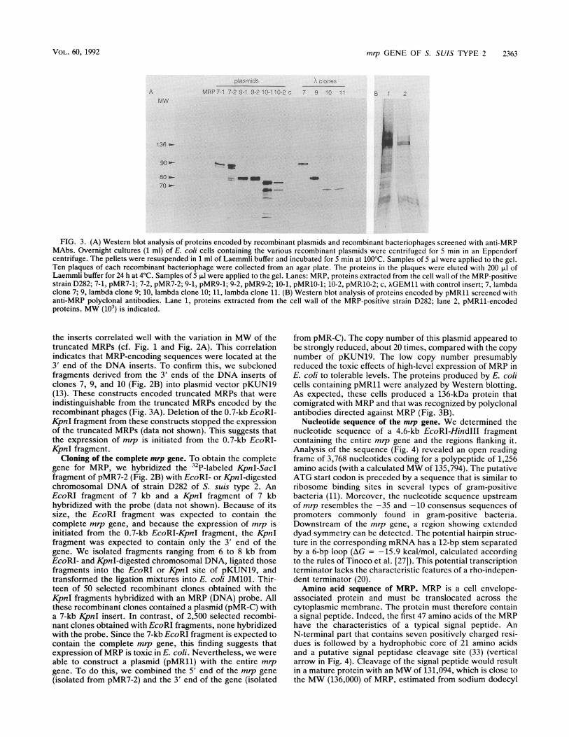
FIG. 1. Western blot analysis of proteins eluted from plaques ofrecombinant bacteriophages screened with anti-MRP MAbs. Tenplaques of each recombinant bacteriophage were collected from anagar plate. The proteins in the plaques were eluted with 200 RI ofLaemmli buffer for 24 h at 4°C. Samples of 5 ,ul were applied to thegel. Lanes: c, proteins extracted from the cell wall of an MRP-negative S. suis strain; MRP, proteins extracted from the cell wall ofan MRP-positive strain, D282; 7, proteins extracted from clone 7; 9,clone 9; 10, clone 10; 11, clone 11; 12, clone 12; MW (10') isindicated.
Wis.). Recombinant phages were plated on E. coli LE392and incubated for 16 h at 37°C. Nitrocellulose filters (Schlei-cher & Schuell, Inc., Dassel, Germany) were placed on theplaques, and the plates were further incubated for 2 h at37°C. Recombinants that produce MRP were visualized byuse of anti-MRP monoclonal antibodies (MAbs) (30). Boundantibodies were detected with anti-mouse serum conjugatedwith alkaline phosphatase (Zymed Laboratories, Inc., SanFrancisco, Calif.) as described by Maniatis et al. (15).Selected clones were purified by several rounds of single-plaque isolation and immunological screening.Sodium dodecyl sulfate-polyacrylamide gel electrophoresis
and Western blot (immunoblot) analysis. Proteins were sep-arated by sodium dodecyl sulfate-polyacrylamide gel elec-trophoresis in which 4% stacking and 6% separation gelswere used (14). The separated proteins were transferred tonitrocellulose by use of a Semi-Dry transfer cell (Bio-RadLaboratories, Richmond, Calif.). Specific proteins were vi-sualized by use of anti-MRP polyclonal antibodies (31) oranti-MRP MAbs (30) and anti-rabbit or anti-mouse serumconjugated with alkaline phosphatase (Zymed Laboratories).
Binding to fibronectin. To detect binding of proteins tofibronectin, we incubated Western blots with human plasmafibronectin (Sigma Chemical Co., St. Louis, Mo.) (25 ,ug/ml)for 2 h at 37°C (1, 26). Fibronectin-binding proteins werevisualized by use of rabbit antifibronectin immunoglobulinsconjugated with peroxidase (DAKOPATTS, Glostrup, Den-mark).
Nucleotide sequence analysis. DNA sequences were deter-mined by the dideoxy-chain termination method (21). DNAand protein sequences were analyzed by two softwaresystems: PCGENE (Intelligenetics Corp., Mountain View,Calif.) and Wisconsin GCG (University of Wisconsin). Themrp sequence has been assigned GenBank accession numberX64450.
RESULTS
Construction and screening of the library. ChromosomalDNA isolated from strain D282 of S. suis type 2 was partiallydigested with the restriction enzyme Sau3A. A DNA librarywas then constructed in the bacteriophage XGEM11 replace-ment vector. We obtained approximately 5 x 10⁵ recombi-nants per pug of DNA. An MAb directed against MRP wasused to screen 1,400 recombinant plaques for the presence ofantigenic determinants of MRP. Five recombinant plaqueswere reactive with the MAb.
Characterization of immunoreactive recombinants. Tostudy the expression of MRP by the five selected recombi-nant bacteriophages, we used Western blotting to analyzethe proteins eluted from the plaques. All five recombinantsencoded proteins that were recognized by MAbs directedagainst MRP (Fig. 1). These proteins, however, had lowermolecular weights (MW) than the MRP. Two clones encodeda protein of approximately 70 kDa (Fig. 1, clones 10 and 11),two clones encoded a protein of approximately 80 kDa (Fig.1, clones 9 and 12), and one clone encoded a protein ofapproximately 90 kDa (Fig. 1, clone 7). Therefore, weconcluded that the five recombinants did not contain thecomplete genetic information for MRP.To confirm this conclusion, we used restriction enzyme
analysis to compare the DNA inserts of the five recombi-nants (data not shown). All clones shared a DNA region ofabout 17 kb (Fig. 2A). The DNA inserts differed, however, atthe 3' and 5' ends. The variation in length at the 3' ends of
A7
S
9 -C
If H
H H
H H
10
11
B
H H
pMR7-1
pMR7-2
pMR9-1
pMR9-2
pMR1O-1
pMRIO-2
E E KX S
E E KX S
E E KX S
E E KX S
E E KX S
E KX S
E EKX S
E KX S
EE E KX S
IE KX S
1 kb
FIG. 2. (A) Restriction maps of the DNA inserts of putativeMRP-positive recombinant bacteriophages. The bar indicates theDNA region common to all these clones. Restriction sites: E,EcoRI; H, HindlIl; X, XbaI; K, KpnI; and S, Sacl. The Sacl sitesare derived from the lambda vector. (B) Parts of the DNA insertssubcloned in the plasmid vector pKUN19 (13).
II I...,
INFECT. IMMUN.
s
s
mrp GENE OF S. SUIS TYPE 2 2363
A
plas rnids ,cC ones
MRP7-1 7-2 9-1 9-2 10-110-2 c 7 9 10 11MlW
136
90 0-
80 *-
70
B 1 2
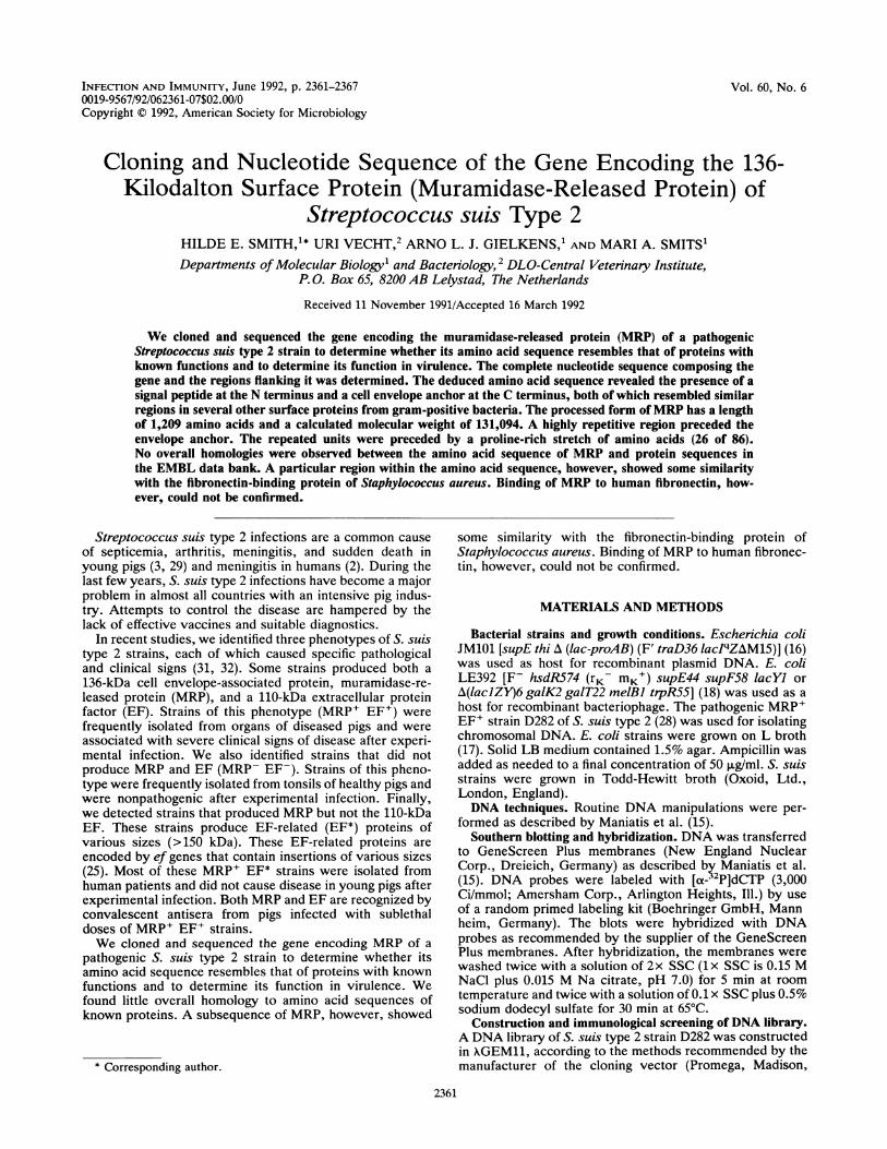
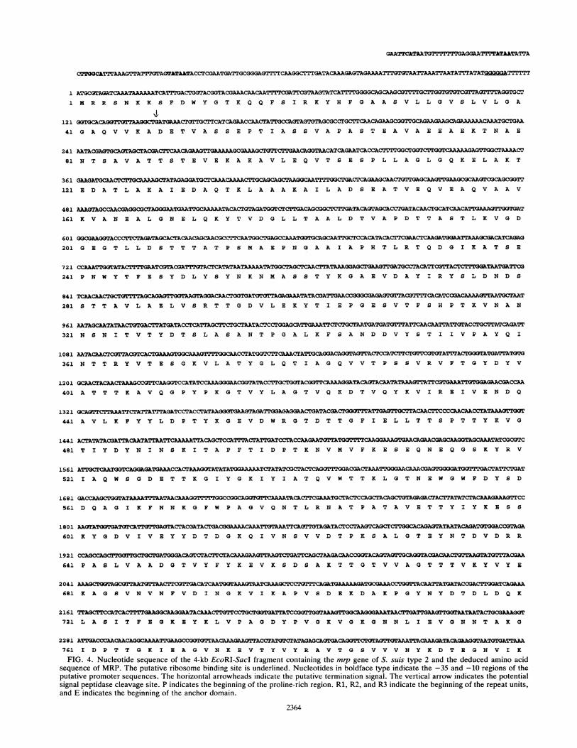
FIG. 3. (A) Western blot analysis of proteins encoded by recombinant plasmids and recombinant bacteriophages screened with anti-MRPMAbs. Overnight cultures (1 ml) of E. coli cells containing the various recombinant plasmids were centrifuged for 5 min in an Eppendorfcentrifuge. The pellets were resuspended in 1 ml of Laemmli buffer and incubated for 5 min at 100°C. Samples of 5 p1 were applied to the gel.Ten plaques of each recombinant bacteriophage were collected from an agar plate. The proteins in the plaques were eluted with 200 ,ul ofLaemmli buffer for 24 h at 4°C. Samples of 5 ,ul were applied to the gel. Lanes: MRP, proteins extracted from the cell wall of the MRP-positivestrain D282; 7-1, pMR7-1; 7-2, pMR7-2; 9-1, pMR9-1; 9-2, pMR9-2; 10-1, pMR10-1; 10-2, pMR10-2; c, XGEM11 with control insert; 7, lambdaclone 7; 9, lambda clone 9; 10, lambda clone 10; 11, lambda clone 11. (B) Western blot analysis of proteins encoded by pMR11 screened withanti-MRP polyclonal antibodies. Lane 1, proteins extracted from the cell wall of the MRP-positive strain D282; lane 2, pMR11-encodedproteins. MW (103) is indicated.
the inserts correlated well with the variation in MW of thetruncated MRPs (cf. Fig. 1 and Fig. 2A). This correlationindicates that MRP-encoding sequences were located at the3' end of the DNA inserts. To confirm this, we subclonedfragments derived from the 3' ends of the DNA inserts ofclones 7, 9, and 10 (Fig. 2B) into plasmid vector pKUN19(13). These constructs encoded truncated MRPs that wereindistinguishable from the truncated MRPs encoded by therecombinant phages (Fig. 3A). Deletion of the 0.7-kb EcoRI-KpnI fragment from these constructs stopped the expressionof the truncated MRPs (data not shown). This suggests thatthe expression of mrp is initiated from the 0.7-kb EcoRI-I4pnI fragment.
Cloning of the complete mrp gene. To obtain the completegene for MRP, we hybridized the 32P-labeled KpnI-SacIfragment of pMR7-2 (Fig. 2B) with EcoRI- or KpnI-digestedchromosomal DNA of strain D282 of S. suis type 2. AnEcoRI fragment of 7 kb and a KpnI fragment of 7 kbhybridized with the probe (data not shown). Because of itssize, the EcoRI fragment was expected to contain thecomplete m-p gene, and because the expression of mrp isinitiated from the 0.7-kb EcoRI-KpnI fragment, the KpnIfragment was expected to contain only the 3' end of thegene. We isolated fragments ranging from 6 to 8 kb fromEcoRI- and KpnI-digested chromosomal DNA, ligated thosefragments into the EcoRI or KpnI site of pKUN19, andtransformed the ligation mixtures into E. coli JM101. Thir-teen of 50 selected recombinant clones obtained with theKpnI fragments hybridized with an MRP (DNA) probe. Allthese recombinant clones contained a plasmid (pMR-C) witha 7-kb ApnI insert. In contrast, of 2,500 selected recombi-nant clones obtained with EcoRI fragments, none hybridizedwith the probe. Since the 7-kb EcoRI fragment is expected tocontain the complete mrp gene, this finding suggests thatexpression of MRP is toxic in E. coli. Nevertheless, we wereable to construct a plasmid (pMR11) with the entire mipgene. To do this, we combined the 5' end of the mip gene(isolated from pMR7-2) and the 3' end of the gene (isolated
from pMR-C). The copy number of this plasmid appeared tobe strongly reduced, about 20 times, compared with the copynumber of pKUN19. The low copy number presumablyreduced the toxic effects of high-level expression of MRP inE. coli to tolerable levels. The proteins produced by E. colicells containing pMR11 were analyzed by Western blotting.As expected, these cells produced a 136-kDa protein thatcomigrated with MRP and that was recognized by polyclonalantibodies directed against MRP (Fig. 3B).
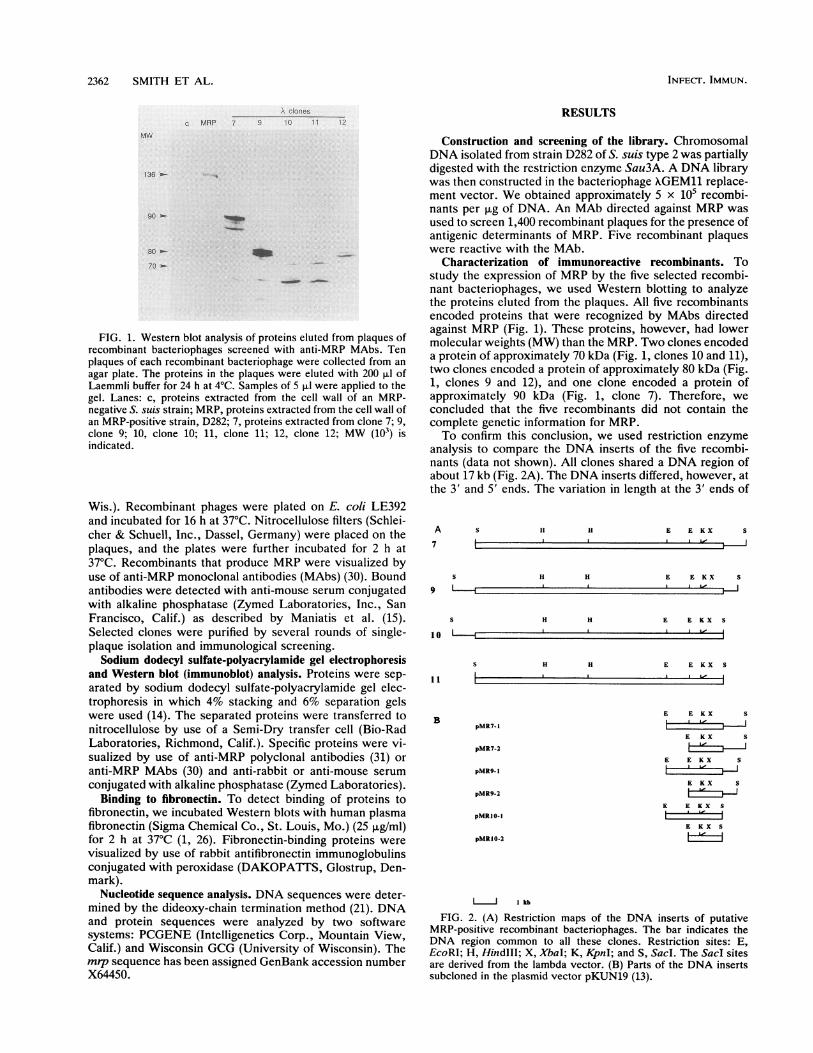
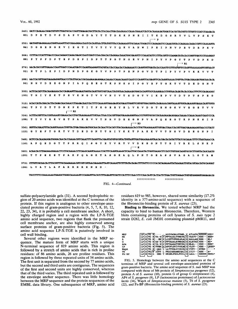
Nucleotide sequence of the mrp gene. We determined thenucleotide sequence of a 4.6-kb EcoRI-HindIII fragmentcontaining the entire mip gene and the regions flanking it.Analysis of the sequence (Fig. 4) revealed an open readingframe of 3,768 nucleotides coding for a polypeptide of 1,256amino acids (with a calculated MW of 135,794). The putativeATG start codon is preceded by a sequence that is similar toribosome binding sites in several types of gram-positivebacteria (11). Moreover, the nucleotide sequence upstreamof mip resembles the -35 and -10 consensus sequences ofpromoters commonly found in gram-positive bacteria.Downstream of the mrp gene, a region showing extendeddyad symmetry can be detected. The potential hairpin struc-ture in the corresponding mRNA has a 12-bp stem separatedby a 6-bp loop (AG = -15.9 kcal/mol, calculated accordingto the rules of Tinoco et al. [27]). This potential transcriptionterminator lacks the characteristic features of a rho-indepen-dent terminator (20).Amino acid sequence of MRP. MRP is a cell envelope-
associated protein and must be translocated across thecytoplasmic membrane. The protein must therefore containa signal peptide. Indeed, the first 47 amino acids of the MRPhave the characteristics of a typical signal peptide. AnN-terminal part that contains seven positively charged resi-dues is followed by a hydrophobic core of 21 amino acidsand a putative signal peptidase cleavage site (33) (verticalarrow in Fig. 4). Cleavage of the signal peptide would resultin a mature protein with an MW of 131,094, which is close tothe MW (136,000) of MRP, estimated from sodium dodecyl
VOL. 60, 1992
I i
GArCkTAA.'IT1'1'I'rGAGGAATTITAT&&TA¶TA
11
121
ATGCGTAGATCAAATAAAAAATCATTTGACTGGFACGGTACGAAACAACAATTTTCGATTCGTAAGTATCATTTTGGGGCAGCAAGM R R S N X K S F D W Y G T K Q Q F S I R K Y H F G A A S V L L G V S L V L G A
G3IGCACAGGTTGTTAAGGCTGATGAAACTGTTGCTTCATCAGAACCAACTATTGCCAGrAGTGTAGCGCCTGCTTCAACAGAAGCGGTTGCAGAAGAAGCAGAAAAAACAAATGCTGAA41 G A Q V V K A D E T V A S S E P T I A S S V A P A S T B A V A E E A E K T N A E
241 AATACGAGTGCAGT AGCTACGACTTCAACAGAAGTTGAAAAAGCGAAAGCTGTTCTTGAACAGGAACATCAGAATCA CT
81 N T S A V A T T S T B V E K A K A V L B Q V T S B S P L L A G L G Q K E L A K T
361 GAAGATGCAACTCII CAAAAGCTATAGAGGATGCTCAAACAAAAAC AGAAGAAGTCGCAGCGGTT
121 B D A T L A K A I B D A Q T K L A A A K A I L A D S E A T V E Q V E A Q V A A V
481 =AGCC%ACGAGGCGCTAGGAATGAATTGCAAATCTGA GATG2CTCTTG G ATA CAACATTGAAA:TTGGT
161 K V A N B A L G N E L Q K Y T V D G L L T A A L D T V A P D T T A S T L K V G D
601 GGCGAAGOTACCCTTCTAGATAGCACTACAACAGCAACGCCTTCAATGGCTAGCCAAATGGTGCOCAATTGCTCCACATACA201 G B G T L L D S T T T A T P S M A E P N G A A I A P H T L R T Q D G I K A T S E
721 CCAAATTWrATACTTTAGTATCTACGATTTGTACTCATATATAAAGG TGCCTACATTCG
241 P N W Y T F B S Y D L Y S Y N K N M A S S T Y K G A E V D A Y I R Y S L D N D S
841 CAAAAATAATGCTAAT
281 S T T A V L A B L V S R T T G D V L E K Y T I E P G E S V T F S H P T K V N A N
961 AATAGCAATATAACTGTGACTTATGATACCTCATTAGCTTCTGTAATACTCCTGGAGCATGAAATTTCTGCTAATGAT"GATTGATTCAACAATTAACCTATCAGATT321 N S N I T V T Y D T S L A S A N T P G A L K F S A N D D V Y S T I I V P A Y Q I
1081 AATACAACTCGIACTCACTGA AGTGGCACCTATGGTCTTCAATCTATTGCAIGAtAGGTAGAlACTCCATCTT361 N T T R Y V T E S G K V L A T Y G L Q T I A G Q V V T P S S V R V F T G Y D Y V
1201 GCAACT ACAACTAAGCCGTTCAAGGTATATC CGTGAAAT GGAGACACCA
401 A T T T K A V Q G P Y P K G T V Y L A G T V Q K D T V Q Y K V I R E I V E N D Q
1321 GCAGTTTAAATTCTATTATTTAGATCCTACCTAT CCCAACAACTAAAT
441 A V L K F Y Y L D P T Y K G E V D W R G T D T T G F I E L L T T S P T T Y K V G
1441 ACTATATACGATTACAATATTAATTCAAA TTACAGCTCCATTTACTATTGATCCTAC GAGCAAXTAGCAAATATCGCGTC
481 T I Y D Y N I N S K I T A P F T I D P T K N V M V F K E S E Q N E Q G S K Y R V
1561 ATTGCTCAATOGTCAGAGATGAAACCACTAAAGGrATATATGGAAAAATCTATATCGCTACTCAGOTTTGGACGACTAAATTGGGAACAAACG:CnC-KETrnwrl'GACTATTCTGAT521 I A Q W S G D B T T K G I Y G K I Y I A T Q V W T T K L G T N E W G W F D Y S D
1681 GACCAAG=TATAAAATTTAATAACAAGGTTTrTGGCCG T CAACTACTCCAGCTACAGCTGGAGACrACTTATATCTACAAAGAAAGTrCC561 D Q A G I K F N N K G F W P A G V Q N T L R N A T P A T A V B T T Y I Y K E S S
1801 AAGTATGGTGAToTCATTGTTSAOTACTACGATACrGACCTAAGTCACTTGG AGATACTCAGA CCGTAGA
601 K Y G D V I V B Y Y D T D G K Q I V N S V V D T P K S A L G T B Y N T D V D R R
1921641 P A S L V A A D G T V Y F Y K E V K S D S A K T T G T V V A G T T T V K Y V Y E
2041 CTTGCGTAATOTTAACTCGTTGTCAATGAAAAATCAACGCTCTTTGAAC CACTTaGATCAQAA681 K A G S V N V N F V D I N G K V I K A P V S D B K D A K P G Y N Y D T D L D Q K
2161 TTAGCTTC
721 L A S I T F B G K B Y K L V P A G D Y P V G K V G K G N N L I B V G N N T A K G
2281 ATACCCAC CA ACCTACAAAGTACClAC CTACAGKT761 I D P T T G K I B A G V N K B V T Y V Y R A V T G S V V V N Y K D T B G N V I K
FIG. 4. Nucleotide sequence of the 4-kb EcoRI-SacI fragment containing the mrp gene of S. suis type 2 and the deduced amino acidsequence of MRP. The putative ribosome binding site is underlined. Nucleotides in boldface type indicate the -35 and -10 regions of theputative promoter sequences. The horizontal arrowheads indicate the putative termination signal. The vertical arrow indicates the potentialsignal peptidase cleavage site. P indicates the beginning of the proline-rich region. Rl, R2, and R3 indicate the beginning of the repeat units,and E indicates the beginning of the anchor domain.
2364
CTTGGCILTTTAAAGTTATTTGTAOTATAATACCTCGAATGAT'TGCGGGAGTTTTCAAGGCTTTGATACAAAGAGTAGAAAATTTGTOTAATTAAATTAATATTTATAT9929MTTTTTT
mrp GENE OF S. SUIS TYPE 2 2365
GATCCAGiAAACGGTGTTCTAYTGCwACr,GTTGA5GATCTTAACrACAACTGICAAGACCAACABAACAATCATCACAAAAGATGGATCACGCTATTCTTlTCCATCrAAGACAD P E T D V S D A P V G D A Y T T T D K K P N E I I T K D G S R Y V L V P S K T
GATGGAGGAAAATGQAAAGTTATCGAAGGAA AACAATCACAGrAA TC G C ATCCAGAGATTCCAAATGTACCAGAACGACCGTCC3lAAAGTAD G E E N G K V I E G T I T V T Y V Y Q K V A N W I P E I P N V P E T D R P K V
CCTrACCCATTTGACCCAACAGAGCCAGACGKGCCAATCGATCCAACGACACCAGGAACAAATGGCGAGGTTCCAAATATTCCTTACGTrCCAGGkATATACACCGGTTGATCCTAAGGATP Y P F D P T E P D E P I D P T T P G T N G E V P N I P Y V P G Y T P V D P K D
AACACGCCGTTGvAAACCAATTGATCCAAATGATrCCAGGTAAGGGTTATGrACCALCCAACACCAGAA.AArCCAOMTGTTGATACACCAATTrr TTTCAGanuuu;FwrAAAAAT ACTN T P L K P I D P N D P G K G Y V P P T P E N P G V D T P I P Y V P V K K V V T
AACACoTBTGL BA aoAACCTsTGCCoCAAGAAGAGGGAACAAAAACCAAACAAATCAATCCCAGGSTTACGAOTTCACAoG rAACGTACGCGA_K;ACACAN H V D E E G N P I A P Q E E G T K P N K S I P G YE F T G K T V T D E D G N T
ACTCACATCTACAAGAAAACAC___CAAGGAACCTt3TAACAGATACACCAACTTrCTCCAGAAGG3CT H I Y K K T P E V K N G T V V V N Y V T E D G T V I K E P V T D T P T S P E G
ACACCAAGA_CACGCkAAACTAAGATCATTCAAGTGAAGAGTT P Y D T T D N K P K T I T F K G B B Y E L V R V D G T E N G K V V E G E T V V
(---> R2
ACTTACGrTTTACCGT AAAGITOC3AAACACC GCTAAGSAAA T GTAACTAACA OGAGAGAGGAA CGCAAAGG_ L.CAACAAACAAATCkACCCAT Y V Y R K V E T P A K K V V T N H V D E E G N P V A P Q E E G T K P N K S I P
[---> R3
GTTACGAATTrACAGGT _ ACAA GAAAACAC _ T A TACCTATTG Y E F T G K T V T D B D G N T T H I Y K K T P A K K V V T N H V D EE G N P I(}CTC ACA
A P Q E D G T T P K R Q I S G Y E Y V R T V V D E E G N T T H I Y R K L S N K P
r---> EACAACGGAGAATCG AACCACAOAAAACCCrAGOTAAAGTCAATCCAAATACTGGTGAGGCTTV O_AsATT T P E K E T P A K P Q A G K T A S G K A Q L P N T G E A S S V A G A L G T A M
TTPEKETPA=PQAGKTASGKAQTTNTGAATAVAGGTACATL V A T L A F A R K R R R N E D -
TGCCTTCCGAAAAAATGAG AAAAATCCAGAGTTACATCTTGATTCGCTCCATT4AACCAGCACeTd
FIG. 4Continued.
sulfate-polyacrylamide gels (31). A second hydrophobic re-
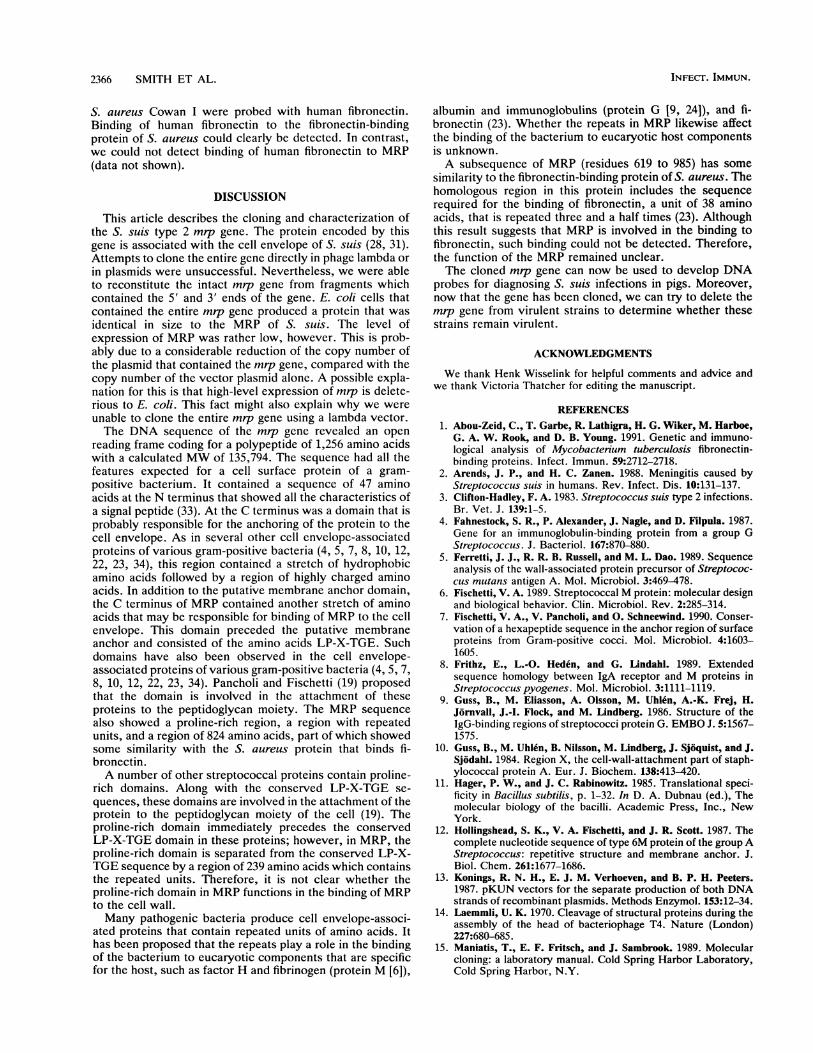
gion of 20 amino acids was identified at the C terminus of theprotein. If this region is analogous to other envelope-asso-ciated proteins of gram-positive bacteria (4, 5, 7, 8, 10, 12,22, 23, 34), it is probably a cell membrane anchor. A short,highly charged region and a region with the LP-X-TGEamino acid sequence, two regions that flank the presumedcell membrane anchor, are also highly conserved amongsurface proteins of gram-positive bacteria (Fig. 5). Theamino acid sequence LP-X-TGE is putatively involved incell wall binding.
Several other regions were identified in the MRP se-
quence. The mature form of MRP starts with a uniqueN-terminal sequence of 819 amino acids. This region isfollowed by a stretch of amino acids that is rich in prolineresidues: of 86 amino acids, 26 are proline residues. Thisregion is followed by three repeated units of 54 amino acids.The first unit is separated from the second by 77 amino acids,but the second and third units are contiguous. The sequencesof the first and second units are highly conserved, whereasthat of the third varies. The third repeated unit is followed bythe envelope anchor sequence. There was little homologybetween the MRP sequence and the protein sequences of theEMBL data library. One subsequence of MRP, amino acid
residues 619 to 985, however, shared some similarity (17.2%identity in a 377-amino-acid sequence) with a sequence ofthe fibronectin-binding protein of S. aureus (23).
Binding to fibronectin. We tested whether MRP had thecapacity to bind to human fibronectin. Therefore, Westernblots containing proteins of cell lysates of S. suis type 2strain D282, E. coli JM101 containing plasmid pMR11, and
MRP LP N TG E ---- - ASSVAGALGTAML V ATLAFA RKRRR NED*M6 LP S TG E TA-N P FFTAAALTVMATA G VAAVV- KRK- - EEN*A LP E 1G E - - EN P LIGTVFGGLSLA G AALLAG RR- EL*G LP S TG E GS-N PIIFTAAALAVMAGA G ALAVAS KRK- - ED*AP4 LP S TG E TA-N P FFTAAAATVMVSA G MLAL- - KRR- -EEN*LP LP K TG E ITERIP AFGFLVIWSLM G VLGV-- KRK-- QREE*WapA LP S TG E -QAG L LLTIVGLVIVAVA G VYFY- - RTRR- _ *
T6 LP S 1G IGTY L FKAIGSAAMIGAI G IYIV-- RRK-- A*Fn-BP LP E TG G -EES T NKGMLFGGLFSIL G LALL-- RRNKK NHKA*
FIG. 5. Homology between the amino acid sequences at the Cterminus of MRP and several cell envelope-associated proteins ofgram-positive bacteria. The amino acid sequence of S. suis MRP was
compared with those of M6 protein of Streptococcus pyogenes (12),protein A of S. aureus (10), protein G of group G streptococci (4),AP4 of S. pyogenes (8), LP (lactococcus proteinase) of Lactococcuslactis (34), WapA of Streptococcus mutans (5), T6 of S. pyogenes(22), and Fn-BP (fibronectin-binding protein) of S. aureus (23).
VOL. 60, 1992
2401801
2521841
2641881
2761921
2881961
30011001
31211041
32411081
33611121
34811161
36011201
37211241
2366 SMITH ET AL.
S. aureus Cowan I were probed with human fibronectin.Binding of human fibronectin to the fibronectin-bindingprotein of S. aureus could clearly be detected. In contrast,we could not detect binding of human fibronectin to MRP(data not shown).
DISCUSSION
This article describes the cloning and characterization ofthe S. suis type 2 mrp gene. The protein encoded by thisgene is associated with the cell envelope of S. suis (28, 31).Attempts to clone the entire gene directly in phage lambda orin plasmids were unsuccessful. Nevertheless, we were ableto reconstitute the intact mrp gene from fragments whichcontained the 5' and 3' ends of the gene. E. coli cells thatcontained the entire mrp gene produced a protein that wasidentical in size to the MRP of S. suis. The level ofexpression of MRP was rather low, however. This is prob-ably due to a considerable reduction of the copy number ofthe plasmid that contained the mrp gene, compared with thecopy number of the vector plasmid alone. A possible expla-nation for this is that high-level expression of mrp is delete-rious to E. coli. This fact might also explain why we wereunable to clone the entire mrp gene using a lambda vector.The DNA sequence of the mrp gene revealed an open
reading frame coding for a polypeptide of 1,256 amino acidswith a calculated MW of 135,794. The sequence had all thefeatures expected for a cell surface protein of a gram-positive bacterium. It contained a sequence of 47 aminoacids at the N terminus that showed all the characteristics ofa signal peptide (33). At the C terminus was a domain that isprobably responsible for the anchoring of the protein to thecell envelope. As in several other cell envelope-associatedproteins of various gram-positive bacteria (4, 5, 7, 8, 10, 12,22, 23, 34), this region contained a stretch of hydrophobicamino acids followed by a region of highly charged aminoacids. In addition to the putative membrane anchor domain,the C terminus of MRP contained another stretch of aminoacids that may be responsible for binding of MRP to the cellenvelope. This domain preceded the putative membraneanchor and consisted of the amino acids LP-X-TGE. Suchdomains have also been observed in the cell envelope-associated proteins of various gram-positive bacteria (4, 5, 7,8, 10, 12, 22, 23, 34). Pancholi and Fischetti (19) proposedthat the domain is involved in the attachment of theseproteins to the peptidoglycan moiety. The MRP sequencealso showed a proline-rich region, a region with repeatedunits, and a region of 824 amino acids, part of which showedsome similarity with the S. aureus protein that binds fi-bronectin.A number of other streptococcal proteins contain proline-
rich domains. Along with the conserved LP-X-TGE se-quences, these domains are involved in the attachment of theprotein to the peptidoglycan moiety of the cell (19). Theproline-rich domain immediately precedes the conservedLP-X-TGE domain in these proteins; however, in MRP, theproline-rich domain is separated from the conserved LP-X-TGE sequence by a region of 239 amino acids which containsthe repeated units. Therefore, it is not clear whether theproline-rich domain in MRP functions in the binding of MRPto the cell wall.Many pathogenic bacteria produce cell envelope-associ-
ated proteins that contain repeated units of amino acids. Ithas been proposed that the repeats play a role in the bindingof the bacterium to eucaryotic components that are specificfor the host, such as factor H and fibrinogen (protein M [6]),
albumin and immunoglobulins (protein G [9, 24]), and fi-bronectin (23). Whether the repeats in MRP likewise affectthe binding of the bacterium to eucaryotic host componentsis unknown.A subsequence of MRP (residues 619 to 985) has some
similarity to the fibronectin-binding protein of S. aureus. Thehomologous region in this protein includes the sequencerequired for the binding of fibronectin, a unit of 38 aminoacids, that is repeated three and a half times (23). Althoughthis result suggests that MRP is involved in the binding tofibronectin, such binding could not be detected. Therefore,the function of the MRP remained unclear.The cloned mrp gene can now be used to develop DNA
probes for diagnosing S. suis infections in pigs. Moreover,now that the gene has been cloned, we can try to delete themrp gene from virulent strains to determine whether thesestrains remain virulent.
ACKNOVVLEDGMENTS
We thank Henk Wisselink for helpful comments and advice andwe thank Victoria Thatcher for editing the manuscript.
REFERENCES1. Abou-Zeid, C., T. Garbe, R. Lathigra, H. G. Wiker, M. Harboe,
G. A. W. Rook, and D. B. Young. 1991. Genetic and immuno-logical analysis of Mycobacterium tuberculosis fibronectin-binding proteins. Infect. Immun. 59:2712-2718.
2. Arends, J. P., and H. C. Zanen. 1988. Meningitis caused byStreptococcus suis in humans. Rev. Infect. Dis. 10:131-137.
3. Clifton-Hadley, F. A. 1983. Streptococcus suis type 2 infections.Br. Vet. J. 139:1-5.
4. Fahnestock, S. R., P. Alexander, J. Nagle, and D. Filpula. 1987.Gene for an immunoglobulin-binding protein from a group GStreptococcus. J. Bacteriol. 167:870-880.
5. Ferretti, J. J., R. R. B. Russell, and M. L. Dao. 1989. Sequenceanalysis of the wall-associated protein precursor of Streptococ-cus mutans antigen A. Mol. Microbiol. 3:469-478.
6. Fischetti, V. A. 1989. Streptococcal M protein: molecular designand biological behavior. Clin. Microbiol. Rev. 2:285-314.
7. Fischetti, V. A., V. Pancholi, and 0. Schneewind. 1990. Conser-vation of a hexapeptide sequence in the anchor region of surfaceproteins from Gram-positive cocci. Mol. Microbiol. 4:1603-1605.
8. Frithz, E., L.-O. Heden, and G. Lindahl. 1989. Extendedsequence homology between IgA receptor and M proteins inStreptococcus pyogenes. Mol. Microbiol. 3:1111-1119.
9. Guss, B., M. Eliasson, A. Olsson, M. Uhlen, A.-K. Frej, H.Jornvall, J.-I. Flock, and M. Lindberg. 1986. Structure of theIgG-binding regions of streptococci protein G. EMBO J. 5:1567-1575.
10. Guss, B., M. Uhlen, B. Nilsson, M. Lindberg, J. Sjoquist, and J.Sjodahl. 1984. Region X, the cell-wall-attachment part of staph-ylococcal protein A. Eur. J. Biochem. 138:413-420.
11. Hager, P. W., and J. C. Rabinowitz. 1985. Translational speci-ficity in Bacillus subtilis, p. 1-32. In D. A. Dubnau (ed.), Themolecular biology of the bacilli. Academic Press, Inc., NewYork.
12. Hollingshead, S. K., V. A. Fischetti, and J. R. Scott. 1987. Thecomplete nucleotide sequence of type 6M protein of the group AStreptococcus: repetitive structure and membrane anchor. J.Biol. Chem. 261:1677-1686.
13. Konings, R. N. H., E. J. M. Verhoeven, and B. P. H. Peeters.1987. pKUN vectors for the separate production of both DNAstrands of recombinant plasmids. Methods Enzymol. 153:12-34.
14. Laemmli, U. K. 1970. Cleavage of structural proteins during theassembly of the head of bacteriophage T4. Nature (London)227:680-685.
15. Maniatis, T., E. F. Fritsch, and J. Sambrook. 1989. Molecularcloning: a laboratory manual. Cold Spring Harbor Laboratory,Cold Spring Harbor, N.Y.
INFECT. IMMUN.
mrp GENE OF S. SUIS TYPE 2 2367
16. Messing, J. 1979. A multipurpose cloning system based on thesingle-stranded DNA bacteriophage M13. Recombinant DNAtechnical bulletin. NIH publication no. 79-99, 2, no. 2, p. 43-48.National Institutes of Health, Bethesda, Md.
17. Miller, J. 1972. Experiments in molecular genetics. Cold SpringHarbor Laboratory, Cold Spring Harbor, N.Y.
18. Murray, N. E., W. J. Brammer, and K. Murray. 1977. Lamboidphages that simplify the recovery of in vitro recombinants. Mol.Gen. Genet. 150:53-58.
19. Pancholi, V., and V. A. Fischetti. 1988. Isolation and character-ization of the cell-associated region of group A streptococcal M6protein. J. Bacteriol. 170:2618-2624.
20. Platt, T. 1986. Transcription termination and the regulation ofgene expression. Annu. Rev. Biochem. 55:339-372.
21. Sanger, F., S. Nicklen, and A. R. Coulson. 1977. DNA sequenc-ing with chain-terminating inhibitors. Proc. Natl. Acad. Sci.USA 74:5463-5467.
22. Schneewind, O., K. F. Jones, and V. A. Fischetti. 1990. Se-quence and structural characteristics of the trypsin-resistant T6surface protein of group A streptococci. J. Bacteriol. 172:3310-3317.
23. Signas, C., G. Raucci, K. Jonsson, P.-E. Lindgren, G. M.Anantharamaiah, M. Hook, and M. Lindberg. 1989. Nucleotidesequence of the gene for a fibronectin-binding protein fromStaphylococcus aureus: use of this peptide sequence in thesynthesis of biologically active peptides. Proc. NatI. Acad. Sci.USA 86:699-703.
24. Sjobring, U., C. Falkenberg, E. Nielson, B. Akerstrom, and L.Bjorck. 1988. Isolation and characterization of a 14-kDa albu-min-binding fragment of streptococcal protein G. J. Immunol.140:1595-1599.
25. Smith, H. E., et al. Unpublished data.
26. Talay, S. R., E. Ehrenfeld, G. S. Chhatwal, and K. N. Timmis.1991. Expression of the fibronectin-binding components ofStreptococcus pyogenes in Escherichia coli demonstrates thatthey are proteins. Mol. Microbiol. 5:1727-1734.
27. Tinoco, I., Jr., P. N. Borer, B. Dengler, M. D. Devine, 0. C.Uhlenbeck, D. M. Crothers, and J. Gralla. 1973. Improvedestimation of secondary structure in ribonucleic acids. Nature(London) New Biol. 246:40-41.
28. Vecht, U., J. P. Arends, E. J. van der Molen, and L. A. M. G.van Leengoed. 1989. Differences in virulence between twostrains of Streptococcus suis type 2 after experimentally in-duced infection of newborn germfree pigs. Am. J. Vet. Res.50:1037-1043.
29. Vecht, U., L. A. M. G. van Leengoed, and E. R. M. Verheyen.1985. Streptococcus suis infections in pigs in The Netherlands(part one). Vet. Q. 7:315-321.
30. Vecht, U., H. J. Wisselink, J. Annakotta, and H. E. Smith.Submitted for publication.
31. Vecht, U., H. J. Wisselink, M. L. Jellema, and H. E. Smith. 1991.Identification of two proteins associated with virulence ofStreptococcus suis type 2. Infect. Immun. 59:3156-3162.
32. Vecht, U., H. J. Wisselink, J. E. van Dijk, and H. E. Smith. 1992.Virulence of Streptococcus suis type 2 strains in newborngermfree pigs depends on phenotype. Infect. Immun. 60:550-556.
33. Von Heijne, G. 1986. A new method for predicting signalsequence cleavage sites. Nucleic Acids Res. 14:4683-4690.
34. Vos, P., G. Simons, R. J. Siezen, and W. M. de Vos. 1989.Primary structure and organization of the gene for a procaryoticcell envelope-located serine proteinase. J. Biol. Chem. 264:13579-13585.
VOL. 60, 1992