Embed Size (px)
Citation preview

Regular paper
Disruption of Trichoderma reesei gene encoding protein O-mannosyl-transferase I results in a decrease of the enzyme activity and alteration
of cell wall composition
Wioletta Górka-Nieć, Michał Pniewski, Anna Kania, Urszula Perlińska-Lenart, Grażyna Palamarczyk and Joanna S. Kruszewska
Institute of Biochemistry and Biophysics, Polish Academy of Sciences, Warszawa, Poland
Received: 01 February, 2008; revised: 14 April, 2008; accepted: 25 April, 2008 available on-line: 26 May, 2008
In fungi transfer of the first mannosyl residue to proteins during their O-glycosylation is cata-lyzed by protein O-mannosyltransferases encoded by pmt genes. Disruption of the pmt1 gene in Trichoderma caused a significant decrease in the total activity of protein O-mannosyltransferases. Moreover, disruption of the pmt1 gene also led to osmotic sensitivity of the strain, indicating an essential role of the PMTI protein activity for cell wall synthesis. At the same time, the strain was defective in septa formation, producing only half the number of septa per unit length of hypha compared with the wild type. Disruption of the pmt1 gene decreased protein secretion but had no effect on glycosylation of secreted proteins, which suggests that PMTI protein O-manno-
syltranferase does not take part in glycosylation of these proteins.
Keywords: Trichoderma reesei, pmt1 gene disruption, protein glycosylation, cell wall composition
INTRODUCTION
Trichoderma species are widely exploited in biotechnology for protein production owing to their exceptional protein synthesis and secretory capabil-ity. Our previous study indicated that protein pro-duction and secretion could be closely related to the activity of the O-glycosylation pathway (Kruszewska et al., 1999; Perlińska-Lenart et al., 2005; 2006b).
In fungi the direct reaction of O-mannosyla-tion is catalyzed by protein O-mannosyltransferases (EC 2.4.1.109) and consists in transfer of a mannosyl residue from dolichyl phosphate mannose (DPM) to the serine/threonine OH group of the protein. It is known that in Saccharomyces cerevisiae protein O-mannosyltransferases are encoded by seven PMT genes and they are classified in three subfamilies PMT1, PMT2 and PMT4 (Gentzsch & Tanner, 1996; 1997). Members of the PMT1 and PMT2 subfamilies, Pmt1p, and Pmt5p, and Pmt2p, Pmt3p, and Pmt6,
respectively, form enzymaticaly active heterodimers such as Pmt1-Pmt2 and Pmt3-Pmt5. Deletion of a PMT gene encoding a protein from these groups re-sulted in the formation of less active complexes such as Pmt1-Pmt3 or Pmt2-Pmt5. The PMT4 family has one member only, Pmt4p, and this protein forms an active homodimer. Moreover, protein O-mannosyl-transferases are substrate-specific (Gentzsch & Tan-ner, 1997).
In yeast, O-glycosylation was shown to be es-sential for cell wall rigidity and cell integrity. A lack of activity of two (Pmt2p, Pmt3p or Pmt2p, Pmt4p) or three (Pmt1p, Pmt2p, Pmt3p) protein O-manno-syltransferases made the strains unable to grow in normal conditions, however, they could be saved by osmotic stabilization with 1 M sorbitol (Gentzsch & Tanner, 1996). On the other hand, some other triple deletions of PMT genes such as PMT1, PMT2, PMT4 or PMT2, PMT3, PMT4 could not be rescued by osmotic stabilization (Strahl-Bolsinger et al., 1999).
Corresponding author: Joanna S. Kruszewska, Institute of Biochemistry and Biophysics, Polish Academy of Sciences, A. Pawińskiego 5a, 02-106 Warszawa, Poland; phone: (48 22) 592 1209; fax: (48 22) 658 4636; e-mail: [email protected]: DPM, dolichyl phosphate mannose; DTT, dithiotheritol; GDP-mannose, guanosine 5’-diphospho-d-man-nose; PMT, protein O-mannosyltransferase; PMTI, protein O-mannosyltransferase I.
Vol. 55 No. 2/2008, 251–259
on-line at: www.actabp.pl

252 2008W. Górka-Nieć and others
Since protein O-mannosyltransferases are substrate-specific those results suggested that the vital role of protein O-mannosylation is connected with O-man-nosylation of some cell wall proteins.
In Schizosaccharomyces pombe the protein O-mannosyltransferase family is not as redundant as in S. cerevisiae and has only three members Oma1p, Oma2p and Oma4p, one in each PMT subfamily (Willer et al., 2005). The oma1∆ and oma4∆ mutants revealed some changes in their cell wall composition while deletion of the oma2 gene turned out to be le-thal (Willer et al., 2005).
Disruption of the pmtA gene in Aspergillus awamori gave a similar cell wall fragile phenotype as observed for yeasts. The disruptant exhibited swol-len hyphae formation and increased sensitivity to Congo Red and high temperature (Oka et al., 2005). Analysis of cell wall composition of the mutant re-vealed a lower content of alkali-insoluble fraction which is believed to be responsible for fungal cell wall rigidity, and simultaneous an increase of chitin level.
Our previous study pointed at a fundamental role of cell wall permeability for protein secretion in Trichoderma (Perlińska-Lenart et al., 2006b). Removal of the cell wall from Trichoderma strains carrying the yeast DPM1 gene, encoding DPM synthase resulted in a four-fold increase of protein secretion compared to mycelia which suggested that the cell wall made a barrier for secretion in these strains. This effect, however, was only observed for DPM1-transformed strains characterized by efficient processing of newly synthesized glycoproteins, but not for the control strain, showing that not only cell wall made a bar-rier for secretion but also the limited modification of secretory proteins (Perlińska-Lenart et al., 2006b).
Up to now only one pmt gene encoding pro-tein O-mannosyltransferase in Trichoderma has been cloned (NCBI accession number AF526877) (Zakrze-wska et al., 2003a). Analysis of the predicted protein sequence of the Trichoderma PMTI protein showed the highest 51% identity with S. cerevisiae Pmt4p. On the other hand, expression of the Trichoderma PMTI protein in S. cerevisiae pmt mutants revealed its func-tional similarity to the yeast Pmt2 protein. The Tri-choderma PMTI protein was able to form an active O-mannosyltransferase complex with yeast Pmt1p and partially rescued the defective glycosylation pat-tern of chitinase (Zakrzewska et al., 2003a) and com-pletely restored glycosylation of heterologously ex-pressed cellobiohydrolase II from Trichoderma in the pmt2∆ S. cerevisiae mutant (Górka-Nieć et al., 2007).
In this study we presented influence of dis-ruption of the pmt1 gene in T. reesei on the glyco-sylation, production and secretion of proteins and on cell wall composition. The strain carrying pmt1 disruption needed an osmotic stabilizer (e.g. 1 M
sorbitol) for growth in liquid medium. On the other hand, it was able to grow on the agar plates, albeit very slowly.
Disruption of the pmt1 gene resulted in a sig-nificant decrease in the total activity of protein O-mannosyltransferases and caused a decrease of pro-tein secretion but did not alter O- and N-glycosyla-tion of secreted proteins. Since protein O-mannosyl-transferases are substrate-specific and the lack of the PMTI activity did not change O-mannosylation of the secretory proteins, we conclude that PMTI does not take part in the O-glycosylation of these pro-teins.
MATERIALS AND METHODS
Strains and growth conditions. T. reesei TU-6 (Harman & Kubicek, 1998) was used as a recipient strain for transformation. Escherichia coli strain JM 109 was used for plasmid propagation (Yanish-Per-ron et al., 1985). T. reesei was cultivated at 30°C on a rotary shaker (250 r.p.m.) in 2 l shake flasks con-taining 1 l of minimal medium (MM): 1 g MgSO4 × 7H2O, 6 g (NH4)2SO4, 10 g KH2PO4, 3 g sodium citrate × 2H2O, and trace elements (25 mg FeSO4 × 7H2O, 2.7 mg MnCl2 × 4H2O, 6.2 mg ZnSO4 × 7H2O, 14 mg CaCl2 × 2H2O) per liter and 1% (w/v) lactose as a carbon source. The flasks were inoculated with 42 × 106 konidia/l medium.
Disruption of the pmt1 gene in T. reesei. The plasmid for pmt1 disruption was constructed as fol-lows. DNA fragment of about 4000 bp containing genomic pmt1 (NCBI accession number AY515299) and flanking sequences was amplified from genomic DNA by PCR, using The Expand High Fidelity PCR System (Boehringer, Mannheim) and pmt1-U2 (5’ CGC CAG CGA ATG ATT CGA CGG AGG 3’) and pmt1-L2 (5’ CAA GGT GGC TTC TTG TTG CGA CGA 3’) primers. The PCR product was cloned into the pGEM–T Easy Vector (Promega), digested with EcoRI and cloned in pRS316 (NCBI accession number U03442) devoid of SalI restriction site. Next the pmt1 gene was cut with SalI in the middle and the T. ree-sei pyr4 coding sequence cut out from the pGF1 vec-tor (Gruber et al., 1990) was cloned there. About 6500 bp of DNA was cut out from the resulting plasmid with EcoRI and used for T. reesei TU-6 transforma-tion. The pmt1 disruption in the pyr4+ transformants was confirmed by PCR using Pmt554U (5’ TTG GCT GGC TGG TTG GCT ACG AC 3’) and Pmt1730L (5’ GCT GGG AGT GAT TTG CTT GTT GC 3’) primers. For Southern blot analysis, DNA from disruptants and the parental strain was digested with PvuII and XhoI, loaded onto agarose gel, blotted and hybrid-ized with the 1.8 kb PvuII/XhoI fragment of T. reesei pmt1. Radioactive probe was prepared using [α-32P]

Vol. 55 253Disruption of pmt1 gene in Trichoderma
dATP and the Fermentas HexaLabel Plus DNA labe-ling system according to the standard protocol.
Molecular biology methods. Chromosomal DNA was isolated from T. reesei using the Promega Wizard Genomic DNA Purification kit. Other molec-ular biology procedures were performed according to standard protocols (Sambrook et al., 1989).
Biochemical methods
Membrane fraction preparation. Mycelium was harvested by filtration, washed with water and suspended in 50 mM Tris/HCl, pH 7.4, containing 15 mM MgCl2 and 9 mM β-mercaptoethanol. Cells were homogenized in a beadbeater with glass beads (0.5 mm) and the homogenate was centrifuged at 5000 × g for 10 min to remove cell debris and unbroken cells. The supernatant was centrifuged at 100 000 × g for 1 h. The membrane pellet was homogenized in 50 mM Tris/HCl, pH 7.4, containing 3.5 mM MgCl2 and 6 mM β-mercaptoethanol, and used as the source of enzymes. The whole procedure was per-formed at 4oC (Pless & Palamarczyk, 1987).
Activity of protein O-mannosyltransferases. Protein O-mannosyltransferase activity was assayed in the pelleted membrane fraction by 1 h incuba-tion at 30oC with GDP[14C]-mannose (sp. act. 288 Ci/mol, Amersham) and 5 ng of dolichyl phosphate (Dol-P), according to Kruszewska et al. (1989). Total membrane proteins (about 300 µg) were used as the sugar acceptor. Since only the transfer of the first mannosyl residue from dolichyl phosphate mannose (DPM) to the hydroxyl group of serine or threonine catalyzed by Pmt proteins was to be measured, the reaction mixture was supplemented with 10 mM MgCl2. Under these conditions elongation of the O-linked sugar chain does not occur (Sharma et al., 1974). The protein O-mannosyltransferase activity was expressed in pmoles of [14C]mannose incorpo-rated into 1 mg of membrane protein during 1 h.
N-acetylglucosamine transferase activity. N-acetylglucosamine transferase activity was measured in the membrane fraction by 30 min incubation at 30oC of 200 µg of membrane proteins in a total vol-ume of 50 µl containing 1 × 10⁵ c.p.m. UDP[14C]N-acetylglucosamine (sp. act. 249 Ci/mol, Amersham) and 5 ng of Dol-P in 40 mM Tris/HCl buffer, pH 7.4, with 10 mM MgCl2 and 0.1% Nonidet P-40 (Palamarczyk & Hemming, 1975). The reaction was stopped by addition of 4 ml of chloroform/methanol (3 : 2, v/v). Formation of radioactive dolichyl diphos-phate N-acetylglucosamine and dolichyl diphosphate chitobiose was measured in the organic fraction by a scintillation counter.
Concentration of saccharides bound to se-creted proteins. Saccharides bound to proteins iso-lated from T. reesei culture filtrates were assayed by
the phenol-sulfuric acid procedure (Dubois et al., 1956). Secreted proteins were precipitated with two volumes of ethanol, washed twice with 70% etha-nol and dissolved in distilled water. O-linked sug-ars were cleaved by mild alkaline hydrolysis and then remaining proteins containing N-linked sugars were precipitated with two volumes of ethanol and centrifuged. The concentration of O-linked carbohy-drates was measured in the supernatant after evapo-ration of ethanol, and N-linked sugars were assayed in the pellet. A calibration curve was prepared with d-mannose.
Protein concentration assay. Protein concen-trations were estimated according to Lowry et al. (1951).
Quantification of fungal dry mass. Fungal dry mass was quantified by filtering culture samples through G1 sintered glass funnels, washing the bio-mass with a threefold volume of tap water, and dry-ing to constant weight at 110oC.
Colony growth rate. Colony growth rates were measured as described by Oka et al. (2004). Co-nidia were point-inoculated into the center of agar MM plates with or without osmotic stabilizers (1 M sorbitol or 0.6 M KCl) or with the antifungal agent Calcofluor white (300 µg ml–1) (Oka et al., 2004) and incubated at 30°C. Colony diameter was measured at 48, 72, 96, 120 and 144 h. Measurements of the growth for all strains were done six times.
Cell wall preparation. T. reesei strains were cultivated for 168 h and the mycelia were harvested by centrifugation, homogenized in a beadbeater with 0.5 mm glass beads in 50 mM Tris/HCl, pH 7.5, with 1 mM DTT and centrifuged at 1500 × g for 10 min.
The resulting pellet containing cell walls was washed with ice-cold 1 M NaCl until disappearance of absorbance at 260–280 nm (Nemcovic & Farkas, 2001).
Determination of cell wall polysaccharides. The amount of glucans in the cell wall was deter-mined as described previously (Oka et al., 2004), with a slight modification. For quantification of al-kali-soluble β-(1,6)glucan, 200 mg of cell walls was suspended in 3% NaOH, heated at 75°C for 1 h and centrifuged. The supernatant was dialyzed overnight at 4oC against distilled water, lyophilized and the amount of alkali-soluble β-(1,6)glucan was estimat-ed by the method described by Dubois et al. (1956). The remaining pellet was washed twice with 0.1 M Tris/HCl, pH 7.4, and once with 10 mM Tris/HCl, pH 7.4, and digested overnight with zymolyase 20T (ICN Biomedicals Inc.) (5 mg/ml in 10 mM Tris/HCl pH 7.4). Then the samples were centrifuged (13 000 r.p.m., 15 min) and the supernatant was used to esti-mate the amount of alkali-insoluble β-(1,3)glucan by the same method (Dubois et al., 1956). The remain-ing pellets were incubated for 16 h with 70% sulfu-

254 2008W. Górka-Nieć and others
ric acid at 4oC, then diluted ten-fold with water and heated at 100oC for 8 h. After neutralization with 2 M NaOH samples were used to estimate the amount of alkali-insoluble β-(1,6)glucan (Dubois et al., 1956).
For chitin measurements alkaline hydrolysis of cell walls was performed in 6% KOH for 90 min at 80oC in order to release cell wall proteins. After neutralization with acetic acid, the cell walls were washed with phosphate-buffered saline and chi-tinase buffer, pH 6.0, containing 18 mM citric acid and 60 mM dibasic sodium phosphate. Subsequent-ly, the cell walls were treated with chitinase C (In-terSpex Products) for 3 h at 37oC. The level of chitin was measured with Ehrlich’s reagent as described (Reissig et al., 1955).
Microscopic analysis of mycelia. T. reesei transformants and control strains were harvested by centrifugation, washed twice with sterile water, re-suspended in warm 1.5% agarose (A-5030 Type IX from Sigma), allowed to cool and observed under a Nicon Eclipse E 6800 fluorescence microscope. The hyphae diameter and distances between septa were calculated from about one hundred pictures of each strain using Lucia G program.
RESULTS
Disruption of the pmt1 gene in T. reesei
T. reesei TU-6, a ∆pyr4 mutant of T. reesei QM9414, was transformed with a DNA fragment of about 6.5 kb containing T. reesei pyr4 gene flanked with pmt1 sequences, as described in Methods. Pro-totrophic transformants were selected and isolated by three rounds of transfer from selective to nonse-lective medium and screened by Southern blotting for the genomic copy of pmt1. To this aim genomic DNA of the transformants and the control strain was digested with PvuII/XhoI and hybridized with a pmt1 probe cut out from pGEM with the same restriction enzymes. The pmt1 gene contains one restriction site for each of the enzymes, so its intact copy should yield a 1.98 kb fragment hybridizing with the probe. Since T. reesei pyr4 has many PvuII and XhoI restric-tion sites, DNA obtained from the disruptants gave two fragments of 1.35 and 1.26 kb hybridizing with the pmt1 probe (Fig. 1A, B).
Effect of pmt1 gene disruption on the total activity of protein O-mannosyltransferases
Protein O-mannosyltransferase I (PMTI pro-tein) encoded by the pmt1 gene catalyzes transfer of the mannosyl residue from dolichyl phosphate mannose (DPM) to a serine or threonine OH group
in the protein. To determined the influence of pmt1 disruption on the total activity of direct O-glyco-sylation we measured the combined activity of all PMT proteins. The experiment revealed a signifi-cant decrease of the total activity of protein O-man-nosyltransferases, to 67% of the control value (Fig. 1C).
Effect of the lower activity of protein O-mannosyl-transferases on the growth of Trichoderma, protein secretion and glycosylation of secreted proteins
Trichoderma strains were cultivated on agar plates containing MM medium with glucose or lac-tose as a carbon source, supplemented with 1 M sorbitol or 0.6 M KCl as osmotic stabilizers (Oka et al., 2004). Every 24 h the diameter of the colony was measured and the results are presented as millilitre of growth per hour (Table 1). Colony growth of the strain carrying disruption of the pmt1 gene was very slow and reached 17% of the control when cultivat-ed on the MM medium without osmotic stabilizers. Addition of 1 M sorbitol or 0.6 M KCl to the cultiva-tion medium inhibited growth of the control strain and the level of inhibition depended on the carbon source. The most pronounced effect on growth of the control strain was found on lactose-containing medium supplemented with sorbitol. An opposite effect was observed for the pmt1 disruptant which grew nearly twice better with osmotic stabilization, nonetheless, the growth still remained slower com-pared to the growth of the control strain on medium without supplements.
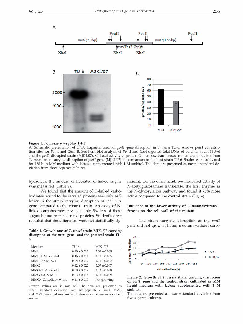
We also examined growth of the strains in liq-uid MM medium with lactose and it turned out that the strain MJK1/07 carrying pmt1 disruption was not able to growth without osmotic stabilization. There-fore, Trichoderma strains were cultivated with 1 M sorbitol and every 24 h mycelia were collected and growth of the strains was presented as an increase of dry mass of mycelia during cultivation (Fig. 2). Rather unexpectedly the pmt1 disruptant turned out to grow better than the control strain. At the same time we measured the amount of proteins liberated to the cultivation medium during 288 h of cultiva-tion and found that the strain carrying disruption of the pmt1 gene secreted less protein compared to the control strain (Fig. 3).
Since we found that the activity of protein O-mannosyltransferases in the examined strain was decreased we expected changes in the intensity of O-glycosylation of secreted protein. Since changes in O-glycosylation could influence N-glycosylation (Ecker et al., 2003) we characterized the amount of both O- and N-linked sugars bound to the secreted proteins. To this end, the proteins from the cultiva-tion medium were isolated and after mild alkaline
Vol. 55 255Disruption of pmt1 gene in Trichoderma
hydrolysis the amount of liberated O-linked sugars was measured (Table 2).
We found that the amount of O-linked carbo-hydrates bound to the secreted proteins was only 14% lower in the strain carrying disruption of the pmt1 gene compared to the control strain. An assay of N-linked carbohydrates revealed only 5% less of these sugars bound to the secreted proteins. Student’s t-test revealed that the differences were not statistically sig-
nificant. On the other hand, we measured activity of N-acetylglucosamine transferase, the first enzyme in the N-glycosylation pathway and found it 78% more active compared to the control strain (Fig. 4).
Influence of the lower activity of O-mannosyltrans-ferases on the cell wall of the mutant
The strain carrying disruption of the pmt1 gene did not grow in liquid medium without sorbi-
Figure 1. Poproszę o wspólny tytułA. Schematic presentation of DNA fragment used for pmt1 gene disruption in T. reesei TU-6. Arrows point at restric-tion sites for PvuII and XhoI. B. Southern blot analysis of PvuII and XhoI digested total DNA of parental strain (TU-6) and the pmt1 disrupted strain (MJK1/07). C. Total activity of protein O-mannosyltransferases in membrane fraction from T. reesei strain carrying disruption of pmt1 gene (MJK1/07) in comparison to the host strain TU-6. Strains were cultivated for 168 h in MM medium with lactose supplemented with 1 M sorbitol. The data are presented as mean ± standard de-viation from three separate cultures.
Table 1. Growth rate of T. reesei strain MJK1/07 carrying disruption of the pmt1 gene and the parental strain TU-6.
Medium TU-6 MJK1/07MML 0.40 ± 0.017 0.07 ± 0.005MML+1 M sorbitol 0.16 ± 0.011 0.11 ± 0.005MML+0.6 M KCl 0.25 ± 0.012 0.11 ± 0.007 MMG 0.42 ± 0.022 0.07 ± 0.007MMG+1 M sorbitol 0.30 ± 0.019 0.12 ± 0.008MMG+0.6 MKCl 0.33 ± 0.016 0.12 ± 0.009MMG+ Calcofluor white 0.41 ± 0.015 not growing
Growth values are in mm h–1. The data are presented as mean ± standard deviation from six separate cultures. MMG and MML, minimal medium with glucose or lactose as a carbon source.
Figure 2. Growth of T. reesei strain carrying disruption of pmt1 gene and the control strain cultivated in MM liquid medium with lactose supplemented with 1 M sorbitol.The data are presented as mean ± standard deviation from five separate cultures.

256 2008W. Górka-Nieć and others
tol, which suggested that the decreased activity of the PMTI O-mannosyltransferase could alter the composition of the cell wall in this strain. Moreover, the pmt1 mutant did not grow in the presence of the antifungal agent Calcofluor white while growth of the control strain was not inhibited in these condi-tions (Table 1).
To study the results of the disruption of the pmt1 gene on the cell wall composition the mutant strain and the control were cultivated in media with an osmotic stabilizer, the cell wall was isolated and the content of glucans and chitin measured. The level of alkali-soluble β-(1,6)glucan observed for the MJK1/07 strain carrying disruption of the pmt1 gene was decreased to 89% of the control value (Table 3). No detectable amount of β-(1,3)glucan or alkali-in-soluble β-(1,6)glucan was found in either strain. Moreover, an assay of the chitin content in the cell wall revealed no differences between the transform-ant and the control.
Microscopic analysis of mycelia
Our T. reesei mutant exhibited significant changes in the composition of its cell wall. Since a weak cell wall could influence the structure of myc-elia we examined our strain under a phase-contrast microscope and measured the diameter of hyphae and the distance between septa using the Lucia G program. One-hundred pictures of transformant and control were analyzed, and we found that the
distance between septa was more than twice longer in the disruptant MJK1/07 compared to the control (Table 4). Moreover, mycelia of the strain bearing disruption of the pmt1 gene were slightly narrower compared to the control.
DISCUSSION
Our earlier studies showed that elevation of DPM synthase, guanyltransferase or cis-prenyltrans-ferase activity in Trichoderma or Aspergillus caused significant changes in the activity of other enzymes engaged in the O-glycosylation pathway and altered protein secretion or glycosylation of secreted pro-teins (Zakrzewska et al., 2003b; Perlińska-Lenart et al., 2005; 2006a; 2006b). This study concentrates on protein O-mannosyltransferase I (PMTI) catalyzing the direct transfer of mannosyl residue to proteins.
Our results showed that disruption of the pmt1 gene encoding PMTI protein led to a signifi-cant decrease in the total activity of protein O-man-nosyltransferases. Simultaneously with the lower activity of protein O-mannosyltransferases in the strain bearing disruption of the pmt1 gene, we found so profound alteration of the cell wall composition
Figure 3. Amount of secreted proteins in cultivation me-dium from MJK1/07 strain carrying disruption of pmt1 gene and its host TU-6 strain.Strains were cultivated in MM with lactose supple-mented with 1 M sorbitol. The data are presented as mean ± standard deviation from five separate cultures.
Table 2. Carbohydrates bound to the secreted proteins in the pmt1 disrupted strain MJK1/07 compared to the host strainTU-6
µg sugar/mg proteinStrain O-linked N-linkedTU-6 4.64 ± 0.91 77.63 ± 6.92MJK1/07 4.00 ± 0.76 73.63 ± 6.92
The data are presented as mean ± standard deviation from three separate cultures.
Figure 4. Activity of N-acetylglucosamine transferase in membrane fraction from T. reesei strain with disruption of pmt1 gene (MJK1/07) in comparison to the host strain TU-6.Strains were cultivated for 168 h in MM medium with lactose supplemented with 1 M sorbitol. The data are pre-sented as mean ± standard deviation from three separate cultures.
Table 3. Cell wall composition of T. reesei strains carry-ing pmt1 disruption (MJK1/07) compared to the parental strain TU-6
Amount of carbohydrate (µg/mg dry cell wall)Strain
Alkali soluble β-(1,6)glucan ChitinTU-6 149.54 ± 11.32 2.74 ± 0.48MJK1/07 133.28 ± 13.25 2.93 ± 0.51
The data are presented as mean ± standard deviation from three separate cultures. The cell walls were isolated from the strains af-ter 168 h of growth in lactose MM medium with sorbitol.

Vol. 55 257Disruption of pmt1 gene in Trichoderma
that this strain could not grow in a liquid medium without sorbitol. The strain was able to grow very slowly on agar plates without osmotic stabilizers, however, osmotic stabilization by 1 M sorbitol or 0.6 M KCl clearly enhanced its growth. Furthermore, the pmt1 mutant was sensitive to Calcofluor white which is known to be adsorbed on cell wall polysac-charides and exhibits an antifungal effect.
In A. nidulans, disruption of the pmtA gene re-sulted in a more than two-fold decrease in growth rate in liquid medium compared to the wild-type strain and the growth of both strains became simi-lar after supplementation of the medium with an osmotic stabilizer (Oka et al., 2004). These data sug-gested that changes in the cell wall of the A. nidu-lans pmtA disruptant were not as dramatic as those found in the Trichoderma MJK1/07 strain. The differ-ence could be caused by an increased production of chitin in the Aspergillus pmtA strain compared to the control, which was not observed in the Trichoderma disruptant. The cell wall of Aspergillus was rescued by induction of a cell wall compensatory mechanism manifested by higher production of chitin (Oka et al., 2004; 2005). To induce this mechanism, sensor pro-teins present in the cell wall detect and transmit the cell wall status to a signaling pathway comprised of a cascade of MAP kinases (Levin, 2005). The extra-cellular domains of these sensors are highly O-man-nosylated, and their limited mannosylation could result in a loss of function (Lommel et al., 2004). Since the PMT proteins are substrate-specific, it is possible that the PMTA protein from Aspergillus and the PMTI protein from Trichoderma differ in their substrate specificity and that PMTI from Trichoderma normally glycosylates cell wall sensors, in contrast to PMTA which glycosylates other substrates.
The Trichoderma PMTI protein, although struc-turally similar to S. cerevisiae Pmt4p, functionally could replace the Pmt2 protein from S. cerevisiae (Za-krzewska et al., 2003a) and is a homolog of S. pombe Oma2p. It has been shown that in S. pombe Oma2p takes part in glycosylation of the cell wall sensor protein Wsc1p and disruption of the oma2 gene is le-thal (Willer et al., 2005).
Analysis of the cell wall composition of our transformants showed that the level of β-(1,3)glucan
and alkali-insoluble β-(1,6)glucan was not detectable and we could only measure the amount of alkali-soluble β-(1,6)glucan and chitin.
Furthermore, microscopic studies of our strains showed a significant decrease in the number of septa in the mycelia of the mutant. The troubles with septa formation could be connected with the lack of β-(1,3)glucan. It has been reported that in S. pombe disruption of β-(1,3)glucan synthase gene (bgs1) resulting in a decreased amount of β-(1,3)glu-can causes defects in septa formation (Carlos et al., 2007).
On the other hand, the troubles with the syn-thesis of cell wall components could be a result of cultivation of the strains in a medium supplemented with sorbitol. A similar effect was observed for Neu-rospora crassa cultivated with an addition of sorbitol, where the cell wall components were synthesized in a limited amount and the activity of β-(1,3)glucan synthase was decreased. In consequence, the fungus grew solely in the form of protoplasts (da Silva et al., 1994).
In this study we also examined the influence of a decreased activity of protein O-mannosyltrans-ferases on the secretion of proteins and their glyco-sylation. We found that the strain carrying disrup-tion of the pmt1 gene secreted a lower amount of proteins compared to the control. A lower secretion was also observed for a Hansenula polymorpha pmt mutant, however, only secretion of O-mannosylated proteins was inhibited, as shown for chitinase which was secreted in a very limited amount (Agaphonov et al., 2005). Invertase or heterologously expressed human urinary type plasminogen activator, which are normally N-glycosylated, were secreted in ele-vated amounts, although they were unglycosylated.
In T. reesei the majority of secretory proteins are highly glycosylated with both N- and O-linked glycans (Palamarczyk et al., 1998). The lack of activ-ity of the PMTI protein O-mannosyltransferase in the strain carrying the pmt1 disruption only slight-ly decreased or even not altered O- and N-glyco-sylation of secreted proteins. These results suggest that the protein O-mannosyltransferase PMTI only marginally participates in glycosylation of secret-ed proteins. On the other hand, the activity of N-acetylglucosamine transferase was enhanced in the pmt1 disruptant suggesting that N-glycosylation of some proteins, although not the secretory ones, should be stimulated in this strain.
It has been shown for an S. cerevisiae pmt4∆ mutant that a lack of O-linked sugar bound to the serine or threonine in the vicinity of the N-glyco-sylation site enables N-glycosylation, while in the wild type strain when the O-glycosylation site is oc-cupied the N-glycosylation site remains not glyco-sylated (Ecker et al., 2003).
Table 4. Hyphae diameter and distance between septa in T. reesei strains carrying pmt1 disruption (MJK1/07) com-pared to the parental strains TU-6
Strain Hyphae diameter (µm)
Distance between septa (µm)
TU-6 3.70 ± 0.51 21.98 ± 4.21MJK1/07 3.37 ± 0.48 54.40 ± 8.44
The data are mean ± standard deviation from one hundred meas-urements.

258 2008W. Górka-Nieć and others
In the present paper we showed the conse-quences of a lack of the PMTI protein O-manno-sytransferase on the survival of Trichoderma cells, protein glycosylation and secretion and on the cell wall formation. We also showed that PMTI protein O-mannosyltransferase did not take part in the O-glycosylation of the secreted proteins. Moreover, the increased activity of N-acetylglucosamine transferase observed in the pmt1 disruptant could confirm an interdependence between protein O- and N-glyco-sylation.
Acknowledgements
We wish to thank Prof. Robert Mach from Technical University of Vienna for the pGF1plas-mid.
This work was partially supported by the State Committee for Scientific Research (KBN, Po-land), grant No. 6P04B00621 to J.S.K.
REFERENCES
Agaphonov MO, Sokolov S, Romanova NV, Sohn J, Kim SY, Kalebina TS, Choi ES, Ter-Avanesyan MD (2005) Mutation of the protein-O-mannosyltransferase enhanc-es secretion of the human urokinase-type plasminogen activator in Hansenula polymorpha. Yeast 22: 1037–1047.
Carlos J, Cortes G, Konomi M, Martins IM, Munoz J, Moreno B, Osumi M, Duran A, Ribas JC (2007) The (1,3)β-d-glucan synthase subunit Bgs1p is responsible for the fission yeast primary septum formation. Mol Microbiol 65: 201–217.
da-Silva MM, Polizeli ML, Jorge JA, Terenzi HF (1994) Cell wall deficiency in “slime” strains of Neurospora crassa: osmotic inhibition of cell wall synthesis and beta-d-glucan synthase activity. Braz J Biol Res 27: 2843–2857.
Dubois M, Gilles K, Hamilton JK, Rebers PA, Smith F (1956) Colorimetric method for determination of sugar and related substrates. Anal Biochem 28: 350–356.
Ecker M, Mrsa V, Hagen I, Deutzmann R, Strahl S, Tanner W (2003) O-mannosylation precedes and potentially controls the N-glycosylation of a yeast cell wall glyco-protein. EMBO Rep 4: 628–632.
Gentzsch M, Tanner W (1996) The PMT gene family: pro-tein O-glycosylation in Saccharomyces cerevisiae is vital. EMBO J 15: 5752–5759.
Gentzsch M, Tanner W (1997) Protein-O-glycosylation in yeast: protein-specific mannosyltransferases. Glycobiol-ogy 7: 481–486.
Górka-Nieć W, Bańkowska R, Palamarczyk G, Krotkiewski H, Kruszewska JS (2007) Protein glycosylation in pmt mutants of Saccharomyces cerevisiae. Influence of heter-ologously expressed cellobiohydrolase II of Trichoderma reesei and elevated levels of GDP-mannose and cis-prenyltransferase activity. Biochim Biophys Acta 1770: 774–780.
Gruber F, Visser J, Kubicek CP, de Graaff LH (1990) Clon-ing of the Trichoderma reesei pyrG gene and its use as a homologous marker for a high-frequency transforma-tion system. Curr Genet 18: 447–451.
Harman GE, Kubicek CP (1998) Enzymes, biocontrol and commercial applications. In Trichoderma and Glocladi-
um. Harman EG, Kubicek CP, eds, vol. 2, pp 129–163. Taylor and Francis Ltd., London.
Kruszewska J, Messner R, Kubicek CP, Palamarczyk G (1989) O-glycosylation of proteins by membrane frac-tion of Trichoderma reesei QM9414. J Gen Microbiol 135: 301–307.
Kruszewska J, Butterweck AH, Kurzątkowski W, Migdal-ski A, Kubicek CP, Palamarczyk G (1999) Overexpres-sion of the Saccharomyces cerevisiae mannosylphos-phodolichol synthase — encoding gene in Trichoderma reesei results in an increased level of protein secretion and abnormal cell ultrastructure. Appl Environm Micro-biol 65: 2382–2387.
Levin DE (2005) Cell wall integrity signaling in Saccharo-myces cerevisiae. Microbiol Mol Biol Rev 69: 262–291.
Lommel M, Bognat M, Strahl S (2004) Aberrant processing of the WSC family and Mid2p cell surface sensors in cell death of Saccharomyces cerevisiae O-mannosylation mutants. Mol Cell Biol 24: 46–57.
Lowry OH, Rosebrough NJ, Farr AL, Randall RJ (1951) Protein measurement with the Folin phenol reagent. J Biol Chem 193: 265–275.
Nemcovic M, Farkas V (2001) Cell wall composition and polysaccharide synthase activity changes following photoinduction in Trichoderma viride. Acta Biol Hung 52: 281–288.
Oka T, Hamaguchi T, Sameshima Y, Goto M, Furukawa K (2004) Molecular characterization of protein O-man-nosylation and its involvement in cell-wall synthesis in Aspergillus nidulans. Microbiol 150: 1973–1982.
Oka T, Sameshima Y, Koga T, Kim H, Goto M, Furukawa K (2005) Protein O-mannosyltransferase A of Aspergil-lus awamori is involved in O-mannosylation of glu-coamylase I. Microbiol 151: 3657–3667.
Palamarczyk G, Hemming FW (1975) The formation of mono-N-acetylhexosamine derivatives of dolichol phos-phate by pig liver microsomal fractions. Biochem J 148: 243–251.
Palamarczyk G, Maras M, Contreras R, Kruszewska J (1998) Protein secretion and glycosylation in Trichode-rma. In Trichoderma and Glocladium. Kubicek CP, Har-man GE, eds, vol 1, pp 121–138. Taylor and Francis Ltd, London.
Perlińska-Lenart U, Kurzątkowski W, Janas P, Kopińska A, Palamarczyk G, Kruszewska JS (2005) Protein produc-tion and secretion in an Aspergillus nidulans mutant im-paired in glycosylation. Acta Biochim Polon 52: 195–205.
Perlińska-Lenart U, Bańkowska R, Palamarczyk G, Krusze-wska JS (2006a) Overexpression of the Saccharomyces cerevisiae RER2 gene in Trichoderma reesei affects doli-chol dependent enzymes and protein glycosylation. Fungal Genet Biol 43: 422–429.
Perlińska-Lenart U, Orłowski J, Laudy AE, Zdebska E, Palamarczyk G, Kruszewska JS (2006b) Glycoprotein hypersecretion alters the cell wall in Trichoderma reesei strains expressing the Saccharomyces cerevisiae dolichyl-phosphate mannose synthase gene. Appl Environ Micro-biol 72: 7778–7784.
Pless DD, Palamarczyk G (1987) Comparison of polyprenol derivatives in yeast glycosyl transfer reactions. Biochim Biophys Acta 529: 21–28.
Reissig JL, Storminger JL, Leloir LF (1955) A modified colorimetric method for the estimation of N-acetylami-no sugars. J Biol Chem 217: 959–966.
Sambrook J, Fritsch EF, Maniatis T (1989) Molecular clon-ing: a laboratory manual, 2nd edn, vol 1, pp 7.37–7.52. Cold Spring Harbor Laboratory, Cold Spring Harbor, N.Y.

Vol. 55 259Disruption of pmt1 gene in Trichoderma
Sharma CB, Babczinski P, Lehle L, Tanner W (1974) The role of dolicholmonophosphate in glycoprotein bio-synthesis in Saccharomyces cerevisiae. Eur J Biochem 46: 35–41.
Strahl-Bolsinger S, Gentzsch M, Tanner W (1999) Protein O-mannosylation. Biochim Biophys Acta 1426: 297–307.
Willer T, Brandl M, Sipiczki M, Strahl S (2005) Protein O-mannosylation is crucial for cell wall integrity, sep-tation and viability in fission yeast. Mol Microbiol 57: 156–170.
Yanish-Perron C, Vieira J, Messing J (1985) Improved M13 phage cloning vectors and host strains: nucleotide se-quences of the M13mp18 and pUC 19 vectors. Gene 33: 103–119.
Zakrzewska A, Migdalski A, Saloheimo M, Penttilä ME, Palamarczyk G, Kruszewska JS (2003a) cDNA encod-ing protein O-mannosyltransferase from the filamen-tous fungus Trichoderma reesei; functional equivalence to Saccharomyces cerevisiae PMT2. Curr Genet 43: 11–16.
Zakrzewska A, Palamarczyk G, Krotkiewski H, Zdebska E, Saloheimo M, Penttilä M, Kruszewska JS (2003b) Over-expression of the gene encoding GTP-mannose-1–phos-phate guanyltransferase, mpg1, increases cellular GDP-mannose levels and protein mannosylation in Trichode-rma reesei. Appl Environ Microbiol 69: 4383–4389.
![CellulasesfromThermophilicFungi:RecentInsightsand … · 2019. 7. 31. · NR [21, 22] Melanocarpus albomyces cel7b 7 Trichoderma reesei 6–8 4.23 NR NR 50.0 [23] Melanocarpus albomyces](https://img.pdfslide.us/doc/110x75/60d40ab91b88ac6c62145ad0/cellulasesfromthermophilicfungirecentinsightsand-2019-7-31-nr-21-22-melanocarpus.jpg)


![RESEARCH Open Access Single cell oil of oleaginous fungi ... · and two fungi (Aspergillus niger and Trichoderma reesei) as feedstock for various industrial fermentations [3]. Several](https://img.pdfslide.us/doc/110x75/5d29831d88c993f3778d6d09/research-open-access-single-cell-oil-of-oleaginous-fungi-and-two-fungi-aspergillus.jpg)