Embed Size (px)
Citation preview
Chapter 15The Urinary System

Functions of the Urinary SystemFunctions of the Urinary System
Elimination of waste products Nitrogenous wastes
Toxins
Drugs
Regulate aspects of homeostasis Volume and chemical makeup of
the blood
Water and electrolyte balance
Acid-base balance in the blood
Produce hormones Renin: regulates blood pressure
and kidney function
Erythropoietin: red blood cell production

Organs of the Urinary systemOrgans of the Urinary system Kidneys
Filters about 200 liters of fluid daily (47 gallons!)
Major excretory organs
Ureters Transport urine from kidneys to
bladder
Urinary bladder Temporary storage reservoir for
urine
Urethra Transports urine from bladder to
the external environment
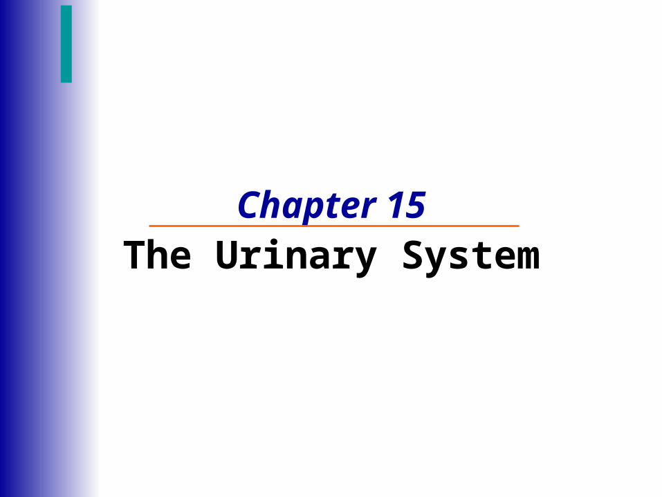
Location of the KidneysLocation of the Kidneys Bean-shaped organ
Lies in the superior lumbar region
Extends from T12 to L3
Right kidney is slightly lower than the left
Average dimensions (about the size of a bar of soap)
12 cm long; 6 cm wide; 3 cm thick
Lateral surface is convex
Medial surface is concave
Renal hilum
The ureter, blood vessels, lymphatic vessels and nerves all join the kidney here
Atop each kidney is an adrenal gland

Regions of the KidneyRegions of the Kidney Renal cortex
Outer region
Renal medulla
Inside the cortex
Exhibit medullary pyramids
Renal columns separate the pyramids
Renal pelvis
Inner funnel-shaped tube
Continuous with the ureter leaving the hilum
Figure 15.2b
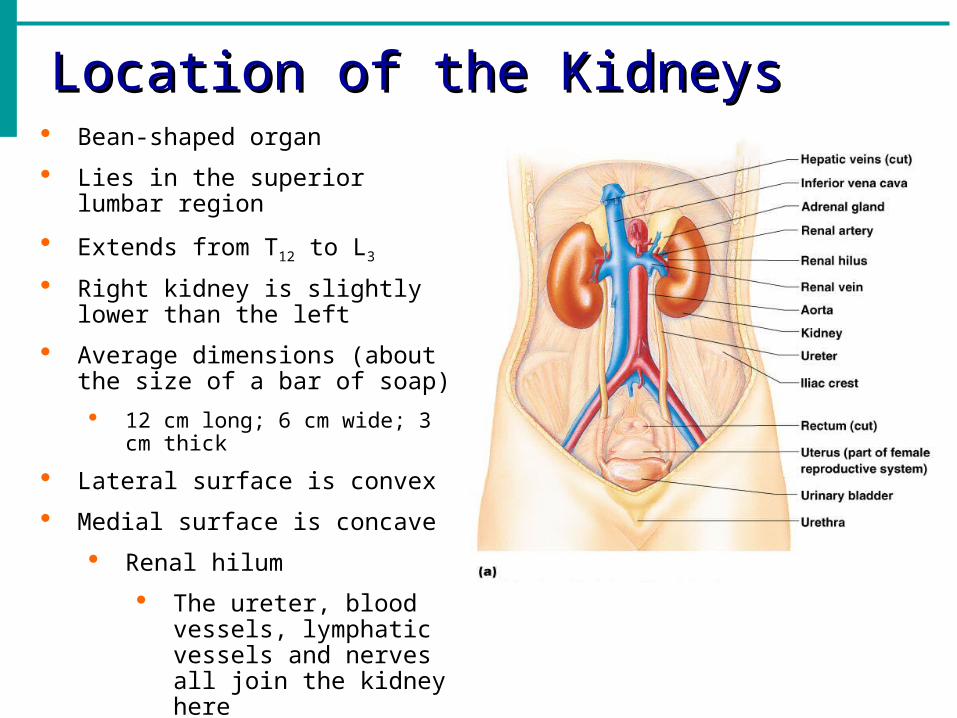
Kidney StructuresKidney Structures
Medullary pyramids
Triangular regions of tissue in the medulla
Calyces
Cup-shaped structures that collect urine from the medullary pyramids and empty it into the renal pelvis
Major calyces and minor calyces

Blood Flow in the KidneysBlood Flow in the Kidneys As each renal artery approaches a kidney, it divides into five segmental
arteries
Each segmental artery branches further to form lobar arteries and then interlobar arteries
The interlobar arteries branch into the arcuate arteries that arch over the bases of the medullary pyramids
Small interlobular arteries radiate outward from the arcuate arteries to supply the cortical tissue
Afferent arterioles branching from the interlobular arteries turn into microscopic blood vessels called the glomerulus, which is the key element of kidney function

Blood Flow in the KidneysBlood Flow in the Kidneys Veins trace the pathway of arterial supply in reverse
Blood leaving the renal cortex (efferent arteriole) drains into the interlobular veins, arcuate veins, interlobar vein and then renal vein(notice no segmental veins) and then the renal vein empties into the inferior vena cava

Blood Flow in the KidneysBlood Flow in the Kidneys

NephronsNephrons The structural and functional units of the kidneys
Kidneys contains over 1 million of these tiny blood-processing units
Responsible for forming urine
Each nephron consists of a glomerulus (capillaries) and renal tubule
The renal tubule has a cup-shaped end called the glomerular capsule or Bowman’s capsule
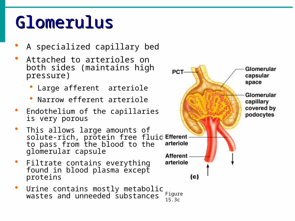
GlomerulusGlomerulus A specialized capillary bed
Attached to arterioles on both sides (maintains high pressure) Large afferent arteriole
Narrow efferent arteriole
Endothelium of the capillaries is very porous
This allows large amounts of solute-rich, protein free fluid to pass from the blood to the glomerular capsule
Filtrate contains everything found in blood plasma except proteins
Urine contains mostly metabolic wastes and unneeded substances
Figure 15.3c
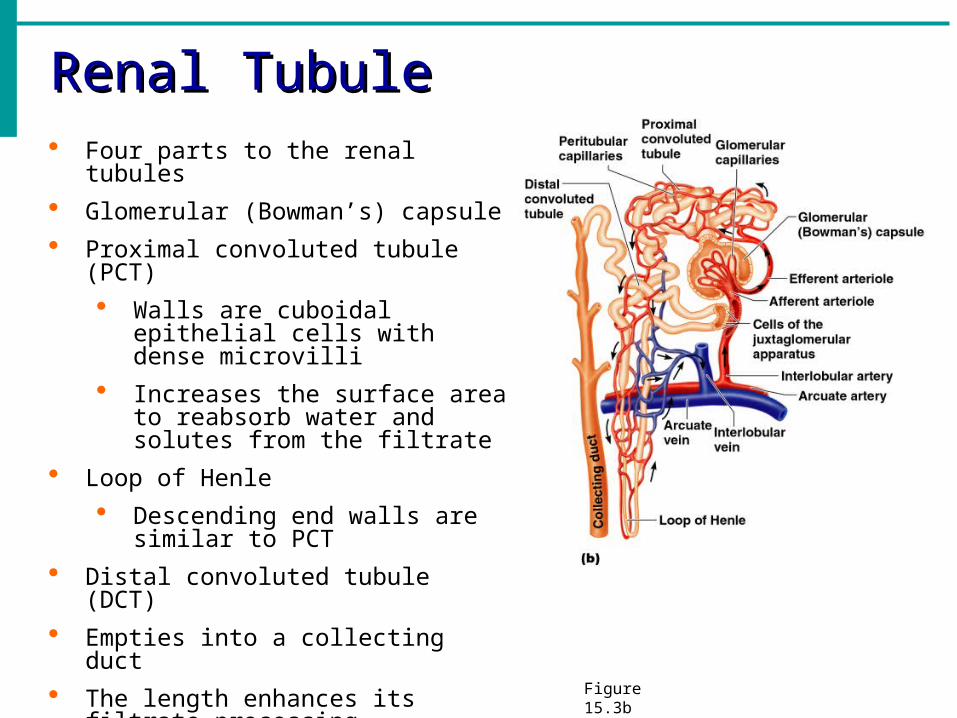
Renal TubuleRenal Tubule Four parts to the renal tubules
Glomerular (Bowman’s) capsule
Proximal convoluted tubule (PCT)
Walls are cuboidal epithelial cells with dense microvilli
Increases the surface area to reabsorb water and solutes from the filtrate
Loop of Henle
Descending end walls are similar to PCT
Distal convoluted tubule (DCT)
Empties into a collecting duct
The length enhances its filtrate processing capabilities
Figure 15.3b

Renal TubuleRenal Tubule Collecting ducts
Receive filtrate from many nephrons
Run through the medullary pyramids
As they reach the renal pelvis, a couple fuse together and deliver urine into the minor calyces
Figure 15.3b

Nephron Capillary BedsNephron Capillary Beds
The renal tubule of every nephron is closely associated with two capillary beds
Glomerulus
Produces the filtrate
Peritubular capillaries
Reclaims most of the filtrate
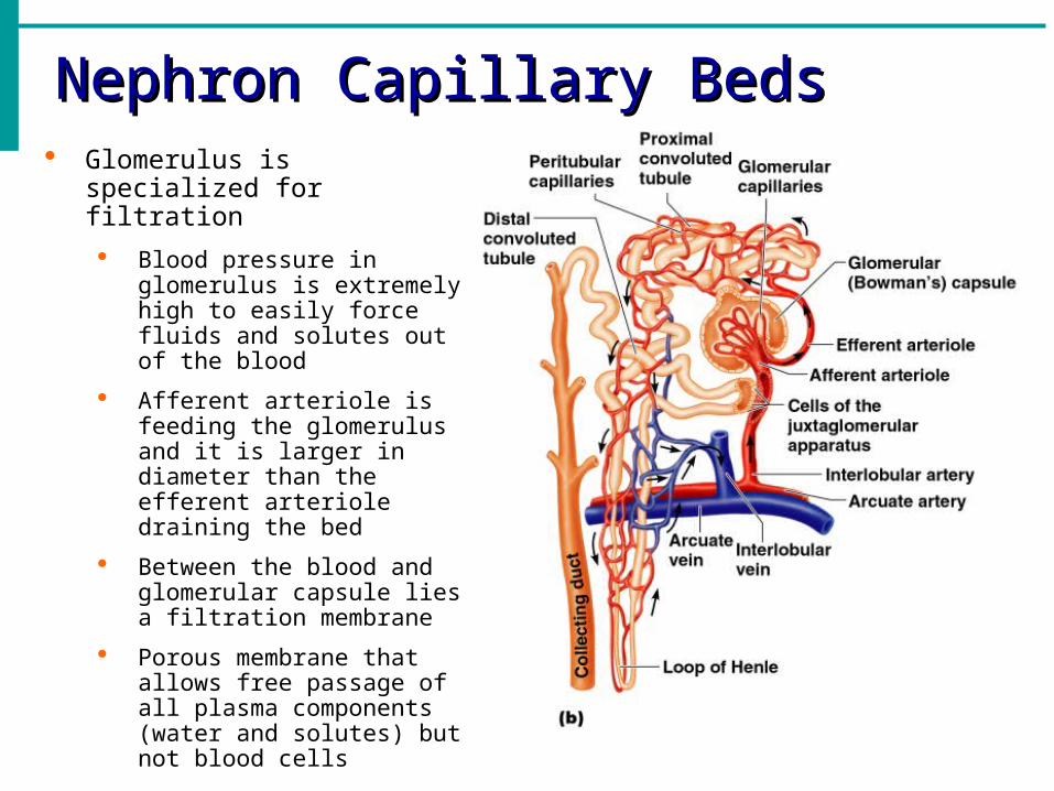
Nephron Capillary BedsNephron Capillary Beds Glomerulus is specialized for
filtration
Blood pressure in glomerulus is extremely high to easily force fluids and solutes out of the blood
Afferent arteriole is feeding the glomerulus and it is larger in diameter than the efferent arteriole draining the bed
Between the blood and glomerular capsule lies a filtration membrane
Porous membrane that allows free passage of all plasma components (water and solutes) but not blood cells

Nephron Capillary BedsNephron Capillary Beds Peritubular capillaries
Arise from efferent arteriole of the glomerulus
Cling close to the renal tubule and empty into nearby venules
Normal, low pressure capillaries
Readily absorb solutes and water from collecting tubes
Most of the resulting filtrate (99%) is reabsorbed by the renal tubule and returned to the blood in the peritubular capillaries
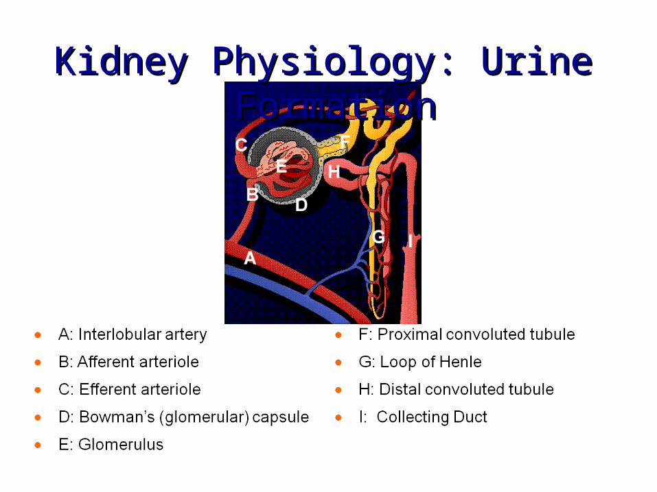
Kidney Physiology: Urine FormationKidney Physiology: Urine Formation

Kidney Physiology: Urine FormationKidney Physiology: Urine Formation
The total plasma filters into the renal tubules about every 22 minutes
All of our plasma would be drained away as urine in less than 30 minutes were it not for the fact that most of the tubule contents are quickly reclaimed and returned to the blood
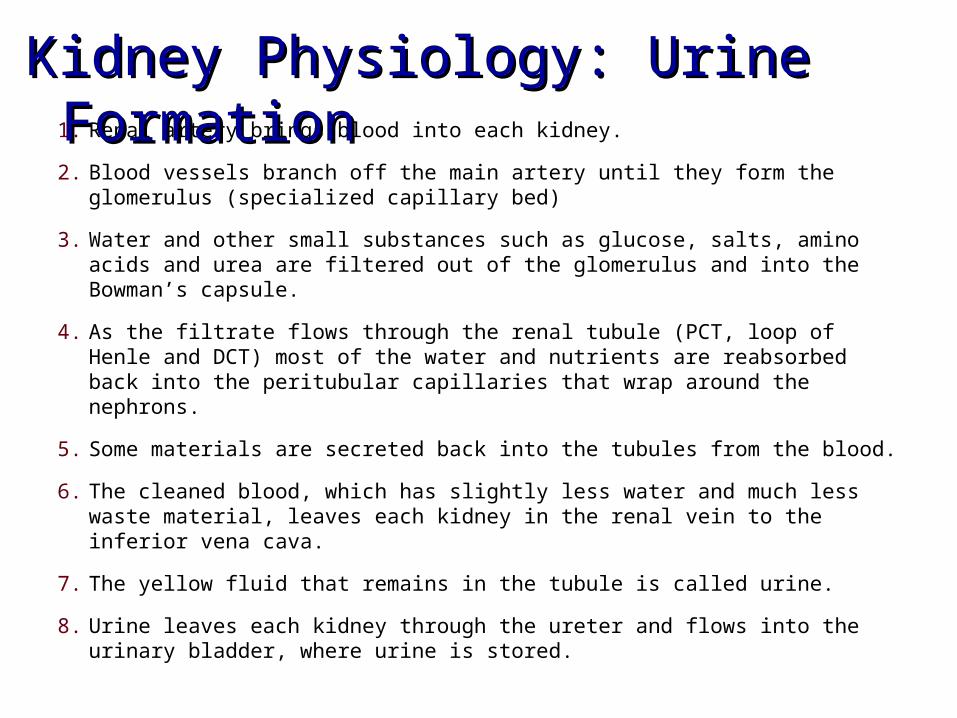
1. Renal artery brings blood into each kidney.
2. Blood vessels branch off the main artery until they form the glomerulus (specialized capillary bed)
3. Water and other small substances such as glucose, salts, amino acids and urea are filtered out of the glomerulus and into the Bowman’s capsule.
4. As the filtrate flows through the renal tubule (PCT, loop of Henle and DCT) most of the water and nutrients are reabsorbed back into the peritubular capillaries that wrap around the nephrons.
5. Some materials are secreted back into the tubules from the blood.
6. The cleaned blood, which has slightly less water and much less waste material, leaves each kidney in the renal vein to the inferior vena cava.
7. The yellow fluid that remains in the tubule is called urine.
8. Urine leaves each kidney through the ureter and flows into the urinary bladder, where urine is stored.
Kidney Physiology: Urine FormationKidney Physiology: Urine Formation

• Pathway of Urine
• Bowman’s capsule (filtrate)Proximal convoluted tubule (filtrate) loop of Henle (filtrate) distal convoluted tubule (filtrate) collecting duct (urine) minor calyces (urine) major calyces (urine) ureter (urine) bladder (urine) urethra(urine)
Kidney Physiology: Urine FormationKidney Physiology: Urine Formation

Kidney Physiology: Urine FormationKidney Physiology: Urine Formation Kidneys form urine in the nephrons and adjust
the blood composition with three major processes
Glomerular filtration (#1)
Dump filtrate into renal tubules
Filters about 200 L daily and only 1.5L leaves the body as urine
Tubular reabsorption (#2)
Kidneys reclaim what the body needs
Almost all the filtrate (99%)
Water, salt, glucose and amino acids
Not reabsorbed is uric acid, creatinine, urea
Anything not reabsorbed becomes urine
Tubular secretion (#3)
Fine-tuning the body’s chemical balance

Kidney Physiology: Urine FormationKidney Physiology: Urine Formation Step 1: Glomerular Filtration
Passive, nonselective process
Glomerular blood pressure is extremely high
Pressure forces fluids and solutes through a membrane
Small molecules such as water, salts, bicarbonate, hydrogen ions, urea, glucose, amino acids and some drugs
Blood cells and large molecules cannot pass through the wall
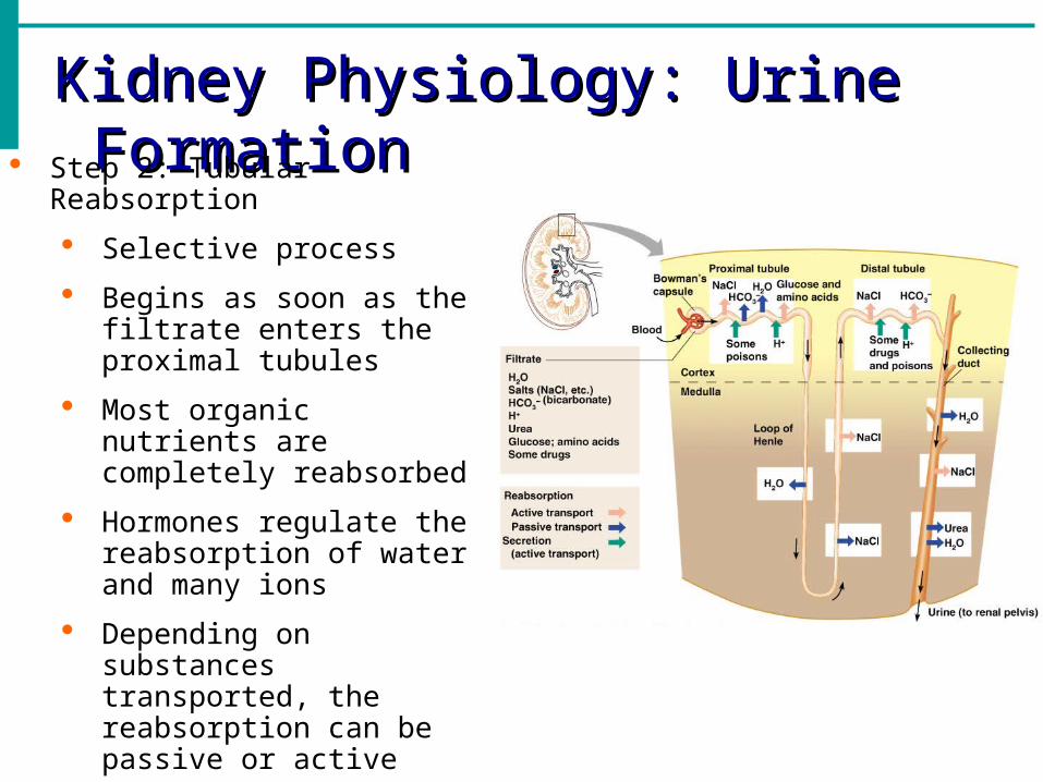
Kidney Physiology: Urine FormationKidney Physiology: Urine Formation Step 2: Tubular Reabsorption
Selective process
Begins as soon as the filtrate enters the proximal tubules
Most organic nutrients are completely reabsorbed
Hormones regulate the reabsorption of water and many ions
Depending on substances transported, the reabsorption can be passive or active
Sodium ions are the single most abundant cation in the filtrate

Kidney Physiology: Urine FormationKidney Physiology: Urine Formation Step 2: Tubular Reabsorption
Reabsorptive abilities of regions of the renal tubules
Proximal convoluted tubule (PCT) Most active reabsorbing area
Sodium (Na+), bicarbonate (HCO3-),
chlorine (Cl-) and water
Loop of Henle Water is salts are reabsorbed
Vital role in kidneys ability to form dilute or concentrated urine
Distal convoluted tubule (DCT) NaCl and water
Most reabsorption at this time depends on the body’s needs

Kidney Physiology: Urine FormationKidney Physiology: Urine Formation Step 2: Tubular Reabsorption
Regulated by hormones
Aldosterone
Released when blood pressure decreases or Na+ concentration drops
Antidiuretic hormone (ADH)
Reabsorption of water
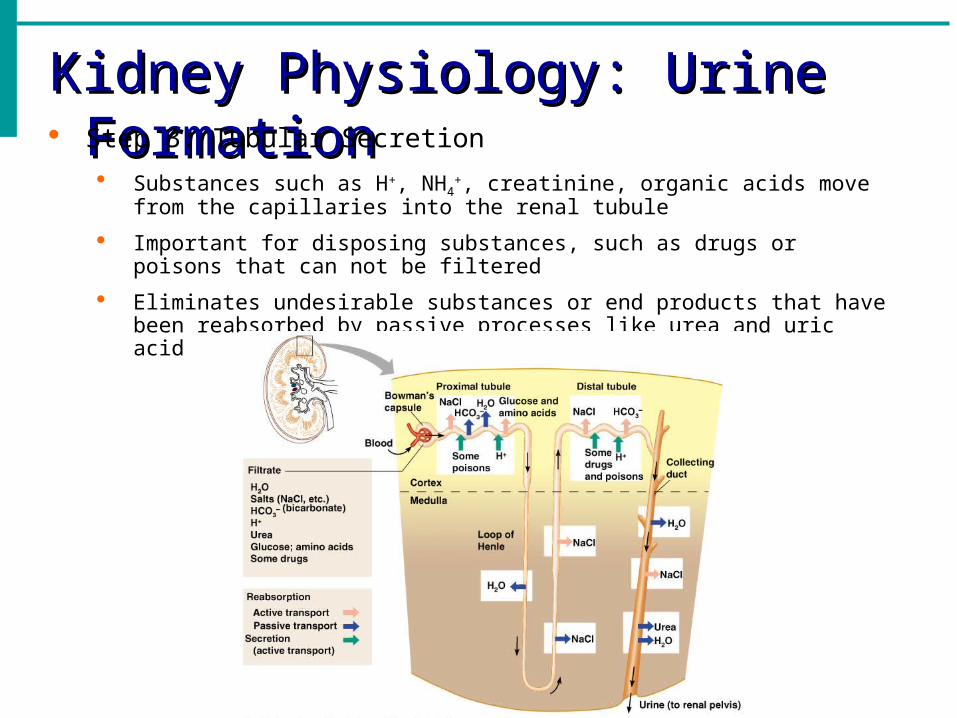
Kidney Physiology: Urine FormationKidney Physiology: Urine Formation Step 3: Tubular Secretion
Substances such as H+, NH4+, creatinine, organic acids move from the
capillaries into the renal tubule
Important for disposing substances, such as drugs or poisons that can not be filtered
Eliminates undesirable substances or end products that have been reabsorbed by passive processes like urea and uric acid

Physical Characteristics of UrinePhysical Characteristics of Urine
Color
Clear to deep yellow
Yellow color is due to the pigment urochrome (from the destruction of hemoglobin)
More concentrated the urine, the deeper the yellow color
An abnormal color such as pink or brown may result from eating certain foods (beets, rhubarb), the presence of bile pigments or blood, or from some commonly prescribed drugs and vitamins
Cloudy urine may indicated a urinary tract infection
Odor
Slightly aromatic
If allowed to stand, it develops an ammonia odor as bacteria metabolize its urea solutes
Physical Characteristics of UrinePhysical Characteristics of Urine
pH
Slightly acidic (around pH 6)
Acidic diet that contains large amounts of protein and whole wheat products, diabetes mellitus and starvation produces acidic urine
Vegetarian diet, prolonged vomiting, and bacterial infection of the urinary tract all can cause the urine to become alkaline
Specific gravity
Ratio of the mass of a substance to the mass of an equal volume of distilled water
Urine is water plus solutes
Distilled water specific gravity is 1.00
Urine specific gravity ranges from 1.001 to 1.035 depending on its solutes
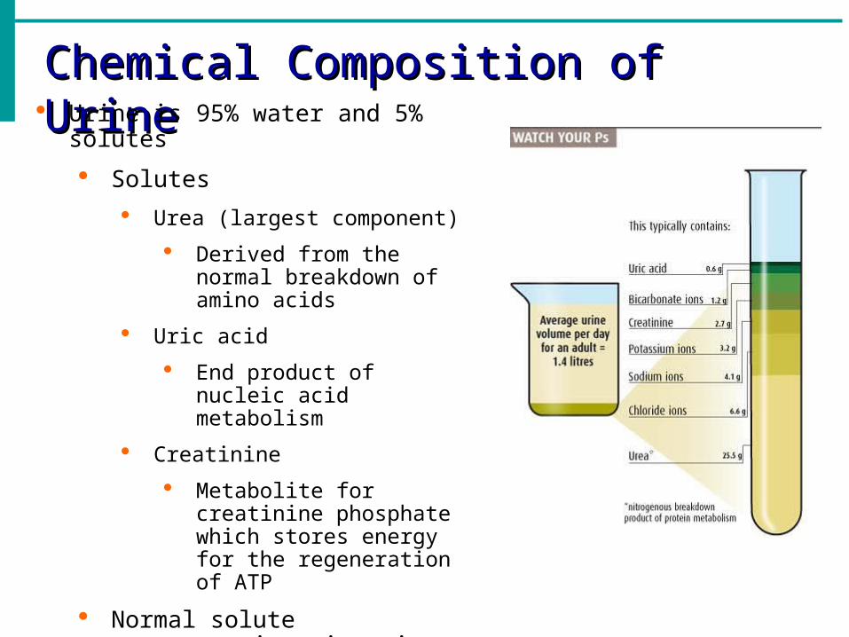
Chemical Composition of UrineChemical Composition of Urine Urine is 95% water and 5% solutes
Solutes
Urea (largest component)
Derived from the normal breakdown of amino acids
Uric acid
End product of nucleic acid metabolism
Creatinine
Metabolite for creatinine phosphate which stores energy for the regeneration of ATP
Normal solute concentrations in urine from high to low
Urea sodium potassium phosphate sulfate creatinine uric acid

Chemical Composition of UrineChemical Composition of Urine Abnormal Urinary Constituents
Glucose (glycosuria) Benedict’s solution and heat
Causes: diabetes mellitus
Proteins (proteinuria) Biuret’s solution
Causes: Non-pathological: excessive physical exertion, pregnancy, high-protein diet; Pathological: heart failure, severe hypertension, renal disease
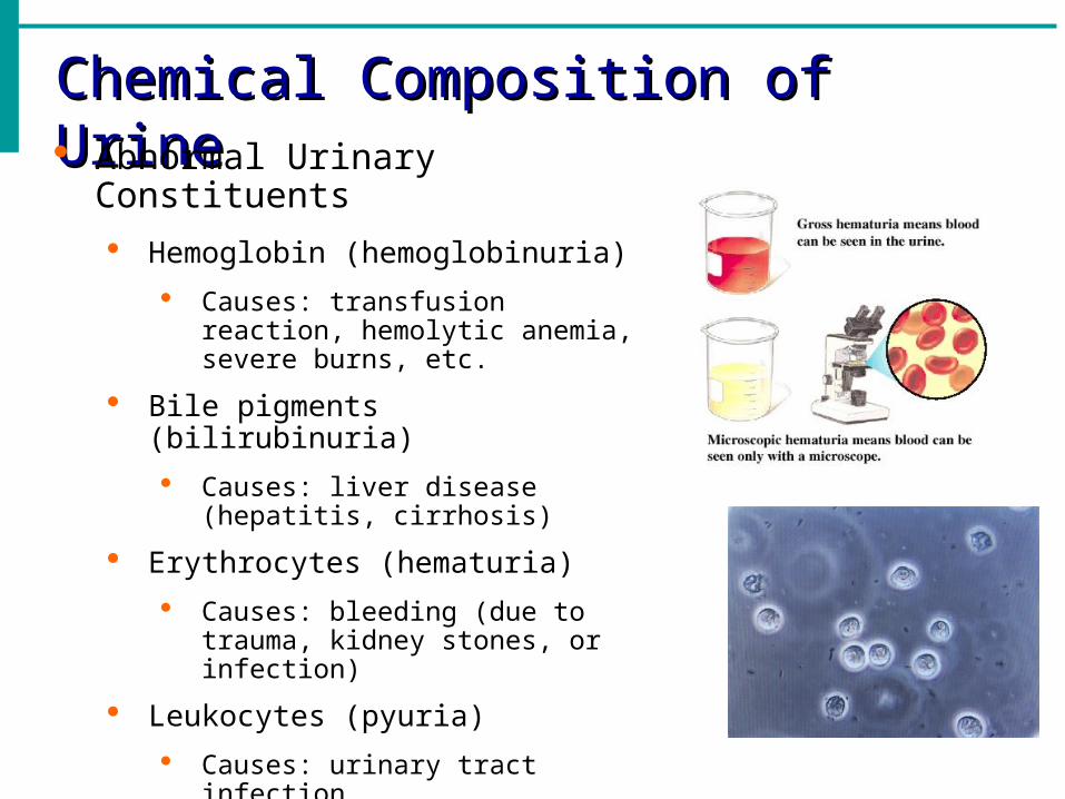
Chemical Composition of UrineChemical Composition of Urine Abnormal Urinary Constituents
Hemoglobin (hemoglobinuria)
Causes: transfusion reaction, hemolytic anemia, severe burns, etc.
Bile pigments (bilirubinuria)
Causes: liver disease (hepatitis, cirrhosis)
Erythrocytes (hematuria)
Causes: bleeding (due to trauma, kidney stones, or infection)
Leukocytes (pyuria)
Causes: urinary tract infection

UretersUreters Slender tubes that carry urine from the kidneys to the bladder
Composed of transitional epithelium
Peristalsis aids gravity in urine transport
Homeostatic Imbalance
Kidney stones
Calcium, magnesium, or uric acid salts in urine may crystallize and precipitate in the renal pelvis
Most are under 5 mm in diameter and pass through the urinary tract without causing problems
Larger stones can obstruct a ureter and block urine drainage
Increasing pressure in the kidney causes excruciating pain
Treatment includes shock wave lithiotripsy a noninvasive procedure that uses ultrasonic shock waves to shatter the stone
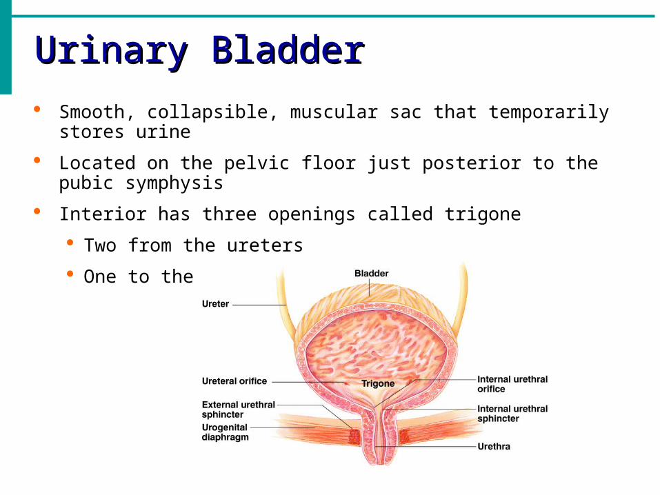
Urinary BladderUrinary Bladder
Smooth, collapsible, muscular sac that temporarily stores urine
Located on the pelvic floor just posterior to the pubic symphysis
Interior has three openings called trigone
Two from the ureters
One to the urethra

Urinary BladderUrinary Bladder
When empty, the bladder collapses and its walls are thick and have folds (rugae)
Bladder can expand significantly
A full bladder is about 12 cm (5 inches) long and holds approximately 500 mL (1 pint) of urine, but it can hold nearly double that if necessary
Maximum capacity of the bladder is 800-1000 mL and when it is overdistended, it may burst
Urine is formed continuously by the kidneys but it is stored in the bladder until it is convenient to release

UrethraUrethra
Thin-walled muscular tube that drains urine from the bladder to the outside of the body
Release of urine (micturition or voiding)is controlled by two sphincters
Internal urethral sphincter (involuntary)
External urethral sphincter (voluntary)
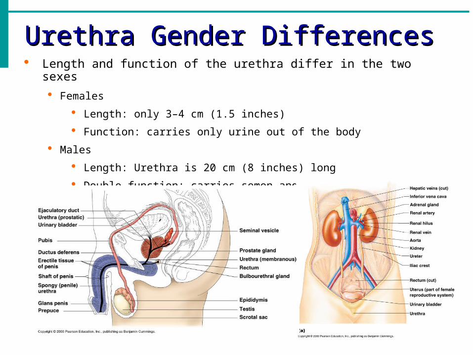
Urethra Gender DifferencesUrethra Gender Differences Length and function of the urethra differ in the two sexes
Females
Length: only 3–4 cm (1.5 inches)
Function: carries only urine out of the body
Males
Length: Urethra is 20 cm (8 inches) long
Double function: carries semen and urine out of the body


Maintaining Water BalanceMaintaining Water Balance
Normal amount of water in the human body
Young adult females – 50% because more body fat
Young adult males – 60% because more muscles
Babies – 75% because of low body fat and low bone mass
Old age – 45%
Water is necessary for many body functions and levels must be maintained
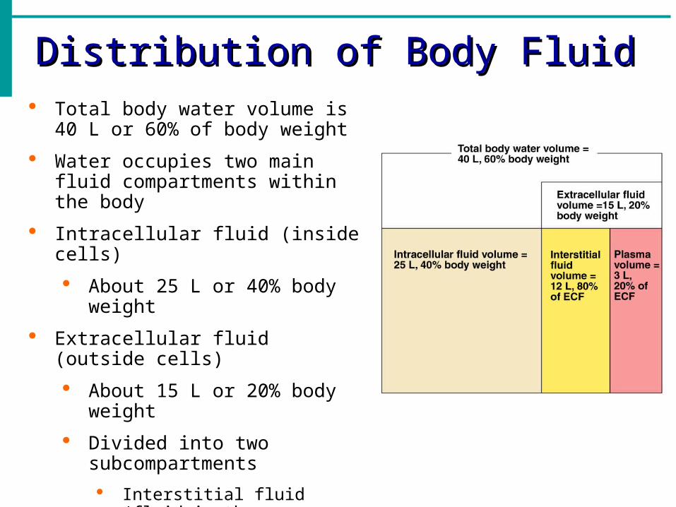
Distribution of Body FluidDistribution of Body Fluid Total body water volume is 40 L or
60% of body weight
Water occupies two main fluid compartments within the body
Intracellular fluid (inside cells)
About 25 L or 40% body weight
Extracellular fluid (outside cells)
About 15 L or 20% body weight
Divided into two subcompartments
Interstitial fluid (fluid in the microscopic spaces between tissue cells)
Blood plasma (fluid portion of blodd)

Composition of Body FluidsComposition of Body Fluids Water is the universal solvent in which a variety of solutes are dissolved
Solutes can be classified into electrolytes and nonelectrolytes
Nonelectrolytes have bonds and cannot dissociate in solution
Organic molecules such as glucose, lipids, creatinine and urea
Electrolytes are chemical compounds that do dissociated into ions in water
Inorganic and organic acids and bases and some proteins
Have the greatest ability to make fluid shifts down their gradients
Most abundant solutes in body fluids
Extracellular fluids have high sodium and chloride ions
Intracellular fluids contains only small amounts of sodium and chloride ions; its most abundant cation is potassium anion is phosphate (HPO42-) as well as high amounts of proteins
Fluid Moving Among CompartmentsFluid Moving Among Compartments
Continuous exchange and mixing of fluids are regulated by osmotic and hydrostatic pressures
Water moves freely between the compartments along osmotic gradients
Solutes are unequally distributed because of their size, electrical charge, or dependence on transport proteins
Changes in electrolyte balance causes water to move from one compartment to another
Maintaining Water BalanceMaintaining Water Balance Body must remain properly hydrated water intake must equal water
output Water intake is typically about 2500 mL a day in adults
Water enters the body through ingested liquids (60%), solid foods (30%) and produced from metabolic processes (10%
Water output occurs by several routes Vaporization out of the lungs and skin (28%)
Perspiration of skin (8%)
Leaves the body in the feces (4%)
The balance (about 60%) is excreted by the kidneys in urine

Maintaining Water BalanceMaintaining Water Balance
A rise in plasma concentration causes thirst (prompts us to drink water) and release of antidiuretic hormone (ADH) which causes the kidneys to conserve water and excrete concentrated urine
A decline in plasma concentration inhibits thirst and ADH release and causes output of large volumes of dilute urine

Regulation of WaterRegulation of Water Water intake is controlled by the thirst mechanism
An increase in plasma concentrations
A dry mouth occurs
Less saliva production
Decrease in blood pressure
Water Output of certain amounts of water is unavoidable
Reason why we cannot live without drinking
Solute concentration and volume of urine excreted depend on fluid intake, diet and water loss via other avenues
Regulation is primarily by hormones Antidiuretic hormone (ADH) prevents excessive water loss in urine
Aldosterone regulates sodium ion content of extracellular fluid
Osmoreceptor cells in the kidneys are active monitors
Regulation of WaterRegulation of Water Dehydration
When water output exceeds intake over a period of time and the body is in negative fluid balance
Commonly follows hemorrhage, severe burns, prolonged vomiting or diarrhea, profuse sweating, and diuretic abuse
Signs are sticky oral mucosa, thirst, dry flushed skin, and decreased water output (oliguria)

Electrolyte BalanceElectrolyte Balance Refers to the salt balance in the body
Important in controlling fluid movements and crucial for cellular activity
Salts enter the body in foods and fluids
Salts are lost from the body in perspiration, feces and urine
Sodium holds a central position in fluid and electrolyte balance and overall body homeostasis
Water follows salt
A change in plasma sodium levels affects not only plasma volume and blood pressure but also the ICF and IF volumes
Regulation is linked to blood pressure and aldosterone
When aldosterone is high all the sodium is reabsorbed in the DCT
Water follows sodium and maintains blood pressure
When aldosterone is inhibited none of the sodium is reabsorbed
Goal of aldosterone is to decrease urinary output and increase blood volume
Acid-Base Balance Acid-Base Balance All biochemical reactions are influenced by the pH of their fluid
environment
The acid-base balance of body fluids is closely regulated
pH measures the amount of H+ ions in solution
Acids are proton donors
Blood normally ranges between pH 7.35 and pH 7.45
If the pH rises above 7.45 a person has alkalosis
If the pH drops below 7.35 a person has acidosis
H+ concentration regulation
Chemical buffers resist changes within a fraction of a second
Respiratory rate changes within 1-3 minutes
Kidneys requires hours to a day to effect changes in blood pH

Acid-Base Balance Acid-Base Balance Respiratory acidosis
Most common cause of acid-base imbalance
Caused when a person breathes shallow or when gas exchange is hampered by diseases
CO2 accumulates in the blood and causes the pH to fall
Respiratory alkalosis
Results from carbon dioxide being eliminated faster than it is produced otherwise known as hyperventilation
Metabolic acidosis
Second most common cause of acid-base imbalance
Low blood pH and HCO3- levels
Caused when a person ingests too much alcohol and excessive loss of HCO3- as a result of excessive diarrhea
Metabolic alkalosis
Rising blood pH and HCO3- levels
Caused by vomiting and intake of excess base
Acid-Base Balance Acid-Base Balance Effects of acidosis and alkalosis
Absolute blood pH limits for life are a low of 7.0 and a high of 7.8
When the pH falls below 7.0 the CNS is so depressed that the person goes into coma and death
When blood pH rises above 7.8, the nervous system is overexcited and leads to muscle spasms, extreme nervousness, and convulsions; death usually results from respiratory arrest