Embed Size (px)
Citation preview

.
MDufilho
Chapter 25
The Urinary
System
10/30/2013 1

.
Kidney Functions
• Removal of toxins, metabolic wastes, and excess ions
from the blood
• Regulation of blood volume, chemical composition, and
pH
• Gluconeogenesis during prolonged fasting
• Endocrine functions
• Renin: regulation of blood pressure and kidney
function
• Erythropoietin: regulation of RBC production
• Activation of vitamin D
10/30/2013 2 MDufilho

Figure 25.5
10/30/2013 MDufilho 3

MDufilho
Figure 25.9 A schematic, uncoiled nephron showing the three major renal processes that adjust plasma composition.
Cortical radiate artery
Afferent arteriole Glomerular
capillaries
Efferent arteriole
Glomerular capsule
Renal tubule and collecting duct containing filtrate
Peritubular capillary
To cortical radiate vein
Three major
renal processes: Urine Glomerular filtration Tubular reabsorption
Tubular secretion
1
2
3
1
2
3 10/30/2013 4

MDufilho
Figure 25.5 Location and structure of nephrons. (3 of 7)
Basement membrane
Podocyte
Fenestrated endothelium of the glomerulus
Glomerular capsule: visceral layer
10/30/2013 5

MDufilho
Figure 25.10a The filtration membrane.
Efferent arteriole
Afferent arteriole
Glomerular capillary covered by podocytes that form the visceral layer of glomerular capsule
Parietal layer of glomerular capsule
Proximal convoluted tubule
Glomerular capsular space
Cytoplasmic extensions of podocytes
Filtration slits
Podocyte cell body
Fenestrations (pores)
Glomerular capillary endothelium (podocyte covering and basement membrane removed)
Foot processes of podocyte
Glomerular capillaries and the
visceral layer of the glomerular
capsule
10/30/2013 6

MDufilho
Figure 25.10c The filtration membrane.
Capillary
Fenestration (pore)
Filtration membrane
• Capillary endothelium
• Basement membrane
• Foot processes of podocyte of glomerular capsule
Filtration slit
Slit diaphragm
Three layers of the filtration membrane
Foot processes of podocyte
Filtrate in capsular space
Plasma
10/30/2013 7

.
10/30/2013 MDufilho 8
Terminology
• Renal Fraction = % of blood flow passing
through kidneys = 20 – 25%
• Filtration Fraction = % plasma (in renal
fraction) that passes through the filtration
membrane to become filtrate = 20%

.
Net Filtration Pressure (NFP)
• The pressure responsible for filtrate formation
(10 mm Hg)
• Determined by
• Glomerular hydrostatic pressure (HPg) the
chief force
• Two opposing forces:
• Colloid osmotic pressure of glomerular blood
(OPg)
• Capsular hydrostatic pressure (HPc)
NFP = HPg – (OPg + HPc)
10/30/2013 9 MDufilho

Figure 25.11 Forces determining net filtration pressure (NFP).
Efferent arteriole
Glomerular
capsule
Afferent
arteriole
NFP = Net filtration pressure
= outward pressures – inward pressures
= (HPgc) – (HPcs + OPgc)
= (55) – (15 + 30)
= 10 mm Hg
HPgc = 55 mm Hg
OPgc = 30 mm Hg
HPcs = 15 mm Hg
MDufilho 10/30/2013 10
.
Glomerular Filtration Rate (GFR)
• Volume of filtrate formed per minute by the
kidneys (120–125 ml/min)
• Governed by (and directly proportional to)
• Total surface area available for filtration
• Filtration membrane permeability
• NFP
10/30/2013 11 MDufilho
.
10/30/2013 MDufilho 12
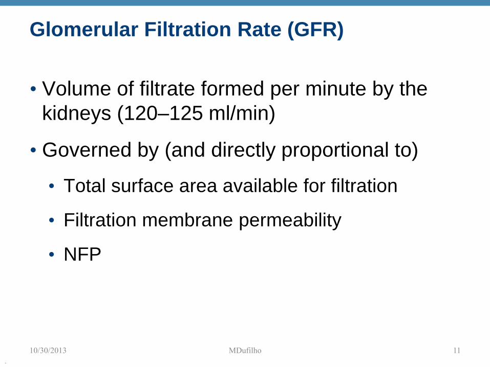
Tubular Maximum
• Also called transport maximum
• When tubular load exceeds tubular maximum,
the substances show up in urine
• e. g. Glucosuria
.
10/30/2013 MDufilho 13
Clinical Applications – What if….. • Hemorrhage –
- Blood Pressure?
- NFP?
- GFR?
- Result
• Fight or flight situation
- Sympathetic stimulation?
- Blood Pressure?
- Result?
• Kidney Infection
- Result

.
Regulation of Glomerular Filtration
• GFR is tightly controlled by two types of
mechanisms
• Intrinsic controls (renal autoregulation)
• Act locally within the kidney
• Extrinsic controls
• Nervous and endocrine mechanisms that
maintain blood pressure, but affect kidney
function
10/30/2013 14 MDufilho

.
Intrinsic Controls
• Maintains a nearly constant GFR when MAP
is in the range of 80–180 mm Hg
• Two types of renal autoregulation
• Myogenic mechanism (Chapter 19)
• Tubuloglomerular feedback mechanism, which
senses changes in the juxtaglomerular
apparatus
10/30/2013 15 MDufilho

Figure 25.8 Juxtaglomerular complex (JGC) of a nephron.
Glomerular capsule
Efferent arteriole
Afferent arteriole
Glomerulus
Efferent arteriole
Juxtaglomerular
complex
Macula densa
cells of the ascending limb of nephron loop
• Extraglomerular
• Granular
cells Afferent arteriole
Parietal layer of glomerular capsule
Capsular space
Foot processes of podocytes
Podocyte cell body (visceral layer)
Red blood cell
Proximal tubule cell
Lumens of glomerular capillaries
Endothelial cell of glomerular capillary
Glomerular mesangial cells
Renal corpuscle Juxtaglomerular complex�
mesangial cells
•
MDufilho 10/30/2013 16

.
Intrinsic Controls: Tubuloglomerular
Feedback Mechanism
Macula densa cells monitor Na+ and Cl- in
filtrate
• Macula densa cells of the JGA respond to
NaCl by releasing a vasoconstricting
chemical that acts on the afferent arteriole
GFR
• The opposite occurs if GFR decreases.
10/30/2013 17 MDufilho

.
10/30/2013 MDufilho 18
Intrinsic Controls
• Compensate for MAP of 80 – 180 mmHg to
maintain GFR at 125 ml/min (+/- 30%)
• Kidney function suffers if MAP < 80 mmHg
• Then extrinsic controls are needed

.
Extrinsic Controls: Sympathetic Nervous
System
• Under extreme stress
• Norepinephrine is released by the sympathetic
nervous system
• Epinephrine is released by the adrenal
medulla
• Both cause constriction of afferent arterioles,
inhibiting filtration and triggering the release of
renin
10/30/2013 19 MDufilho

.
Extrinsic Controls: Renin-Angiotensin
Mechanism
• Triggered when the granular cells of the JGA
release renin
angiotensinogen (a plasma globulin)
renin
angiotensin I
angiotensin converting
enzyme (ACE)
angiotensin II
10/30/2013 20 MDufilho

.
Other Factors Affecting GFR
• Prostaglandin E2
• Vasodilator that counteracts vasoconstriction
by norepinephrine and angiotensin II
• Prevents renal damage when peripheral
resistance is increased
• Intrarenal angiotensin II
• Reinforces the effects of hormonal
angiotensin II
• Adenosine
• A vasoconstrictor of renal vasculature
10/30/2013 21 MDufilho

MDufilho
Figure 25.12 Physiological mechanisms regulating glomerular filtration rate (GFR) in the kidneys.
SYSTEMIC BLOOD PRESSURE
Blood pressure in afferent arterioles; GFR
GFR Granular cells of juxtaglomerular
complex of kidney
Inhibits baroreceptors
in blood vessels of
systemic circulation
Sympathetic nervous system
Release
Renin
Catalyzes cascade
resulting in
formation of
Angiotensin II
Targets
Filtrate flow
and�NaCl in
ascending�limb of
nephron loop
Stretch of smooth
�muscle in walls of
�afferent arterioles
Vasodilation of
afferent arterioles
Macula densa cells
�of juxtaglomerular
�complex of kidney
Aldosterone
secretion by
adrenal cortex
Vasoconstriction of
systemic arterioles;
peripheral resistance
Release of vasoactive
chemicals inhibited
Vasodilation of
afferent arterioles
Na+ reabsorption
by kidney tubules;
water follows
GFR
Blood volume
Systemic
blood pressure
Myogenic mechanism
of autoregulation
Tubuloglomerular
mechanism
of�autoregulation
Hormonal (renin-angiotensin-
aldosterone)�mechanism
Neural controls
Extrinsic mechanisms indirectly regulate GFR
by maintaining systemic blood pressure, which
drives filtration in the kidneys.
Intrinsic mechanisms directly regulate GFR despite
moderate changes in blood pressure (between 80
and 180 mm Hg mean arterial pressure).
10/30/2013 22

.
10/30/2013 MDufilho 23
Regulation of Urine Concentration and
Volume – Really ECF
• Osmolality - number of solute particles in 1Kg of
solvent
• Osmolarity – number of solute particles in 1 liter of
solvent
• Both terms reflect the solution’s ability to cause
osmosis
• Body fluids are measured in milliosmols (mOsm)
• The kidneys keep the solute load of body fluids
constant at about 285 - 300 mOsm
• This is accomplished by the countercurrent
mechanism

.
Countercurrent Mechanism
• Occurs when fluid flows in opposite directions
in two adjacent segments of the same tube
• Filtrate flow in the loop of Henle
(countercurrent multiplier)
• Blood flow in the vasa recta (countercurrent
exchanger)
10/30/2013 MDufilho 24
MDufilho
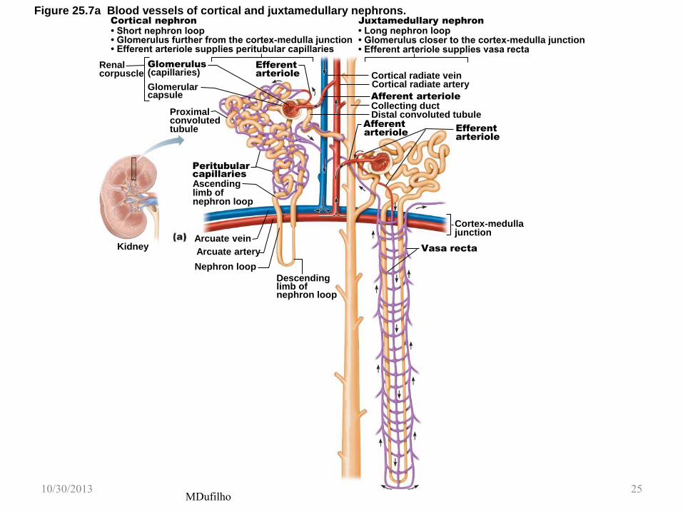
Figure 25.7a Blood vessels of cortical and juxtamedullary nephrons. Cortical nephron
• Short nephron loop • Glomerulus further from the cortex-medulla junction • Efferent arteriole supplies peritubular capillaries
Juxtamedullary nephron
• Long nephron loop • Glomerulus closer to the cortex-medulla junction • Efferent arteriole supplies vasa recta
Renal corpuscle
Glomerulus� (capillaries)
Glomerular� capsule
Proximal convoluted tubule�
Peritubular
capillaries
Ascending limb of nephron loop
Kidney Arcuate vein
Arcuate artery
Nephron loop
Efferent
arteriole
Descending limb of nephron loop
Cortical radiate vein
Afferent arteriole
Distal convoluted tubule
Efferent
arteriole
Vasa recta
Cortex-medulla junction
Cortical radiate artery
Collecting duct
Afferent
arteriole
10/30/2013 25
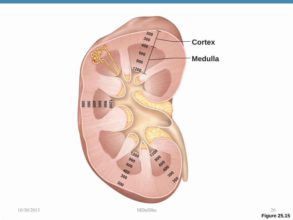
. Figure 25.15
Cortex
Medulla
10/30/2013 MDufilho 26

MDufilho
Figure 25.16a Juxtamedullary nephrons create an osmotic gradient within the renal medulla that allows the kidney to
produce urine of varying concentration. (4 of 4)
(continued) As water and solutes are reabsorbed, the loop first concentrates the filtrate, then dilutes it.
Active transport
Passive transport
Water impermeable
Cortex 300
400
600
900
1200
Osm
ola
lity
of
inte
rsti
tial fl
uid
(m
Osm
)
Outer
medulla
Inner
medulla Nephron loop
300 300
300 100
100
200
400
700
400
600
900
1
2
5
4
3
Filtrate entering the nephron loop is isosmotic to both blood plasma and cortical interstitial fluid.
Water moves out of the filtrate in the descending limb down its osmotic gradient. This concentrates the filtrate.
Filtrate is at its most dilute as it leaves the nephron loop. At 100 mOsm, it is hypo-osmotic to the interstitial fluid.
Na+ and Cl- are pumped out of the filtrate. This increases the interstitial fluid osmolality.
Filtrate reaches its highest concentration at the bend of the loop.
1200
10/30/2013 27
.
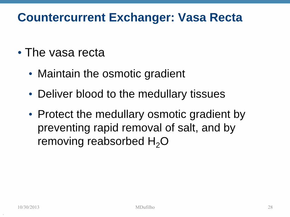
Countercurrent Exchanger: Vasa Recta
• The vasa recta
• Maintain the osmotic gradient
• Deliver blood to the medullary tissues
• Protect the medullary osmotic gradient by
preventing rapid removal of salt, and by
removing reabsorbed H2O
10/30/2013 MDufilho 28

MDufilho
Vasa recta preserve the gradient.
The entire length of the vasa recta is highly permeable to water
and solutes. Due to countercurrent exchanges between each
section of the vasa recta and its surrounding interstitial fluid, the
blood within the vasa recta remains nearly isosmotic to the
surrounding fluid. As a result, the vasa recta do not undo the
osmotic gradient as they remove reabsorbed water and solutes.
Blood from efferent arteriole
To vein
Vasa recta
300
300
400
600
900
1200
325
400
600
900
The countercurrent
flow of fluid moves
through two adjacent
parallel sections of
the vasa recta.
Figure 25.16b Juxtamedullary nephrons create an osmotic gradient within the renal medulla that allows the kidney to
produce urine of varying concentration.
10/30/2013 29

.
Formation of Dilute Urine
• Filtrate is diluted in the ascending loop of
Henle
• In the absence of ADH, dilute filtrate
continues into the renal pelvis as dilute urine
• Na+ and other ions may be selectively
removed in the DCT and collecting duct,
decreasing osmolality to as low as 50 mOsm
10/30/2013 MDufilho 30

MDufilho
Figure 25.17a Mechanism for forming dilute or concentrated urine.
If we were so overhydrated we had no ADH...
Osmolality of extracellular fluids
ADH release from posterior pituitary
Number of aquaporins (H2O channels) in collecting duct
H2O reabsorption from collecting duct
Large volume of dilute urine
Descending limb of nephron loop
Collecting duct
Cortex
Outer medulla
Inner medulla
Large volume
of dilute urine
Active transport
Passive transport
Os
mo
lali
ty o
f in
ters
titi
al
flu
id (
mO
sm
)
DCT
100
100
100
300
100 600
900
400
700
100
300
600
900
Urea
1200 1200
300
10/30/2013 31

.
Formation of Concentrated Urine
• Depends on the medullary osmotic gradient
and ADH
• ADH triggers reabsorption of H2O in the
collecting ducts
• Facultative water reabsorption occurs in the
presence of ADH so that 99% of H2O in filtrate
is reabsorbed
10/30/2013 MDufilho 32

MDufilho
Figure 25.17b Mechanism for forming dilute or concentrated urine.
If we were so dehydrated we had maximal ADH...
Osmolality of extracellular fluids
ADH release from posterior pituitary
Number of aquaporins (H2O channels) in collecting duct
H2O reabsorption from collecting duct
Small volume of concentrated urine
Collecting duct
Descending limb of nephron loop
Cortex
Outer medulla
Urea
Inner medulla
Small volume of
concentrated urine O
sm
ola
lity
of
inte
rsti
tia
l fl
uid
(m
Os
m)
Urea
DCT
100
1200 1200
400
700
300
600
900 900
600
400
300
300
100 150
300
600
900
1200
300
Urea contributes to the osmotic gradient. ADH increases its recycling.
10/30/2013 33

.
Diuretics
• Chemicals that enhance the urinary output
• Osmotic diuretics: substances not reabsorbed,
(e.g., high glucose in a diabetic patient)
• ADH inhibitors such as alcohol
• Substances that inhibit Na+ reabsorption and
obligatory H2O reabsorption such as caffeine,
Lasix, Diuril, HCTZ (hydrochlorothiazide) and
others
10/30/2013 MDufilho 34

.
10/30/2013 MDufilho 35
Solvent Drag
• Collecting ducts – water reabsorbed toward
hypertonic medullary ISF
• What is happening to the remaining filtrate?
• What happens with concentration gradient?
• Urea, toxins and lipid soluble drugs can be
dragged back into blood by water
reabsorption – solvent drag
.
Renal Clearance
• Volume of plasma cleared of a particular
substance in a given time
• Renal clearance tests are used to
• Determine GFR
• Detect glomerular damage
• Follow the progress of renal disease
10/30/2013 MDufilho 36

.
Renal Clearance
RC = UV/P
RC = renal clearance rate (ml/min)
U = concentration (mg/ml) of the substance in
urine
V = flow rate of urine formation (ml/min)
P = concentration of the same substance in
plasma
10/30/2013 MDufilho 37

.
Renal Clearance
• For any substance freely filtered and neither
reabsorbed nor secreted by the kidneys (e.g., inulin),
RC = GFR = 125 ml/min
Kidneys have cleared all inulin present in 125ml
plasma in 1 min
• If RC < 125 ml/min, the substance is reabsorbed
• If RC = 0, the substance is completely reabsorbed
• If RC > 125 ml/min, the substance is secreted (most
drug metabolites)
10/30/2013 MDufilho 38


![7 Catheter-associated Urinary Tract Infection (CAUTI) · UTI Urinary Tract Infection (Catheter-Associated Urinary Tract Infection [CAUTI] and Non-Catheter-Associated Urinary Tract](https://img.pdfslide.us/doc/110x75/5c40b88393f3c338af353b7f/7-catheter-associated-urinary-tract-infection-cauti-uti-urinary-tract-infection.jpg)