Embed Size (px)
Citation preview

Physiology of the Urinary System
Functions of the Urinary System
Major Nitrogenous Wastes
Formation of Urine
Filtration
Reabsorption
Secretion

Functions of the Urinary System
Remove waste products from blood Maintain water balance Maintain salt balance Regulate blood pressure
Major Nitrogen-containing Wastes
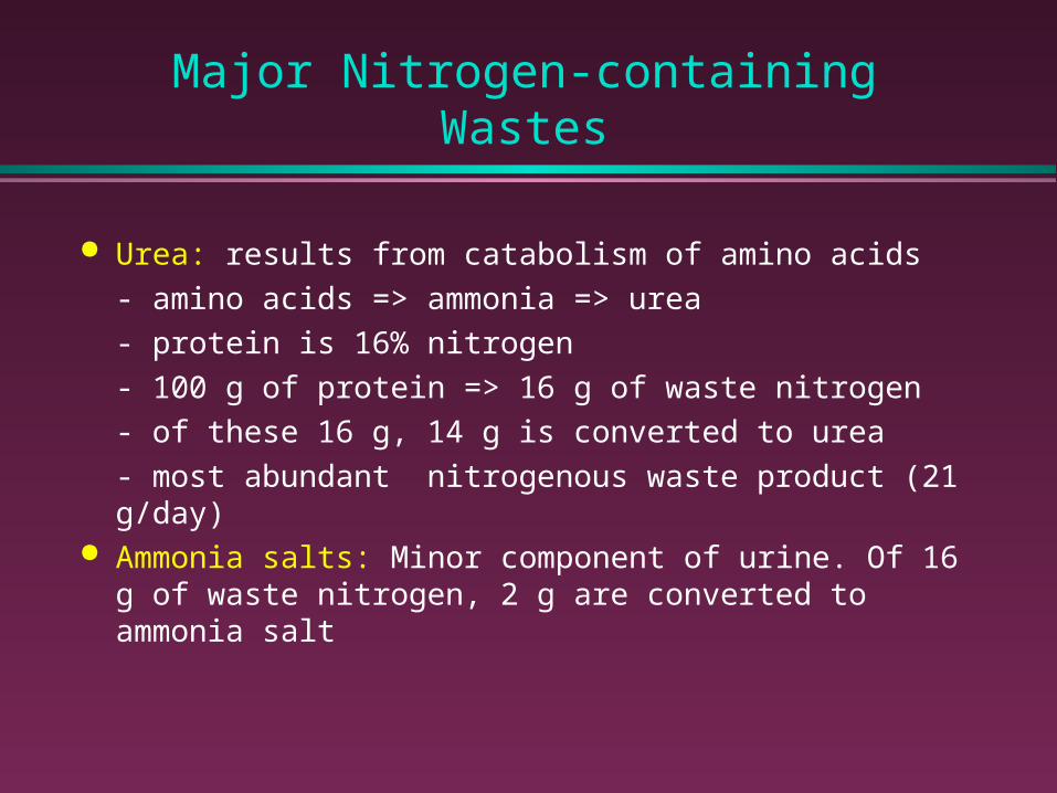
Urea: results from catabolism of amino acids
- amino acids => ammonia => urea
- protein is 16% nitrogen
- 100 g of protein => 16 g of waste nitrogen
- of these 16 g, 14 g is converted to urea
- most abundant nitrogenous waste product (21 g/day)
Ammonia salts: Minor component of urine. Of 16 g of waste nitrogen, 2 g are converted to ammonia salt
Major Nitrogen-containing Wastes (cont.)
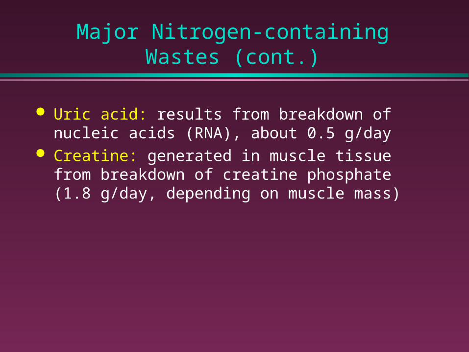
Uric acid: results from breakdown of nucleic acids (RNA), about 0.5 g/day
Creatine: generated in muscle tissue from breakdown of creatine phosphate (1.8 g/day, depending on muscle mass)
Processes involved in Urine Formation
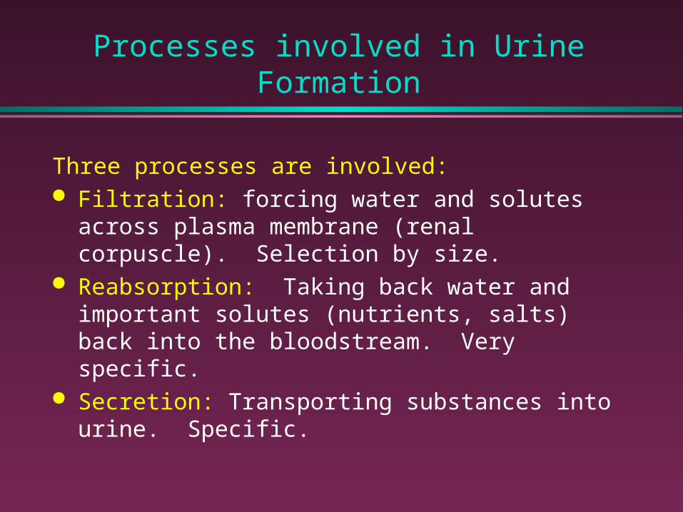
Three processes are involved: Filtration: forcing water and solutes across plasma
membrane (renal corpuscle). Selection by size. Reabsorption: Taking back water and important solutes
(nutrients, salts) back into the bloodstream. Very specific. Secretion: Transporting substances into urine. Specific.


Filtration
All filtration occurs at the renal corpuscle Recall that the corpuscle if formed from the glomerulus
(capillary) and Bowman’s capsule (continuous with the tubules of the nephron)
parietal epithelium
podocyte
glomerularcapillary
afferentarteriole
efferentarteriole
DCT
macula densa
juxtaglomerularcells

Process of Filtration
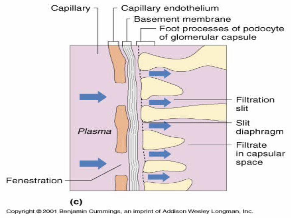
Blood pressure forces water across the glomerular endothelium, basement membrane, and filtration slits of podocyte cells (visceral layer of Bowman’s capsule) into the capsular space
Podocytes
filtration slits
basement membraneendothelial wall
of capillary
fenestra
capsular space
filtration
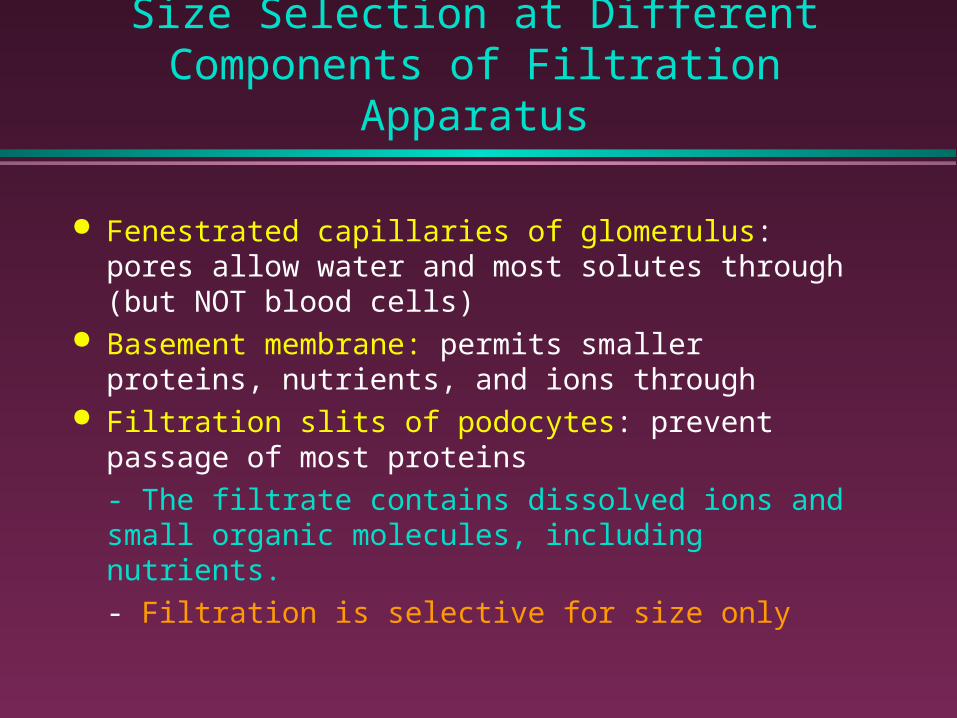
Size Selection at Different Components of Filtration Apparatus
Fenestrated capillaries of glomerulus: pores allow water and most solutes through (but NOT blood cells)
Basement membrane: permits smaller proteins, nutrients, and ions through
Filtration slits of podocytes: prevent passage of most proteins
- The filtrate contains dissolved ions and small organic molecules, including nutrients.
- Filtration is selective for size only
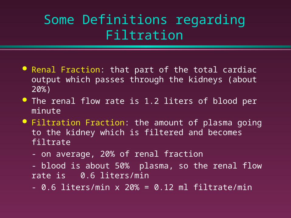
Some Definitions regarding Filtration
Renal Fraction: that part of the total cardiac output which passes through the kidneys (about 20%)
The renal flow rate is 1.2 liters of blood per minute Filtration Fraction: the amount of plasma going to the
kidney which is filtered and becomes filtrate
- on average, 20% of renal fraction
- blood is about 50% plasma, so the renal flow rate is 0.6 liters/min
- 0.6 liters/min x 20% = 0.12 ml filtrate/min
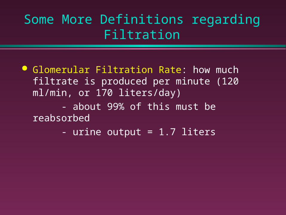
Some More Definitions regarding Filtration
Glomerular Filtration Rate: how much filtrate is produced per minute (120 ml/min, or 170 liters/day)
- about 99% of this must be reabsorbed
- urine output = 1.7 liters

Driving Force of Filtration
The filtration across membranes is driven by the net filtration pressure
The net filtration pressure = net hydrostatic pressure minus the net colloid osmotic pressure
The net hydrostatic pressure is determined by the glomerular hydrostatic pressure (GHP) minus the capsular hydrostatic pressure (CHP)
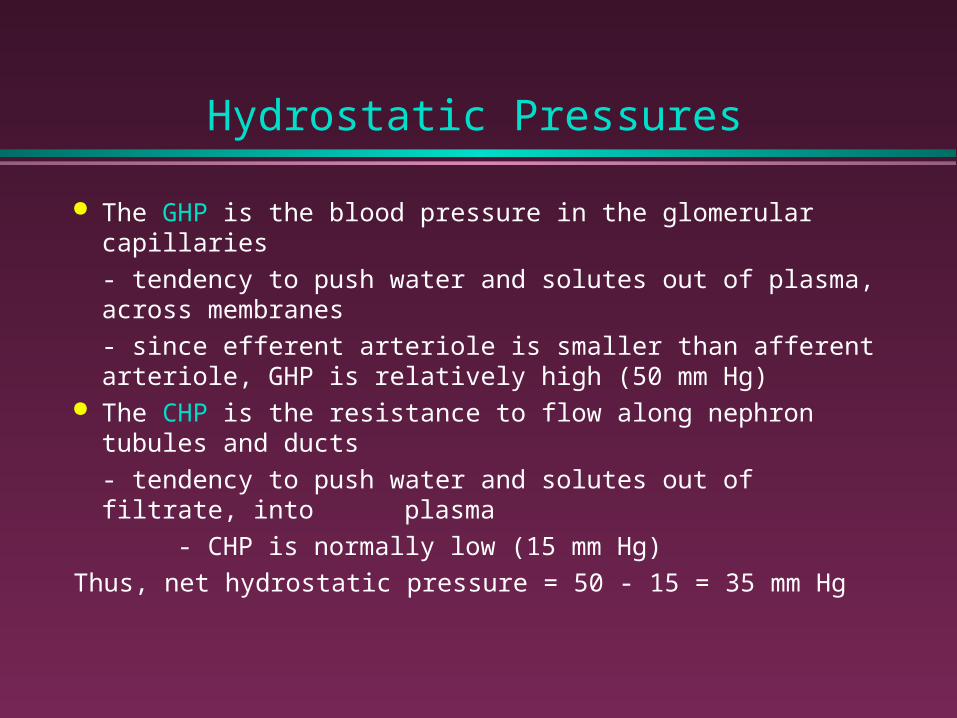
Hydrostatic Pressures
The GHP is the blood pressure in the glomerular capillaries
- tendency to push water and solutes out of plasma, across membranes
- since efferent arteriole is smaller than afferent arteriole, GHP is relatively high (50 mm Hg)
The CHP is the resistance to flow along nephron tubules and ducts
- tendency to push water and solutes out of filtrate, into plasma
- CHP is normally low (15 mm Hg)
Thus, net hydrostatic pressure = 50 - 15 = 35 mm Hg
Colloid Osmotic Pressure (COP)
The colloid osmotic pressure is the osmotic pressure resulting from the presence of proteins in a solution
The COP of blood is about 25 mm Hg The COP of filtrate is normally 0 Thus, total COP is 25 mm Hg

Net Filtration Pressure
Thus, the net filtration pressure =
net hydrostatic pressure - colloid osmotic pressure
= 35 mm Hg - 25 mm Hg = 10 mm Hg Abnormal changes in either net hydrostatic pressure or
colloid osmotic pressure will affect filtration rate
- damage to glomerulus will allow proteins into the filtrate, decreasing net COP, and increasing filtration rate
- increasing capsular hydrostatic pressure (obstruction of tubules, ducts) will markedly decrease net hydrostatic pressure, decreasing filtration rate
Reabsorption
Reabsorption takes place in the proximal convoluted tubule (PCT; 65%), loop of Henle (20%), distal convoluted tubule (DCT; 5%), and collecting ducts (10%)
In the PCT:- Over 99% of organic nutrients (glucose, amino acids) are resorbed- Active ion resorption- Water resorption by osmosis- Other solvents (urea, lipids, Cl- ions) resorbed by solvent drag
At the end of the PCT, filtrate contains no glucose, no amino acids, 12% of NaCl, 25% of volume, and increased urea, uric acid (no change in osmolarity)

Resorption in the Loop of Henle
In the loop of Henle, half of the remaining water and 2/3rds of the remaining NaCl will be resorbed
The loop of Henle utilizes a countercurrent multiplication exchange system to reabsorb water and NaCl
Countercurrent: exchange occurs between fluids moving in opposite directions
Multiplication: the effect of exchange between the limbs increasing as fluid movement occurs

Countercurrent Exchange: Loop of Henle
The walls of the descending and ascending limbs have different permeability characteristics
The descending limb is permeable to water, impermeable to solutes
The ascending limb is impermeable to water and solutes
Na+ and Cl- are actively pumped out of ascending limb
Osmotic concentration of peritubular fluid rises
Water leaves descending limb by osmosis Increased solute concentration causes
increased Na+ and Cl- transport
Results of Countercurrent Exchange
Get resorption of water and NaCl, with filtrate at the end of the loop of Henle with lower osmolarity than at the beginning of the loop
A concentration gradient is built up in the peritubular space, which allows subsequent resorption of water from collecting duct

Reabsorption in the DCT and Collecting Ducts
In the distal convoluted tubule, small adjustments in composition of filtrate take place
- active transport of Na+ and Cl- continues
- water reabsorption occurs under influence of ADH (5% of total water reabsorption)
In the collecting ducts, water and sodium are reabsorbed
- water is reabsorbed under regulation by ADH (10% of water reabsorption)
- sodium reabsorbed under regulation by aldosterone
- some reabsorption of bicarbonate and urea

Secretion into Urinary Filtrate
Secretion plays a relatively small role in production of urine
Occurs in tubules and ducts There is active secretion of a number of substances into the
filtrate:
- potassium, hydrogen ions (in exchange for sodium ions)
- creatinine, penicillin, neurotransmitters, organic acids and bases
Summary
Reviewed major nitrogenous wastes Defined terminology related to filtration Reviewed processes of filtration, absorption, and secretion
(mechanisms, site of action)

Next Lecture......
Regulation of the Urinary System