Embed Size (px)
Citation preview
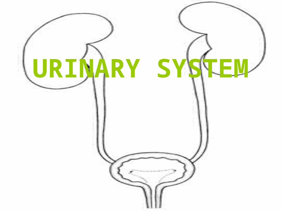
URINARY SYSTEM
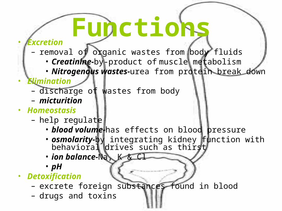
Functions• Excretion
– removal of organic wastes from body fluids• Creatinine-by-product of muscle metabolism• Nitrogenous wastes-urea from protein break down
• Elimination– discharge of wastes from body– micturition
• Homeostasis– help regulate
• blood volume-has effects on blood pressure• osmolarity-by integrating kidney function with behavioral
drives such as thirst• ion balance-Na, K & Cl• pH
• Detoxification– excrete foreign substances found in blood– drugs and toxins
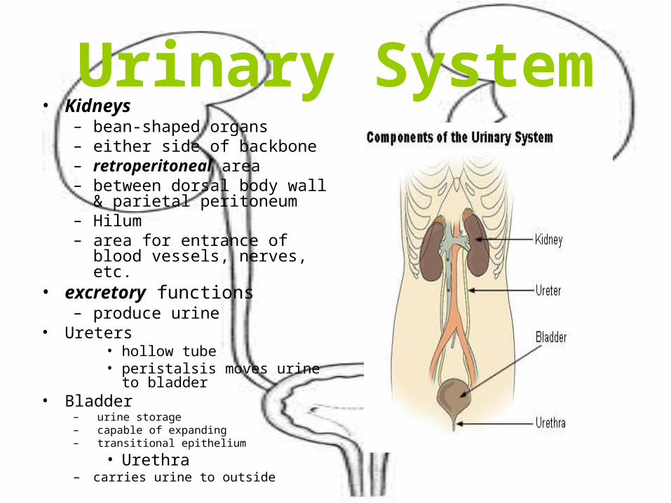
Urinary System• Kidneys
– bean-shaped organs– either side of backbone – retroperitoneal area– between dorsal body wall &
parietal peritoneum– Hilum– area for entrance of blood
vessels, nerves, etc.• excretory functions
– produce urine• Ureters
• hollow tube• peristalsis moves urine to
bladder• Bladder
– urine storage– capable of expanding– transitional epithelium
• Urethra– carries urine to outside
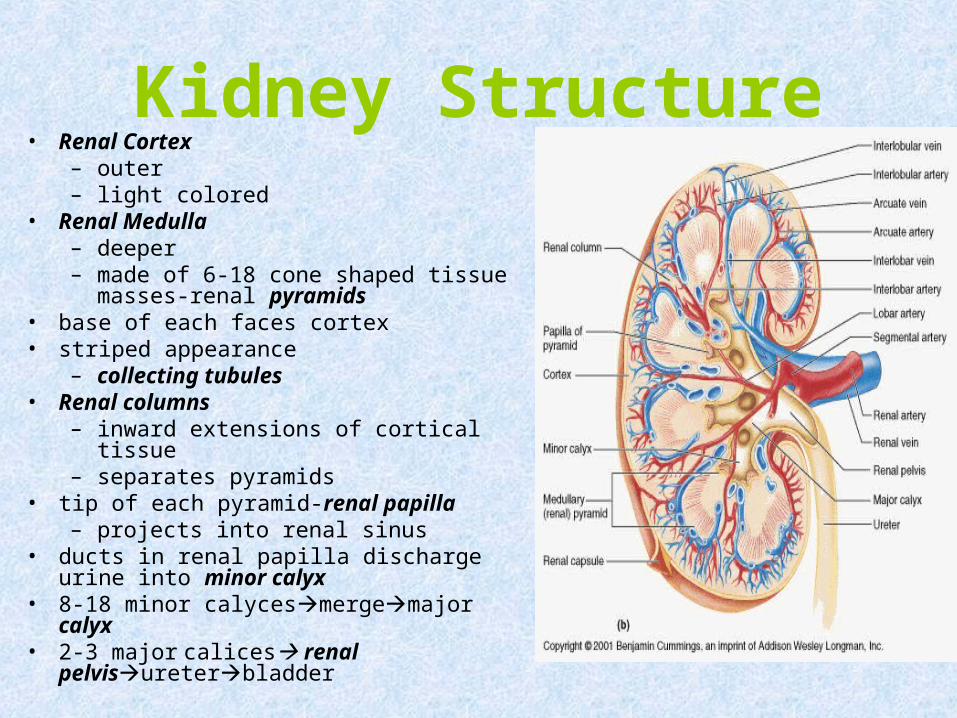
Kidney Structure• Renal Cortex
– outer – light colored
• Renal Medulla– deeper– made of 6-18 cone shaped tissue
masses-renal pyramids• base of each faces cortex• striped appearance
– collecting tubules• Renal columns
– inward extensions of cortical tissue– separates pyramids
• tip of each pyramid-renal papilla– projects into renal sinus
• ducts in renal papilla discharge urine into minor calyx
• 8-18 minor calycesmergemajor calyx• 2-3 major calices renal
pelvisureterbladder
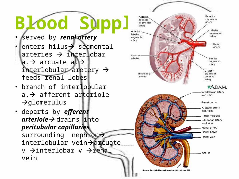
Blood Supply• served by renal artery
• enters hilus segmental arteries interlobar a. arcuate a. interlobular aretery feeds renal lobes
• branch of interlobular a. afferent arteriole glomerulus
• departs by efferent arteriole drains into peritubular capillaries surrounding nephron interlobular veinarcuate v interlobar v renal vein
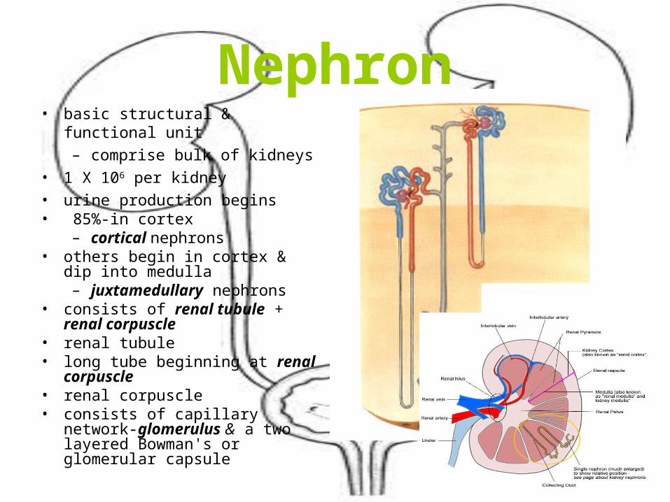
Nephron• basic structural & functional unit
– comprise bulk of kidneys
• 1 X 10⁶ per kidney
• urine production begins• 85%-in cortex
– cortical nephrons• others begin in cortex & dip into
medulla– juxtamedullary nephrons
• consists of renal tubule + renal corpuscle
• renal tubule• long tube beginning at renal
corpuscle• renal corpuscle• consists of capillary network-
glomerulus & a two layered Bowman's or glomerular capsule
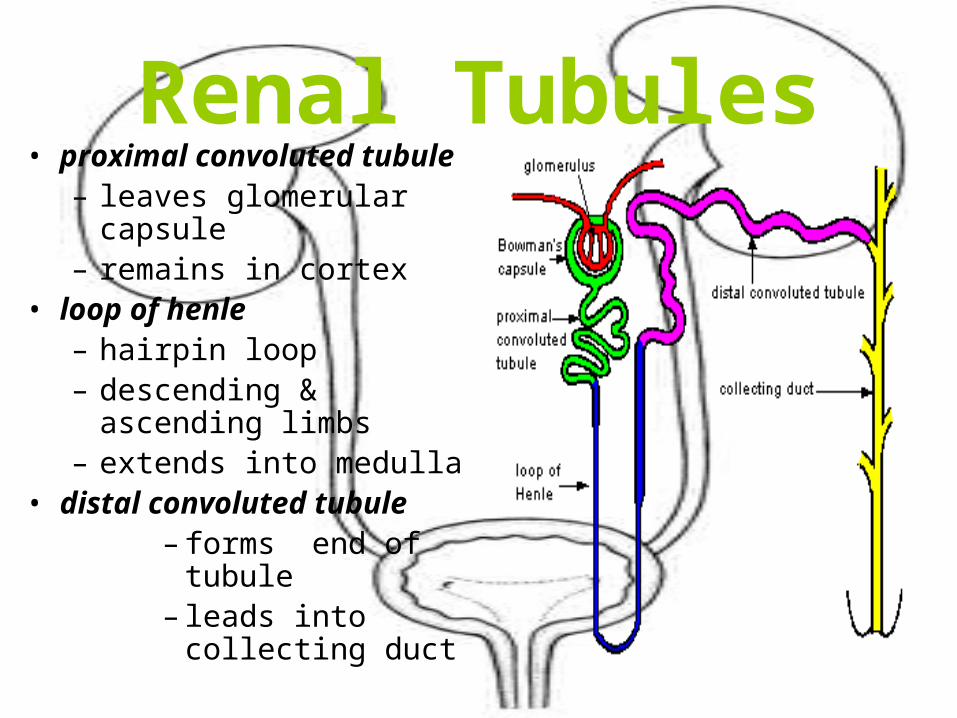
Renal Tubules• proximal convoluted
tubule– leaves glomerular capsule– remains in cortex
• loop of henle– hairpin loop– descending & ascending
limbs– extends into medulla
• distal convoluted tubule– forms end of
tubule– leads into collecting
duct
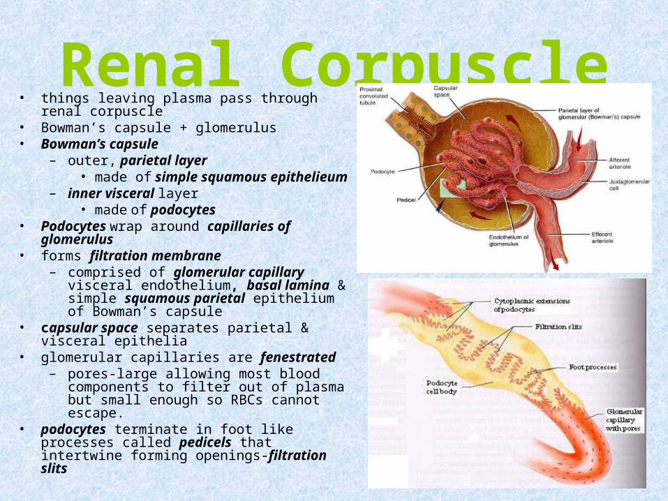
Renal Corpuscle• things leaving plasma pass through renal
corpuscle• Bowman’s capsule + glomerulus• Bowman’s capsule
– outer, parietal layer• made of simple squamous
epithelieum– inner visceral layer
• made of podocytes• Podocytes wrap around capillaries of
glomerulus• forms filtration membrane
– comprised of glomerular capillary visceral endothelium, basal lamina & simple squamous parietal epithelium of Bowman’s capsule
• capsular space separates parietal & visceral epithelia
• glomerular capillaries are fenestrated– pores-large allowing most blood
components to filter out of plasma but small enough so RBCs cannot escape.
• podocytes terminate in foot like processes called pedicels that intertwine forming openings-filtration slits
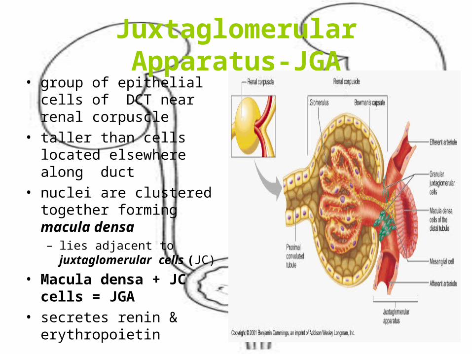
Juxtaglomerular Apparatus-JGA• group of epithelial cells of
DCT near renal corpuscle• taller than cells located
elsewhere along duct• nuclei are clustered
together forming macula densa– lies adjacent to
juxtaglomerular cells (JC)
• Macula densa + JC cells = JGA
• secretes renin & erythropoietin
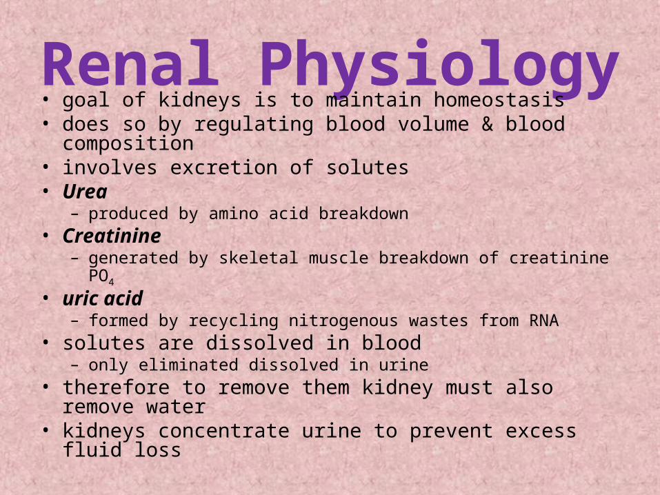
Renal Physiology• goal of kidneys is to maintain homeostasis• does so by regulating blood volume & blood composition• involves excretion of solutes• Urea
– produced by amino acid breakdown• Creatinine
– generated by skeletal muscle breakdown of creatinine PO4
• uric acid– formed by recycling nitrogenous wastes from RNA
• solutes are dissolved in blood– only eliminated dissolved in urine
• therefore to remove them kidney must also remove water
• kidneys concentrate urine to prevent excess fluid loss
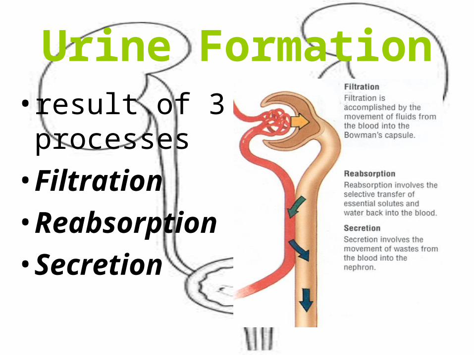
Urine Formation• result of 3
processes
• Filtration
• Reabsorption
• Secretion
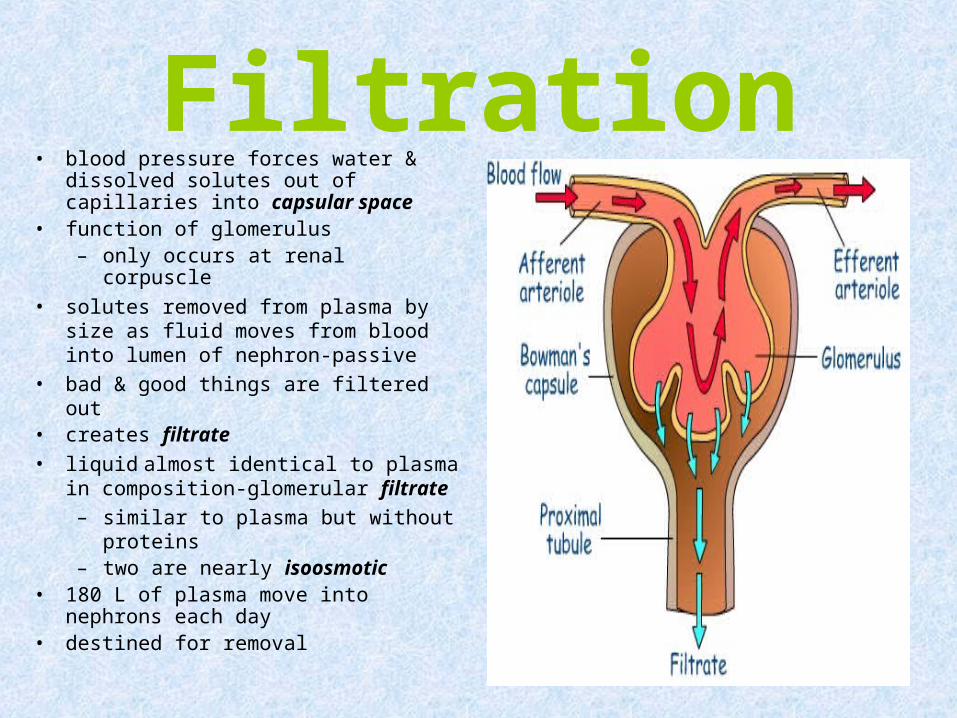
Filtration• blood pressure forces water &
dissolved solutes out of capillaries into capsular space
• function of glomerulus– only occurs at renal corpuscle
• solutes removed from plasma by size as fluid moves from blood into lumen of nephron-passive
• bad & good things are filtered out• creates filtrate
• liquid almost identical to plasma in composition-glomerular filtrate
– similar to plasma but without proteins
– two are nearly isoosmotic• 180 L of plasma move into nephrons
each day• destined for removal
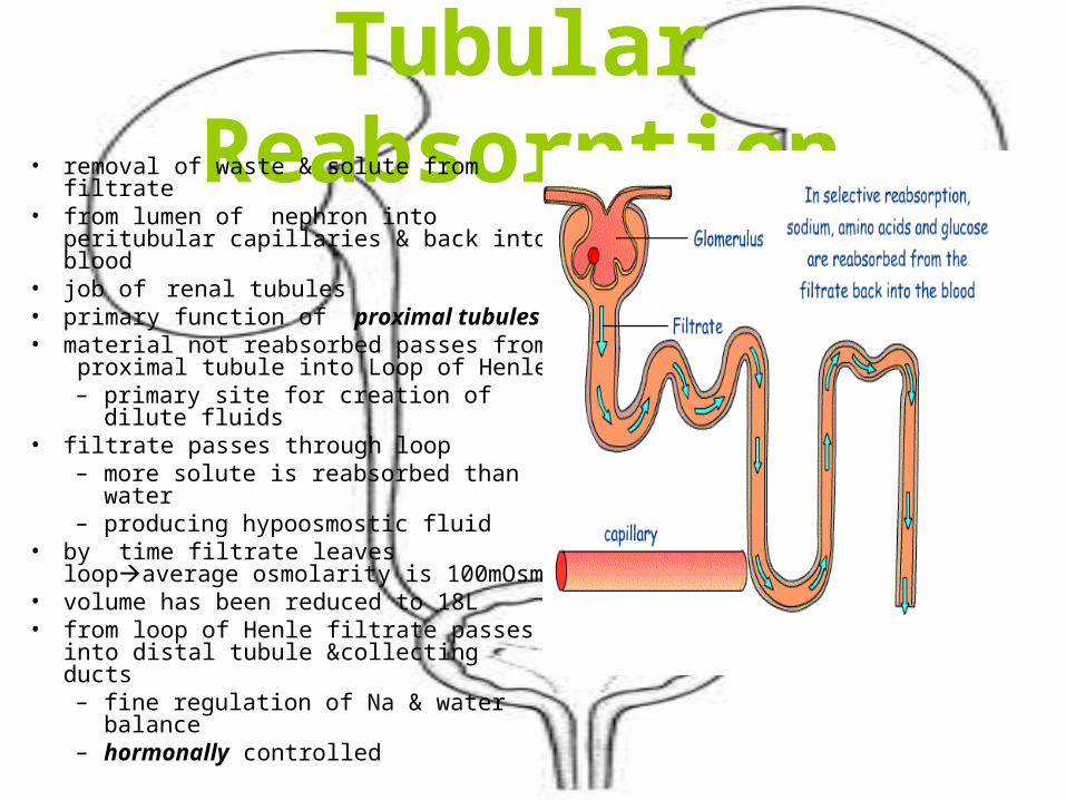
Tubular Reabsorption• removal of waste & solute from filtrate• from lumen of nephron into peritubular
capillaries & back into blood• job of renal tubules• primary function of proximal tubules• material not reabsorbed passes from
proximal tubule into Loop of Henle– primary site for creation of dilute
fluids• filtrate passes through loop
– more solute is reabsorbed than water– producing hypoosmostic fluid
• by time filtrate leaves loopaverage osmolarity is 100mOsm
• volume has been reduced to 18L• from loop of Henle filtrate passes into
distal tubule &collecting ducts– fine regulation of Na & water balance– hormonally controlled
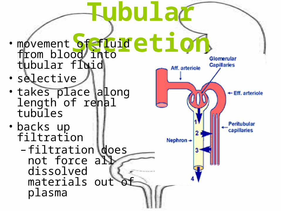
Tubular Secretion• movement of fluid
from blood into tubular fluid
• selective• takes place along
length of renal tubules
• backs up filtration– filtration does not
force all dissolved materials out of plasma
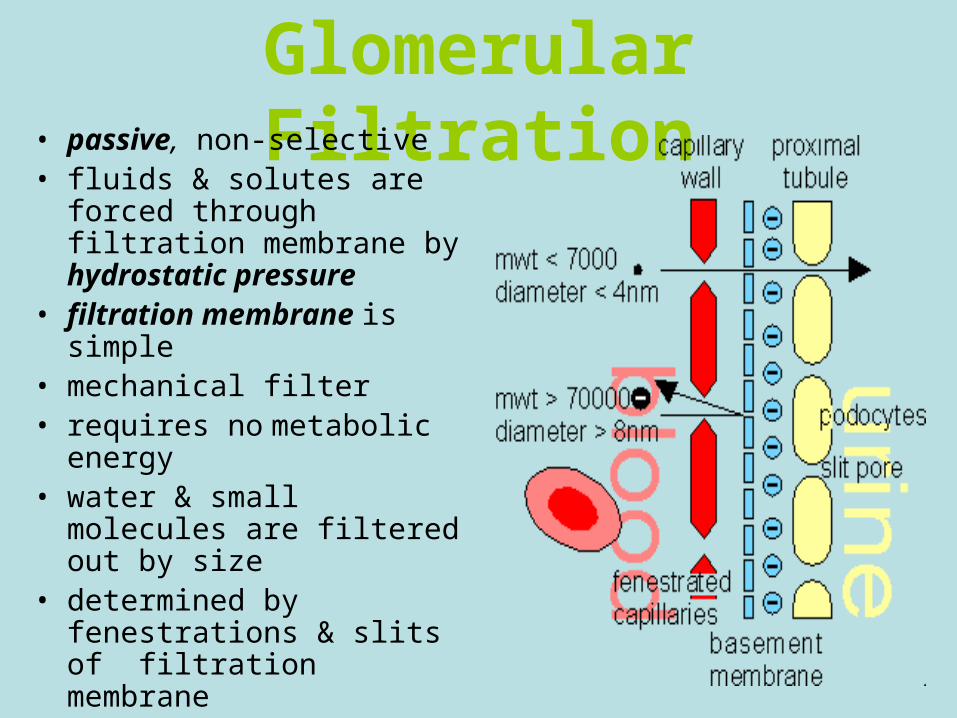
Glomerular Filtration• passive, non-selective• fluids & solutes are forced
through filtration membrane by hydrostatic pressure
• filtration membrane is simple
• mechanical filter• requires no metabolic
energy• water & small molecules are
filtered out by size• determined by fenestrations
& slits of filtration membrane
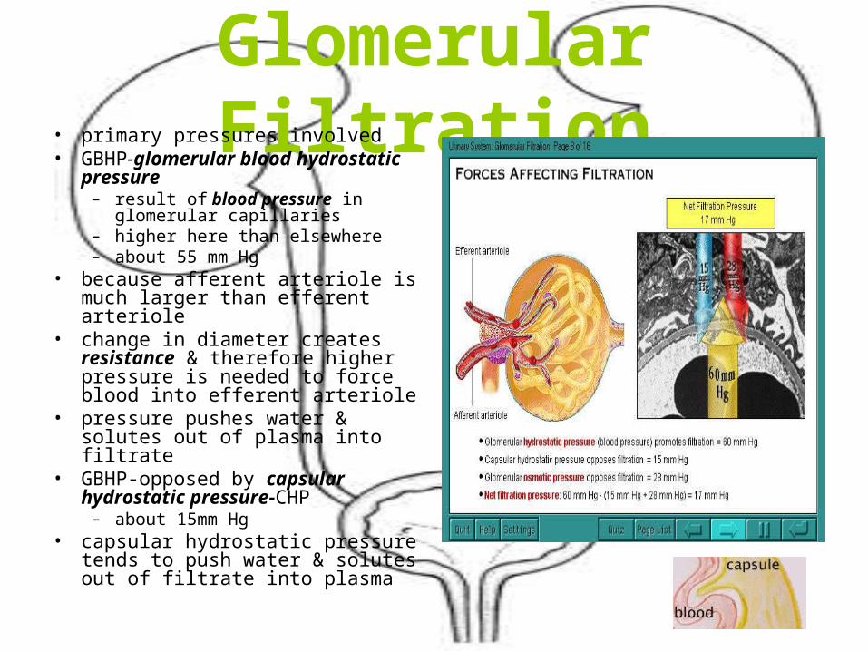
Glomerular Filtration• primary pressures involved• GBHP-glomerular blood
hydrostatic pressure– result of blood pressure in
glomerular capillaries– higher here than elsewhere– about 55 mm Hg
• because afferent arteriole is much larger than efferent arteriole
• change in diameter creates resistance & therefore higher pressure is needed to force blood into efferent arteriole
• pressure pushes water & solutes out of plasma into filtrate
• GBHP-opposed by capsular hydrostatic pressure-CHP
– about 15mm Hg• capsular hydrostatic pressure
tends to push water & solutes out of filtrate into plasma
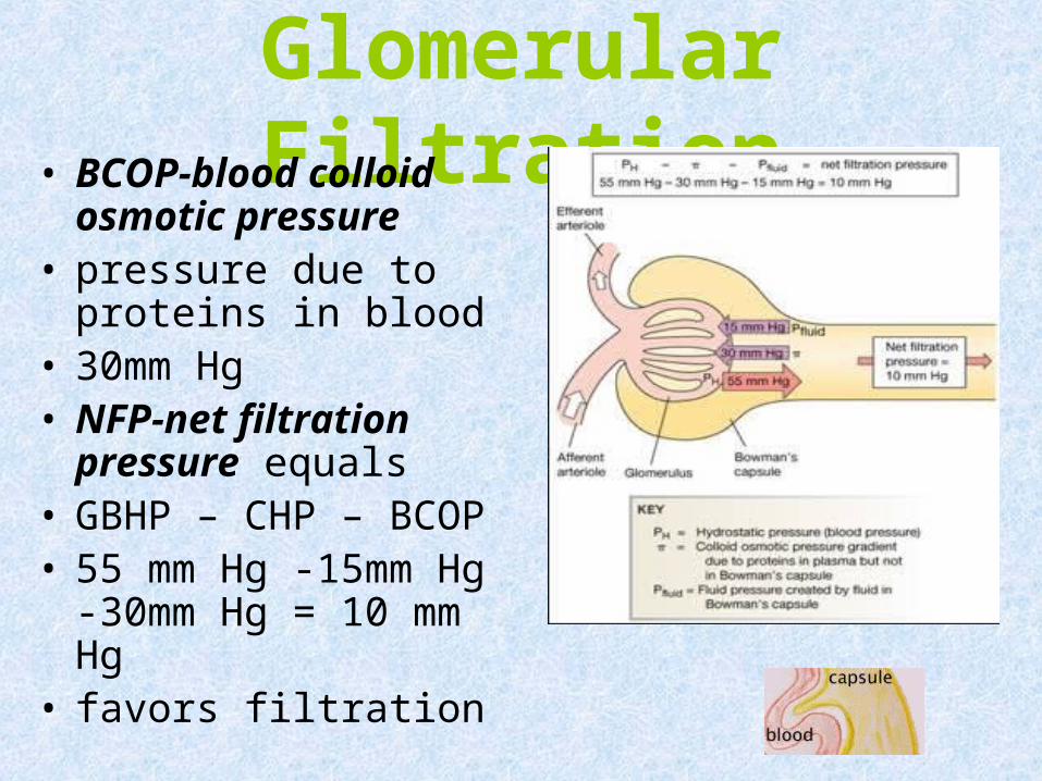
Glomerular Filtration• BCOP-blood colloid
osmotic pressure • pressure due to
proteins in blood• 30mm Hg• NFP-net filtration
pressure equals• GBHP – CHP – BCOP• 55 mm Hg -15mm Hg -
30mm Hg = 10 mm Hg • favors filtration
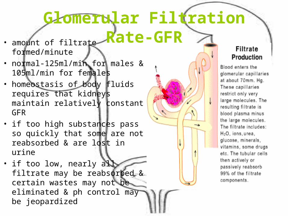
Glomerular Filtration Rate-GFR• amount of filtrate formed/minute• normal-125ml/min for males &
105ml/min for females• homeostasis of body fluids
requires that kidneys maintain relatively constant GFR
• if too high substances pass so quickly that some are not reabsorbed & are lost in urine
• if too low, nearly all filtrate may be reabsorbed & certain wastes may not be eliminated & ph control may be jeopardized
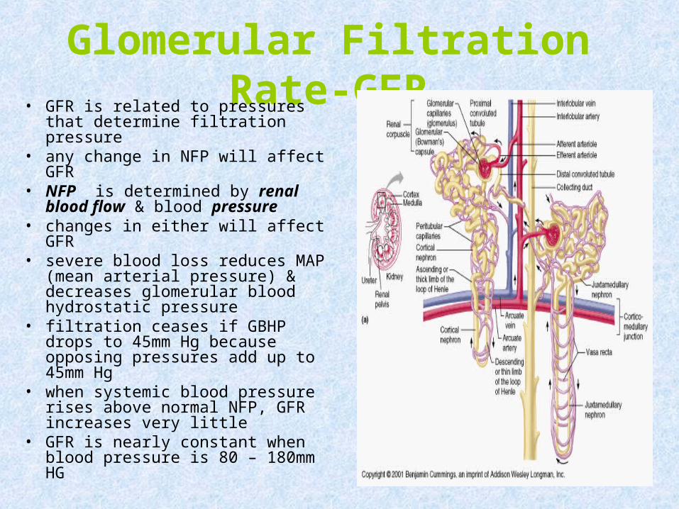
Glomerular Filtration Rate-GFR• GFR is related to pressures that
determine filtration pressure• any change in NFP will affect
GFR• NFP is determined by renal
blood flow & blood pressure • changes in either will affect GFR• severe blood loss reduces MAP
(mean arterial pressure) & decreases glomerular blood hydrostatic pressure
• filtration ceases if GBHP drops to 45mm Hg because opposing pressures add up to 45mm Hg
• when systemic blood pressure rises above normal NFP, GFR increases very little
• GFR is nearly constant when blood pressure is 80 – 180mm HG
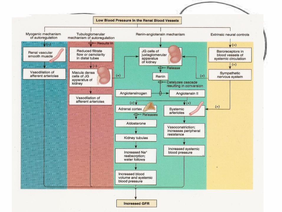
Controlling GFR• regulatory mechanisms ensure GFR is kept within
normal limits• mechanisms that regulate GFR
– adjust blood pressure into & out of the glomerulus– alter glomerular capillary surface area available for
filtration• GFR increases when blood flow into glomerulus
increases• 3 mechanism control GFR• renal autoregulation• nervous regulation• hormonal mechanisms
Renal Autoregulation• ability of nephrons to adjust
own blood flow & GFR
• two mechanisms
• myogenic mechanisms
• tubuloglomerular feedback
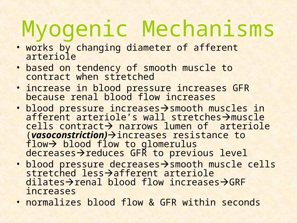
Myogenic Mechanisms• works by changing diameter of afferent arteriole• based on tendency of smooth muscle to contract when
stretched• increase in blood pressure increases GFR because renal
blood flow increases • blood pressure increasessmooth muscles in afferent
arteriole’s wall stretchesmuscle cells contract narrows lumen of arteriole (vasoconstriction)increases resistance to flow blood flow to glomerulus decreasesreduces GFR to previous level
• blood pressure decreasessmooth muscle cells stretched lessafferent arteriole dilatesrenal blood flow increasesGRF increases
• normalizes blood flow & GFR within seconds
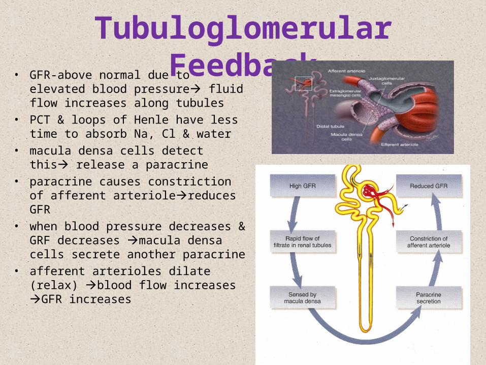
Tubuloglomerular Feedback• GFR-above normal due to elevated
blood pressure fluid flow increases along tubules
• PCT & loops of Henle have less time to absorb Na, Cl & water
• macula densa cells detect this release a paracrine
• paracrine causes constriction of afferent arteriolereduces GFR
• when blood pressure decreases & GRF decreases macula densa cells secrete another paracrine
• afferent arterioles dilate (relax) blood flow increases GFR increases
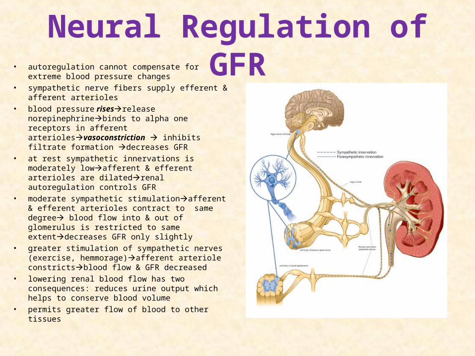
Neural Regulation of GFR• autoregulation cannot compensate for extreme
blood pressure changes
• sympathetic nerve fibers supply efferent & afferent arterioles
• blood pressure risesrelease norepinephrinebinds to alpha one receptors in afferent arteriolesvasoconstriction inhibits filtrate formation decreases GFR
• at rest sympathetic innervations is moderately lowafferent & efferent arterioles are dilatedrenal autoregulation controls GFR
• moderate sympathetic stimulationafferent & efferent arterioles contract to same degree blood flow into & out of glomerulus is restricted to same extentdecreases GFR only slightly
• greater stimulation of sympathetic nerves (exercise, hemmorage)afferent arteriole constrictsblood flow & GFR decreased
• lowering renal blood flow has two consequences: reduces urine output which helps to conserve blood volume
• permits greater flow of blood to other tissues
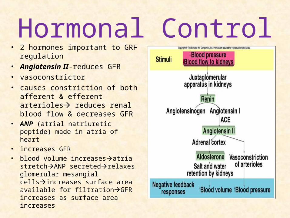
Hormonal Control• 2 hormones important to GRF
regulation• Angiotensin II-reduces GFR• vasoconstrictor• causes constriction of both
afferent & efferent arterioles reduces renal blood flow & decreases GFR
• ANP (atrial natriuretic peptide) made in atria of heart
• increases GFR
• blood volume increasesatria stretchANP secretedrelaxes glomerular mesangial cellsincreases surface area available for filtrationGFR increases as surface area increases
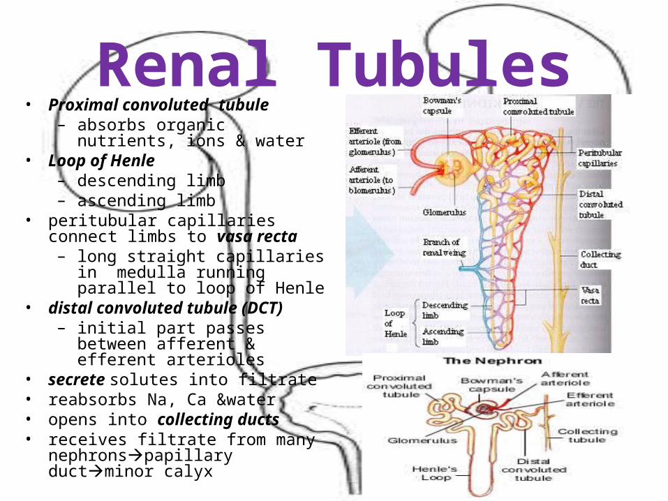
Renal Tubules• Proximal convoluted tubule
– absorbs organic nutrients, ions & water
• Loop of Henle– descending limb– ascending limb
• peritubular capillaries connect limbs to vasa recta– long straight capillaries in medulla
running parallel to loop of Henle• distal convoluted tubule (DCT)
– initial part passes between afferent & efferent arterioles
• secrete solutes into filtrate• reabsorbs Na, Ca &water• opens into collecting ducts• receives filtrate from many
nephronspapillary ductminor calyx
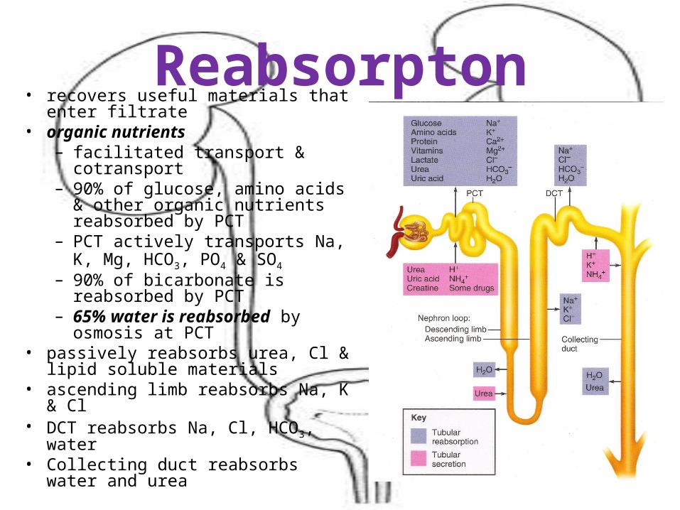
Reabsorpton• recovers useful materials that enter
filtrate• organic nutrients
– facilitated transport & cotransport– 90% of glucose, amino acids &
other organic nutrients reabsorbed by PCT
– PCT actively transports Na, K, Mg, HCO3, PO4 & SO4
– 90% of bicarbonate is reabsorbed by PCT
– 65% water is reabsorbed by osmosis at PCT
• passively reabsorbs urea, Cl & lipid soluble materials
• ascending limb reabsorbs Na, K & Cl
• DCT reabsorbs Na, Cl, HCO3, water• Collecting duct reabsorbs water and
urea
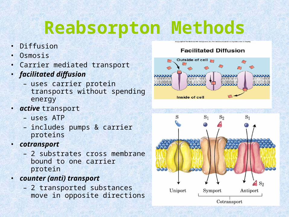
Reabsorpton Methods• Diffusion• Osmosis• Carrier mediated transport• facilitated diffusion
– uses carrier protein transports without spending energy
• active transport– uses ATP– includes pumps & carrier
proteins• cotransport
– 2 substrates cross membrane bound to one carrier protein
• counter (anti) transport– 2 transported substances move
in opposite directions
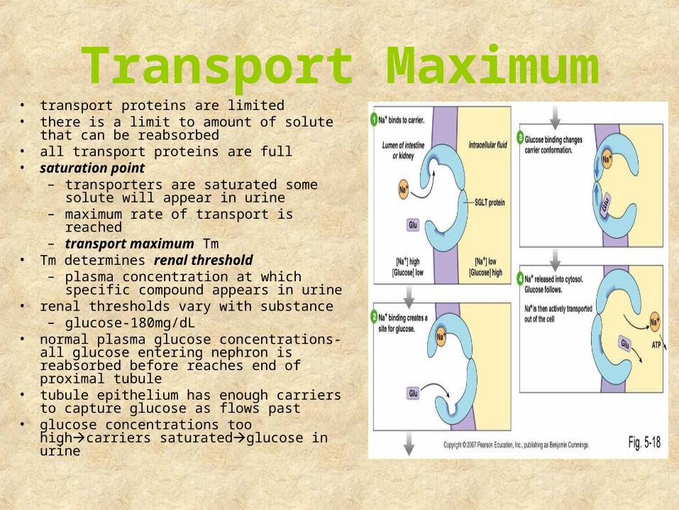
Transport Maximum• transport proteins are limited• there is a limit to amount of solute that
can be reabsorbed• all transport proteins are full• saturation point
– transporters are saturated some solute will appear in urine
– maximum rate of transport is reached– transport maximum Tm
• Tm determines renal threshold– plasma concentration at which
specific compound appears in urine• renal thresholds vary with substance
– glucose-180mg/dL• normal plasma glucose concentrations- all
glucose entering nephron is reabsorbed before reaches end of proximal tubule
• tubule epithelium has enough carriers to capture glucose as flows past
• glucose concentrations too highcarriers saturatedglucose in urine
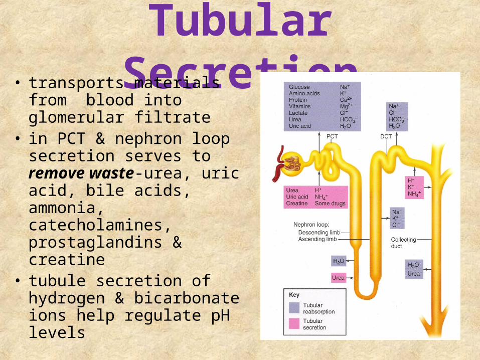
Tubular Secretion• transports materials from
blood into glomerular filtrate
• in PCT & nephron loop secretion serves to remove waste-urea, uric acid, bile acids, ammonia, catecholamines, prostaglandins & creatine
• tubule secretion of hydrogen & bicarbonate ions help regulate pH levels
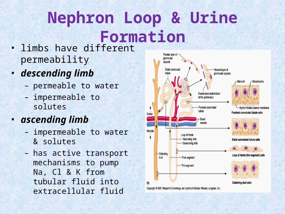
Nephron Loop & Urine Formation• limbs have different
permeability• descending limb
– permeable to water– impermeable to solutes
• ascending limb– impermeable to water &
solutes– has active transport
mechanisms to pump Na, Cl & K from tubular fluid into extracellular fluid
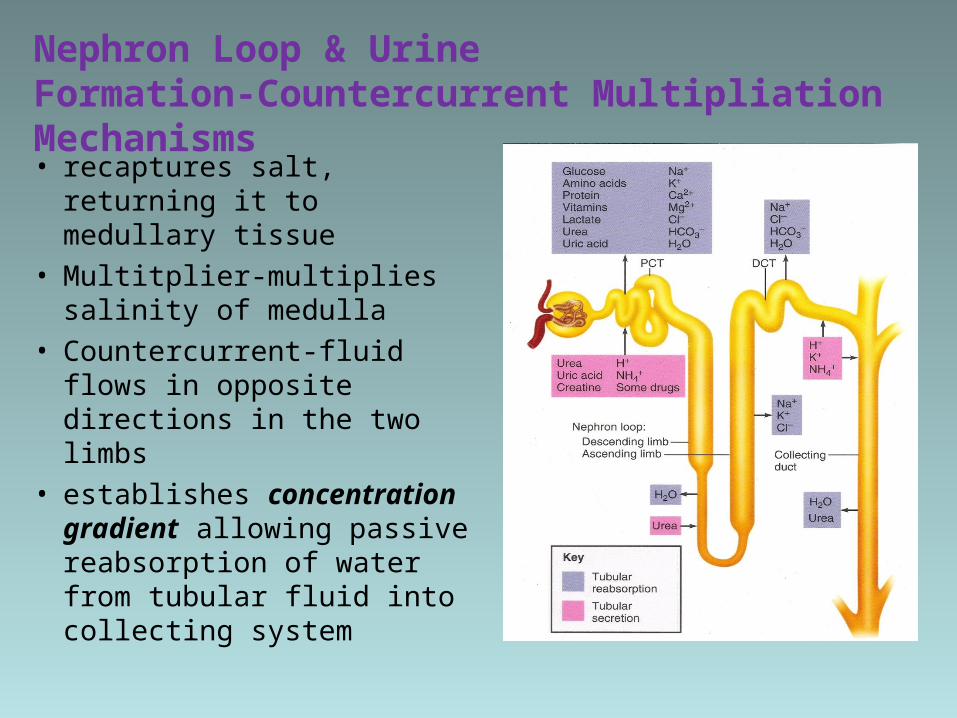
Nephron Loop & Urine Formation-Countercurrent Multipliation Mechanisms• recaptures salt, returning it
to medullary tissue
• Multitplier-multiplies salinity of medulla
• Countercurrent-fluid flows in opposite directions in the two limbs
• establishes concentration gradient allowing passive reabsorption of water from tubular fluid into collecting system
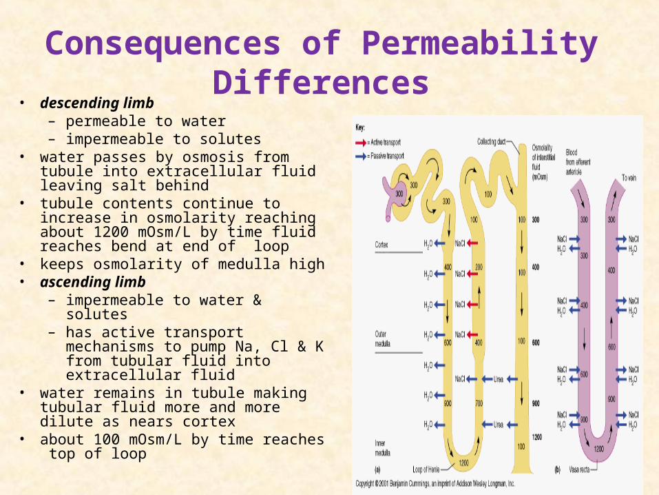
Consequences of Permeability Differences
• descending limb– permeable to water– impermeable to solutes
• water passes by osmosis from tubule into extracellular fluid leaving salt behind
• tubule contents continue to increase in osmolarity reaching about 1200 mOsm/L by time fluid reaches bend at end of loop
• keeps osmolarity of medulla high• ascending limb
– impermeable to water & solutes– has active transport mechanisms
to pump Na, Cl & K from tubular fluid into extracellular fluid
• water remains in tubule making tubular fluid more and more dilute as nears cortex
• about 100 mOsm/L by time reaches top of loop
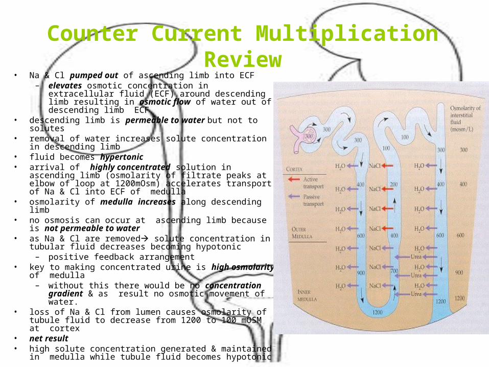
Counter Current MultiplicationReview
• Na & Cl pumped out of ascending limb into ECF– elevates osmotic concentration in extracellular fluid
(ECF) around descending limb resulting in osmotic flow of water out of descending limb ECF
• descending limb is permeable to water but not to solutes• removal of water increases solute concentration in
descending limb• fluid becomes hypertonic• arrival of highly concentrated solution in ascending
limb (osmolarity of filtrate peaks at elbow of loop at 1200mOsm) accelerates transport of Na & Cl into ECF of medulla
• osmolarity of medulla increases along descending limb• no osmosis can occur at ascending limb because is not
permeable to water• as Na & Cl are removed solute concentration in tubular
fluid decreases becoming hypotonic – positive feedback arrangement
• key to making concentrated urine is high osmolarity of medulla
– without this there would be no concentration gradient & as result no osmotic movement of water.
• loss of Na & Cl from lumen causes osmolarity of tubule fluid to decrease from 1200 to 100 mOSM at cortex
• net result• high solute concentration generated & maintained in
medulla while tubule fluid becomes hypotonic

Countercurrent Exchange-Vasa Recta• blood vessels surrounding
nephron-vasa recta• water & solutes which move
into surrounding tissue are removed by vasa recta– freely permeable to water
& salt• return water to blood• maintain high osmolarity of
medulla
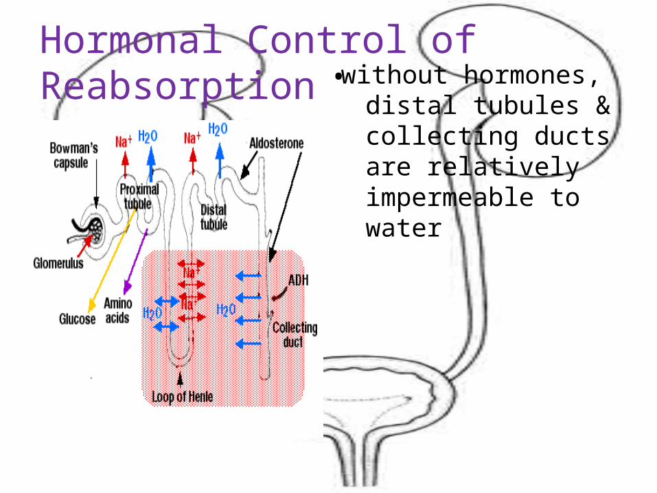
Hormonal Control of Reabsorption without hormones, distal
tubules & collecting ducts are relatively impermeable to water
•
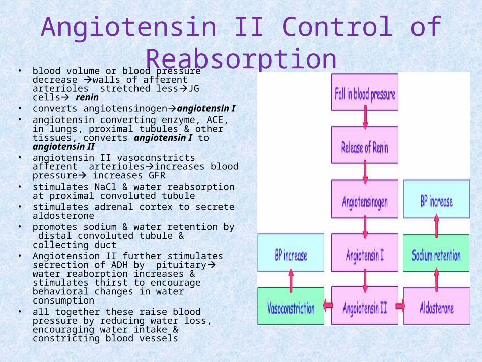
Angiotensin II Control of Reabsorption
• blood volume or blood pressure decrease walls of afferent arterioles stretched lessJG cells renin
• converts angiotensinogenangiotensin I• angiotensin converting enzyme, ACE, in
lungs, proximal tubules & other tissues, converts angiotensin I to angiotensin II
• angiotensin II vasoconstricts afferent arteriolesincreases blood pressure increases GFR
• stimulates NaCl & water reabsorption at proximal convoluted tubule
• stimulates adrenal cortex to secrete aldosterone
• promotes sodium & water retention by distal convoluted tubule & collecting duct
• Angiotension II further stimulates secrection of ADH by pituitary water reaborption increases & stimulates thirst to encourage behavioral changes in water consumption
• all together these raise blood pressure by reducing water loss, encouraging water intake & constricting blood vessels
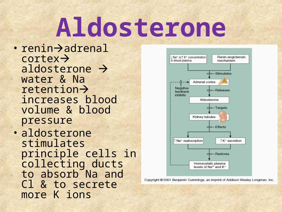
Aldosterone• reninadrenal
cortex aldosterone water & Na retention increases blood volume & blood pressure
• aldosterone stimulates principle cells in collecting ducts to absorb Na and Cl & to secrete more K ions
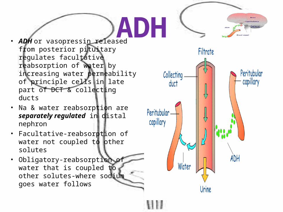
ADH• ADH or vasopressin released
from posterior pituitary regulates facultative reabsorption of water by increasing water permeability of principle cells in late part of DCT & collecting ducts
• Na & water reabsorption are separately regulated in distal nephron
• Facultative-reabsorption of water not coupled to other solutes
• Obligatory-reabsorption of water that is coupled to other solutes-where sodium goes water follows
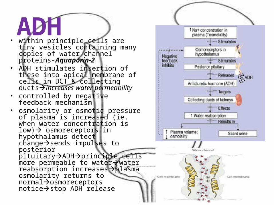
ADH• within principle cells are tiny
vesicles containing many copies of water channel proteins-Aquaporin-2
• ADH stimulates insertion of these into apical membrane of cells in DCT & collecting ductsincreases water permeability
• controlled by negative feedback mechanism
• osmolarity or osmotic pressure of plasma is increased (ie. when water concentration is low) osmoreceptors in hypothalamus detect changesends impulses to posterior pituitaryADHprinciple cells more permeable to waterwater reabsorption increasesplasma osmolarity returns to normalosmoreceptors noticestop ADH release
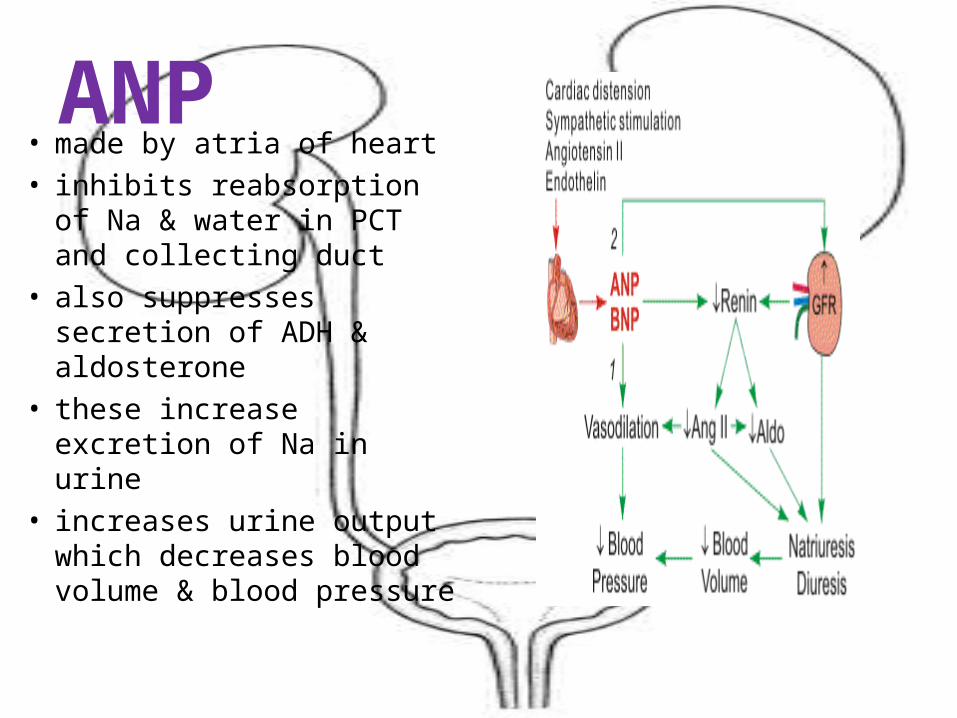
ANP• made by atria of heart• inhibits reabsorption of
Na & water in PCT and collecting duct
• also suppresses secretion of ADH & aldosterone
• these increase excretion of Na in urine
• increases urine output which decreases blood volume & blood pressure
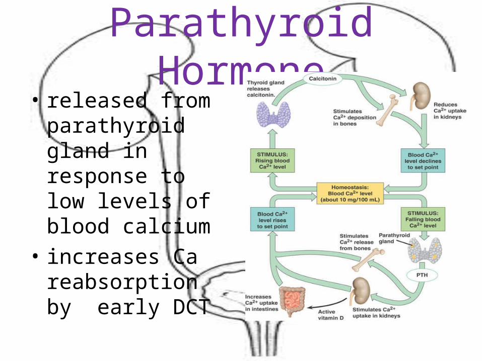
Parathyroid Hormone• released from
parathyroid gland in response to low levels of blood calcium
• increases Ca reabsorption by early DCT
Evaluation of Kidney Function• several ways to determine how effectively kidneys are
functioning
• most used & easiest-urinalysis
• urine is evaluated for volume, physical & chemical properties
• renal clearance
– way to access functions & to access renal function indirectly
– volume of blood cleaned or cleared of a substance per unit time (ml/min)
– assesses renal function by using urine &blood values
• Renal Clearance = S = U X V/P U=concentration of substance in urine, P=concentration of substance in plasma and V= urine flow in ml/minute
Renal Clearance• Renal clearance (C) = UV/P• U = urea concentration in urine• V = rate of urine output• P = urea concentration in plasma• U = 6.0 mg/ml, V = 2ml/min and P = 0.2 mg/ml
then C = 60ml/min• means 60ml of blood is completely cleared of urea
each minute • estimates GFR• cannot be exactly determined by urea excretion• some urea is secreted into renal tubule and not
filtered by glomerulus• some urea that is filtered by glomerulus not
reabsorbed
Renal Clearance• depends on 3 basic processes: filtration, secretion &
reabsorption • for a substance that is filter but not reabsorbed or secreted
clearance = GFR• all the molecules that pass filtration membrane appear in
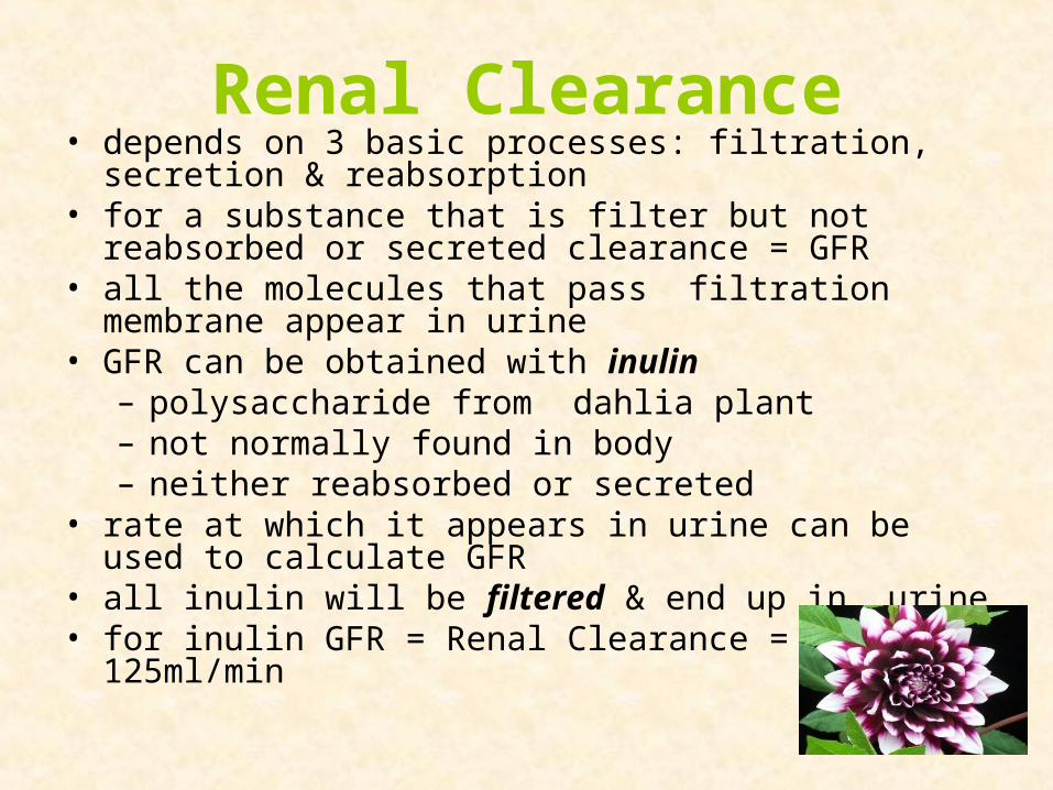
urine• GFR can be obtained with inulin
– polysaccharide from dahlia plant– not normally found in body– neither reabsorbed or secreted
• rate at which it appears in urine can be used to calculate GFR
• all inulin will be filtered & end up in urine• for inulin GFR = Renal Clearance = 125ml/min
Creatinine Clearance Test• can be used to estimate GFR• compares blood & urine creatinine concentrations• creatinine-breakdown product of phosphocreatine
– energy storage compound in muscle• produced & removed at constant rate from blood• filtered & not reabsorbed in significant amounts (15%)• only 2 ways for substance to be in urine• filtered at glomerulus or secreted from peritubular capillaries into
tubules• GFR = amount of substance eliminated divided by amount of
substance in plasma• Kidneys eliminate 84mg of creatinine/hour & plasma creatinine =
1.4mg/dL• 84/1.4 = 60dL/hr = 100ml/min• because nearly all creatinine appears in urinechange in rate of
creatinine excretion may reflect renal disorder
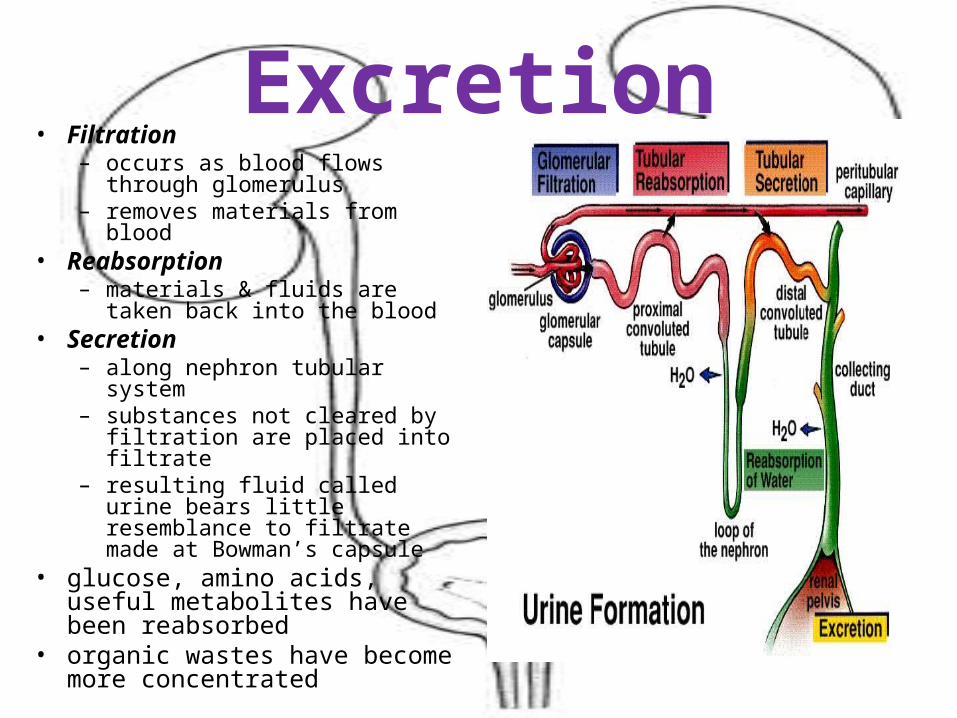
Excretion• Filtration
– occurs as blood flows through glomerulus
– removes materials from blood• Reabsorption
– materials & fluids are taken back into the blood
• Secretion– along nephron tubular system– substances not cleared by
filtration are placed into filtrate– resulting fluid called urine bears
little resemblance to filtrate made at Bowman’s capsule
• glucose, amino acids, useful metabolites have been reabsorbed
• organic wastes have become more concentrated
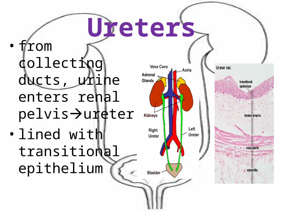
Ureters• from collecting
ducts, urine enters renal pelvisureter
• lined with transitional epithelium
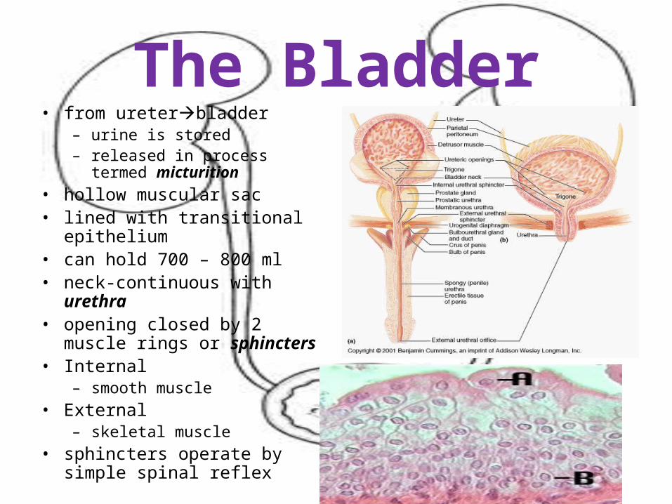
The Bladder• from ureterbladder
– urine is stored– released in process termed
micturition
• hollow muscular sac• lined with transitional
epithelium• can hold 700 – 800 ml• neck-continuous with urethra• opening closed by 2 muscle
rings or sphincters• Internal
– smooth muscle
• External– skeletal muscle
• sphincters operate by simple spinal reflex
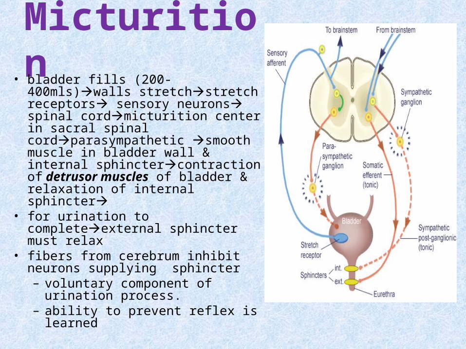
Micturition• bladder fills (200-400mls)walls
stretchstretch receptors sensory neurons spinal cordmicturition center in sacral spinal cordparasympathetic smooth muscle in bladder wall & internal sphinctercontraction of detrusor muscles of bladder & relaxation of internal sphincter
• for urination to completeexternal sphincter must relax
• fibers from cerebrum inhibit neurons supplying sphincter– voluntary component of
urination process. – ability to prevent reflex is
learned