Embed Size (px)
Citation preview

Chapter 15 Lecture
HUMAN ANATOMYFifth Edition
Chapter 15The Nervous System:
The Brain and Cranial NervesFrederic Martini
Michael TimmonsRobert Tallitsch
Copyright © 2005 Pearson Education, Inc., publishing as Benjamin Cummings

Introduction
• The brain is far more complex than thespinal cord.
• The brain contains roughly 20 billionneurons.- Excitatory and inhibitory interactions amongthe extensively interconnected neuronal poolsensure that the response can vary to meetchanging circumstances.
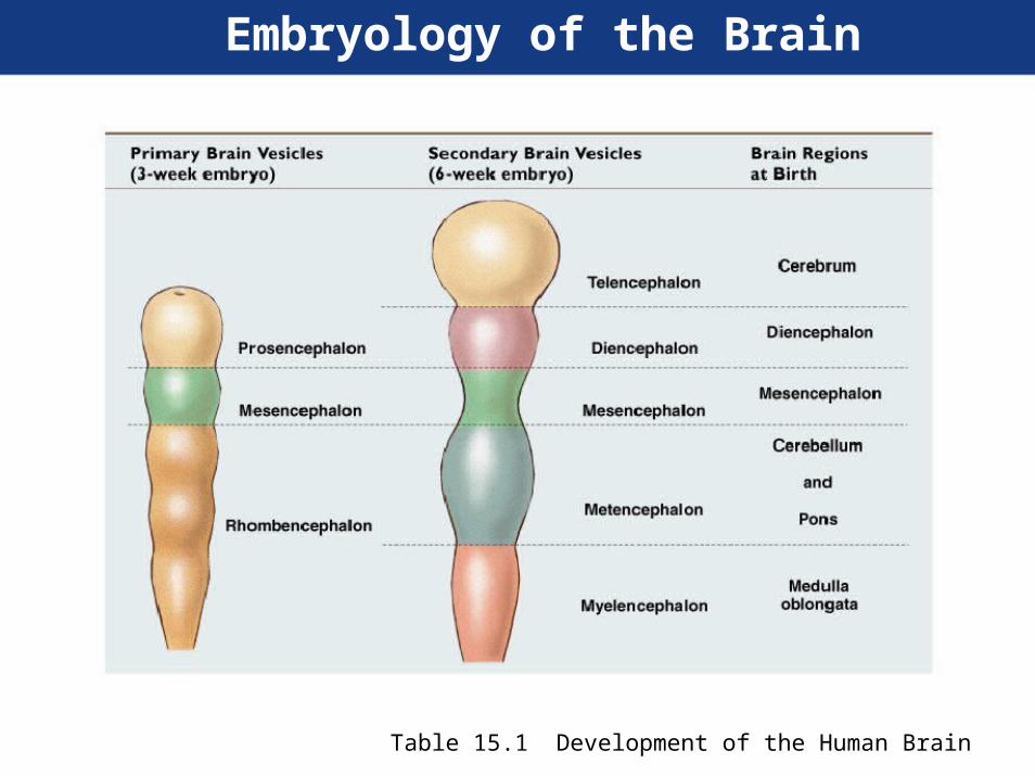
Embryology of the Brain
Table 15.1 Development of the Human Brain
Fig
15.1

Fig
15.11White matter
Grey matter

VentriclesFig
4 fluid filled cavities in the brain
Lined by ependymal cells
Contain cerebrospinal fluid
15.2
Protection and support of the brain
• Bones of the skull
• cranial meninges
• cerebrospinal fluid
• blood-brain barrier

Cranial Meninges
• Protective layers of the brain & spinal cord- Provide physical stability and shock absorption
• Outermost- Dura mater-Tough fibrous layer
• Middle- Arachnoid
• Innermost- Pia mater

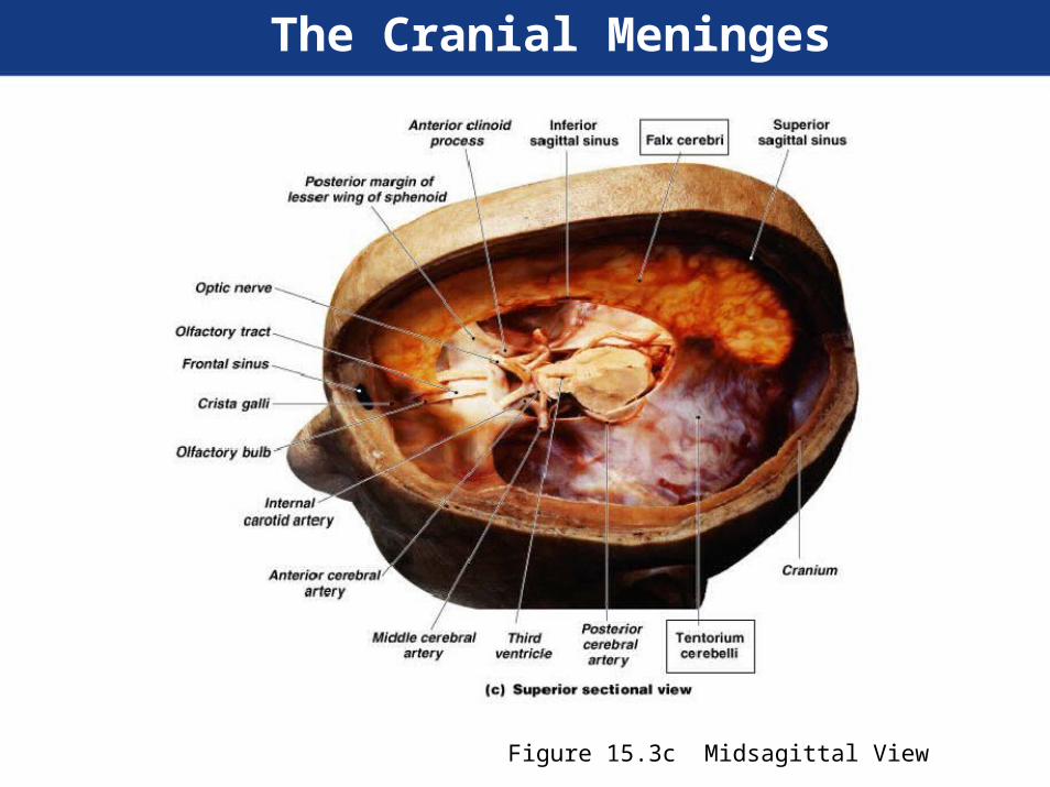
The Cranial Meninges
Figure 15.4a Superior Cut away
The Cranial Meninges
Figure 15.3c Midsagittal View
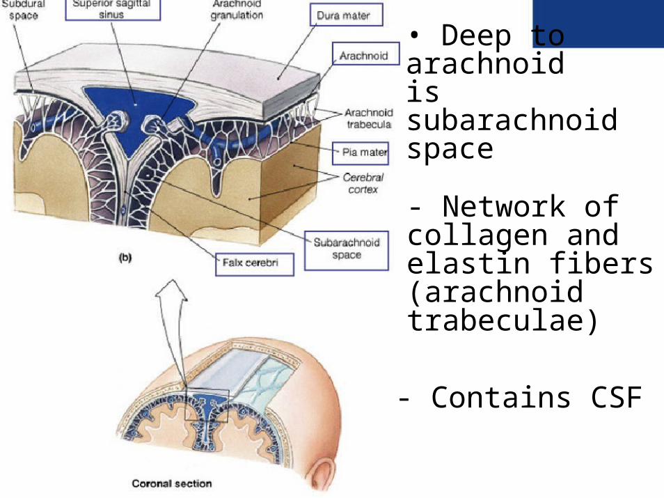
• Deep to arachnoidis subarachnoidspace
- Network ofcollagen andelastin fibers(arachnoidtrabeculae)
- Contains CSF
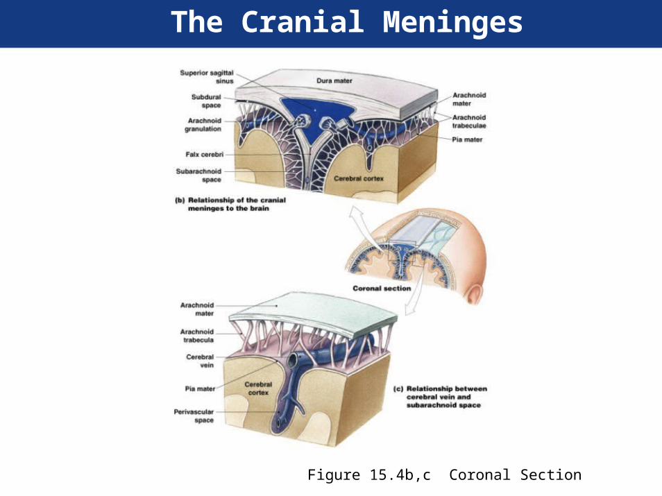
The Cranial Meninges
Figure 15.4b,c Coronal Section
Cerebral Spinal Fluid
• Cushions the CNS
• Supports the brain-the brains is floating inthe CSF
• Transport nutrient/wastes etc.

Choroid plexus
• Produces CSF 500 ml/day
• Composed of ependymal cells andcapillaries (CSF is very different from plasma)
• Found in each ventricle
• Floor of lateral ventricles (2)
• Roof of 3rd ventricle
• Roof of 4th ventricle

Fig
15.5
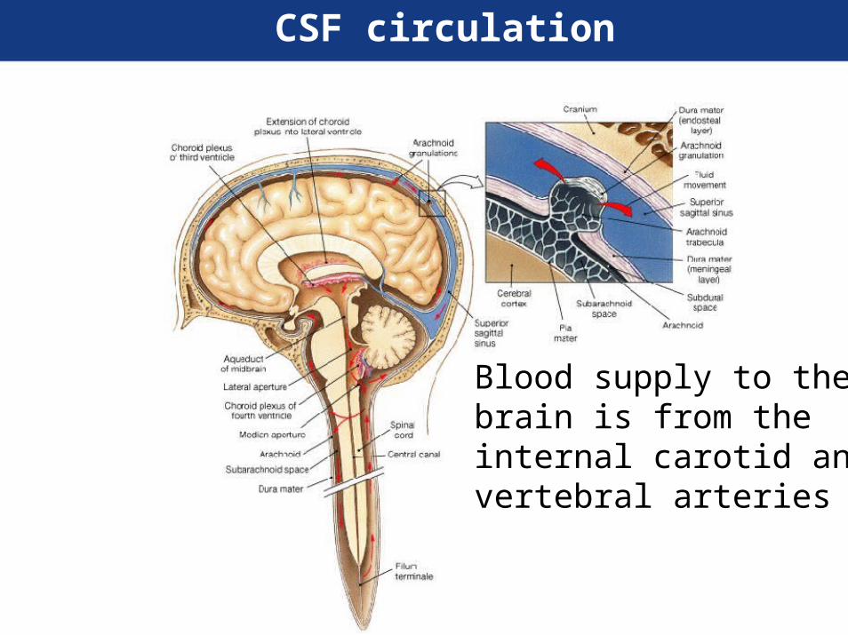
CSF circulation
Blood supply to thebrain is from theinternal carotid andvertebral arteries
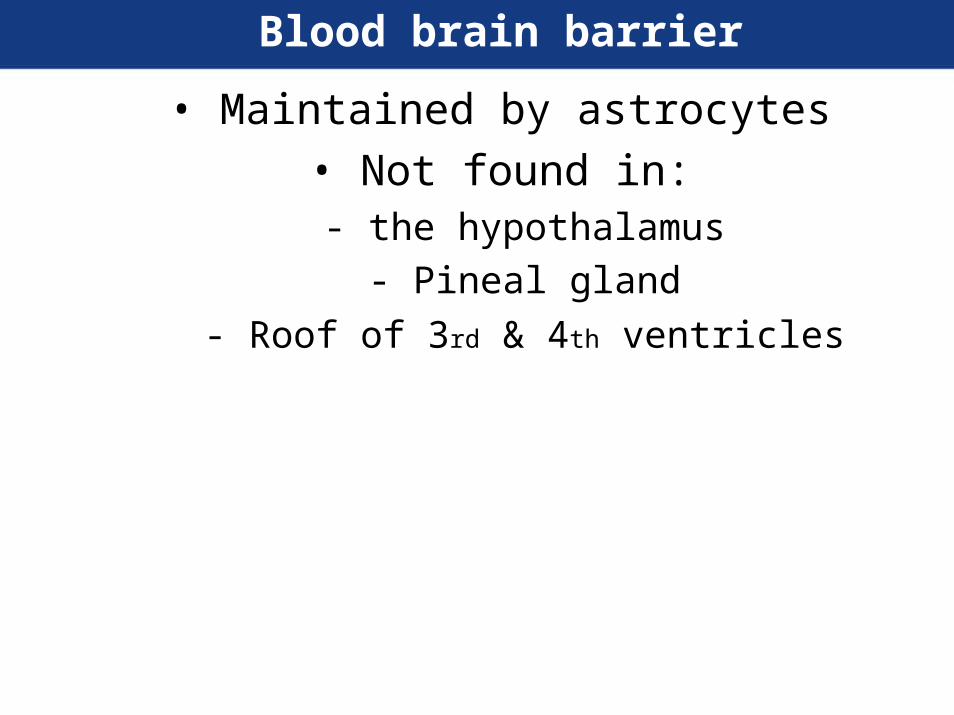
Blood brain barrier
• Maintained by astrocytes
• Not found in:- the hypothalamus
- Pineal gland
- Roof of 3rd & 4th ventricles

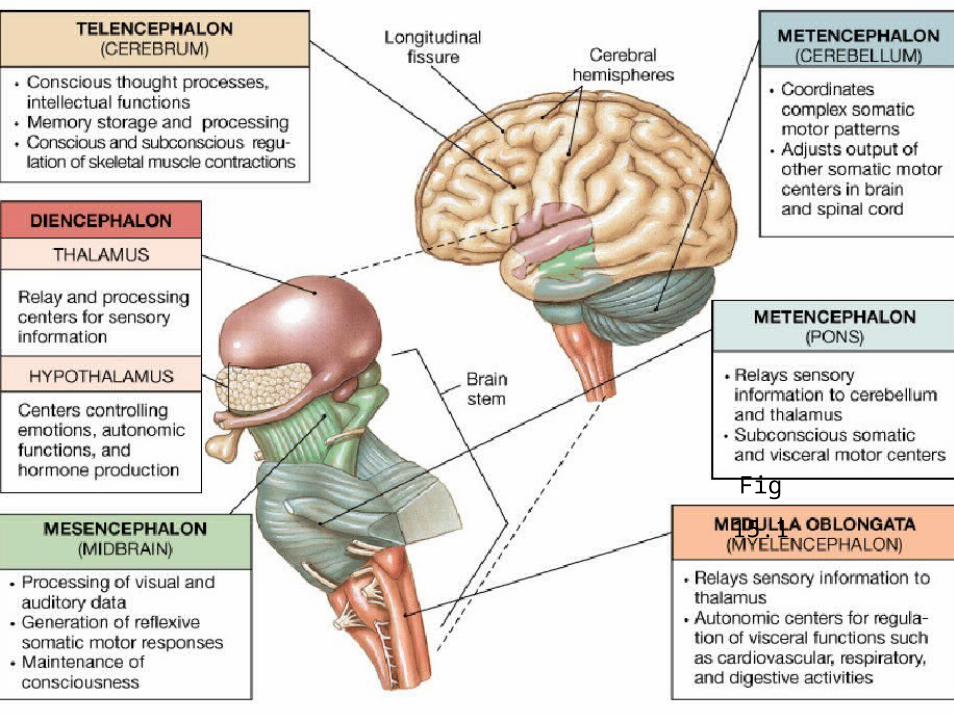
The Cerebrum
Figure 15.8a,b The Cerebral Hemispheres, Superior and Anterior Views

The Cerebrum
Figure 15.8c Posterior View Figure 15.9a Lateral View
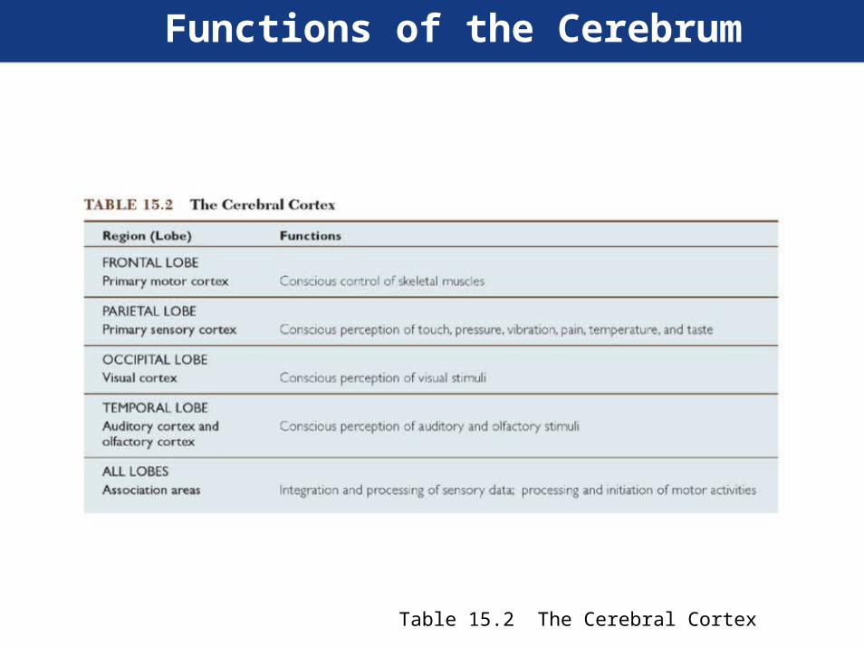
Functions of the Cerebrum
Table 15.2 The Cerebral Cortex

Motor and Sensory Areasof the Cerebral Cortex
Figure 15.9b Functional Areas of the Cortex

Central White Matter of the Brain
Figure 15.10a Lateral View Figure 15.10b Anterior View

Basal Nuclei
Figure 15.11b,c Coronal View
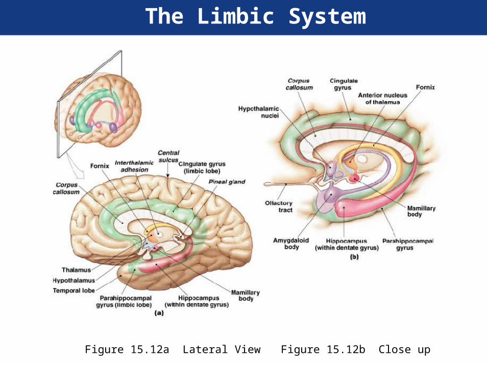
The Limbic System
Figure 15.12a Lateral View Figure 15.12b Close up
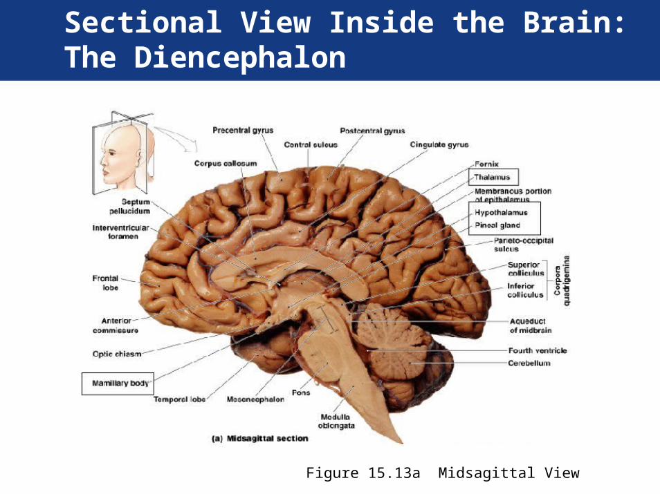
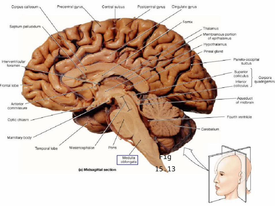
Sectional View Inside the Brain:The Diencephalon
Figure 15.13a Midsagittal View
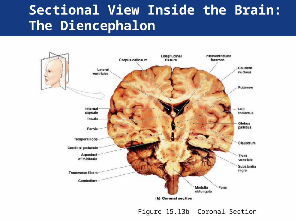
Sectional View Inside the Brain:The Diencephalon
Figure 15.13b Coronal Section
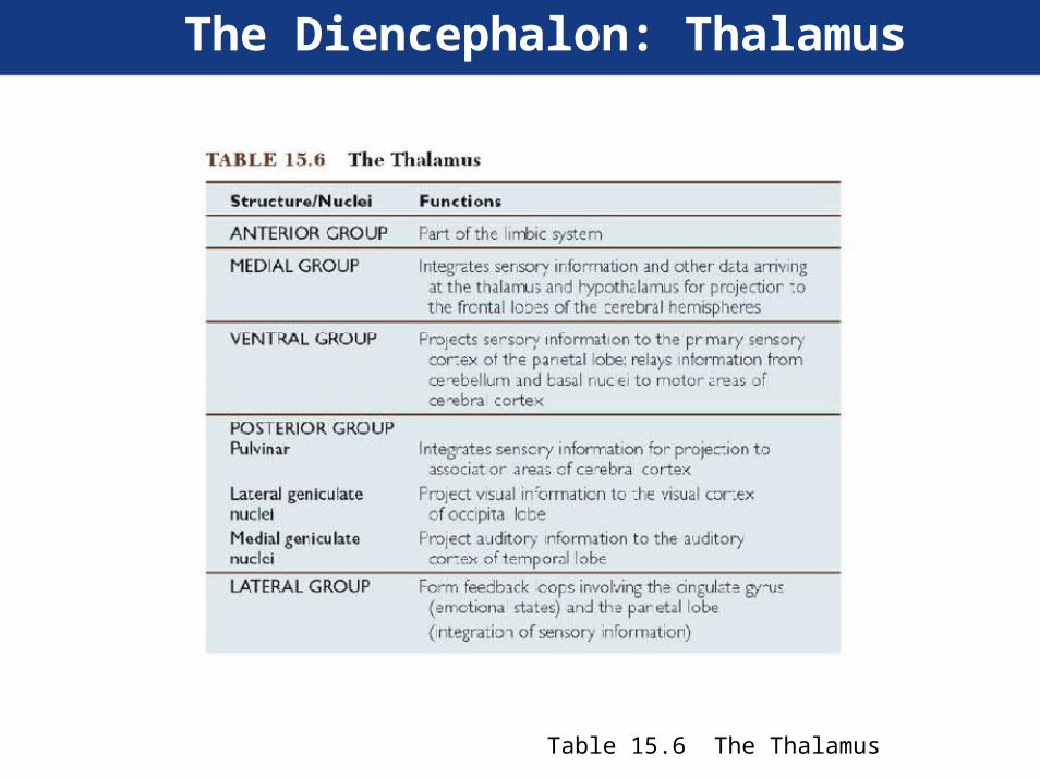
The Diencephalon: Thalamus
Table 15.6 The Thalamus

The Mesencephalon
Figure 15.16a Lateral View Figure 15.16c Posterior View

Copora
quadrigemina
Fig
15.15
Aqueduct of midbrainor
Cerebralpeduncles Cerebral aqueduct
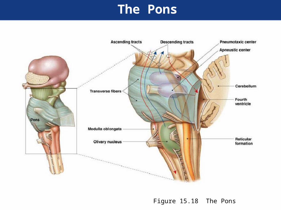
The Pons
Figure 15.18 The Pons

The Cerebellum
Figure 15.19a Posterior, Superior Surface
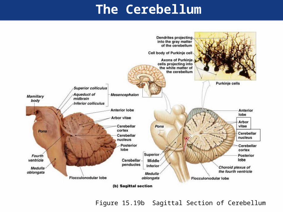
The Cerebellum
Figure 15.19b Sagittal Section of Cerebellum
Fig
15.13
The Cranial Nerves
• Cranial nerves are components of theperipheral nervous system that connect tothe brain rather than to the spinal cord.
- There are twelve pairs of cranial nerves.
- Cranial nerves are numbered using Romannumerals.
• Each cranial nerve attaches to the brainnear the associated sensory or motornuclei.

1-12Old
12 pairs ofCranial
nerves OwlsFig
On15.21
Tree
TopsAre
Forever
Viewing
Green
Valleys
And12 Hills
11

The Olfactory Nerve (N I)
• Primary function:- Special sensory (smell)
• Origin:- Receptors of olfactory epithelium
Figure 15.22 The Olfactory Nerve
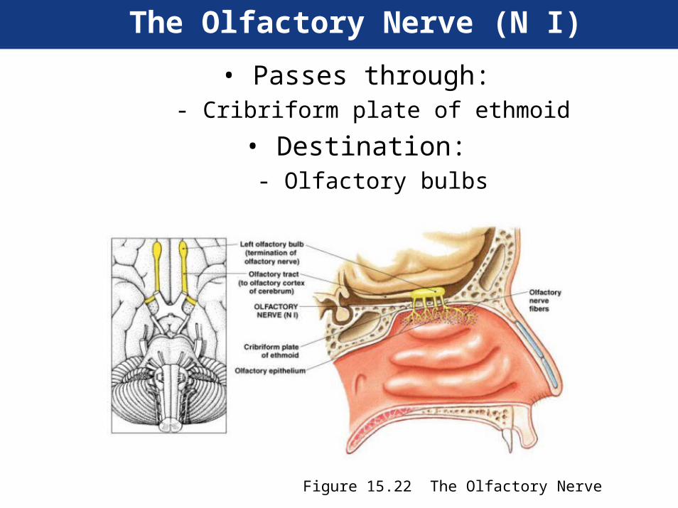
The Olfactory Nerve (N I)
• Passes through:- Cribriform plate of ethmoid
• Destination:- Olfactory bulbs
Figure 15.22 The Olfactory Nerve

The Optic Nerve (N II)
• Primary function:- Special sensory (vision)
• Origin:- Retina of eye
• Passes through:- Optic canal of sphenoid
• Destination:- Diencephalon by way of the optic chiasm

The Optic Nerve (N II)
Figure 15.23 The Optic Nerve
The Oculomotor Nerve (N III)
• Primary function:- Motor, eye movements
• Origin:- Mesencephalon
• Passes through:- Superior orbital fissure of sphenoid

The Oculomotor Nerve (N III)
• Destination:- Somatic motor:
• Superior, inferior, and medial rectus muscles; theinferior oblique muscle; the levator palpebraesuperioris muscle
- Visceral motor:• Intrinsic eye muscles

The Oculomotor Nerve (N III)
Figure 15.24 The Oculomotor Nerve

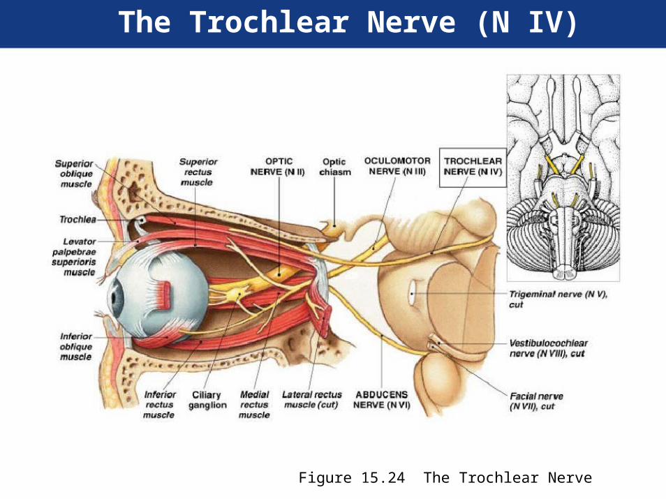
The Trochlear Nerve (N IV)
• Primary function:- Motor, eye movements
• Origin:- Mesencephalon
• Passes through:- Superior orbital fissure of sphenoid
• Destination:- Superior oblique muscle
The Trochlear Nerve (N IV)
Figure 15.24 The Trochlear Nerve

The Trigeminal Nerve (N V)
• Primary function:- Mixed (sensory and motor)
- Ophthalmic and maxillary branches sensory
- Mandibular branch mixed
The Trigeminal Nerve (N V)
• Origin:- Ophthalmic branch (sensory):
• Orbital structures, nasal cavity, skin of forehead,superior eyelid, eyebrow, and part of the nose
- Maxillary branch (sensory):• Inferior eyelid, upper lip, gums, and teeth; cheek;nose, palate, and part of the pharynx
- Mandibular branch (mixed):• Sensory from lower gums, teeth, and lips; palateand tongue (part); motor from motor nuclei of pons

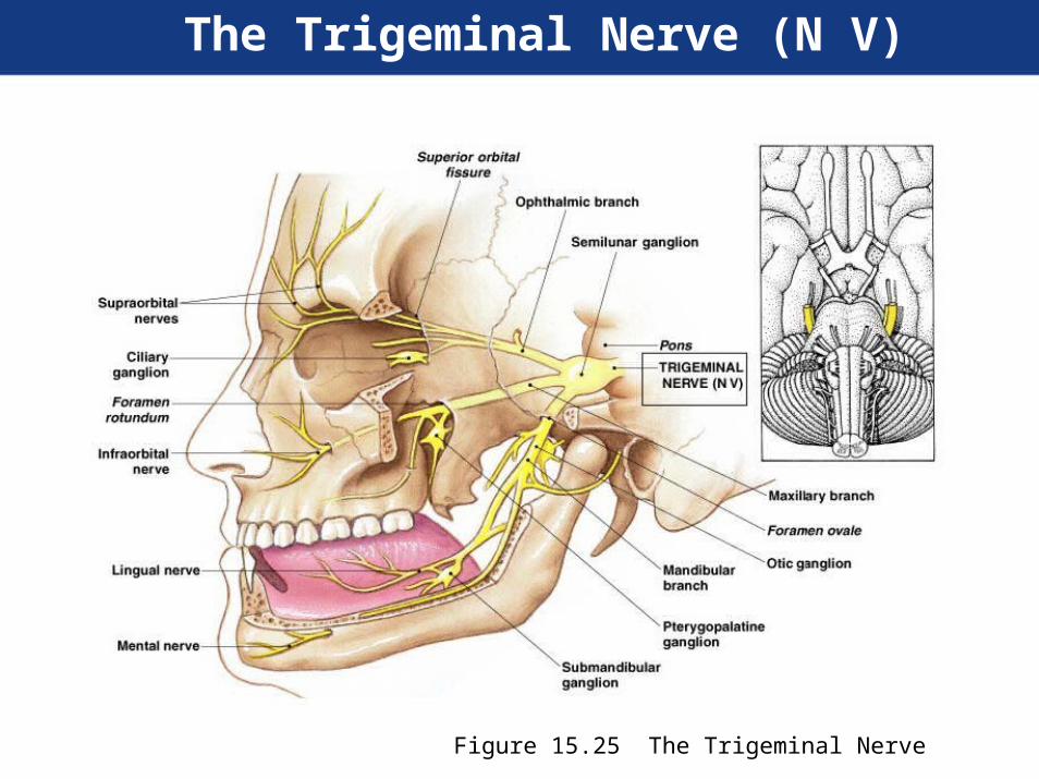
The Trigeminal Nerve (N V)
• Passes through:- Ophthalmic branch through superior orbitalfissure
- Maxillary branch through foramen rotundum
- Mandibular branch through foramen ovale
• Destination:- Ophthalmic, maxillary, and mandibularbranches to sensory nuclei in the pons
- Mandibular branch also innervates muscles ofmastication
The Trigeminal Nerve (N V)
Figure 15.25 The Trigeminal Nerve
The Abducens Nerve (N VI)
• Primary function:- Motor, eye movements
• Origin:- Pons
• Passes through:- Superior orbital fissure of sphenoid
• Destination:- Lateral rectus muscle

The Abducens Nerve (N VI)
Figure 15.2 The Abducens Nerve

The Facial Nerve (N VII)
• Primary function:- Mixed (sensory and motor)
• Origin:- Sensory from taste receptors on anterior two-thirds of tongue
- Motor from motor nuclei of pons
• Passes through:- Internal acoustic meatus of temporal bone,along facial canal to reach stylomastoidforamen
The Facial Nerve (N VII)
• Destination:- Sensory to sensory nuclei of pons
- Somatic motor: muscles of facial expression
- Visceral motor: lacrimal (tear) gland and nasalmucous glands via pterygopalatine ganglion;submandibular and sublingual salivary glandsvia submandibular ganglion

The Facial Nerve (N VII)
Figure 15.26 The Facial Nerve
The Vestibulocochlear Nerve (N VIII)
• Primary function:- Special sensory:
• Balance and equilibrium (vestibular branch) andhearing (cochlear branch)
• Origin:- Receptors of the inner ear (vestibule andcochlea)
• Passes through:- Internal acoustic meatus of the temporal bone

The Vestibulocochlear Nerve (N VIII)
• Destination:- Vestibular and cochlear nuclei of pons andmedulla oblongata
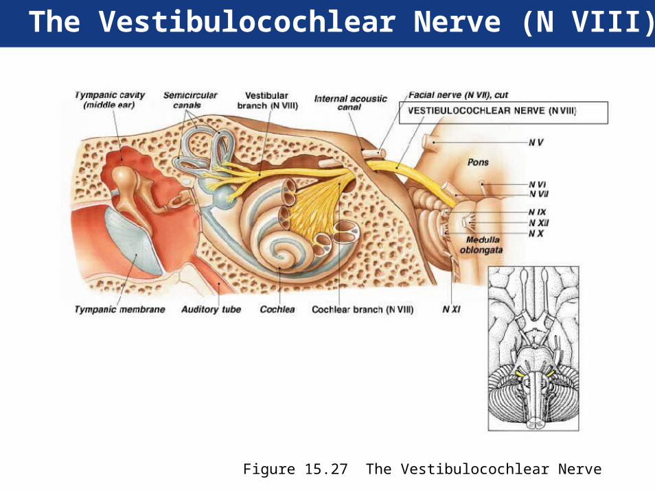
The Vestibulocochlear Nerve (N VIII)
Figure 15.27 The Vestibulocochlear Nerve
The Glossopharyngeal Nerve (N IX)
• Primary function:- Mixed (sensory and motor)
• Origin:- Sensory from posterior one-third of thetongue, part of the pharynx and palate, thecarotid arteries of the neck
- Motor from motor nuclei of medulla oblongata
• Passes through:- Jugular foramen between occipital andtemporal bones
The Glossopharyngeal Nerve (N IX)
• Destination:- Sensory fibers to sensory nuclei of medullaoblongata
- Somatic motor:• Pharyngeal muscles involved in swallowing
- Visceral motor:• Parotid salivary gland, after synapsing in the oticganglion
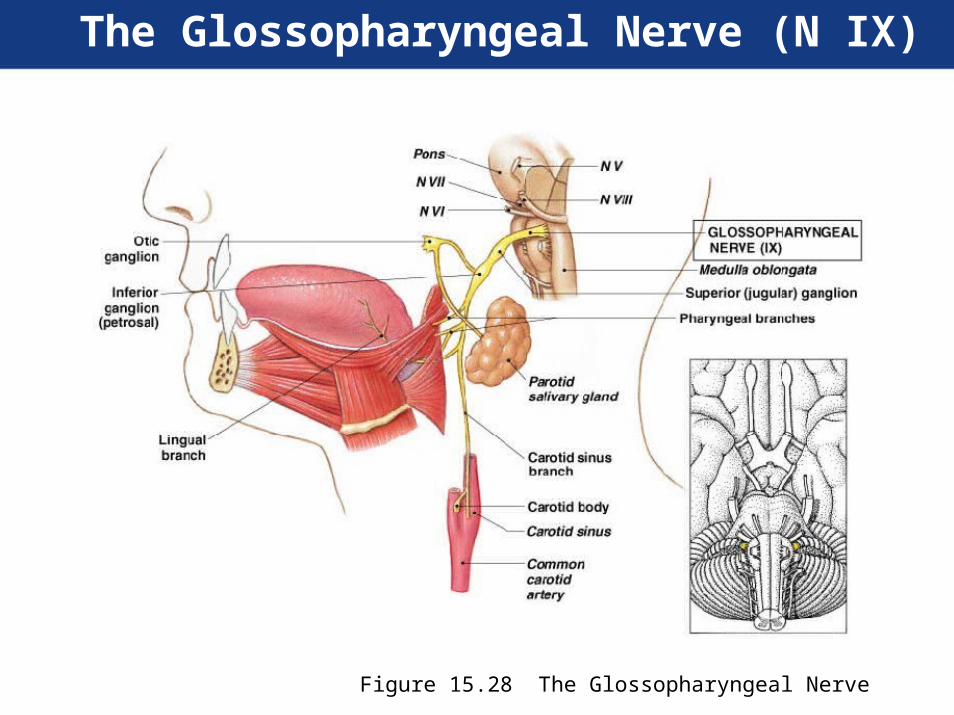
The Glossopharyngeal Nerve (N IX)
Figure 15.28 The Glossopharyngeal Nerve
The Vagus Nerve (N X)
• Primary function:- Mixed (sensory and motor)
• Origin:- Visceral sensory from pharynx (part), auricle,external acoustic meatus, diaphragm, andvisceral organs in thoracic and
abdominopelvic cavities
- Visceral motor from motor nuclei in themedulla oblongata
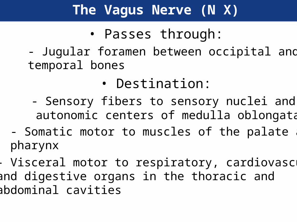
The Vagus Nerve (N X)
• Passes through:- Jugular foramen between occipital andtemporal bones
• Destination:- Sensory fibers to sensory nuclei andautonomic centers of medulla oblongata
- Somatic motor to muscles of the palate andpharynx
- Visceral motor to respiratory, cardiovascular,and digestive organs in the thoracic andabdominal cavities
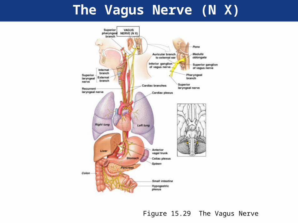
The Vagus Nerve (N X)
Figure 15.29 The Vagus Nerve

The Accessory Nerve (N XI)
• Primary function:- Motor
• Origin:- Motor nuclei of spinal cord and medullaoblongata
• Passes through:- Jugular foramen between occipital andtemporal bones

The Accessory Nerve (N XI)
• Destination:- Internal branch innervates voluntary musclesof palate, pharynx, and larynx
- External branch controls sternocleidomastoidand trapezius muscles

The Accessory Nerve (N XI)
Figure 15.30 The Accessory Nerve

The Hypoglossal Nerve (N XII)
• Primary function:- Motor, tongue movements
• Origin:- Motor nuclei of the medulla oblongata
• Passes through:- Hypoglossal canal of occipital bone
• Destination:- Muscles of the tongue
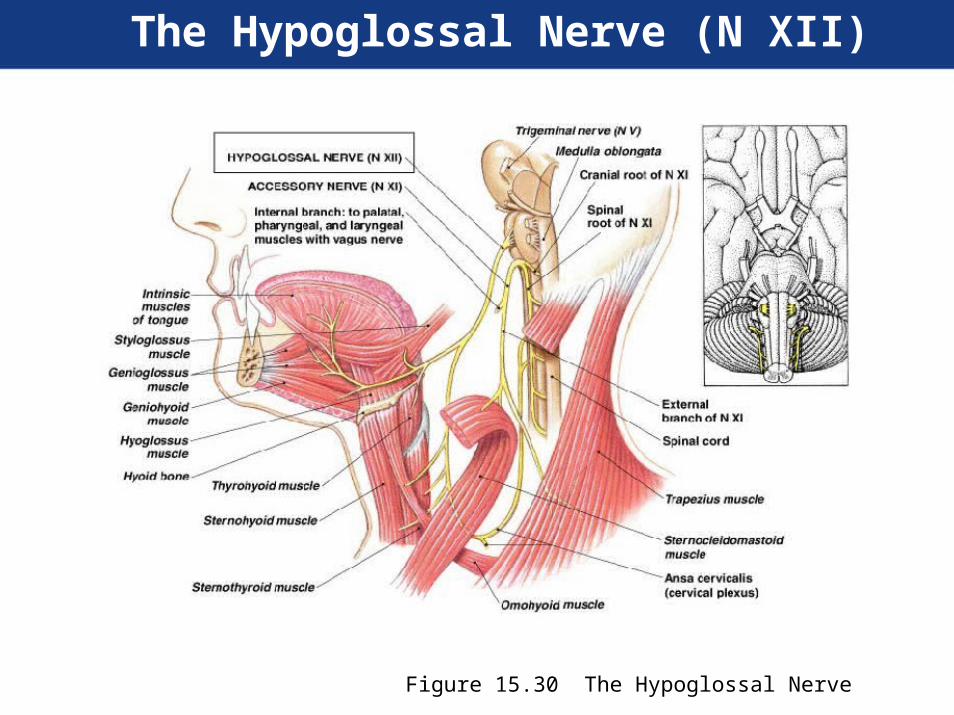
The Hypoglossal Nerve (N XII)
Figure 15.30 The Hypoglossal Nerve