Embed Size (px)
Citation preview
1
Essentials of Human Anatomy & Physiology
Copyright © 2003 Pearson Education, Inc. publishing as Benjamin Cummings
Slides 11.1 – 11.19
Seventh Edition
Elaine N. Marieb
Chapter 11
The Cardiovascular System
Lecture Slides in PowerPoint by Jerry L. Cook
INTERESTING FACTS ABOUT THE HEART
. . .
• http://www.webmd.com/heart/features/amazing-
facts-about-heart-health-and-heart-disease_
The Cardiovascular SystemThe Cardiovascular System
Slide 11.1Copyright © 2003 Pearson Education, Inc. publishing as Benjamin Cummings
• A closed system of the heart and blood vessels
• The heart pumps blood
• Blood vessels allow blood to circulate to all parts of the body
• The function of the cardiovascular system is to deliver oxygen and nutrients and to remove carbon dioxide and other waste products
The HeartThe Heart
Slide 11.2aCopyright © 2003 Pearson Education, Inc. publishing as Benjamin Cummings
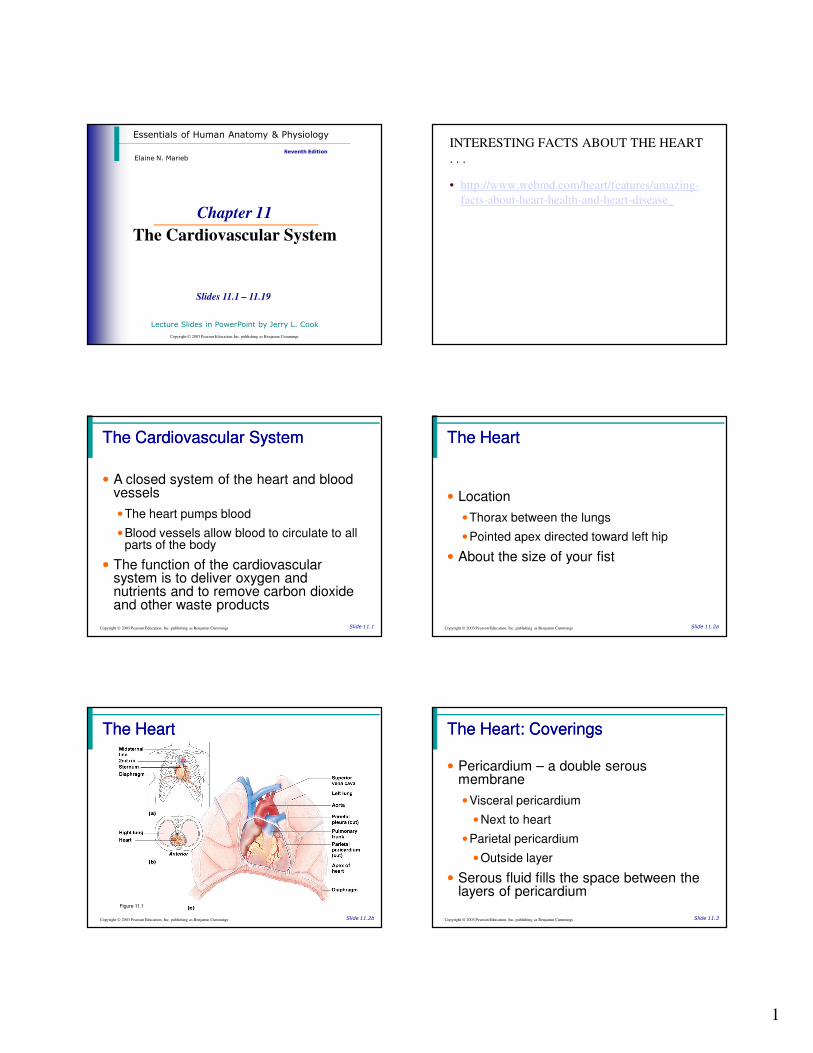
• Location
• Thorax between the lungs
• Pointed apex directed toward left hip
• About the size of your fist
The HeartThe Heart
Slide 11.2bCopyright © 2003 Pearson Education, Inc. publishing as Benjamin Cummings
Figure 11.1
The Heart: CoveringsThe Heart: Coverings
Slide 11.3Copyright © 2003 Pearson Education, Inc. publishing as Benjamin Cummings
• Pericardium – a double serous membrane
• Visceral pericardium
• Next to heart
• Parietal pericardium
• Outside layer
• Serous fluid fills the space between the layers of pericardium
2
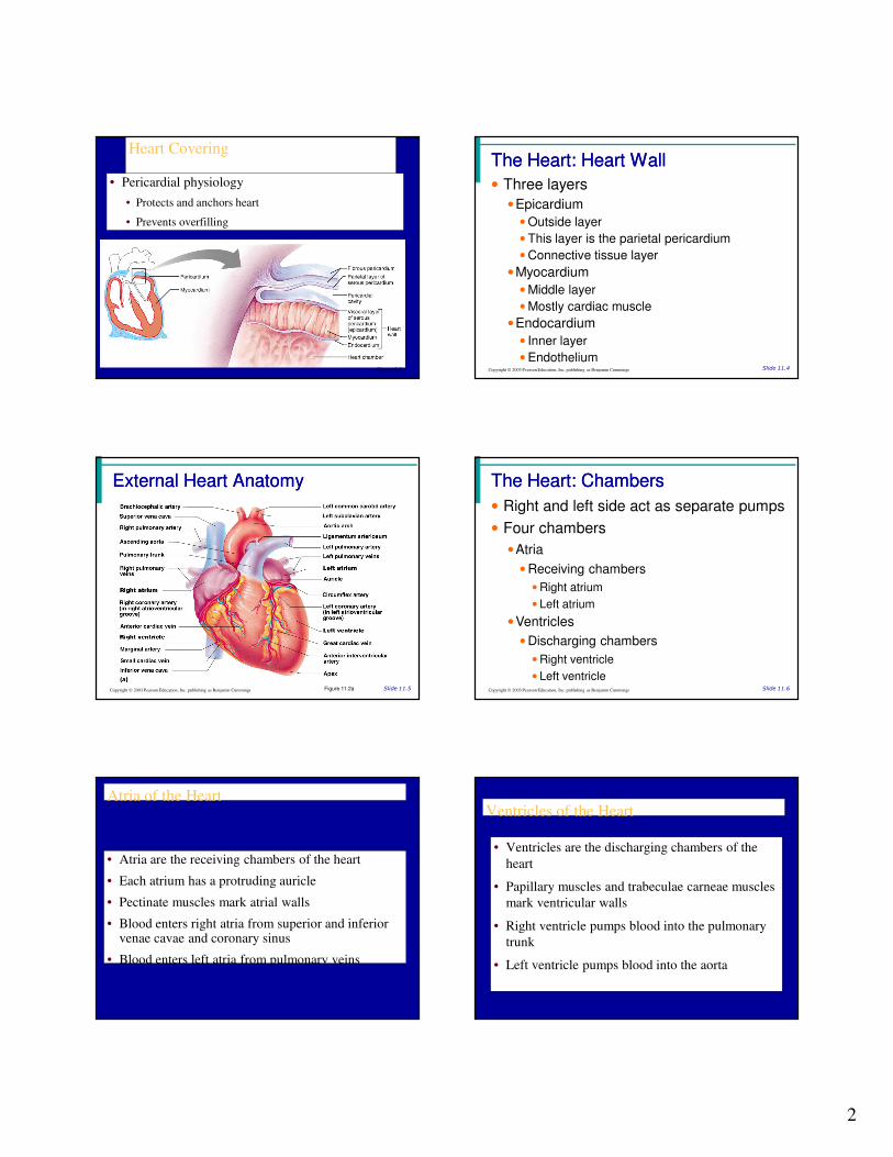
Heart Covering
• Pericardial physiology
• Protects and anchors heart
• Prevents overfilling
Figure 19.2
The Heart: Heart WallThe Heart: Heart Wall
Slide 11.4Copyright © 2003 Pearson Education, Inc. publishing as Benjamin Cummings
• Three layers
• Epicardium
• Outside layer
• This layer is the parietal pericardium
• Connective tissue layer
• Myocardium
• Middle layer
• Mostly cardiac muscle
• Endocardium
• Inner layer
• Endothelium
External Heart AnatomyExternal Heart Anatomy
Slide 11.5Copyright © 2003 Pearson Education, Inc. publishing as Benjamin Cummings Figure 11.2a
The Heart: ChambersThe Heart: Chambers
Slide 11.6Copyright © 2003 Pearson Education, Inc. publishing as Benjamin Cummings
• Right and left side act as separate pumps
• Four chambers
• Atria
• Receiving chambers
• Right atrium
• Left atrium
• Ventricles
• Discharging chambers
• Right ventricle
• Left ventricle
Atria of the Heart
• Atria are the receiving chambers of the heart
• Each atrium has a protruding auricle
• Pectinate muscles mark atrial walls
• Blood enters right atria from superior and inferior venae cavae and coronary sinus
• Blood enters left atria from pulmonary veins
Ventricles of the Heart
• Ventricles are the discharging chambers of the
heart
• Papillary muscles and trabeculae carneae muscles
mark ventricular walls
• Right ventricle pumps blood into the pulmonary
trunk
• Left ventricle pumps blood into the aorta
3
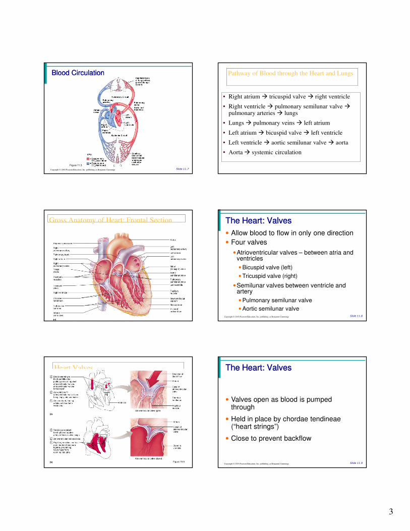
Blood CirculationBlood Circulation
Slide 11.7Copyright © 2003 Pearson Education, Inc. publishing as Benjamin Cummings
Figure 11.3
Pathway of Blood through the Heart and Lungs
• Right atrium � tricuspid valve � right ventricle
• Right ventricle � pulmonary semilunar valve �pulmonary arteries � lungs
• Lungs � pulmonary veins � left atrium
• Left atrium � bicuspid valve � left ventricle
• Left ventricle � aortic semilunar valve � aorta
• Aorta � systemic circulation
Gross Anatomy of Heart: Frontal Section
Figure 19.4e
The Heart: ValvesThe Heart: Valves
Slide 11.8Copyright © 2003 Pearson Education, Inc. publishing as Benjamin Cummings
• Allow blood to flow in only one direction
• Four valves
• Atrioventricular valves – between atria and ventricles
• Bicuspid valve (left)
• Tricuspid valve (right)
• Semilunar valves between ventricle and artery
• Pulmonary semilunar valve
• Aortic semilunar valve
Heart Valves
Figure 19.9
The Heart: ValvesThe Heart: Valves
Slide 11.9Copyright © 2003 Pearson Education, Inc. publishing as Benjamin Cummings
• Valves open as blood is pumped through
• Held in place by chordae tendineae (“heart strings”)
• Close to prevent backflow
4
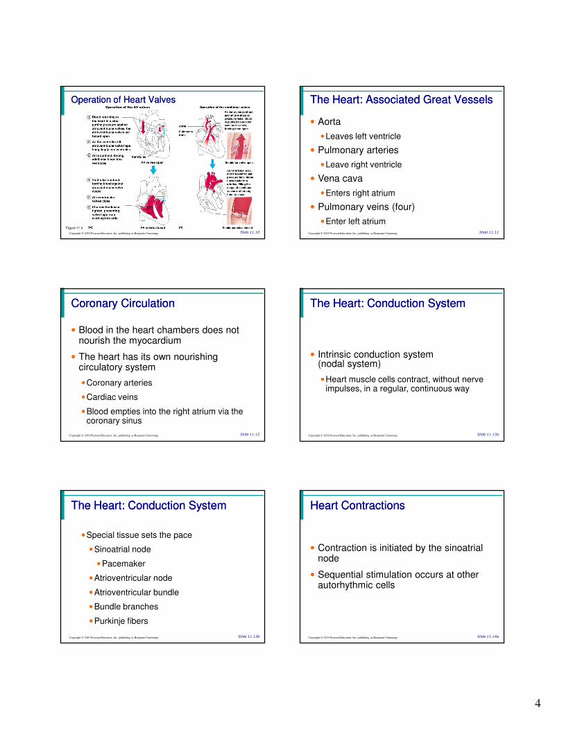
Operation of Heart ValvesOperation of Heart Valves
Slide 11.10Copyright © 2003 Pearson Education, Inc. publishing as Benjamin Cummings
Figure 11.4
The Heart: Associated Great VesselsThe Heart: Associated Great Vessels
Slide 11.11Copyright © 2003 Pearson Education, Inc. publishing as Benjamin Cummings
• Aorta
• Leaves left ventricle
• Pulmonary arteries
• Leave right ventricle
• Vena cava
• Enters right atrium
• Pulmonary veins (four)
• Enter left atrium
Coronary CirculationCoronary Circulation
Slide 11.12Copyright © 2003 Pearson Education, Inc. publishing as Benjamin Cummings
• Blood in the heart chambers does not nourish the myocardium
• The heart has its own nourishing circulatory system
• Coronary arteries
• Cardiac veins
• Blood empties into the right atrium via the coronary sinus
The Heart: Conduction SystemThe Heart: Conduction System
Slide 11.13aCopyright © 2003 Pearson Education, Inc. publishing as Benjamin Cummings
• Intrinsic conduction system (nodal system)
• Heart muscle cells contract, without nerve impulses, in a regular, continuous way
The Heart: Conduction SystemThe Heart: Conduction System
Slide 11.13bCopyright © 2003 Pearson Education, Inc. publishing as Benjamin Cummings
• Special tissue sets the pace
• Sinoatrial node
• Pacemaker
• Atrioventricular node
• Atrioventricular bundle
• Bundle branches
• Purkinje fibers
Heart ContractionsHeart Contractions
Slide 11.14aCopyright © 2003 Pearson Education, Inc. publishing as Benjamin Cummings
• Contraction is initiated by the sinoatrial node
• Sequential stimulation occurs at other autorhythmic cells
5
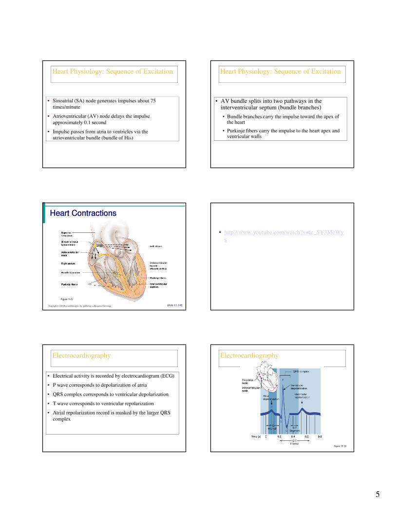
Heart Physiology: Sequence of Excitation
• Sinoatrial (SA) node generates impulses about 75
times/minute
• Atrioventricular (AV) node delays the impulse
approximately 0.1 second
• Impulse passes from atria to ventricles via the
atrioventricular bundle (bundle of His)
Heart Physiology: Sequence of Excitation
• AV bundle splits into two pathways in the interventricular septum (bundle branches)
• Bundle branches carry the impulse toward the apex of the heart
• Purkinje fibers carry the impulse to the heart apex and ventricular walls
Heart ContractionsHeart Contractions
Slide 11.14bCopyright © 2003 Pearson Education, Inc. publishing as Benjamin Cummings
Figure 11.5
• http://www.youtube.com/watch?v=te_SY3MeWy
s
Electrocardiography
• Electrical activity is recorded by electrocardiogram (ECG)
• P wave corresponds to depolarization of atria
• QRS complex corresponds to ventricular depolarization
• T wave corresponds to ventricular repolarization
• Atrial repolarization record is masked by the larger QRS
complex
Electrocardiography
Figure 19.16
6
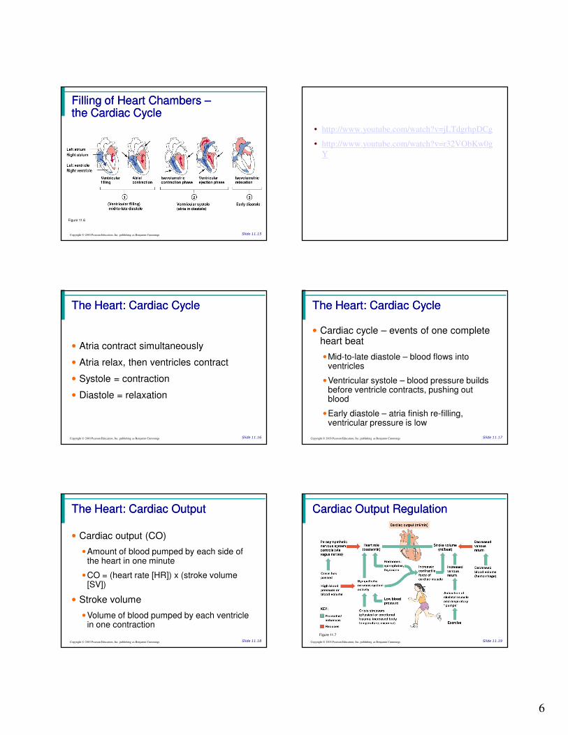
Filling of Heart Chambers Filling of Heart Chambers ––the Cardiac Cyclethe Cardiac Cycle
Slide 11.15Copyright © 2003 Pearson Education, Inc. publishing as Benjamin Cummings
Figure 11.6
• http://www.youtube.com/watch?v=jLTdgrhpDCg
• http://www.youtube.com/watch?v=r32VObKw0g
Y
The Heart: Cardiac CycleThe Heart: Cardiac Cycle
Slide 11.16Copyright © 2003 Pearson Education, Inc. publishing as Benjamin Cummings
• Atria contract simultaneously
• Atria relax, then ventricles contract
• Systole = contraction
• Diastole = relaxation
The Heart: Cardiac CycleThe Heart: Cardiac Cycle
Slide 11.17Copyright © 2003 Pearson Education, Inc. publishing as Benjamin Cummings
• Cardiac cycle – events of one complete heart beat
• Mid-to-late diastole – blood flows into ventricles
• Ventricular systole – blood pressure builds before ventricle contracts, pushing out blood
• Early diastole – atria finish re-filling, ventricular pressure is low
The Heart: Cardiac OutputThe Heart: Cardiac Output
Slide 11.18Copyright © 2003 Pearson Education, Inc. publishing as Benjamin Cummings
• Cardiac output (CO)
• Amount of blood pumped by each side of the heart in one minute
• CO = (heart rate [HR]) x (stroke volume [SV])
• Stroke volume
• Volume of blood pumped by each ventricle in one contraction
Cardiac Output RegulationCardiac Output Regulation
Slide 11.19Copyright © 2003 Pearson Education, Inc. publishing as Benjamin Cummings
Figure 11.7