Embed Size (px)
Citation preview
© Essentials Education 20162
TOPIC 1 CELLS AND MICROORGANISMS LIVING THINGS CONSIST OF CELLS
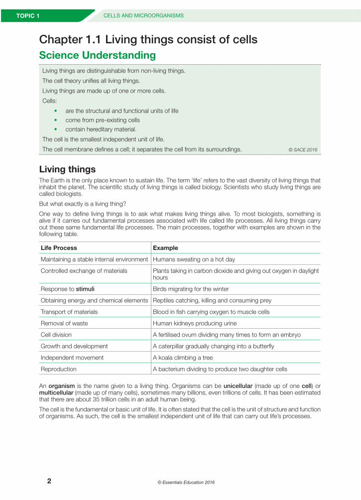
Chapter 1.1 Living things consist of cellsScience UnderstandingLiving things are distinguishable from non-living things.
The cell theory unifies all living things.
Living things are made up of one or more cells.
Cells:
• are the structural and functional units of life• come from pre-existing cells• contain hereditary material.
The cell is the smallest independent unit of life.
The cell membrane defines a cell; it separates the cell from its surroundings. © SACE 2016
Living thingsThe Earth is the only place known to sustain life. The term ‘life’ refers to the vast diversity of living things that inhabit the planet. The scientific study of living things is called biology. Scientists who study living things are called biologists.
But what exactly is a living thing?
One way to define living things is to ask what makes living things alive. To most biologists, something is alive if it carries out fundamental processes associated with life called life processes. All living things carry out these same fundamental life processes. The main processes, together with examples are shown in the following table.
Life Process Example
Maintaining a stable internal environment Humans sweating on a hot day
Controlled exchange of materials Plants taking in carbon dioxide and giving out oxygen in daylight hours
Response to stimuli Birds migrating for the winter
Obtaining energy and chemical elements Reptiles catching, killing and consuming prey
Transport of materials Blood in fish carrying oxygen to muscle cells
Removal of waste Human kidneys producing urine
Cell division A fertilised ovum dividing many times to form an embryo
Growth and development A caterpillar gradually changing into a butterfly
Independent movement A koala climbing a tree
Reproduction A bacterium dividing to produce two daughter cells
An organism is the name given to a living thing. Organisms can be unicellular (made up of one cell) or multicellular (made up of many cells), sometimes many billions, even trillions of cells. It has been estimated that there are about 35 trillion cells in an adult human being.
The cell is the fundamental or basic unit of life. It is often stated that the cell is the unit of structure and function of organisms. As such, the cell is the smallest independent unit of life that can carry out life’s processes.
FINAL Workbook C.indb 2 21/09/2016 9:19:02 PM
© Essentials Education 2016 3
CHAPTER 1.1LIVING THINGS CONSIST OF CELLS
1.1
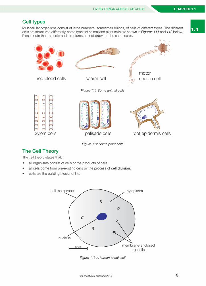
Cell typesMulticellular organisms consist of large numbers, sometimes billions, of cells of different types. The different cells are structured differently, some types of animal and plant cells are shown in Figures 111 and 112 below. Please note that the cells and structures are not drawn to the same scale.
Fig
red blood cells sperm cellmotor neuron cell
ure 111 Some animal cells
xylem cells palisade cells root epidermis cells
Figure 112 Some plant cells
The Cell Theory The cell theory states that:
• all organisms consist of cells or the products of cells.• all cells come from pre-existing cells by the process of cell division.• cells are the building blocks of life.
cell membrane cytoplasm
nucleus
10 µm membrane-enclosedorganelles
Figure 113 A human cheek cell
FINAL Workbook C.indb 3 21/09/2016 9:19:02 PM
© Essentials Education 20164
TOPIC 1 CELLS AND MICROORGANISMS LIVING THINGS CONSIST OF CELLS
CellsCells consist of a volume of fluid enclosed by a membrane. The membrane is called the cell membrane and the enclosed fluid is called the cytoplasm. The cytoplasm is mostly made up of water in which substances are dissolved and some insoluble proteins. In animal and plant cells the cytoplasm also has structures in it called membrane-enclosed organelles that have specific functions; for example a nucleus that controls the overall function of the cell. An example of a cheek cell is shown in Figure 113, a scale bar is shown to help estimate the size of the cell. N.B. 1000 micrometres (µm) = 1mm
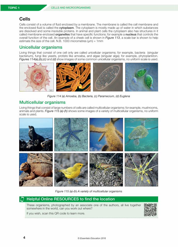
Unicellular organismsLiving things that consist of one cell only are called unicellular organisms; for example, bacteria (singular bacterium), fungi like yeasts, protists like amoeba, and algae (singular alga); for example, phytoplankton. Figures 114(a),(b),(c) and (d) show images of some common unicellular organisms, no uniform scale is used.
Figure 114 (a) Amoeba, (b) Bacteria, (c) Paramecium, (d) Euglena
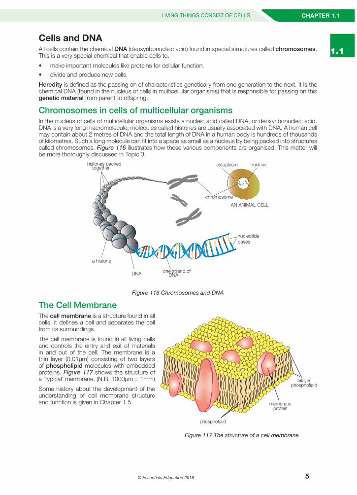
Multicellular organismsLiving things that consist of large numbers of cells are called multicellular organisms; for example, mushrooms, animals and plants. Figure 115 (a)-(h) shows some images of a variety of multicellular organisms, no uniform scale is used.
Figure 115 (a)-(h) A variety of multicellular organisms
Helpful Online RESOURCES to find the locationThese organisms, photographed by an associate one of the authors, all live together somewhere in the world, can you work out where?
If you wish, scan this QR code to learn more.
FINAL Workbook C.indb 4 21/09/2016 9:19:09 PM
© Essentials Education 2016 5
CHAPTER 1.1LIVING THINGS CONSIST OF CELLS
1.1Cells and DNA All cells contain the chemical DNA (deoxyribonucleic acid) found in special structures called chromosomes. This is a very special chemical that enable cells to:
• make important molecules like proteins for cellular function.• divide and produce new cells.
Heredity is defined as the passing on of characteristics genetically from one generation to the next. It is the chemical DNA (found in the nucleus of cells in multicellular organisms) that is responsible for passing on this genetic material from parent to offspring.
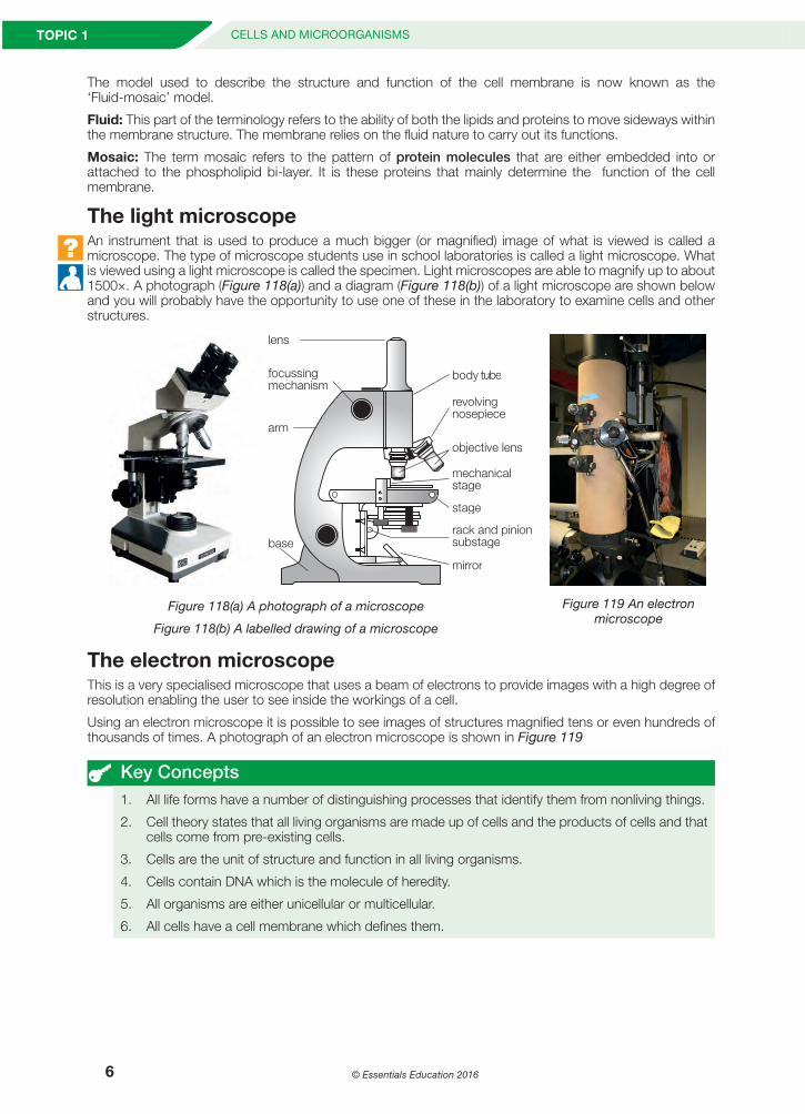
Chromosomes in cells of multicellular organismsIn the nucleus of cells of multicellular organisms exists a nucleic acid called DNA, or deoxyribonucleic acid. DNA is a very long macromolecule; molecules called histones are usually associated with DNA. A human cell may contain about 2 metres of DNA and the total length of DNA in a human body is hundreds of thousands of kilometres. Such a long molecule can fit into a space as small as a nucleus by being packed into structures called chromosomes. Figure 116 illustrates how these various components are organised. This matter will be more thoroughly discussed in Topic 3.
one strand ofDNA
nucleotidebases
DNA
a histone
chromosome
nucleuscytoplasm
AN ANIMAL CELL
histones packedtogether
Figure 116 Chromosomes and DNA
The Cell MembraneThe cell membrane is a structure found in all cells; it defines a cell and separates the cell from its surroundings.
The cell membrane is found in all living cells and controls the entry and exit of materials in and out of the cell. The membrane is a thin layer (0.01µm) consisting of two layers of phospholipid molecules with embedded proteins. Figure 117 shows the structure of a ‘typical’ membrane. (N.B. 1000µm = 1mm)
Some history about the development of the understanding of cell membrane structure and function is given in Chapter 1.5. membrane
protein
bilayerphospholipid
phospholipid
Figure 117 The structure of a cell membrane
FINAL Workbook C.indb 5 21/09/2016 9:19:09 PM
© Essentials Education 20166
TOPIC 1 CELLS AND MICROORGANISMS LIVING THINGS CONSIST OF CELLS
The model used to describe the structure and function of the cell membrane is now known as the ‘Fluid-mosaic’ model.
Fluid: This part of the terminology refers to the ability of both the lipids and proteins to move sideways within the membrane structure. The membrane relies on the fluid nature to carry out its functions.
Mosaic: The term mosaic refers to the pattern of protein molecules that are either embedded into or attached to the phospholipid bi-layer. It is these proteins that mainly determine the function of the cell membrane.
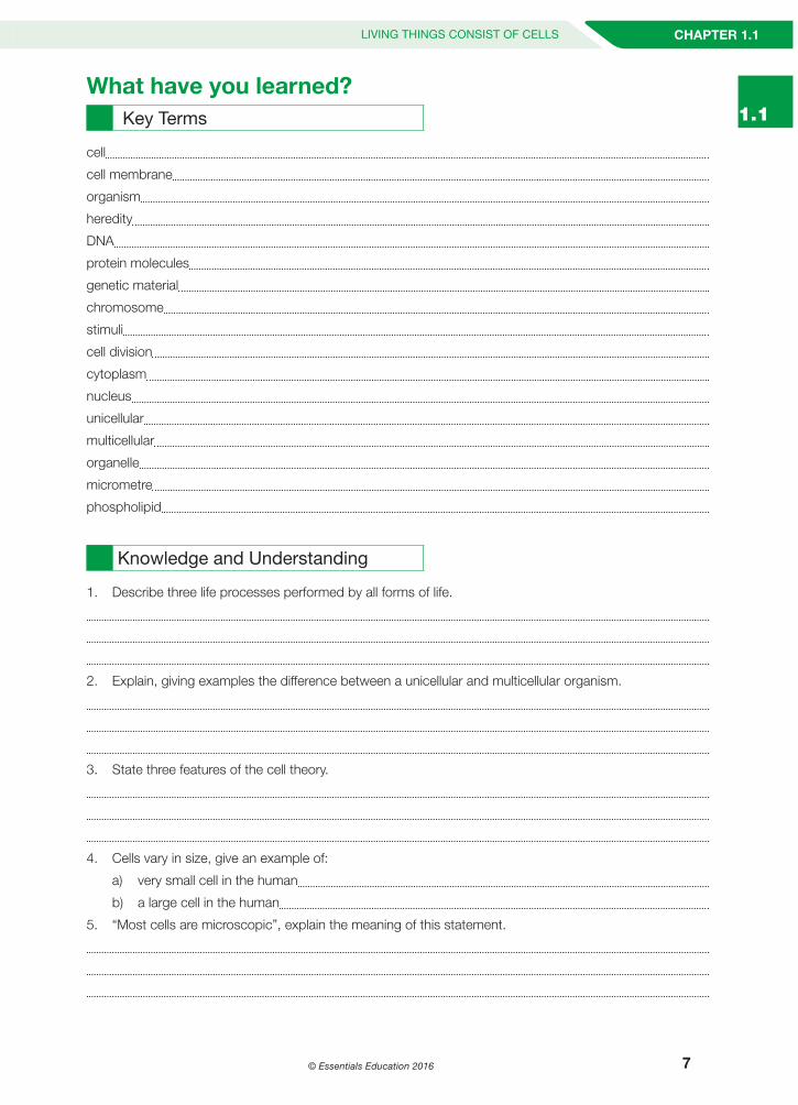
The light microscopeAn instrument that is used to produce a much bigger (or magnified) image of what is viewed is called a microscope. The type of microscope students use in school laboratories is called a light microscope. What is viewed using a light microscope is called the specimen. Light microscopes are able to magnify up to about 1500×. A photograph (Figure 118(a)) and a diagram (Figure 118(b)) of a light microscope are shown below and you will probably have the opportunity to use one of these in the laboratory to examine cells and other structures.
Figure 118(a) A photograph of a microscope
Figure 118(b) A labelled drawing of a microscope
lens
body tube
revolvingnosepiece
objective lens
mechanicalstage
stage
mirror
rack and pinionsubstage
focussingmechanism
arm
base
Figure 119 An electron microscope
The electron microscopeThis is a very specialised microscope that uses a beam of electrons to provide images with a high degree of resolution enabling the user to see inside the workings of a cell.
Using an electron microscope it is possible to see images of structures magnified tens or even hundreds of thousands of times. A photograph of an electron microscope is shown in Figure 119
Key Concepts1. All life forms have a number of distinguishing processes that identify them from nonliving things.
2. Cell theory states that all living organisms are made up of cells and the products of cells and that cells come from pre-existing cells.
3. Cells are the unit of structure and function in all living organisms.
4. Cells contain DNA which is the molecule of heredity.
5. All organisms are either unicellular or multicellular.
6. All cells have a cell membrane which defines them.
FINAL Workbook C.indb 6 21/09/2016 9:19:11 PM
© Essentials Education 2016 7
CHAPTER 1.1LIVING THINGS CONSIST OF CELLS
1.1What have you learned?
Key Terms
cell
cell membrane
organism
heredity
DNA
protein molecules
genetic material
chromosome
stimuli
cell division
cytoplasm
nucleus
unicellular
multicellular
organelle
micrometre
phospholipid
Knowledge and Understanding
1. Describe three life processes performed by all forms of life.
2. Explain, giving examples the difference between a unicellular and multicellular organism.
3. State three features of the cell theory.
4. Cells vary in size, give an example of:
a) very small cell in the human
b) a large cell in the human
5. “Most cells are microscopic”, explain the meaning of this statement.
FINAL Workbook C.indb 7 21/09/2016 9:19:11 PM
© Essentials Education 20168
TOPIC 1 CELLS AND MICROORGANISMS LIVING THINGS CONSIST OF CELLS
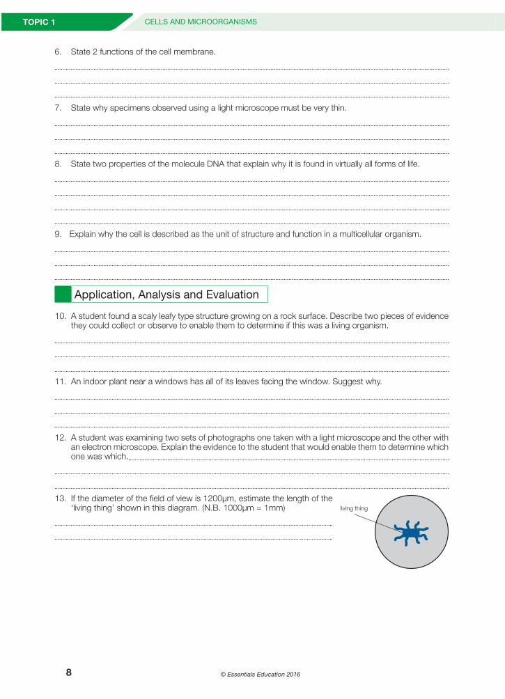
6. State 2 functions of the cell membrane.
7. State why specimens observed using a light microscope must be very thin.
8. State two properties of the molecule DNA that explain why it is found in virtually all forms of life.
9. Explain why the cell is described as the unit of structure and function in a multicellular organism.
Application, Analysis and Evaluation
10. A student found a scaly leafy type structure growing on a rock surface. Describe two pieces of evidence they could collect or observe to enable them to determine if this was a living organism.
11. An indoor plant near a windows has all of its leaves facing the window. Suggest why.
12. A student was examining two sets of photographs one taken with a light microscope and the other with an electron microscope. Explain the evidence to the student that would enable them to determine which one was which.
13. If the diameter of the field of view is 1200µm, estimate the length of the ‘living thing’ shown in this diagram. (N.B. 1000µm = 1mm)
living thing
FINAL Workbook C.indb 8 21/09/2016 9:19:11 PM
© Essentials Education 2016 9
CHAPTER 1.1LIVING THINGS CONSIST OF CELLS
1.1
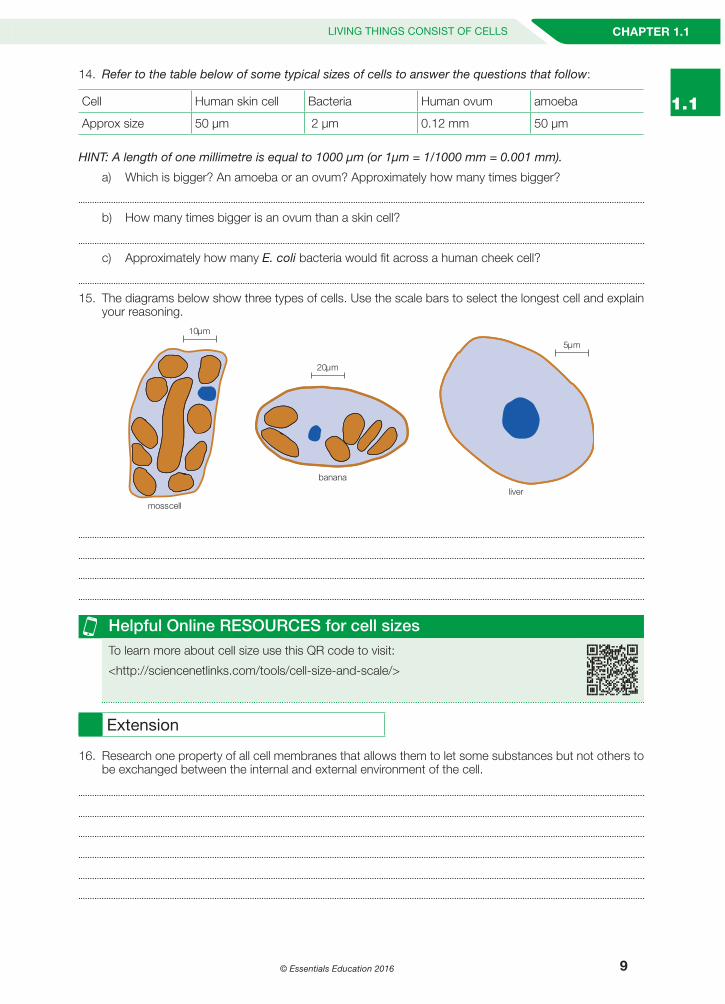
14. Refer to the table below of some typical sizes of cells to answer the questions that follow:
Cell Human skin cell Bacteria Human ovum amoeba
Approx size 50 µm 2 µm 0.12 mm 50 µm
HINT: A length of one millimetre is equal to 1000 µm (or 1µm = 1/1000 mm = 0.001 mm).
a) Which is bigger? An amoeba or an ovum? Approximately how many times bigger?
b) How many times bigger is an ovum than a skin cell?
c) Approximately how many E. coli bacteria would fit across a human cheek cell?
15. The diagrams below show three types of cells. Use the scale bars to select the longest cell and explain your reasoning.
ll
liv
n n
Helpful Online RESOURCES for cell sizesTo learn more about cell size use this QR code to visit:
<http://sciencenetlinks.com/tools/cell-size-and-scale/>
Extension
16. Research one property of all cell membranes that allows them to let some substances but not others to be exchanged between the internal and external environment of the cell.
FINAL Workbook C.indb 9 21/09/2016 9:19:12 PM
© Essentials Education 201610
TOPIC 1 CELLS AND MICROORGANISMS LIVING THINGS CONSIST OF CELLS
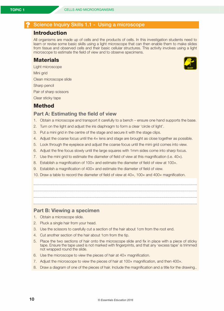
? Science Inquiry Skills 1.1 - Using a microscope
IntroductionAll organisms are made up of cells and the products of cells. In this investigation students need to learn or revise some basic skills using a light microscope that can then enable them to make slides from tissue and observed cells and their basic cellular structures. This activity involves using a light microscope to estimate the field of view and to observe specimens.
MaterialsLight microscope
Mini grid
Clean microscope slide
Sharp pencil
Pair of sharp scissors
Clear sticky tape
MethodPart A: Estimating the field of view1. Obtain a microscope and transport it carefully to a bench – ensure one hand supports the base.
2. Turn on the light and adjust the iris diaphragm to form a clear ‘circle of light’.
3. Put a mini grid in the centre of the stage and secure it with the stage clips.
4. Adjust the coarse focus until the 4× lens and stage are brought as close together as possible.
5. Look through the eyepiece and adjust the coarse focus until the mini grid comes into view.
6. Adjust the fine focus slowly until the large squares with 1mm sides come into sharp focus.
7. Use the mini grid to estimate the diameter of field of view at this magnification (i.e. 40×).
8. Establish a magnification of 100× and estimate the diameter of field of view at 100×.
9. Establish a magnification of 400× and estimate the diameter of field of view.
10. Draw a table to record the diameter of field of view at 40×, 100× and 400× magnification.
Part B: Viewing a specimen1. Obtain a microscope slide.
2. Pluck a single hair from your head.
3. Use the scissors to carefully cut a section of the hair about 1cm from the root end.
4. Cut another section of the hair about 1cm from the tip.
5. Place the two sections of hair onto the microscope slide and fix in place with a piece of sticky tape. Ensure the tape used is not marked with fingerprints, and that any ‘excess tape’ is trimmed not wrapped round the slide.
6. Use the microscope to view the pieces of hair at 40× magnification.
7. Adjust the microscope to view the pieces of hair at 100× magnification, and then 400×.
8. Draw a diagram of one of the pieces of hair. Include the magnification and a title for the drawing..
FINAL Workbook C.indb 10 21/09/2016 9:19:12 PM
© Essentials Education 2016 11
CHAPTER 1.1LIVING THINGS CONSIST OF CELLS
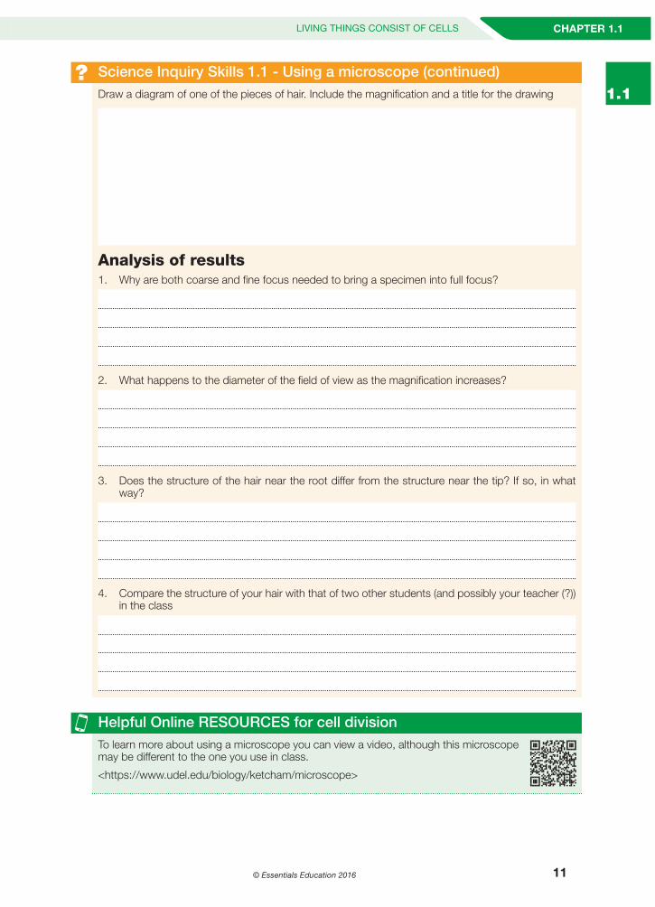
1.1? Science Inquiry Skills 1.1 - Using a microscope (continued)
Draw a diagram of one of the pieces of hair. Include the magnification and a title for the drawing
Analysis of results1. Why are both coarse and fine focus needed to bring a specimen into full focus?
2. What happens to the diameter of the field of view as the magnification increases?
3. Does the structure of the hair near the root differ from the structure near the tip? If so, in what way?
4. Compare the structure of your hair with that of two other students (and possibly your teacher (?)) in the class
Helpful Online RESOURCES for cell divisionTo learn more about using a microscope you can view a video, although this microscope may be different to the one you use in class.
<https://www.udel.edu/biology/ketcham/microscope>
FINAL Workbook C.indb 11 21/09/2016 9:19:12 PM