Embed Size (px)
Citation preview
Cell walls
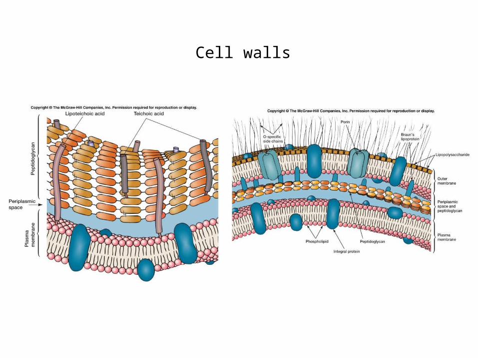
Gram-positive cell walls
Thick layer of peptidoglycan surrounding the plasma membrane
Contain teichoic acids

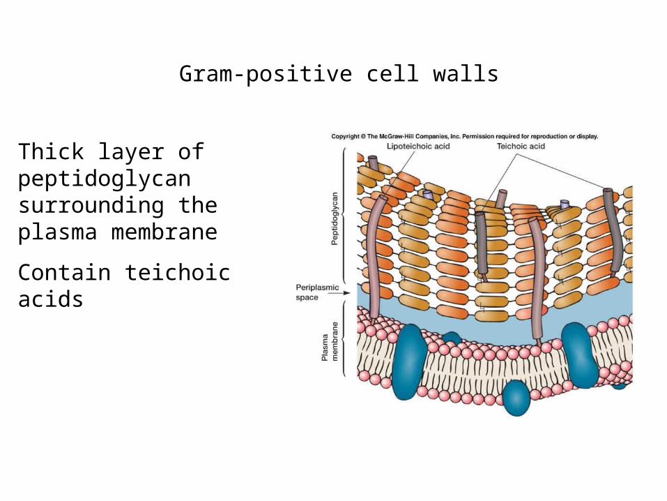
Teichoic acids
Polymers of glycerol or ribitol joined by phosphate groups
Amino acids or sugars are attached to glycerol or ribitol groups
Teichoic acids
Can be attached to either peptidoglycan or membrane lipid lipoteichoic acid
May contribute to negative charge of cell surface
Are not found in gram-negative bacteria
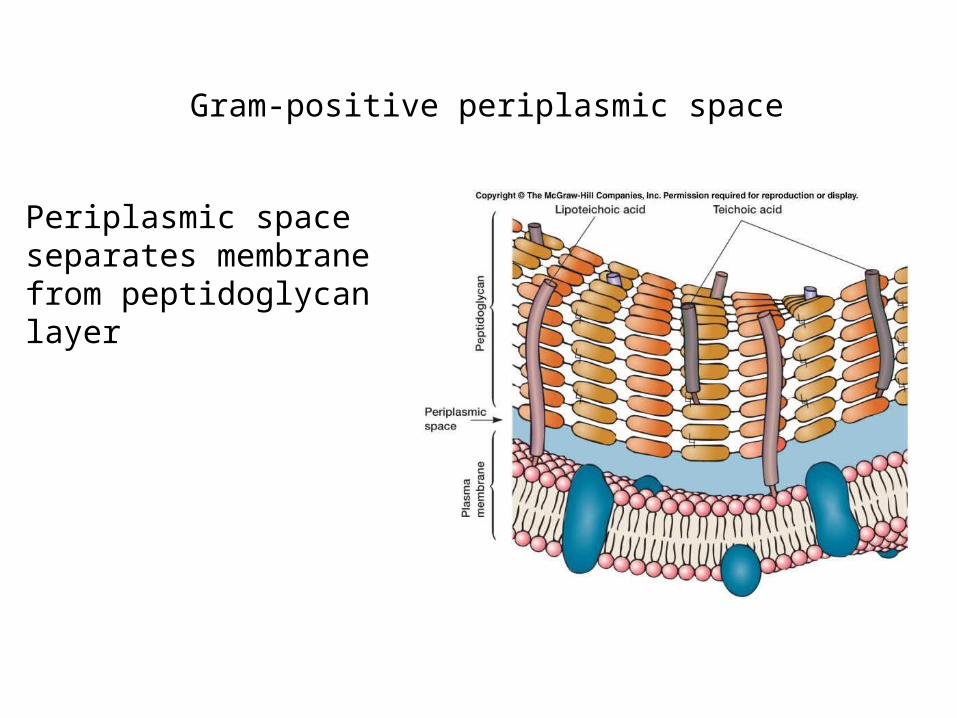
Gram-positive periplasmic space
Periplasmic space separates membrane from peptidoglycan layer
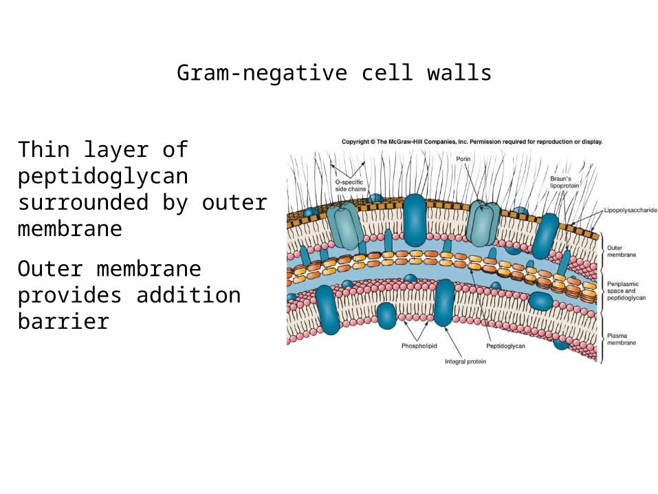
Gram-negative cell walls
Thin layer of peptidoglycan surrounded by outer membrane
Outer membrane provides addition barrier
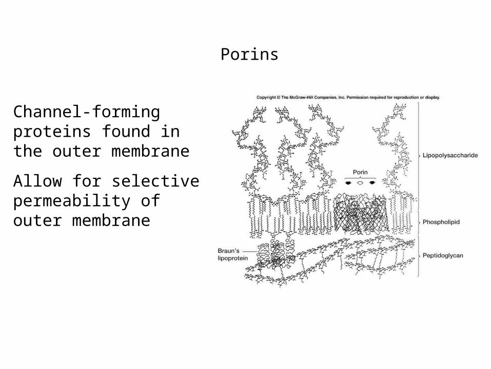
Porins
Channel-forming proteins found in the outer membrane
Allow for selective permeability of outer membrane
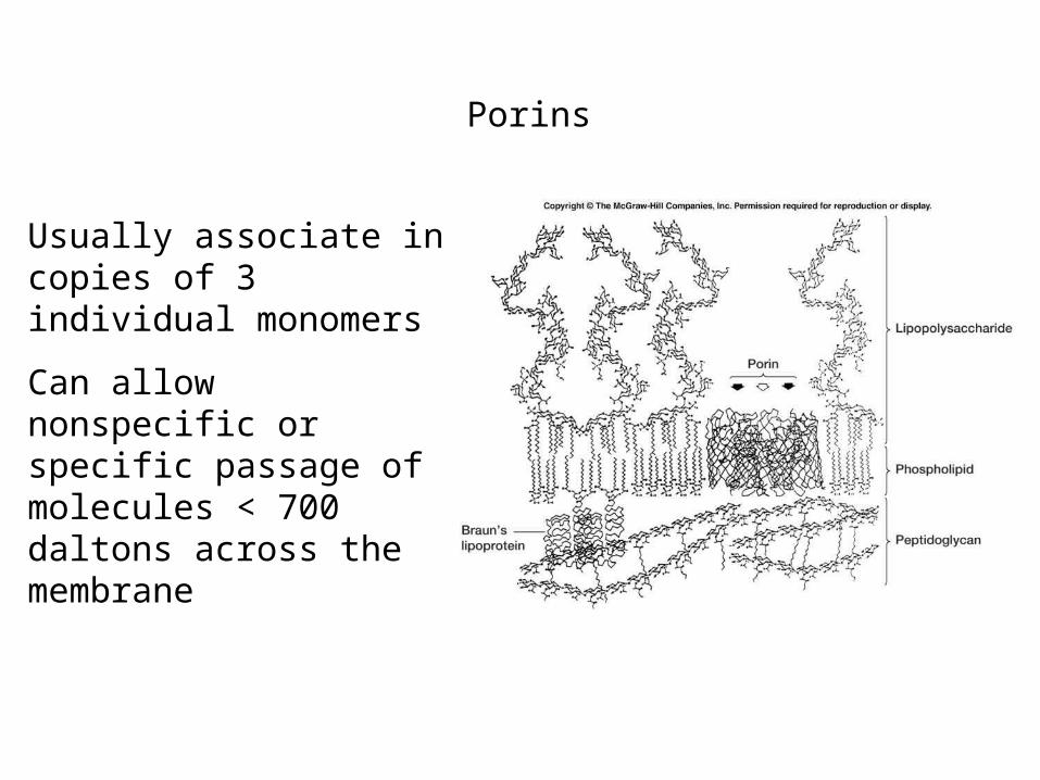
Porins
Usually associate in copies of 3 individual monomers
Can allow nonspecific or specific passage of molecules < 700 daltons across the membrane
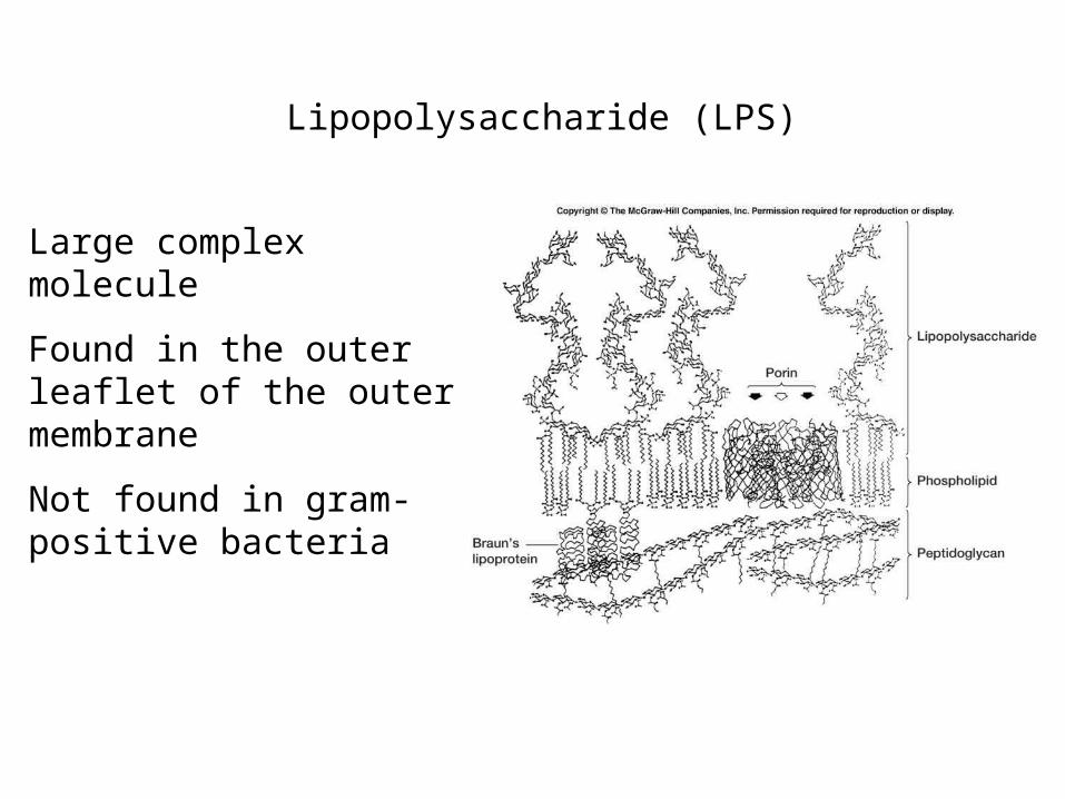
Lipopolysaccharide (LPS)
Large complex molecule
Found in the outer leaflet of the outer membrane
Not found in gram-positive bacteria
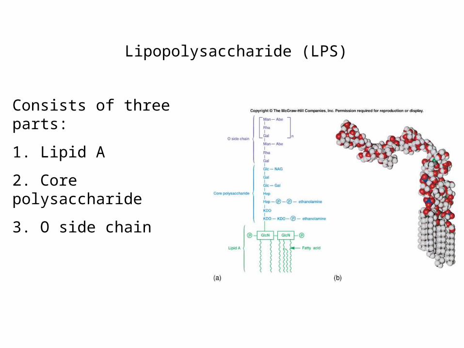
Lipopolysaccharide (LPS)
Consists of three parts:
1. Lipid A
2. Core polysaccharide
3. O side chain
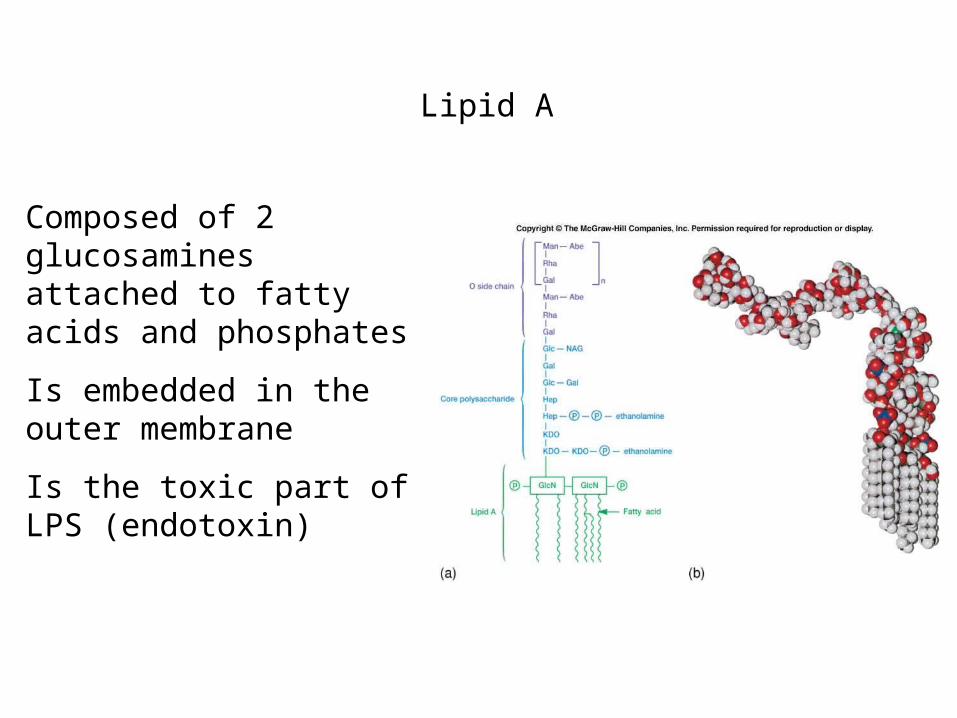
Lipid A
Composed of 2 glucosamines attached to fatty acids and phosphates
Is embedded in the outer membrane
Is the toxic part of LPS (endotoxin)
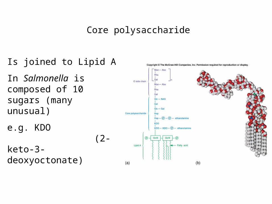
Core polysaccharide
Is joined to Lipid A
In Salmonella is composed of 10 sugars (many unusual)
e.g. KDO (2-keto-3-deoxyoctonate)
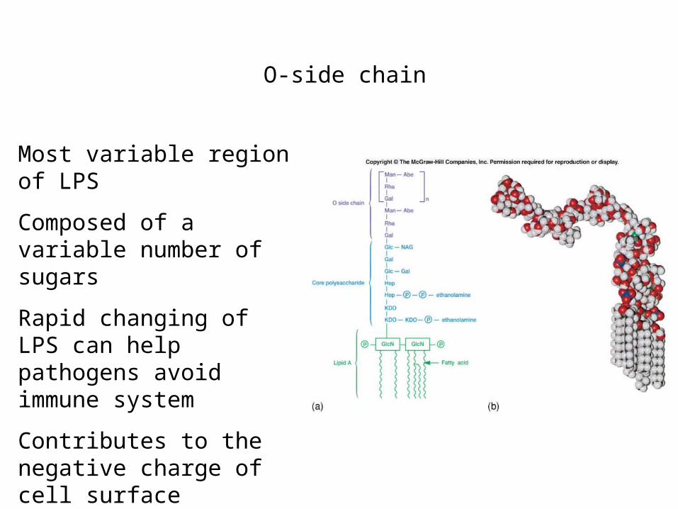
O-side chain
Most variable region of LPS
Composed of a variable number of sugars
Rapid changing of LPS can help pathogens avoid immune system
Contributes to the negative charge of cell surface
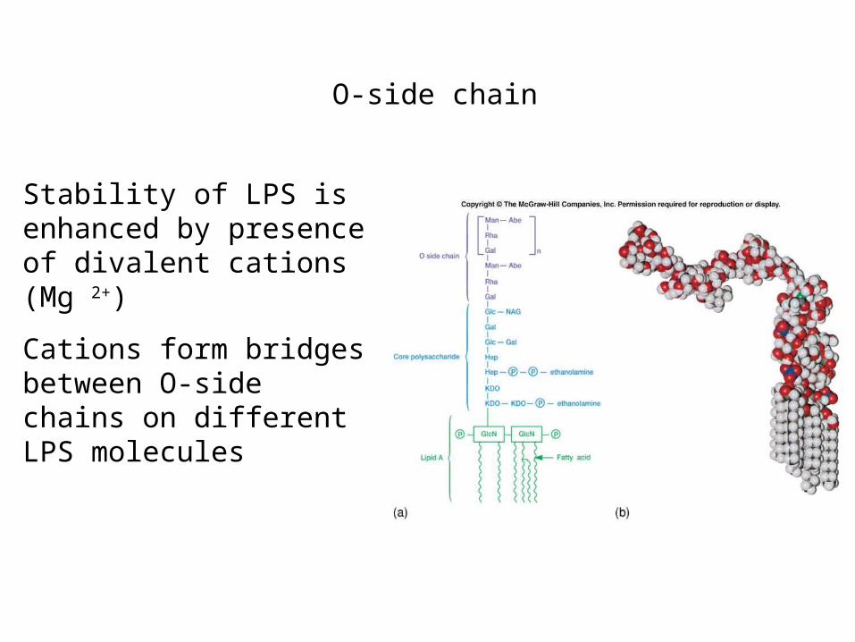
O-side chain
Stability of LPS is enhanced by presence of divalent cations (Mg 2+)
Cations form bridges between O-side chains on different LPS molecules
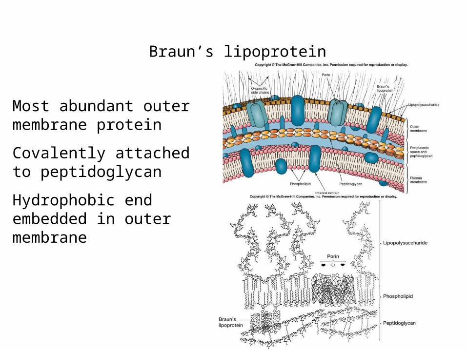
Braun’s lipoprotein
Most abundant outer membrane protein
Covalently attached to peptidoglycan
Hydrophobic end embedded in outer membrane
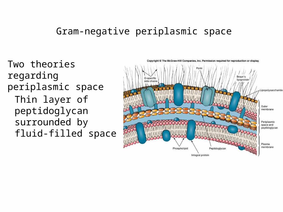
Gram-negative periplasmic space
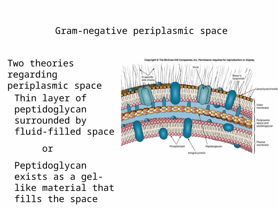
Two theories regarding periplasmic space
Thin layer of peptidoglycan surrounded by fluid-filled space
Gram-negative periplasmic space
Two theories regarding periplasmic space
Thin layer of peptidoglycan surrounded by fluid-filled space
or
Peptidoglycan exists as a gel-like material that fills the space
Peptidoglycan
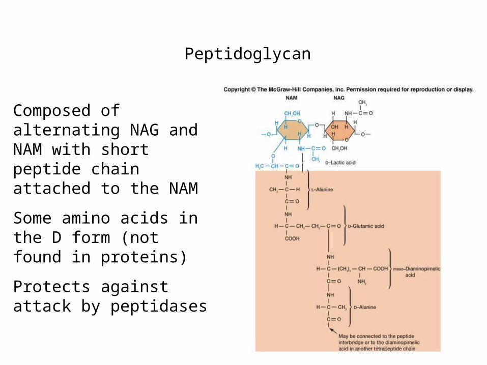
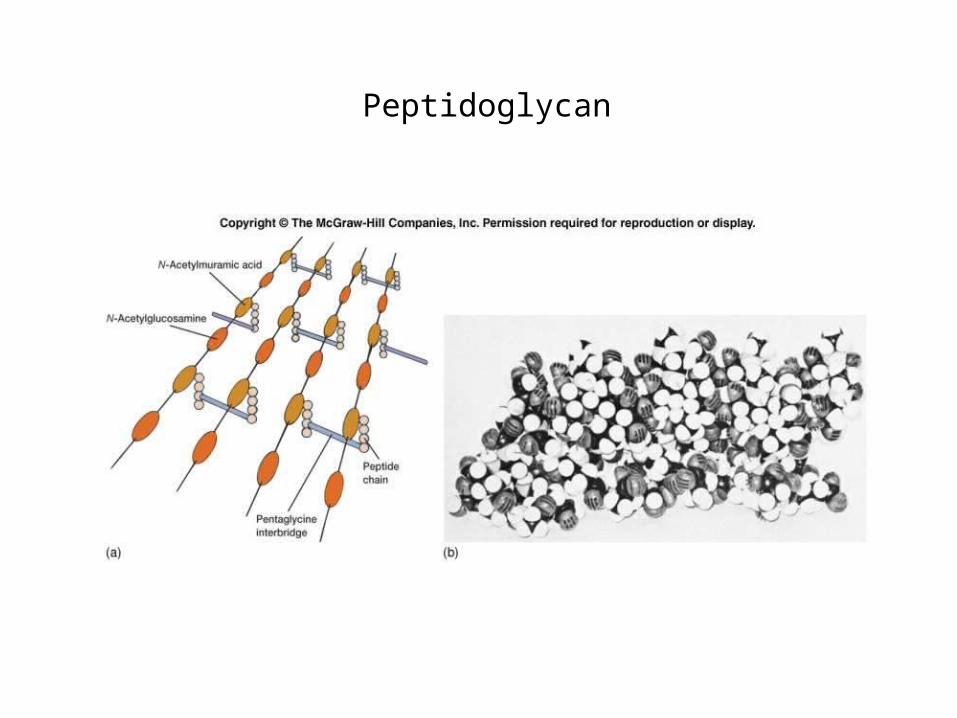
Composed of alternating NAG and NAM with short peptide chain attached to the NAM
Some amino acids in the D form (not found in proteins)
Protects against attack by peptidases
Peptidoglycan
Peptidoglycan synthesis
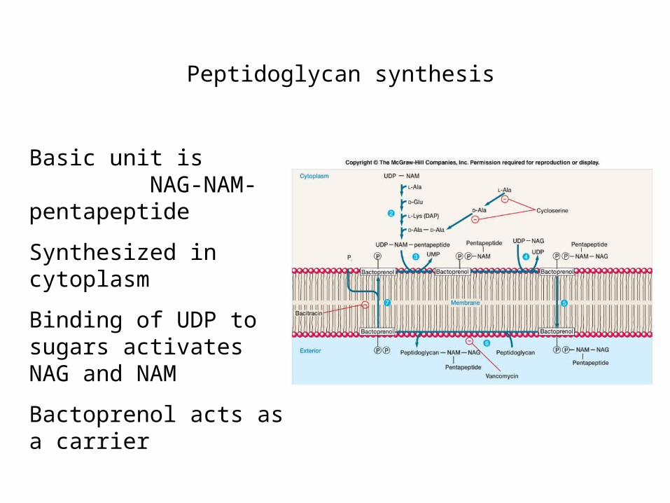
Basic unit is NAG-NAM-pentapeptide
Synthesized in cytoplasm
Binding of UDP to sugars activates NAG and NAM
Bactoprenol acts as a carrier

Bactoprenol
Very hydrophobic molecule
Allows for transport through the interior of the membrane
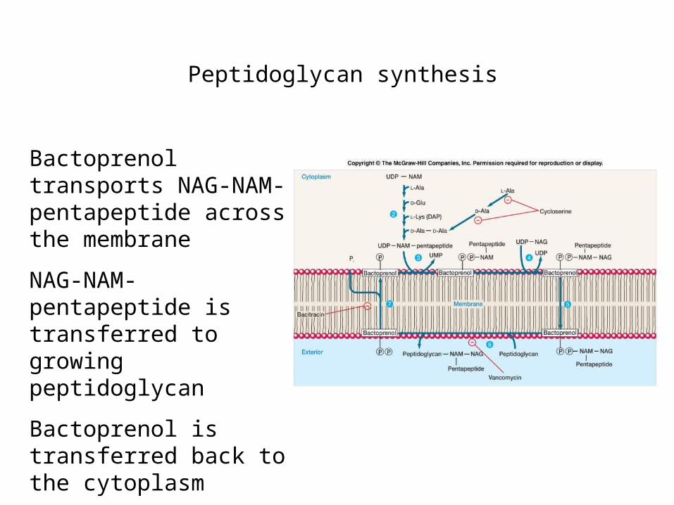
Peptidoglycan synthesis
Bactoprenol transports NAG-NAM-pentapeptide across the membrane
NAG-NAM-pentapeptide is transferred to growing peptidoglycan
Bactoprenol is transferred back to the cytoplasm
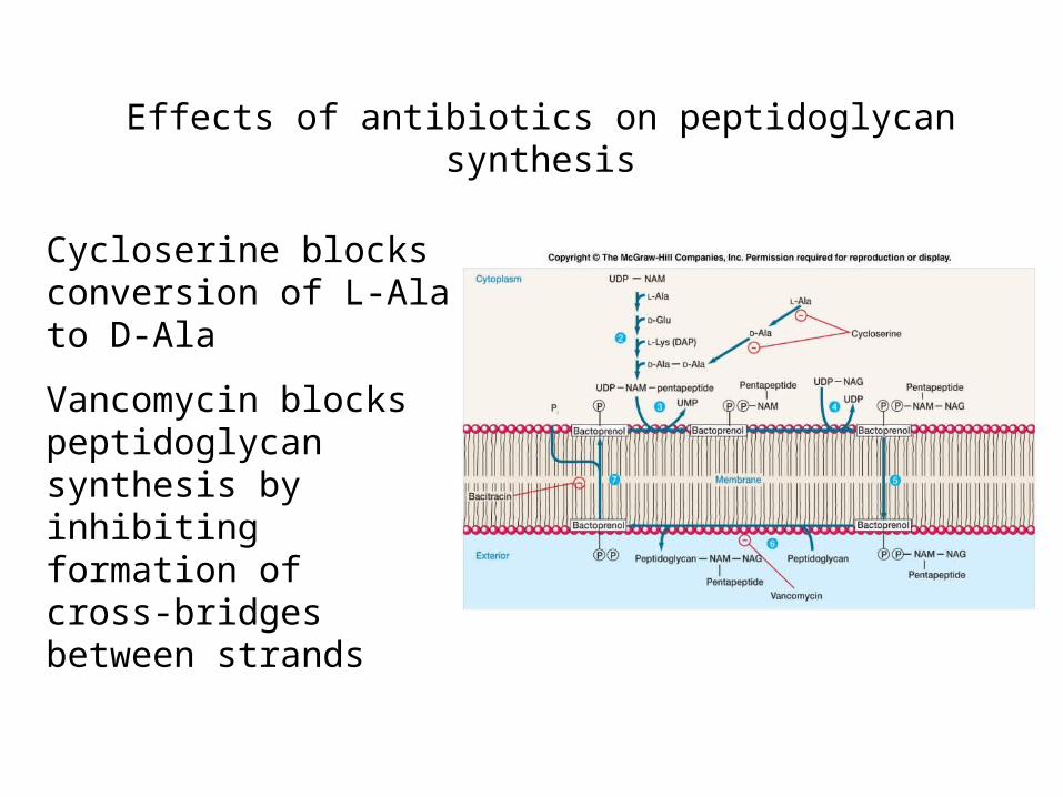
Effects of antibiotics on peptidoglycan synthesis
Cycloserine blocks conversion of L-Ala to D-Ala
Vancomycin blocks peptidoglycan synthesis by inhibiting formation of cross-bridges between strands
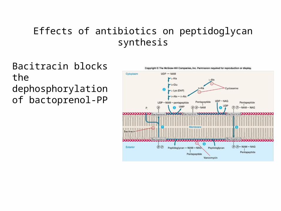
Effects of antibiotics on peptidoglycan synthesis
Bacitracin blocks the dephosphorylation of bactoprenol-PP
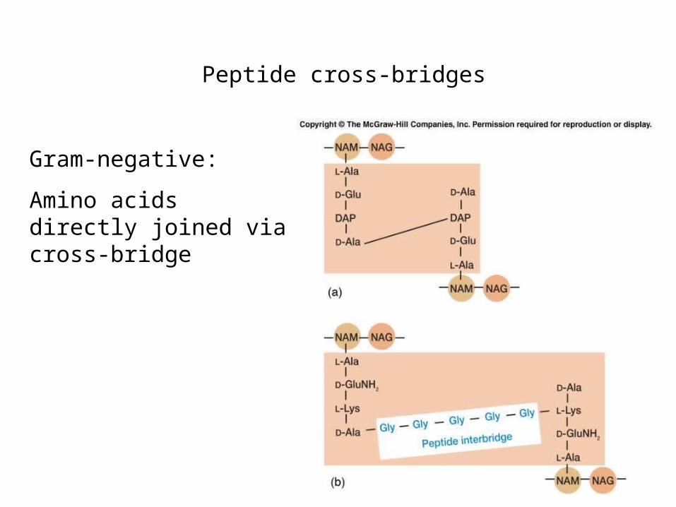
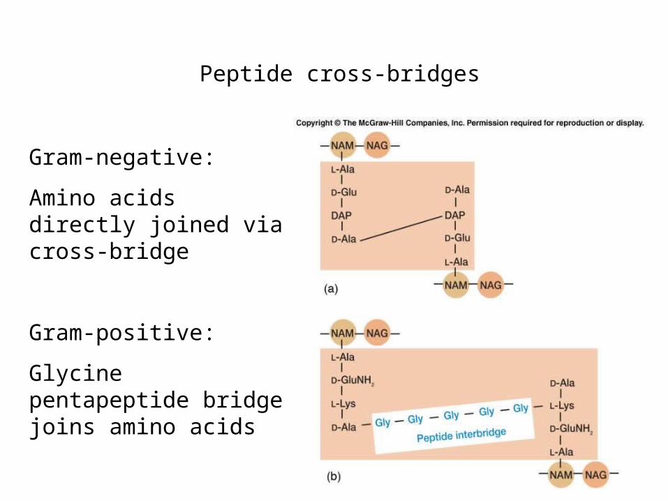
Peptide cross-bridges
Gram-negative:
Amino acids directly joined via cross-bridge
Peptide cross-bridges
Gram-negative:
Amino acids directly joined via cross-bridge
Gram-positive:
Glycine pentapeptide bridge joins amino acids
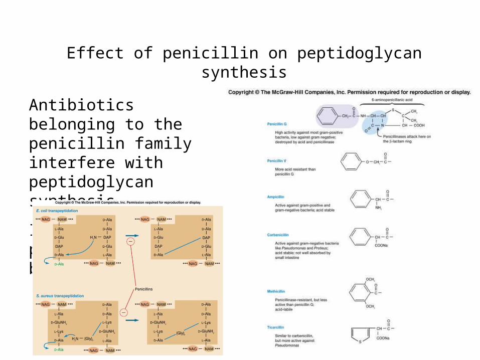
Effect of penicillin on peptidoglycan synthesis
Antibiotics belonging to the penicillin family interfere with peptidoglycan synthesis
Inhibit formation of peptide cross-bridges
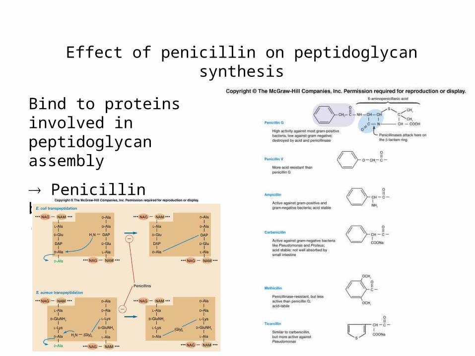
Effect of penicillin on peptidoglycan synthesis
Bind to proteins involved in peptidoglycan assembly
Penicillin binding proteins (PBPs)
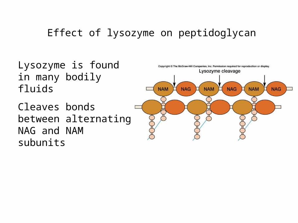
Effect of lysozyme on peptidoglycan
Lysozyme is found in many bodily fluids
Cleaves bonds between alternating NAG and NAM subunits