Embed Size (px)
Citation preview
Role of Wall Teichoic Acids inStaphylococcus aureus Endophthalmitis
The Harvard community has made thisarticle openly available. Please share howthis access benefits you. Your story matters
Citation Suzuki T, Campbell J, Swoboda JG, Walker S, Gilmore MS..2011. Role of wall teichoic acids in Staphylococcus aureusendophthalmitis. Invest Ophthalmol Vis Sci. 52, no. 6: 3187-92. doi:10.1167/iovs.10-6558.
Published Version doi:10.1167/iovs.10-6558
Citable link http://nrs.harvard.edu/urn-3:HUL.InstRepos:33867376
Terms of Use This article was downloaded from Harvard University’s DASHrepository, and is made available under the terms and conditionsapplicable to Other Posted Material, as set forth at http://nrs.harvard.edu/urn-3:HUL.InstRepos:dash.current.terms-of-use#LAA
Role of Wall Teichoic Acids in Staphylococcusaureus Endophthalmitis
Takashi Suzuki,1 Jennifer Campbell,2 Jonathan G. Swoboda,2 Suzanne Walker,2
and Michael S. Gilmore1,2
PURPOSE. Wall teichoic acids (WTAs) are major polyanionicpolymer components of the cell wall of Staphylococcus au-reus. However, little is known about their role at the host–pathogen interface, especially in endophthalmitis. This studywas designed to investigate the extent to which WTAs contrib-ute to the pathogenicity of S. aureus in models of endophthal-mitis and to determine whether there would be value in tar-geting their biosynthesis as a new therapeutic approach.
METHODS. S. aureus RN6390 and its isogenic WTA-null mutant(RN6390�tarO) were used to evaluate the role of WTAs inendophthalmitis. RN6390 and RN6390�tarO were cultured inbovine vitreous humor (VH) in vitro or inoculated into thevitreous chamber of C57B6 mice. Changes in the number ofbacteria, organ function as determined by electroretinography(ERG), and histopathologic changes were assessed throughoutthe course of infection. In addition, the efficacy of WTA bio-synthesis inhibitors in VH in vitro was examined.
RESULTS. It was observed that a component of VH synergizedwith WTA biosynthesis inhibitors in vitro and killed the S.aureus. This effect was also seen when mutants incapable ofexpressing WTA were exposed to VH. The killing activity ofVH was lost on treatment with a protease inhibitor.RN6390�tarO could not survive in mouse eyes and did notaffect organ function, nor was it able to establish endophthal-mitis.
CONCLUSIONS. WTAs are essential cellular constituents for themanifestation of virulence by S. aureus in endophthalmitis,and appears to be a viable target for treating the endophthal-mitis caused by S. aureus strains. (Invest Ophthalmol Vis Sci.2011;52:3187–3192) DOI:10.1167/iovs.10-6558
Bacterial endophthalmitis is a severe and sight-threateningocular infection.1 It is characterized by massive inflamma-
tion and tissue damage caused both by bacterial infection andsubsequent immune response. Bacterial endophthalmitis usu-ally occurs in the context of ocular surgery, trauma, microbialkeratitis, or the hematogenous spread of the organism to theeye. Postoperative endophthalmitis is a severe complication of
ocular surgery, such as cataract surgery, glaucoma surgery, orvitrectomy.1 Although the technical procedures used in sur-gery are improving, the incidence of endophthalmitis has notchanged and may be increasing.2
Staphylococcus aureus is a commensal species that colo-nizes the mucosa and skin adjacent to the eye, and as a result,often contaminates surgical sites and leads to endophthalmi-tis.1,3 Methicillin resistance in S. aureus is on the rise in thecommunity, which in the United States has been associatedwith the proliferation of the USA300 MRSA (methicillin-resis-tant S. aureus) lineage,4,5 which likely has contributed to theincreased incidence of endophthalmitis caused by MRSA.6,7
Although fluoroquinolones and cephalosporins are widelyused for the treatment and prevention of S. aureus endoph-thalmitis, they are less efficacious against MRSA.6,7 Moreover,S. aureus recently acquired resistance to vancomycin, a drugof last resort.8–10 Because of the rapid emergence of resistantstrains, new antibacterial targets that lack cross-resistance withdrugs currently in use to treat S. aureus infection are urgentlyneeded.
A possible therapeutic target that is now being explored isS. aureus wall teichoic acid (WTA) biosynthesis. WTAs areanionic polymers composed of repeating units of ribitol-phos-phate that are covalently linked to the bacterial cell wall inmany Gram-positive pathogens.11 They influence critical prop-erties of the cell envelope, including charge, cation binding,tensile strength, rigidity, and permeability.11 WTAs are notrequired for the growth of S. aureus in vitro, but are needed toestablish infection in most animal models.12–15
S. aureus WTA polymer biosynthesis is performed by theTar enzymes.11,16 Of interest, this biosynthetic pathway con-tains two classes of targets: antivirulence targets and antibac-terial targets. The first two genes in the pathway (tarO or tarA)can be knocked out, and the resulting mutants are viable invitro, making the products of these genes potential antiviru-lence targets. However, genes downstream of tarA in the WTApathway cannot be deleted in a wild-type background,16 mak-ing them antibacterial targets in wild-type strains. However, ifflux through the pathway is diverted, either through geneticmutation (of tarO or tarA) or pharmacologic inhibition of TarOby the natural product tunicamycin, these downstream en-zymes become expendable, suggesting that the essentiality ofthese downstream functions results from the accumulation ofWTA biosynthetic intermediates in the pathway. The charac-terization of the first small-molecule inhibitor of an antibacterialtarget within the WTA biosynthetic pathway was recently re-ported by one of our groups (SW).17 The compound, 1835F03,inhibits TarG, the transmembrane component of the ABC trans-porter that exports WTAs to the cell surface.17 A structure–activity relationship study of 1835F03 led to the discovery of asecond-generation analogue, targocil, which is 10 times morepotent.18 Because blocking polymer synthesis at the beginningof the pathway renders TarG nonessential, these WTA inhibi-tors generate mutants in which WTAs are no longer expressed.
From the Departments of 1Ophthalmology and 2Microbiology andMolecular Genetics, Harvard Medical School, Boston, Massachusetts.
Supported by Public Health and Human Services (PHHS) GrantsEY008289 (MSG), Harvard-wide project AI083214, and by GrantGM078477 (SW), Fellowship Grants F3178727 (JGS) and F32AI084316(JC), the Uehara Memorial Foundation (TS), and the Japanese Eye BankSociety (TS).
Submitted for publication September 10, 2010; revised January 14,2011; accepted January 30, 2011.
Disclosure: T. Suzuki, None; J. Campbell, None; J.G. Swoboda,None; S. Walker, None; M.S. Gilmore, None
Corresponding author: Michael S. Gilmore, Department of Oph-thalmology, Harvard Medical School, 243 Charles Street, Boston, MA02114; [email protected].
Immunology and Microbiology
Investigative Ophthalmology & Visual Science, May 2011, Vol. 52, No. 6Copyright 2011 The Association for Research in Vision and Ophthalmology, Inc. 3187

Thus, they can be regarded as both antibiotics and antiviru-lence factor agents.17
Although the roles of WTAs have been investigated in S.aureus colonization of epithelial and endothelial cells,12–15 therole of WTAs in vivo in eye infection and the extent to whichthis pathway could represent a viable treatment target have notyet been explored. In the present study, we assessed thenecessity for WTA biosynthesis during S. aureus endophthal-mitis, both in vitro and in vivo. The WTA transport inhibitor,targocil, was previously shown to halt S. aureus growththrough a bacteriostatic mechanism.17,18 In this study, weshowed the surprising finding that targocil treatment killed S.aureus in vitro in vitreous humor (VH). Further, we found thata WTA-deficient mutant cannot survive in VH in vitro or invivo, and as a result WTA-deficient mutants are highly attenu-ated, with little capacity to cause endophthalmitis.
MATERIALS AND METHODS
Bacteria and Growth Conditions
The S. aureus strains used in this study are listed in Table 1. TheWTA-deficient tarO mutant (RN6390�tarO) was generated fromRN6390 by allelic exchange with a tetracycline resistance marker.22,23
RN6390�tarO is defective in WTA production because of deletion ofthe first essential enzyme in the pathway. For reasons yet unknown,the mutant was subtly affected in toxin production. Supernatants fromovernight cultures of RN6390�tarO were found to be one half to onefourth that of RN6390. In addition RN4220 PspactarO, a strain thatexpresses tarO under the control of an isopropyl �-D-1-thiogalactopy-ranoside (IPTG)-inducible promoter, was used to verify the role ofTarO.17 S. aureus strains were grown in tryptic soy broth (TSB) at37°C, unless otherwise noted. Tetracycline (Tc; 2.5 �g/mL) and eryth-romycin (Em; 10 �g/mL) were used for selection where appropriate.To inhibit various steps in WTA biosynthesis, targocil and tunicamycinwere dissolved in dimethyl sulfoxide (DMSO) and stored at �20°C. Incontrols and experimental groups, final concentrations of DMSO wereadjusted to 1% in all assays, unless otherwise stated.
Assessment of Activity in Vitreous Humor
Bovine eyes obtained from Sierra for Medical Science (Whittier, CA)were used within 24 hours of enucleation and stored at 4°C before use.VH was aspirated with a syringe fitted with an 18-gauge needle.Liquefied portions of the vitreous were carefully aspirated, taking careto avoid areas of adhesion to the retina and other tissues. The VH wasfiltered through a GD/X sterile 0.45 �m PES syringe filter (Whatman,Clifton, NJ) and stored at �20°C. Each experiment used vitreous froman individual eye.
Animal Care and Use
Female C57BL/6J mice were obtained from the Charles River Labora-tory (Boston, MA). All animals were humanely treated according to theguidelines of the ARVO Statement for the Use of Animals in Ophthal-mic and Vision Research. All procedures involving mice were ap-proved by the Schepens Eye Research Institute Institutional AnimalCare and Use Committee (IACUC). When appropriate, the mice wereanesthetized by intraperitoneal injection of ketamine (62.5 mg/kg) and
xylazine (12.5 mg/kg). The animals were euthanatized at the appropri-ate time points by CO2 asphyxiation after anesthesia.
Bacterial Growth in Vitreous Humor In Vitro
Targocil and tunicamycin were used as WTA biosynthesis inhibitors.Targocil inhibits a late step in WTA biosynthesis and stops S. aureusgrowth bacteriostatically.18 The minimum inhibitory concentration(MIC) of targocil against RN6390 is 1 �g/mL. Tunicamycin is aninhibitor of a large class of enzymes that couple sugar phosphates tomembrane-embedded lipid phosphates,24 and 1 �g/mL tunicamycininhibits WTA production by S. aureus in vitro without affecting bac-terial growth rates.17 An overnight culture of S. aureus RN6390 wasdiluted to approximately 104 CFU in 100 �L of VH or brain–heartinfusion (BHI) medium containing 1% DMSO, 5� MIC (5 �g/mL)targocil, or 1 �g/mL tunicamycin and cultured at 37°C statically for 24hours. Bacteria were enumerated by plating a serial 10-fold dilution. Toexamine rates of growth of the RN6390 and RN6390�tarO strains, wediluted overnight cultures to approximately 105 CFU in 1 mL of VH orBHI medium and cultured the bacteria at 37°C statically. The bacteriawere enumerated by serial dilution at each time point. In additionRN4220 PspactarO was tested for bacterial growth in VH. Dilutions ofan overnight culture of S. aureus RN4220 wild-type or PspactarO wereinoculated into VH, with or without 1 mM IPTG, and bacterial growthwas monitored over a 24-hour period. Experiments were performedthree times independently.
Transmission Electron Microscopy
RN6390�tarO or RN6390 were inoculated to approximately 109 CFUper milliliter of VH and cultured at 37°C for 6 hours. The cells werecollected at 0 or 6 hours, fixed in Karnovsky’s fixative (2% paraformal-dehyde and 2.5% glutaraldehyde in cacodylate buffer [pH 7.4]), andprocessed for transmission electron microscopy (TEM) by using stan-dard procedures described elsewhere.25 For TEM, 60- to 90-Å sectionswere obtained, viewed, and photographed (model 410 microscope;Philips Electronics NV, Eindhoven, The Netherlands). Diameters of allcells within three microscopic fields (50–80 cells per strain) weremeasured with ImageJ software (developed by Wayne Rasband, Na-tional Institutes of Health, Bethesda, MD; available at http://rsb.info.nih.gov/ij/index.html).
Effect of Enzyme or Chemical Treatment on theKilling Ability of VH
Bacterial growth was examined in VH or VH treated in various waysto illuminate the basis for killing. In one case, VH was pretreatedwith 100 �g/mL protease K (New England Biolabs Inc., Beverly,MA) at 37°C overnight and then boiled for 5 minutes to inactivatethe added protease. VH was also pretreated with protease inhibitorsfor 1 hour at 37°C at the following concentrations: 104 mM 4-(2-aminoethyl)benzenesulfonylfluoride (AEBSF), 1.4 mM trans-epoxysuccinyl-L-leucylamido(4-guanidino)butane (E-64), 4 mMbestatin, and 1� protease inhibitor (PI) cocktail, EDTA-free (Sigma-Aldrich, Poole, UK). DMSO, the diluent for all inhibitors, was ad-justed to 1% in VH and used as control. Growth of RN6390�tarO invariously treated VH was examined for 24 hours.
TABLE 1. Bacterial Strains
Strain Genotype and/or Phenotype Reference
RN6390 Prophage-cured derivative of NCTC 8325 19RN4220 A mutant of NCTC 8325–4 that accepts foreign DNA partial agr defect 20RN6390�tarO RN6390 �tarO::tetL This studyRN4220�tarO RN4220 �tarO 21PspactarO RN4220 �tarO::tetL [geh:: (pCL25int-PspactarO) Emr] 17
3188 Suzuki et al. IOVS, May 2011, Vol. 52, No. 6
Murine Model of Endophthalmitis
The vitreous of eyes of 6- to 8-week-old mice were inoculated byinsertion of a borosilicate microcapillary, pulled to a tip size of 50 �m,immediately behind the limbus-parallel conjunctival vessels, corre-sponding to the narrow murine pars plana, essentially as de-scribed.26,27 The right eyes of the mice were injected with 0.5 �L of abacterial suspension containing 5000 CFU of either S. aureus RN6390or RN6390�tarO diluted in physiologic saline. The left eye of eachmouse was left untreated and served as an internal control for electro-retinography (ERG) studies. Experiments were performed with a min-imum of four animals per experimental group and repeated at leasttwice.
Quantification of Bacterial Growth
Eyes were enucleated after euthanatization, and the residual adnexaltissue was trimmed before the eyes were rinsed and placed in coldphosphate-buffered saline (PBS). The enucleated eyes were disruptedand homogenized to release bacteria for enumeration by bead-beating with 1.0-mm glass beads (FastPrep; Thermo Scientific,Waltham, MA) in 1 mL PBS for 1 minute at maximum speed. Thehomogenates were serially diluted, plated onto BHI agar plates, andincubated overnight at 37°C.
Electroretinography
ERG was performed on the mice at 24 and 48 hours after inoculation,using a protocol modified slightly from those previously published.26,27
Briefly, the mice were dark adapted for at least 4 hours and thenanesthetized. The pupils were dilated using 1% tropicamide ophthal-mic solution (Bausch & Lomb, Tampa, FL). After anesthesia, the bodytemperature was maintained at 37°C with a microwave heating pad.Gold wire electrodes (0.25 mm; Alfa Aesar, Ward Hill, MA) were placedon the cornea after application of a hypromellose ophthalmic prismsolution (Akorn, Inc. Buffalo Grove, IL) and connected to a visualelectrodiagnostic system (UTAS-E 3000; LKC Technologies, Gaithers-burg, MD). Needle electrodes placed in the anterior scalp and the tailserved as reference and ground leads, respectively. The b-wave ampli-tude (measured from the trough of the a-wave to the peak of theb-wave) in response to a bright flash in a Ganzfeld illumination spherewas recorded for the injected right eye and the contralateral (internal-control) left eye simultaneously. A total of 30 readings at 0.6 cd-s/m2
flash intensity with a 1-second interval between flashes were taken andaveraged. The retinal function was defined as the ratio of the b-waveamplitude (from the trough of the a-wave to the peak of the b-wave) of
the experimentally treated eye divided by the value for the contralat-eral untreated eye.
Histologic Analysis
The mice were euthanatized 48 hours after infection, and the eyeswere enucleated with Stevens curved, sharp-tip scissors. The eyeswere fixed in 10% buffered formalin, embedded in paraffin, sectioned,and stained with hematoxylin and eosin (HE).
Statistical Analysis
Data were analyzed by Student’s t-test for significance. Values of P �0.05 were considered significant.
RESULTS
Bacterial Growth in Vitreous Humor In Vitro
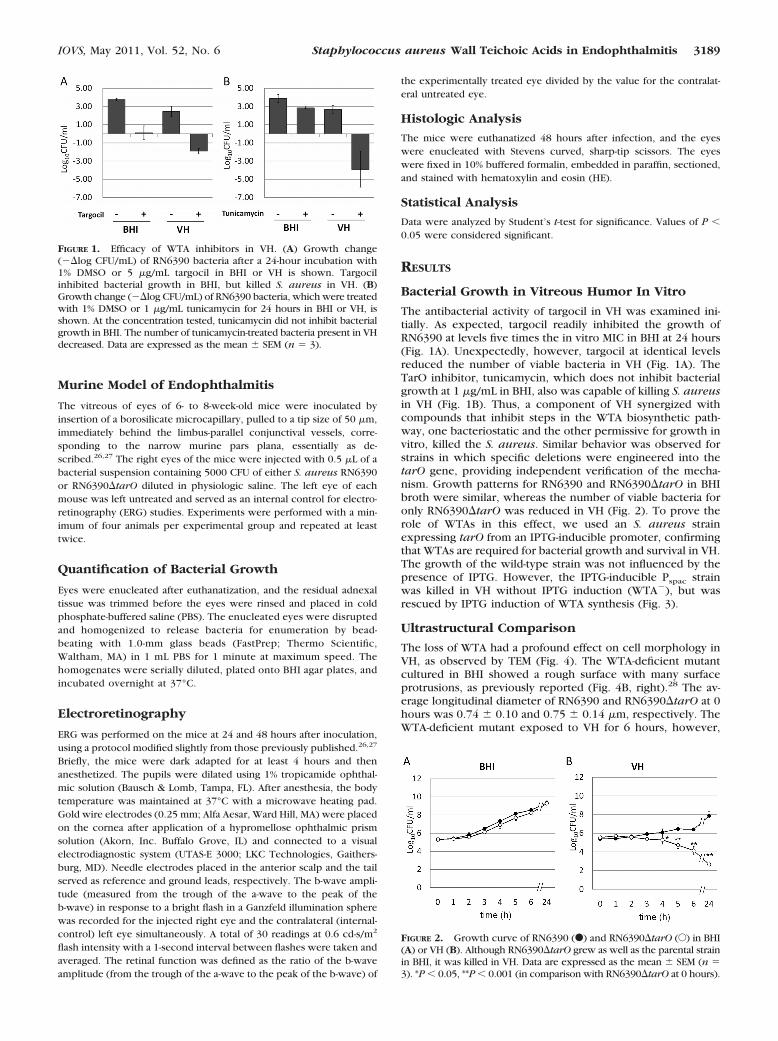
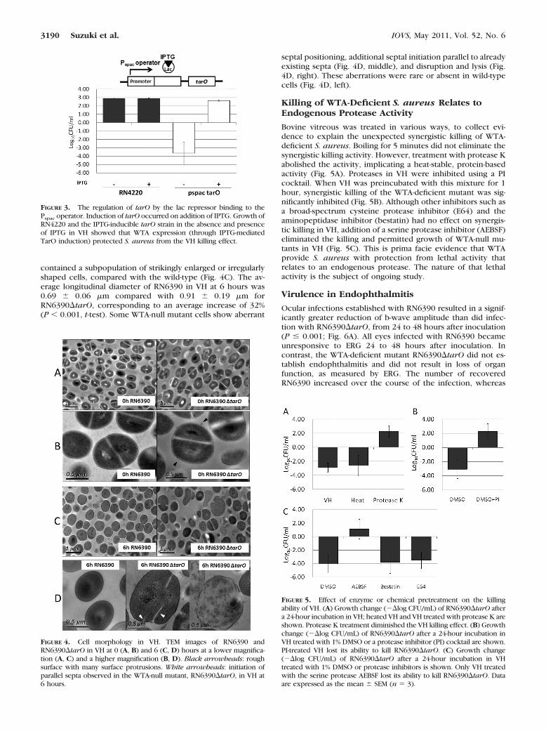
The antibacterial activity of targocil in VH was examined ini-tially. As expected, targocil readily inhibited the growth ofRN6390 at levels five times the in vitro MIC in BHI at 24 hours(Fig. 1A). Unexpectedly, however, targocil at identical levelsreduced the number of viable bacteria in VH (Fig. 1A). TheTarO inhibitor, tunicamycin, which does not inhibit bacterialgrowth at 1 �g/mL in BHI, also was capable of killing S. aureusin VH (Fig. 1B). Thus, a component of VH synergized withcompounds that inhibit steps in the WTA biosynthetic path-way, one bacteriostatic and the other permissive for growth invitro, killed the S. aureus. Similar behavior was observed forstrains in which specific deletions were engineered into thetarO gene, providing independent verification of the mecha-nism. Growth patterns for RN6390 and RN6390�tarO in BHIbroth were similar, whereas the number of viable bacteria foronly RN6390�tarO was reduced in VH (Fig. 2). To prove therole of WTAs in this effect, we used an S. aureus strainexpressing tarO from an IPTG-inducible promoter, confirmingthat WTAs are required for bacterial growth and survival in VH.The growth of the wild-type strain was not influenced by thepresence of IPTG. However, the IPTG-inducible Pspac strainwas killed in VH without IPTG induction (WTA�), but wasrescued by IPTG induction of WTA synthesis (Fig. 3).
Ultrastructural Comparison
The loss of WTA had a profound effect on cell morphology inVH, as observed by TEM (Fig. 4). The WTA-deficient mutantcultured in BHI showed a rough surface with many surfaceprotrusions, as previously reported (Fig. 4B, right).28 The av-erage longitudinal diameter of RN6390 and RN6390�tarO at 0hours was 0.74 � 0.10 and 0.75 � 0.14 �m, respectively. TheWTA-deficient mutant exposed to VH for 6 hours, however,
FIGURE 1. Efficacy of WTA inhibitors in VH. (A) Growth change(��log CFU/mL) of RN6390 bacteria after a 24-hour incubation with1% DMSO or 5 �g/mL targocil in BHI or VH is shown. Targocilinhibited bacterial growth in BHI, but killed S. aureus in VH. (B)Growth change (��log CFU/mL) of RN6390 bacteria, which were treatedwith 1% DMSO or 1 �g/mL tunicamycin for 24 hours in BHI or VH, isshown. At the concentration tested, tunicamycin did not inhibit bacterialgrowth in BHI. The number of tunicamycin-treated bacteria present in VHdecreased. Data are expressed as the mean � SEM (n � 3).
FIGURE 2. Growth curve of RN6390 (F) and RN6390�tarO (E) in BHI(A) or VH (B). Although RN6390�tarO grew as well as the parental strainin BHI, it was killed in VH. Data are expressed as the mean � SEM (n �3). *P � 0.05, **P � 0.001 (in comparison with RN6390�tarO at 0 hours).
IOVS, May 2011, Vol. 52, No. 6 Staphylococcus aureus Wall Teichoic Acids in Endophthalmitis 3189
contained a subpopulation of strikingly enlarged or irregularlyshaped cells, compared with the wild-type (Fig. 4C). The av-erage longitudinal diameter of RN6390 in VH at 6 hours was0.69 � 0.06 �m compared with 0.91 � 0.19 �m forRN6390�tarO, corresponding to an average increase of 32%(P � 0.001, t-test). Some WTA-null mutant cells show aberrant
septal positioning, additional septal initiation parallel to alreadyexisting septa (Fig. 4D, middle), and disruption and lysis (Fig.4D, right). These aberrations were rare or absent in wild-typecells (Fig. 4D, left).
Killing of WTA-Deficient S. aureus Relates toEndogenous Protease Activity
Bovine vitreous was treated in various ways, to collect evi-dence to explain the unexpected synergistic killing of WTA-deficient S. aureus. Boiling for 5 minutes did not eliminate thesynergistic killing activity. However, treatment with protease Kabolished the activity, implicating a heat-stable, protein-basedactivity (Fig. 5A). Proteases in VH were inhibited using a PIcocktail. When VH was preincubated with this mixture for 1hour, synergistic killing of the WTA-deficient mutant was sig-nificantly inhibited (Fig. 5B). Although other inhibitors such asa broad-spectrum cysteine protease inhibitor (E64) and theaminopeptidase inhibitor (bestatin) had no effect on synergis-tic killing in VH, addition of a serine protease inhibitor (AEBSF)eliminated the killing and permitted growth of WTA-null mu-tants in VH (Fig. 5C). This is prima facie evidence that WTAprovide S. aureus with protection from lethal activity thatrelates to an endogenous protease. The nature of that lethalactivity is the subject of ongoing study.
Virulence in Endophthalmitis
Ocular infections established with RN6390 resulted in a signif-icantly greater reduction of b-wave amplitude than did infec-tion with RN6390�tarO, from 24 to 48 hours after inoculation(P � 0.001; Fig. 6A). All eyes infected with RN6390 becameunresponsive to ERG 24 to 48 hours after inoculation. Incontrast, the WTA-deficient mutant RN6390�tarO did not es-tablish endophthalmitis and did not result in loss of organfunction, as measured by ERG. The number of recoveredRN6390 increased over the course of the infection, whereas
FIGURE 3. The regulation of tarO by the lac repressor binding to thePspac operator. Induction of tarO occurred on addition of IPTG. Growth ofRN4220 and the IPTG-inducible tarO strain in the absence and presenceof IPTG in VH showed that WTA expression (through IPTG-mediatedTarO induction) protected S. aureus from the VH killing effect.
FIGURE 4. Cell morphology in VH. TEM images of RN6390 andRN6390�tarO in VH at 0 (A, B) and 6 (C, D) hours at a lower magnifica-tion (A, C) and a higher magnification (B, D). Black arrowheads: roughsurface with many surface protrusions. White arrowheads: initiation ofparallel septa observed in the WTA-null mutant, RN6390�tarO, in VH at6 hours.
FIGURE 5. Effect of enzyme or chemical pretreatment on the killingability of VH. (A) Growth change (��log CFU/mL) of RN6390�tarO aftera 24-hour incubation in VH; heated VH and VH treated with protease K areshown. Protease K treatment diminished the VH killing effect. (B) Growthchange (��log CFU/mL) of RN6390�tarO after a 24-hour incubation inVH treated with 1% DMSO or a protease inhibitor (PI) cocktail are shown.PI-treated VH lost its ability to kill RN6390�tarO. (C) Growth change(��log CFU/mL) of RN6390�tarO after a 24-hour incubation in VHtreated with 1% DMSO or protease inhibitors is shown. Only VH treatedwith the serine protease AEBSF lost its ability to kill RN6390�tarO. Dataare expressed as the mean � SEM (n � 3).
3190 Suzuki et al. IOVS, May 2011, Vol. 52, No. 6
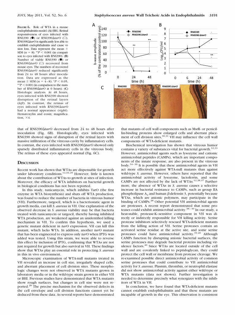
that of RN6390�tarO decreased from 24 to 48 hours afterinoculation (Fig. 6B). Histologically, eyes infected withRN6390 showed signs of disruption of the retinal layers withmassive infiltration of the vitreous cavity by inflammatory cells.In contrast, the eyes infected with RN6390�tarO showed onlysparsely distributed inflammatory cells in the vitreous body.The retinas of these eyes appeared normal (Fig. 6C).
DISCUSSION
Recent work has shown that WTAs are dispensable for growthunder laboratory conditions.12,16,29 However, little is knownabout the contribution of WTAs to growth at sites of infection.Moreover, the efficacy of WTA inhibitors on bacterial growthin biological conditions has not been reported.
In this study, tunicamycin, which inhibits TarO (the firstenzyme in WTA biosynthesis) and shuts off WTA production,was found to reduce the number of bacteria in vitreous humor(VH). Furthermore, targocil, which is a bacteriostatic agent ingrowth media, can kill S. aureus in VH. One explanation of theobserved decrease in S. aureus viability may be that bacteriatreated with tunicamycin or targocil, thereby having inhibitedWTA production, are weakened against an unidentified killingmechanism in VH. To confirm this hypothesis, we used agenetic mutant deficient in tarO expression. VH can kill thismutant, which lacks WTA. In addition, another tarO mutantthat has been engineered to express only tarO when IPTG wasadded was tested. Using this strain, we were able to reversethis effect by inclusion of IPTG, confirming that WTAs are notjust required for growth but also survival in VH. These findingsshow that WTAs play an essential role in protecting S. aureusin this in vivo environment.
Microscopic examination of WTA-null mutants treated inVH revealed an increase in cell size, irregularly shaped cells,and aberrant placement of cell division sites. These morpho-logic changes were not observed in WTA mutants grown inlaboratory media or in the wild-type strain grown in either VHor BHI. Previous studies have demonstrated that WTA mutantsshow rough surfaces, but changes in cell size were not re-ported.28 The precise mechanism for the observed defects inthe cell envelope and cell division functions cannot yet bededuced from these data. As several reports have demonstrated
that mutants of cell wall components such as MsrR- or penicil-lin-binding proteins show enlarged cells and aberrant place-ment of cell division sites,30,31 VH may influence the cell wallcomponents of WTA-deficient mutants.
Biochemical investigation has shown that vitreous humorcontains a variety of substances vital for bacterial growth.32,33
However, antimicrobial agents such as lysozyme and cationicantimicrobial peptides (CAMPs), which are important compo-nents of the innate response, are also present in the vitreousbody.34–36 It is possible that these antimicrobial agents in VHact more effectively against WTA-null mutants than againstwild-type S. aureus. However, others have reported that theantimicrobial activity of lysozyme, lactoferrin, and someCAMPs are not affected by the lack of WTAs.12,28,37 Further-more, the absence of WTAs in S. aureus causes a selectiveincrease in bacterial resistance to CAMPs, such as group IIAphospholipase A2 and human �-defensin 3, potentially becauseWTAs, which are anionic polymers, may participate in thebinding of CAMPs.28 Other potential VH antimicrobial agentsare proteases. A recent report demonstrated that some pro-teases could exhibit antimicrobial activity.38–40 In our study, aheat-stable, protease-K–sensitive component in VH was di-rectly or indirectly responsible for VH killing activity. Serineprotease inhibitors selectively rescued WTA-deficient mutantsfrom the killing action of VH. Serine proteases contain anactivated serine residue at the active site, and some serineproteases could have antimicrobial activity.39,40 AlthoughCAMPs function by disrupting anionic bacterial surfaces, theserine proteases may degrade bacterial proteins including vir-ulence factors.40 Since WTAs are located outside of the cellwall and are covalently linked to peptidoglycan, they couldprotect the cell wall or membrane from protease cleavage. Were-examined possible direct antimicrobial activity of commonserine proteases that could contribute to VH antimicrobialactivity for S. aureus. Plasmin, thrombin, or neutrophil elastasedid not show antimicrobial activity against either wild-type orWTA mutants (data not shown). Further investigation isneeded to determine precisely what synergizes with the inhib-itors of WTA in VH.
In conclusion, we have found that WTA-deficient mutantscannot establish endophthalmitis and that these mutants areincapable of growth in the eye. This observation is consistent
FIGURE 6. Role of WTA in a mouseendophthalmitis model. (A) ERG. Retinalresponsiveness of eyes infected withRN6390 (F) or RN6390�tarO (E).RN6390�tarO is significantly less able toestablish endophthalmitis and cause vi-sion loss. Data represent the mean �SEM (n � 8). **P � 0.001 (in compari-son to eyes infected with RN6390). (B)Number of viable RN6390 (F) orRN6390�tarO (E) recovered frommouse eyes. The numbers of recoveredRN6390�tarO reduced significantlyfrom 24 to 48 hours after inocula-tion. Data are expressed as themean � SEM (n � 4 – 8). *P � 0.05,**P � 0.001 (in comparison to the num-ber of RN6390�tarO at 0 hours). (C)Histologic analysis. At 48 hours,eyes infected with RN6390 showeddisruption of the retinal layers(left). In contrast, the retinas ofeyes infected with RN6390�tarOhad a normal appearance (right).Hematoxylin and eosin; magnifica-tion, �4.
IOVS, May 2011, Vol. 52, No. 6 Staphylococcus aureus Wall Teichoic Acids in Endophthalmitis 3191

with our in vitro data using VH, which collectively show thatWTAs are necessary for growth and survival of S. aureus in theeye. Previous animal experiments have shown that WTAs playcritical roles in tissue adhesion to establish infections.12,13,15
Collectively, these observations implicate the WTA biosyn-thetic pathway in S. aureus as a useful target to treat or preventendophthalmitis and other infections.
Acknowledgments
The authors thank Timothy Meredith for the PspactarO strain andhelpful discussions and Daisuke Todokoro and Patricia Pearson fortechnical expertise.
References
1. Callegan MC, Engelbert M, Parke DW 2nd, Jett BD, Gilmore MS.Bacterial endophthalmitis: epidemiology, therapeutics, and bacte-rium-host interactions. Clin Microbiol Rev. 2002;15:111–124.
2. West ES, Behrens A, McDonnell PJ, Tielsch JM, Schein OD. Theincidence of endophthalmitis after cataract surgery among the U.S.Medicare population increased between 1994 and 2001. Ophthal-mology. 2005;112:1388–1394.
3. Speaker MG, Milch FA, Shah MK, Eisner W, Kreiswirth BN. Role ofexternal bacterial flora in the pathogenesis of acute postoperativeendophthalmitis. Ophthalmology. 98:639–649, 1991; discussion650.
4. Tenover FC, Goering RV. Methicillin-resistant Staphylococcus au-reus strain USA300: origin and epidemiology. J Antimicrob Che-mother. 2009;64:441–446.
5. Rutar T, Chambers HF, Crawford JB et al. Ophthalmic manifesta-tions of infections caused by the USA300 clone of community-associated methicillin-resistant Staphylococcus aureus. Ophthal-mology. 2006;113:1455–1462.
6. Deramo VA, Lai JC, Winokur J, Luchs J, Udell IJ. Visual outcomeand bacterial sensitivity after methicillin-resistant Staphylococcusaureus-associated acute endophthalmitis. Am J Ophthalmol. 2008;145:413–417.
7. Major JC Jr, Engelbert M, Flynn HW Jr, Miller D, Smiddy WE, DavisJL. Staphylococcus aureus endophthalmitis: antibiotic susceptibil-ities, methicillin resistance, and clinical outcomes. Am J Ophthal-mol. 2010;149:278–283.
8. Chang S, Sievert DM, Hageman JC, et al. Infection with vancomy-cin-resistant Staphylococcus aureus containing the vanA resistancegene. N Engl J Med. 2003;348:1342–1347.
9. Peterson DL. Vancomycin-resistant Staphylococcus aureus. InfectMed. 1999;16:235–238.
10. Sievert DM, Rudrik JT, Patel JB, McDonald LC, Wilkins MJ, Hage-man JC. Vancomycin-resistant Staphylococcus aureus in the UnitedStates. 2002–2006. Clin Infect Dis. 2008;46:668–674.
11. Swoboda JG, Campbell J, Meredith TC, Walker S. Wall teichoicacid function, biosynthesis, and inhibition. Chembiochem. 2010:11:35–45.
12. Weidenmaier C, Kokai-Kun JF, Kristian SA, et al. Role of teichoicacids in Staphylococcus aureus nasal colonization, a major riskfactor in nosocomial infections. Nat Med. 2004;10:243–245.
13. Weidenmaier C, Kokai-Kun JF, Kulauzovic E, et al. Differentialroles of sortase-anchored surface proteins and wall teichoic acid inStaphylococcus aureus nasal colonization. Int J Med Microbiol.2008;298:505–513.
14. Weidenmaier C, Peschel A. Teichoic acids and related cell-wallglycopolymers in Gram-positive physiology and host interactions.Nat Rev Microbiol. 2008;6:276–287.
15. Weidenmaier C, Peschel A, Xiong YQ, et al. Lack of wall teichoicacids in Staphylococcus aureus leads to reduced interactions withendothelial cells and to attenuated virulence in a rabbit model ofendocarditis. J Infect Dis. 2005;191:1771–1777.
16. D’Elia MA, Pereira MP, Chung YS, et al. Lesions in teichoic acidbiosynthesis in Staphylococcus aureus lead to a lethal gain offunction in the otherwise dispensable pathway. J Bacteriol. 2006;188:4183–4189.
17. Swoboda JG, Meredith TC, Campbell J, et al. Discovery of a smallmolecule that blocks wall teichoic acid biosynthesis in Staphylo-coccus aureus. ACS Chem Biol. 2009;4:875–883.
18. Lee K, Campbell J, Swoboda JG, Cuny GD, Walker S. Developmentof improved inhibitors of wall teichoic acid biosynthesis withpotent activity against Staphylococcus aureus. Bioorg Med ChemLett. 20:1767–1770.
19. Peng HL, Novick RP, Kreiswirth B, Kornblum J, Schlievert P.Cloning, characterization, and sequencing of an accessory generegulator (agr) in Staphylococcus aureus. J Bacteriol. 1988;170:4365–4372.
20. Kreiswirth BN, Lofdahl S, Betley MJ, et al. The toxic shock syn-drome exotoxin structural gene is not detectably transmitted by aprophage. Nature. 1983;305:709–712.
21. Grundling A, Schneewind O. Cross-linked peptidoglycan mediateslysostaphin binding to the cell wall envelope of Staphylococcusaureus. J Bacteriol. 2006;188:2463–2472.
22. Bae T, Schneewind O. Allelic replacement in Staphylococcus au-reus with inducible counter-selection. Plasmid. 2006;55:58–63.
23. Meredith TC, Swoboda JG, Walker S. Late-stage polyribitol phos-phate wall teichoic acid biosynthesis in Staphylococcus aureus. JBacteriol. 2008;190:3046–3056.
24. Price NP, Tsvetanova B. Biosynthesis of the tunicamycins: a re-view. J Antibiot (Tokyo). 2007;60:485–491.
25. Gipson IK, Grill SM, Spurr SJ, Brennan SJ. Hemidesmosome forma-tion in vitro. J Cell Biol. 1983;97:849–857.
26. Engelbert M, Gilmore MS. Fas ligand but not complement is criticalfor control of experimental Staphylococcus aureus Endophthalmi-tis. Invest Ophthalmol Vis Sci. 2005;46:2479–2486.
27. Whiston EA, Sugi N, Kamradt MC, et al. alphaB-crystallin protectsretinal tissue during Staphylococcus aureus-induced endophthal-mitis. Infect Immun. 2008;76:1781–1790.
28. Koprivnjak T, Weidenmaier C, Peschel A, Weiss JP. Wall teichoicacid deficiency in Staphylococcus aureus confers selective resis-tance to mammalian group IIA phospholipase A(2) and humanbeta-defensin 3. Infect Immun. 2008;76:2169–2176.
29. D’Elia MA, Millar KE, Beveridge TJ, Brown ED. Wall teichoic acidpolymers are dispensable for cell viability in Bacillus subtilis. JBacteriol. 2006;188:8313–8316.
30. Hubscher J, McCallum N, Sifri CD, et al. MsrR contributes to cellsurface characteristics and virulence in Staphylococcus aureus.FEMS Microbiol Lett. 2009;295:251–260.
31. Pereira SF, Henriques AO, Pinho MG, de Lencastre H, Tomasz A.Role of PBP1 in cell division of Staphylococcus aureus. J Bacteriol.2007;189:3525–3531.
32. Coutselinis A, Boukis D, Kalofoutis A. Concentrations of sometrace elements in vitreous humor after death. Clin Chem. 1977;23:915–916.
33. Davey P, Barza M, Peckman C. Spontaneous inhibition of bacterialgrowth in experimental gram-negative endophthalmitis. InvestOphthalmol Vis Sci. 1987;28:867–873.
34. Haynes RJ, McElveen JE, Dua HS, Tighe PJ, Liversidge J. Expressionof human beta-defensins in intraocular tissues. Invest OphthalmolVis Sci. 2000;41:3026–3031.
35. Yamagata M, Rook SL, Sassa Y, et al. Bactericidal/permeability-increasing protein’s signaling pathways and its retinal trophic andanti-angiogenic effects. FASEB J. 2006;20:2058–2067.
36. Stainer GA, Peyman GA, Berkowitz R, Tessler HH. Intraocularlysozyme in experimental uveitis in rabbits: aqueous and vitreousassay. Invest Ophthalmol. 1976;15:312–315.
37. Bera A, Biswas R, Herbert S, et al. Influence of wall teichoic acid onlysozyme resistance in Staphylococcus aureus. J Bacteriol. 2007;189:280–283.
38. Houghton AM, Hartzell WO, Robbins CS, Gomis-Ruth FX, ShapiroSD. Macrophage elastase kills bacteria within murine macro-phages. Nature. 2009;460:637–641.
39. Standish AJ, Weiser JN. Human neutrophils kill Streptococcuspneumoniae via serine proteases. J Immunol. 2009;183:2602–2609.
40. Belaaouaj A, Kim KS, Shapiro SD. Degradation of outer membraneprotein A in Escherichia coli killing by neutrophil elastase. Science.2000;289:1185–1188.
3192 Suzuki et al. IOVS, May 2011, Vol. 52, No. 6