Embed Size (px)
Citation preview

Bacillus velezensis Wall Teichoic Acids Are Required for BiofilmFormation and Root Colonization
Zhihui Xu,a Huihui Zhang,a Xinli Sun,a Yan Liu,a Wuxia Yan,a Weibing Xun,a Qirong Shen,a Ruifu Zhanga,b
aJiangsu Provincial Key Lab for Organic Solid Waste Utilization, National Engineering Research Center for Organic-based Fertilizers, Jiangsu Collaborative InnovationCenter for Solid Organic Waste Resource Utilization, Nanjing Agricultural University, Nanjing, China
bKey Laboratory of Microbial Resource Collection and Preservation, Ministry of Agriculture, Institute of Agricultural Resources and Regional Planning, Chinese Academyof Agriculture Sciences, Beijing, China
ABSTRACT Rhizosphere colonization by plant growth-promoting rhizobacteria(PGPR) along plant roots facilitates the ability of PGPR to promote plant growth andhealth. Thus, an understanding of the molecular mechanisms of the root coloniza-tion process by plant-beneficial Bacillus strains is essential for the use of thesestrains in agriculture. Here, we observed that an sfp gene mutant of the plantgrowth-promoting rhizobacterium Bacillus velezensis SQR9 was unable to form nor-mal biofilm architecture, and differential protein expression was observed by pro-teomic analysis. A minor wall teichoic acid (WTA) biosynthetic protein, GgaA, wasdecreased over 4-fold in the Δsfp mutant, and impairment of the ggaA gene post-poned biofilm formation and decreased cucumber root colonization capabilities. Inaddition, we provide evidence that the major WTA biosynthetic enzyme GtaB is in-volved in both biofilm formation and root colonization. The deficiency in biofilm for-mation of the ΔgtaB mutant may be due to an absence of UDP-glucose, which isnecessary for the synthesis of biofilm matrix exopolysaccharides (EPS). These obser-vations provide insights into the root colonization process by a plant-beneficial Ba-cillus strain, which will help improve its application as a biofertilizer.
IMPORTANCE Bacillus velezensis is a Gram-positive plant-beneficial bacterium whichis widely used in agriculture. Additionally, Bacillus spp. are some of the model or-ganisms used in the study of biofilms, and as such, the molecular networks and reg-ulation systems of biofilm formation are well characterized. However, the molecularprocesses involved in root colonization by plant-beneficial Bacillus strains remainlargely unknown. Here, we showed that WTAs play important roles in the plant rootcolonization process. The loss of the gtaB gene affects the ability of B. velezensisSQR9 to sense plant polysaccharides, which are important environmental cues thattrigger biofilm formation and colonization in the rhizosphere. This knowledge pro-vides new insights into the Bacillus root colonization process and can help improveour understanding of plant-rhizobacterium interactions.
KEYWORDS Bacillus velezensis SQR9, UDP-glucose, biofilm formation, rootcolonization, wall teichoic acids
Rhizobacteria associated with plant root may provide beneficial effects to their hostplants and have been widely used in agriculture (1, 2). Bacillus spp. are typical
biocontrol agents that can suppress soilborne pathogens and promote plant growth(3). Their biological control of soilborne pathogens involves various mechanisms, whichinclude antibiosis, competition for ecological niches or substrates, production of inhib-itory allelochemicals, and the induction of systemic resistance (ISR) (4–8). Bacillusvelezensis SQR9 (formerly Bacillus amyloliquefaciens SQR9) is a well-investigated plantgrowth-promoting rhizobacterial (PGPR) strain with strong root colonization capabili-
Citation Xu Z, Zhang H, Sun X, Liu Y, Yan W,Xun W, Shen Q, Zhang R. 2019. Bacillusvelezensis wall teichoic acids are required forbiofilm formation and root colonization. ApplEnviron Microbiol 85:e02116-18. https://doi.org/10.1128/AEM.02116-18.
Editor Hideaki Nojiri, University of Tokyo
Copyright © 2019 American Society forMicrobiology. All Rights Reserved.
Address correspondence to Ruifu Zhang,[email protected].
Z.X. and H.Z. contributed equally to this paper.
Received 30 August 2018Accepted 3 December 2018
Accepted manuscript posted online 14December 2018Published
ENVIRONMENTAL MICROBIOLOGY
crossm
March 2019 Volume 85 Issue 5 e02116-18 aem.asm.org 1Applied and Environmental Microbiology
20 February 2019
on May 21, 2020 by guest
http://aem.asm
.org/D
ownloaded from
ties and is commercially used as a biocontrol bacterium, being especially efficientagainst soilborne pathogens (9, 10).
Microbial colonization on the plant roots is a crucial step in the ability of rhizobac-teria to exert their beneficial effects on plants, which mainly depend on their ability toform biofilms (11, 12). Biofilms are the conglomeration of multicellular communityattached to a surface and held together in a self-produced polymeric materials (13). Thebiofilm polymeric matrix is extremely important, which provides complex architecturalstructure for multicellular community (14, 15). The major components produced byBacillus species in biofilm are exopolysaccharides (EPS) and amyloid-forming proteinTasA, encoded by the epsA-epsO operon (epsA-O) and tapA-sipW-tasA operon, respec-tively (15). In Bacillus subtilis, biofilm formation is triggered by several environmentalcues (16, 17). It has been reported that plant polysaccharides (arabinogalactan, pectin,and xylan) served as signals and as the substrates for B. subtilis matrix synthesis, whichthen stimulate root colonization (11, 18).
Despite intensive studies of the molecular mechanisms of B. subtilis biofilm forma-tion, its role in plant root colonization has not been fully characterized (11). Detectionand identification of key proteins or genes involved in in situ root colonization canimprove our knowledge of rhizobacterial behavior on the root. Previous studies showthe important role for wall teichoic acids (WTAs) in adherence of the Gram-positivebacteria to host cells in vitro (19). Hussain et al. (20) discovered that teichoic acidsstrengthen the adhesion of Staphylococcus epidermidis to immobilized fibronectin.Moreover, it has been reported that WTAs play a key role in the early stage of biofilmdevelopment and that D-alanine-modified teichoic acids are necessary for adherence topolar and nonpolar surfaces (21). In B. subtilis, WTAs are composed of two kinds offorms, major and minor WTAs (22). The biosynthetic genes of the major WTA have beenidentified, including tagABDEFOP and gtaB (23, 24). The minor WTA form is synthesizedby GgaA and GgaB, which consists of glucosyl-N-acetylgalactosamine 1-phosphate(GlcGalNAcP) (23). The role of WTAs in biofilm formation and root colonization, how-ever, has remained elusive.
The attachment of rhizobacteria to the roots of their host plants is considered theinitial step in the rhizosphere colonization process. Several WTA polymers and surface-exposed proteins have been previously implicated in Staphylococcus aureus attachmentto nasal epithelial cells (23, 25). In this study, we compare the proteomes of thewild-type and sfp mutant strains using the high-throughput isobaric tags for relativeand absolute quantitation (iTRAQ)-based quantitative proteomics approach. We iden-tified two WTAs involved in in situ root attachment. We present evidence that two WTAbiosynthetic enzymes, GgaA and GtaB, are involved in root colonization and biofilmformation. This research enhances the understanding of PGPR root colonization mech-anisms and may help improve the application of Bacillus rhizobacteria as a biofertilizer.
RESULTSMutations in the sfp gene affect biofilm architecture and protein expression.
The sfp gene encodes a 4=-phosphopantetheinyl transferase, an enzyme that catalyzesa necessary processing step in the biosynthesis of nonribosomal synthesized peptides(26, 27). Previous studies demonstrated that Sfp is crucially involved in the productionof cyclic lipopeptides and polyketides in Bacillus spp. (2, 28); laboratory strain PY79carries a mutation in the sfp gene and was shown to form atypical surface-associatedbiofilm structures (15, 29). In addition, the sfp mutant strains of B. velezensis FZB42 weresimultaneously affected in their ability to form biofilm and to colonize plant roots (30).Here, we also observed that an sfp gene mutant of SQR9 was unable to form biofilmwith normal architecture (Fig. 1A). The sfp deletion mutant exhibited significantly lowerlevels of wrinkling on both the colony surface and pellicles (see Fig. S1 in thesupplemental material). Since several studies indicated that the onset of proteinproduction was correlated with the architecture of biofilms in different developmentalstages, we hypothesized that the protein composition would be different between thebiofilms of the wild-type and sfp mutant strains. Therefore, we investigated the profiles
Xu et al. Applied and Environmental Microbiology
March 2019 Volume 85 Issue 5 e02116-18 aem.asm.org 2
on May 21, 2020 by guest
http://aem.asm
.org/D
ownloaded from

of biofilm matrix proteins from the wild-type and sfp mutant strains by SDS-PAGEanalysis. Interestingly, the protein profiles in the biofilm matrix were distinct betweenthe sfp mutant and wild-type strains (Fig. 1B). These results showed that the effect ofthe sfp deletion on the biofilm and protein composition in the biofilm matrix may belinked.
SQR9 wild-type and sfp mutant cells showed different proteomic profiles. Tofurther understand the physiological changes that resulted in the change in biofilmarchitecture in the sfp mutant, we compared the complete proteomes of wild-type andmutant strains by using an iTRAQ analysis. A total of 765 common proteins wereidentified, of which 763 were quantified (see the supplemental material). In total, 87and 107 proteins were upregulated (SQR9 sfp mutant, �4) and downregulated (SQR9sfp mutant, �4) with treatment (P � 0.05), and all proteins were classified in clusters oforthologous groups (COGs) (Tables S1 and S2 in the supplemental material). We nextperformed cluster analysis by combining the protein abundance data using a hierar-chical clustering algorithm according to the program instructions. The distribution ofthese 194 proteins in different functional categories (e.g., cell envelope and cellularprocesses, intermediary metabolism, information pathways, and other functions) forthese 194 proteins is shown in Fig. 2A. Previous studies have revealed the genetic basisof rhizosphere adaptation and the plant-beneficial effects of strain SQR9, and wecompared proteins related to biocontrol (antibiotic production), detoxification, trans-porters, cell motility, and biofilm formation (Fig. 2B). Some proteins involved in anti-biotic production were decreased in the sfp mutant, including DfnJ, BacB, FenA, FenB,and BmyB. However, six proteins with higher fold changes were associated withdetoxification (KatE, YceD, YceE, and YceC), transporters (OppA), and cell motility (Hag).For biofilm formation, four proteins (LuxS, AhpC, SpeE, and GgaA) were decreased inthe sfp mutant. We noted that GgaA is a WTA biosynthetic enzyme, and WTA is involvedin cell division, host cell adhesion, and colonization in many Gram-positive bacteria;GgaA showed a 4-fold decrease in expression in the sfp mutant, which implies thatGgaA is possibly involved in biofilm development in SQR9.
WTA biosynthetic enzyme GgaA is necessary for biofilm formation and rootcolonization. Because the differences in proteome composition of the wild-type andsfp mutant strains may affect biofilm development, we were interested in whetherdistinct biofilm-specific proteins produced in wild-type biofilms might have importantimplications for biofilm development. We therefore tested the biofilm formation ofseven B. velezensis mutant strains with mutations in proteins that displayed a �4-fold
FIG 1 The biofilm architecture and protein profiles were significantly different between the sfp mutantand wild-type strains. (A) The complex colony morphology and microtiter plate assay of biofilmformation by the sfp mutant and wild-type strains in MSgg medium at 24 h. (B) Biofilm matrix proteinprofiles were distinct between the sfp mutant and wild-type strains. SDS-PAGE gel was stained with sliverstaining. Lane Marker, protein molecular weight markers in kilodaltons; lane SQR9, protein extractionfrom biofilm formed by wild-type SQR9; lane Δsfp, protein extraction from biofilm formed by sfp mutant.
Wall Teichoic Acids Involved in Root Colonization Applied and Environmental Microbiology
March 2019 Volume 85 Issue 5 e02116-18 aem.asm.org 3
on May 21, 2020 by guest
http://aem.asm
.org/D
ownloaded from

increase in expression compared to the sfp mutant (Fig. 3A and Table S1). Biofilmformation was indicated by the presence of pellicles (floating pellicles at the medium-air interface). During this process, we observed a ggaA mutant that formed thin and flatpellicles, while other mutants showed a biofilm architecture indistinct from that of thewild type (Fig. 3A). Complementation of the ggaA gene in the ΔggaA mutant restoredits ability to form a normal biofilm (Fig. S2). At the 22-h time point, this patterncorrelated well with the measured values of pellicle biomass (Fig. 3B), thus reinforcingthe observation that the ggaA gene is needed for biofilm development by the SQR9strain.
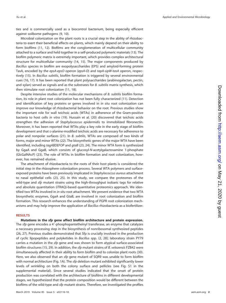
To further investigate whether SQR9 and the ggaA mutant differ in root colonization,we monitored the populations of wild-type and mutant strains colonized on thecucumber roots. After 2 days of incubation in a hydroponic system, green fluorescentprotein (GFP)-labeled B. velezensis mutant strains (SQR9-gfp, ΔggaA-gfp, and C-ΔggaA-gfp) were investigated using confocal laser scanning microscopy (CLSM). The formationof biofilms consisting of wild-type GFP-labeled cells on the root surface could be easilyobserved. Comparatively, only a few small regions of the roots were colonized by ggaAmutant cells, and complementation of the ggaA gene in the ΔggaA mutant restoredalmost all root colonization ability (Fig. 4A). This was further confirmed by quantitativemeasurement of the bacterial population that colonized the plant roots. The resultsshowed that approximately 105 CFU g�1 root of SQR9 cells were detected, but only 103
CFU g�1 root of ggaA mutant cells were colonized on the root. Similarly, there were nosignificant differences in the populations of wild-type SQR9 and the C-ΔggaA-gfpcomplemented strain that colonized on the roots (Fig. 4B).
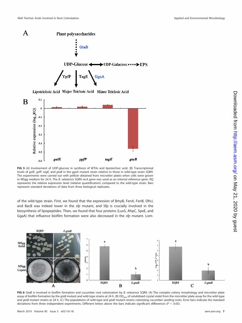
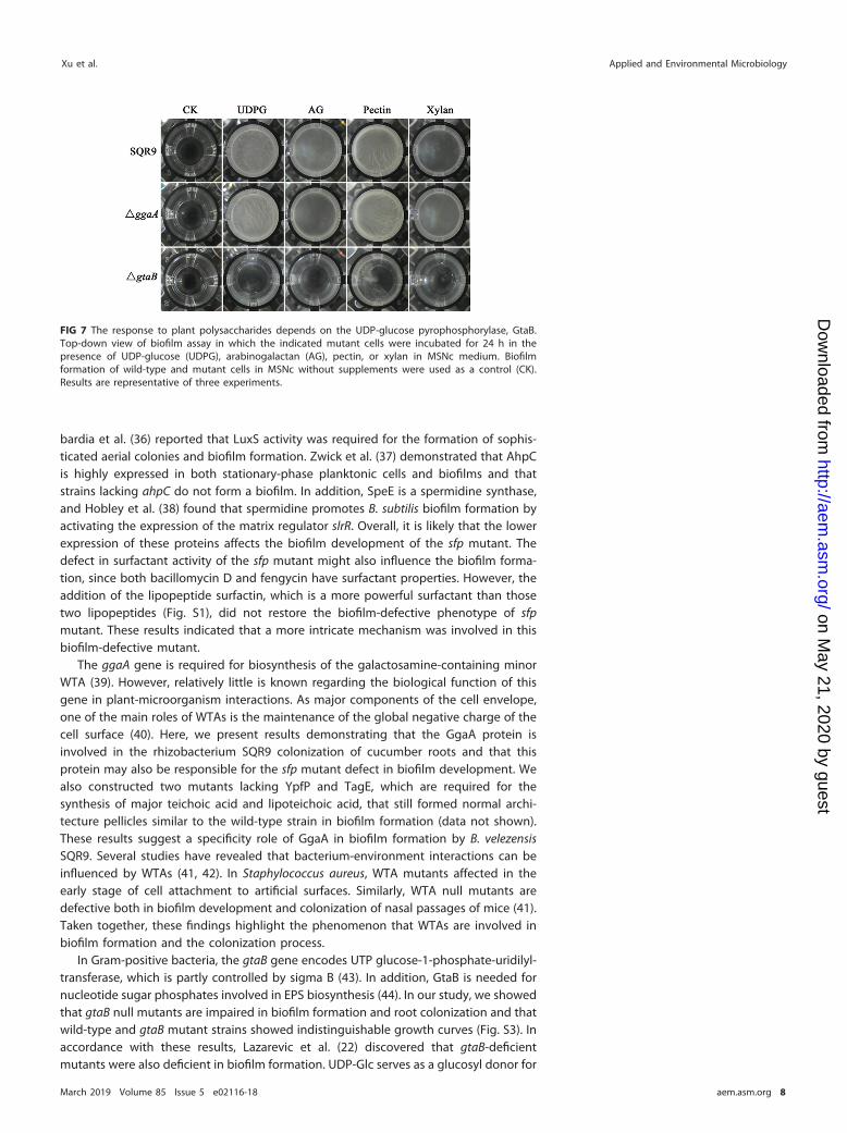
SQR9 lacking gtaB has a significant defect in biofilm formation. According to theaccepted knowledge of WTA synthesis in B. subtilis (11, 22), several genes (galE, ypfP,tagE, and gtaB) are involved in the synthesis of WTAs and lipoteichoic acid (Fig. 5A), andUDP-glucose (UDP-Glc) is a key precursor for WTA polymers. Reverse transcription-quantitative PCR (RT-qPCR) results showed that the transcripts of gtaB were signifi-cantly decreased (4-fold) in the ggaA mutant (Fig. 5B). GtaB is an �-glucose-1-phosphate uridylyltransferase that catalyzes the formation of UDP-Glc and is involvedin the glucosylation of the major and minor WTAs (22, 31). We hypothesized that gtaBmay be involved in biofilm formation in SQR9. We constructed ΔgtaB and ΔgtaB-gfpmutant strains and evaluated their biofilm formation and root colonization abilitiescompared to those of the wild-type strain SQR9. On MSgg solid medium (see Materialsand Methods for ingredients), gtaB mutant colonies were smooth and lacked aerialstructures. Moreover, in MSgg liquid medium, the gtaB mutant formed very fragile
FIG 2 The sfp mutant and wild-type strains showed different proteomic profiles by iTRAQ analysis. (A) Distribution in various functional categories of proteinsaltered in the sfp mutant strain. (B) Distribution of the rhizosphere proteins in the presence of various treatments. The abscissa shows the fold change in theprotein ratio of two treatments (SQR9/sfp mutant), and the ordinate represents various indicated proteins.
Xu et al. Applied and Environmental Microbiology
March 2019 Volume 85 Issue 5 e02116-18 aem.asm.org 4
on May 21, 2020 by guest
http://aem.asm
.org/D
ownloaded from

pellicles that usually split and sank to the bottom of the culture vessel (Fig. 6A).Quantitative analysis of the biofilm biomass showed that the gtaB mutant exhibitedsignificantly lower levels of biofilm biomass (Fig. 6B). In addition, the gtaB mutantpopulation was significantly decreased compared to that of the wild-type strain(Fig. 6C). Overall, these results suggested that the gtaB gene is involved in biofilmformation and root colonization in SQR9.
Response to plant polysaccharides depends on the UDP-glucose pyrophospho-rylase GtaB. Beauregard et al. reported that plant polysaccharides (arabinogalactan,pectin, and xylan) can be used as a carbohydrate source for building the biofilm matrixin Bacillus subtilis and that the production of the EPS requires the synthesis of UDP-galactose (11, 22). We therefore anticipated that a mutation in gtaB would block thesuccessive catalytic steps in UDP-Glc synthesis, which consequently may have beenresponsible for the defect in biofilm development. To investigate this possibility, we
FIG 3 Impairment of the ggaA gene postponed the formation of cellular biofilms and decreased the rootcolonization capability of the B. velezensis SQR9 strain. (A) Microtiter plate assay of biofilm formation by thewild-type and mutant strains. (B) OD600 of solubilized crystal violet from the microtiter plate assay over timefor the wild-type and mutant strains. Error bars indicate the standard deviations based on three differentreplicated experimental values. Different letters above the bars indicate significant differences (P � 0.05),and the subscript numbers differentiate the different time points. Mutant designations Δ2739, Δ9906, andΔ3020 represent mutants with deletions of the gene with the last four numbers of the GI accession numberof each gene in the SQR9 genome in the NCBI database (accession no. CP006890), i.e., V529_33780(https://www.ncbi.nlm.nih.gov/protein/631802739), V529_05450 (https://www.ncbi.nlm.nih.gov/protein/631799906), and V529_36590 (https://www.ncbi.nlm.nih.gov/protein/631803020), respectively.
Wall Teichoic Acids Involved in Root Colonization Applied and Environmental Microbiology
March 2019 Volume 85 Issue 5 e02116-18 aem.asm.org 5
on May 21, 2020 by guest
http://aem.asm
.org/D
ownloaded from
examined the capacity of plant polysaccharides to induce B. velezensis SQR9 and themutant strains to form biofilms in MSNc medium (see Materials and Methods foringredients). First, we tested whether the transcripts of ggaA or gtaB could be inducedby plant polysaccharides. Our results showed that the expression of gtaB can beinduced in the wild-type strain and that xylan had the strongest effect (5-fold) (Fig. S4).Next, the results from our biofilm assay showed that the wild-type SQR9 and ΔggaAmutant strains were able to form biofilm in the presence of plant polysaccharides.These results suggest that plant polysaccharides can serve as an environmental cue forbiofilm formation, as has been reported in B. subtilis (Fig. 7). However, the ΔgtaB mutantformed much weaker pellicles under the same conditions. Additionally, we comparedthe EPS production of the wild-type and mutant strains. Our results showed that boththe ggaA and gtaB mutant strains were deficient in EPS production (Fig. S5). Finally, wetested the attachment abilities of ggaA mutant, gtaB mutant, and wild-type strains onthe cucumber root surfaces. The results showed that wild-type strain SQR9 exhibitedsignificantly higher cell attachment than did the ggaA and gtaB mutant cells (Fig. S6).These observations suggest that plant polysaccharides can be incorporated into theEPS production by gtaB and that EPS is an important biofilm matrix for root attachment.
DISCUSSION
Gram-positive Bacillus species are attractive PGPR that are widely used as a biofer-tilizers (32, 33). The colonization of PGPR strains in the rhizosphere is a prerequisite forthem to execute their specific functions (34). In this study, we have shown that the ggaAand gtaB genes, which are required for the biosynthesis of WTAs in B. velezensis SQR9,are essential for biofilm formation and root colonization. By using a proteomic ap-proach to compare the proteomes of wild-type and sfp mutant strains, we noted thatthe GgaA protein is much more abundant in wild-type cells than in sfp mutant cells.Moreover, we showed that plant polysaccharides are used as a source for the synthesisof biofilm matrix exopolysaccharides by GtaB.
The sfp gene encodes a phosphopantetheinyl transferase that is required for Bacilluscells to produce lipopeptide antibiotics (35). In B. velezensis FZB42, an sfp gene mutantwas found to be impaired in biofilm formation and root colonization (28). Here, wefocused on the proteome of the biofilm produced by the sfp mutant compared to that
FIG 4 The ggaA mutant strain is deficient in cucumber root colonization. (A) CLSM micrographs of cucumber roots colonizationby GFP-tagged wild-type and ggaA mutant strains. Ck is a control which was not inoculated with GFP-tagged SQR9. (B) Thepopulations of wild-type and ggaA mutant strains colonizing cucumber seeding roots. Error bars indicate the standarddeviations from the results from three independent experiments. Different letters above the bars indicate significantdifferences (P � 0.01).
Xu et al. Applied and Environmental Microbiology
March 2019 Volume 85 Issue 5 e02116-18 aem.asm.org 6
on May 21, 2020 by guest
http://aem.asm
.org/D
ownloaded from
of the wild-type strain. First, we found that the expression of BmyB, FenA, FenB, DfnJ,and BacB was indeed lower in the sfp mutant, and Sfp is crucially involved in thebiosynthesis of lipopeptides. Then, we found that four proteins (LuxS, AhpC, SpeE, andGgaA) that influence biofilm formation were also decreased in the sfp mutant. Lom-
FIG 5 (A) Involvement of UDP-glucose in synthesis of WTAs and lipoteichoic acid. (B) Transcriptionallevels of galE, ypfP, tagE, and gtaB in the ggaA mutant strain relative to those in wild-type strain SQR9.The experiments were carried out with pellicle obtained from microtiter plates when cells were grownin MSgg medium for 24 h. The B. velezensis SQR9 recA gene was used as an internal reference gene. RQrepresents the relative expression level (relative quantification) compared to the wild-type strain. Barsrepresent standard deviations of data from three biological replicates.
FIG 6 GtaB is involved in biofilm formation and cucumber root colonization by B. velezensis SQR9. (A) The complex colony morphology and microtiter plateassay of biofilm formation by the gtaB mutant and wild-type strains at 24 h. (B) OD600 of solubilized crystal violet from the microtiter plate assay for the wild-typeand gtaB mutant strains at 24 h. (C) The populations of wild-type and gtaB mutant strains colonizing cucumber seeding roots. Error bars indicate the standarddeviations from three independent experiments. Different letters above the bars indicate significant differences (P � 0.05).
Wall Teichoic Acids Involved in Root Colonization Applied and Environmental Microbiology
March 2019 Volume 85 Issue 5 e02116-18 aem.asm.org 7
on May 21, 2020 by guest
http://aem.asm
.org/D
ownloaded from
bardia et al. (36) reported that LuxS activity was required for the formation of sophis-ticated aerial colonies and biofilm formation. Zwick et al. (37) demonstrated that AhpCis highly expressed in both stationary-phase planktonic cells and biofilms and thatstrains lacking ahpC do not form a biofilm. In addition, SpeE is a spermidine synthase,and Hobley et al. (38) found that spermidine promotes B. subtilis biofilm formation byactivating the expression of the matrix regulator slrR. Overall, it is likely that the lowerexpression of these proteins affects the biofilm development of the sfp mutant. Thedefect in surfactant activity of the sfp mutant might also influence the biofilm forma-tion, since both bacillomycin D and fengycin have surfactant properties. However, theaddition of the lipopeptide surfactin, which is a more powerful surfactant than thosetwo lipopeptides (Fig. S1), did not restore the biofilm-defective phenotype of sfpmutant. These results indicated that a more intricate mechanism was involved in thisbiofilm-defective mutant.
The ggaA gene is required for biosynthesis of the galactosamine-containing minorWTA (39). However, relatively little is known regarding the biological function of thisgene in plant-microorganism interactions. As major components of the cell envelope,one of the main roles of WTAs is the maintenance of the global negative charge of thecell surface (40). Here, we present results demonstrating that the GgaA protein isinvolved in the rhizobacterium SQR9 colonization of cucumber roots and that thisprotein may also be responsible for the sfp mutant defect in biofilm development. Wealso constructed two mutants lacking YpfP and TagE, which are required for thesynthesis of major teichoic acid and lipoteichoic acid, that still formed normal archi-tecture pellicles similar to the wild-type strain in biofilm formation (data not shown).These results suggest a specificity role of GgaA in biofilm formation by B. velezensisSQR9. Several studies have revealed that bacterium-environment interactions can beinfluenced by WTAs (41, 42). In Staphylococcus aureus, WTA mutants affected in theearly stage of cell attachment to artificial surfaces. Similarly, WTA null mutants aredefective both in biofilm development and colonization of nasal passages of mice (41).Taken together, these findings highlight the phenomenon that WTAs are involved inbiofilm formation and the colonization process.
In Gram-positive bacteria, the gtaB gene encodes UTP glucose-1-phosphate-uridilyl-transferase, which is partly controlled by sigma B (43). In addition, GtaB is needed fornucleotide sugar phosphates involved in EPS biosynthesis (44). In our study, we showedthat gtaB null mutants are impaired in biofilm formation and root colonization and thatwild-type and gtaB mutant strains showed indistinguishable growth curves (Fig. S3). Inaccordance with these results, Lazarevic et al. (22) discovered that gtaB-deficientmutants were also deficient in biofilm formation. UDP-Glc serves as a glucosyl donor for
FIG 7 The response to plant polysaccharides depends on the UDP-glucose pyrophosphorylase, GtaB.Top-down view of biofilm assay in which the indicated mutant cells were incubated for 24 h in thepresence of UDP-glucose (UDPG), arabinogalactan (AG), pectin, or xylan in MSNc medium. Biofilmformation of wild-type and mutant cells in MSNc without supplements were used as a control (CK).Results are representative of three experiments.
Xu et al. Applied and Environmental Microbiology
March 2019 Volume 85 Issue 5 e02116-18 aem.asm.org 8
on May 21, 2020 by guest
http://aem.asm
.org/D
ownloaded from

the synthesis of all phosphate-containing anionic envelope polymers, including lipo-teichoic acid, major teichoic acid, and minor teichoic acid (22). The absence of UDP-Glcin the gtaB mutant might explain the deficiency in biofilm formation, since UDP-Glc isan important precursor for the extracellular matrix required for biofilm development. Inmany environmental bacteria, UDP-Glc is necessary for the synthesis of exopolysaccha-rides (EPS) (44, 45). For example, a Bradyrhizobium japonicum mutant that lacks UDP-Glc-4=-epimerase activity produces less extracellular EPS than does the wild-type strain(46). Additionally, in another report, UDP-Glc and UDP-galactose were shown to beinvolved as signaling molecules or as precursors for the synthesis of EPS (44). In ourstudy, EPS production was indeed lower in gtaB-deficient mutants than in the wild-typestrain. In the rhizosphere, the production of extracellular matrix, including both EPS andprotein components, is essential for root colonization, as Bacillus strains carryingknockout mutations in either component failed to colonize on the plant root.
Rhizobacteria in the rhizosphere sense plant extracts and root exudate componentsreleased by plants (47, 48). For instance, organic acids, such as malic acid and citric acid,in root exudates recruit Bacillus spp. in the rhizosphere (49). After that, other moleculesthat PGPR encounter on the plant root include the polysaccharides from the plant cellwall. Beauregard et al. reported that plant polysaccharides (arabinogalactan, pectin,and xylan) act both as an environmental cue and as a substrate for the synthesis of theB. subtilis 3610 biofilm matrix and that these plant polysaccharides promote biofilmdevelopment in various plant growth-promoting Bacillus strains, including B. subtilisGB03 and B. amyloliquefaciens FZB42 (11). In this study, we provide evidence that lossof the gtaB gene affects the ability of SQR9 to sense plant polysaccharides and form abiofilm, which can partially explain the defect of the gtaB strain in cucumber rootcolonization.
Over the past few decades, research on the process of Bacillus biofilm formation hasbeen well studied under laboratory conditions (13, 14, 18). However, relatively little isknown about how this bacterium colonizes plant roots. We showed that WTAs may playimportant roles in the root colonization process. Although there is evidence that twoWTA biosynthetic enzymes, GgaA and GtaB, positively influence biofilm formation androot colonization, determining exactly how WTAs affect the root colonization processwill be an important challenge for the future.
MATERIALS AND METHODSStrains and culture conditions. The strains and plasmids used in this study are listed in Table 1. B.
velezensis strain SQR9 (CGMCC accession no. 5808; China General Microbiology Culture Collection Center)was used throughout this study. Escherichia coli DH5� was used as the host strain for all plasmids. B.
TABLE 1 Microorganisms and plasmids used in this study
Strain or plasmid Description or genotypea Source or reference
StrainsE. coli DH5� �80dlacZΔDM15 recA1 endA1 gyrA96 thi-1 hsdR17
(rK� mK
�) supE44 relA1 deoR Δ(lacZYA-argF)U169 phoAInvitrogen (Shanghai, China)
E. coli BL21Bacillus velezensis SQR9 Wild type CGMCC no. 5808B. velezensis SQR9-gfp GFP-labeled SQR9 (Kanr)B. velezensis SQR9M6 SQR9 Δsfp::cm 60
SQR9 ΔggaA This studyGFP-labeled SQR9 ΔggaA (Cmr) This studySQR9 Δ3020 This studyGFP-labeled SQR9 Δ3020 (Cmr) This studySQR9 ΔgtaB This studyGFP-labeled SQR9 ΔgtaB (Cmr) This study
PlasmidspTPC pMD19-T harboring the Pbc-pheS*-cat(PC) cassette 54pNW33n Cmr; MCS BGSCpNW33n-GFP This study
aKanr, kanamycin resistance; Cmr, chloramphenicol resistance; MCS, multiple-cloning site; Δ3020, mutant with deletion of V529_36590(https://www.ncbi.nlm.nih.gov/protein/631803020).
Wall Teichoic Acids Involved in Root Colonization Applied and Environmental Microbiology
March 2019 Volume 85 Issue 5 e02116-18 aem.asm.org 9
on May 21, 2020 by guest
http://aem.asm
.org/D
ownloaded from

velezensis strains were grown in Luria-Bertani medium and, where appropriate, in minimal medium(MSgg; 5 mM potassium phosphate, 100 mM morpholinepropanesulfonic acid [MOPS] [pH 7], 2 mMMgCl2, 700 �M CaCl2, 50 �M MnCl2, 50 �M FeCl3, 1 �M ZnCl2, 2 mM thiamine, 0.5% glycerol, 0.5%glutamate, 50 �g ml�1 tryptophan, 50 �g ml�1 phenylalanine, and 50 �g ml�1 threonine) (15). Fortransformant selection, colonies were selected on MGY-Cl agar plates (minimal medium-glucose-yeastextract [MGY] medium contained glucose [5 g/liter], yeast extract [4 g/liter], NH4NO3 [1 g/liter], NaCl [0.5g/liter], K2HPO4 [1.5 g/liter], KH2PO4 [0.5 g/liter], and MgSO4 [0.2 g/liter], and MGY-Cl medium is anMGY-based medium supplemented with 5 mM p-Cl-Phe [Sigma]). Biofilm assays were performed in48-well plates with 1 ml of MSNc (5 mM potassium phosphate buffer [pH 7], 0.1 M MOPS [pH 7], 2 mMMgCl2, 0.05 mM MnCl2, 1 �M ZnCl2, 2 �M thiamine, 700 �M CaCl2, 0.2% NH4Cl, and 0.5% cellobiose)medium supplemented or not with the purified plant polysaccharides (11). B. velezensis strains wereincubated at 37°C. Antibiotics were added as required at the following concentrations: 20 �g ml�1
zeocin, 5 �g ml�1 chloramphenicol, and 100 �g ml�1 ampicillin for E. coli strains.Protein extraction analysis. Biofilms were incubated in MSgg medium and harvested at the 24-h
time point. Protein extraction was carried out as described by Kierul et al. (50). The cells were removedby centrifugation (11,000 � g, 4°C, 20 min) and filtered through a 0.22-�m-pore-size membrane (Milli-pore). Trichloroacetic acid was added to the filtered exudates to a final concentration of 10%, and thesolution was stored at 4°C overnight. Subsequently, extracellular proteins were collected by centrifuga-tion (15,000 � g, 4°C, 30 min). The protein pellet was washed three times with 1 ml of ice-cold acetoneand centrifuged (15,000 rpm, 4°C, 20 min). Similarly, two washing steps with 96% ice-cold ethanol wereperformed. The extracellular proteins obtained were stored in 96% ethanol at �20°C.
iTRAQ labeling and automated two-dimensional liquid chromatography with tandem massspectrometry protein identification. The protein samples were resuspended in lysis buffer (7 M urea,2 M thiourea, 0.1% 3-[(3-cholamidopropyl)-dimethylammonio]-1-propanesulfonate [CHAPS]), thoroughlysonicated, and then incubated at 37°C for 30 min. After centrifugation at 20,000 � g at 4°C for 30 min, thesupernatant was collected. Protein concentration and quality were determined using a Bradford assay.Sodium dodecyl sulfate-polyacrylamide gel electrophoresis (SDS-PAGE) was performed using the methoddescribed by Laemmli (51), and the SDS-PAGE gel was stained with sliver staining (52).
Protein digestion and labeling were carried out by the method of Qiu et al. (53), and the peptidesamples were labeled using the iTRAQ reagent multiplex kit (Applied Biosystems, Foster City, CA). Theautomated two-dimensional liquid chromatography with tandem mass spectrometry (2D LC-MS/MS)analysis was similar to what has been previously described (53). Peptides were desalted using ZipTip C18
reverse resin (Millipore Corporation, Billerica, MA) and separated via C18 reversed-phase column byhigh-performance liquid chromatography (HPLC; Agilent Technologies, Santa Clara, CA). MS was per-formed using an LTQ Orbitrap mass spectrometer (Thermo Electron Corp.), and data were collected usingthe Xcalibur software (Thermo Electron).
Protein identification and quantification were performed with the ProteinPilot 4.0 software (AppliedBiosystems, USA). The database used for searching was the B. velezensis SQR9 (GenBank accession no.CP006890) entry in the National Center for Biotechnology Information (NCBI) database. Proteins with foldchanges significantly �4.0 or �0.25 were considered differentially expressed (P � 0.05).
B. velezensis SQR9 mutant construction. The marker-free deletion strains of targeted genes wereconstructed using the Pbc-pheS*-cat (PC) cassette and an overlap PCR-based strategy, essentially as hasbeen previously described (54). To delete the targeted genes in B. velezensis SQR9, 1-kb fragmentslocated upstream and downstream of the target gene were amplified. The 1.1-kb chloramphenicolresistance gene (Cmr) was amplified from pNW33N, and the PC cassette was amplified from pTPC as atemplate. These four fragments were fused using overlap PCR and directly transformed into SQR9. Thetransformants were selected on LB plates containing Cm. Cmr colonies were cultivated to an opticaldensity at 600 nm (OD600) of 1.0 without Cm, and a 100-�l aliquot of a 10-fold dilution of the cultures(approximately 105 cells) was plated on MGY-Cl medium (54). Mutants growing on MGY-Cl were furtherconfirmed by PCR and DNA sequencing. The primers used in this experiment are listed in Table 2.
Complementation of disrupted ggaA and gtaB genes. The ggaA and gtaB gene fragments werecomplemented at the amyE locus in each mutant strain. The left flanking (LF) region (�1,000 bp),complement gene (CG) sequences, and right flanking (RF) region (�1,000 bp) were amplified from theSQR9 strain using the primer pairs ggaALF-F/ggaALF-R and gtaBLF-F/gtaBLF-R, ggaACG-F/ggaACG-R andgtaBCG-F/gtaBCG-R, and ggaARF-F/ggaARF-R and gtaBRF-F/gtaBRF-R, respectively. The Cm region(�1,000 bp) was amplified with the primer pairs ggaACm-F/ggaACm-R and gtaBCm-F/gtaBCm-R usingpNW33N as the template. These four fragments were fused and directly transformed into the ΔggaA andΔgtaB mutant strains. The transformants were selected on LB plates containing Cm.
Biofilm assays of B. velezensis SQR9 and mutants. The biofilm assay was carried out in 48-wellmicrotiter plates in MSgg medium, as described by Hamon and Lazazzera (55). Biofilm formation wasquantified by staining with crystal violet (CV). Cells of a biofilm were stained with CV, and then unboundCV was removed with distilled water. The remaining CV was solubilized with 1 ml of 80% ethanol–20%acetone. The absorbance of CV at 570 nm was measured using the SpectraMax i3x analysis system(Molecular Devices Corporation, CA).
Root colonization assay. The bacterial suspensions of SQR9 and mutants were inoculated intosterile cucumber seedlings in 1/4 Murashige and Skoog (MS) culture medium. After 4 days, the cellscolonized on the cucumber roots were collected and quantified using the method described by Qiuet al. (53).
Congo red binding assay. Strains were incubated at 37°C overnight in Luria-Bertani medium, and1 ml of cells was collected by centrifugation (10,000 � g, 4°C, 2 min). The supernatant was removed, the
Xu et al. Applied and Environmental Microbiology
March 2019 Volume 85 Issue 5 e02116-18 aem.asm.org 10
on May 21, 2020 by guest
http://aem.asm
.org/D
ownloaded from
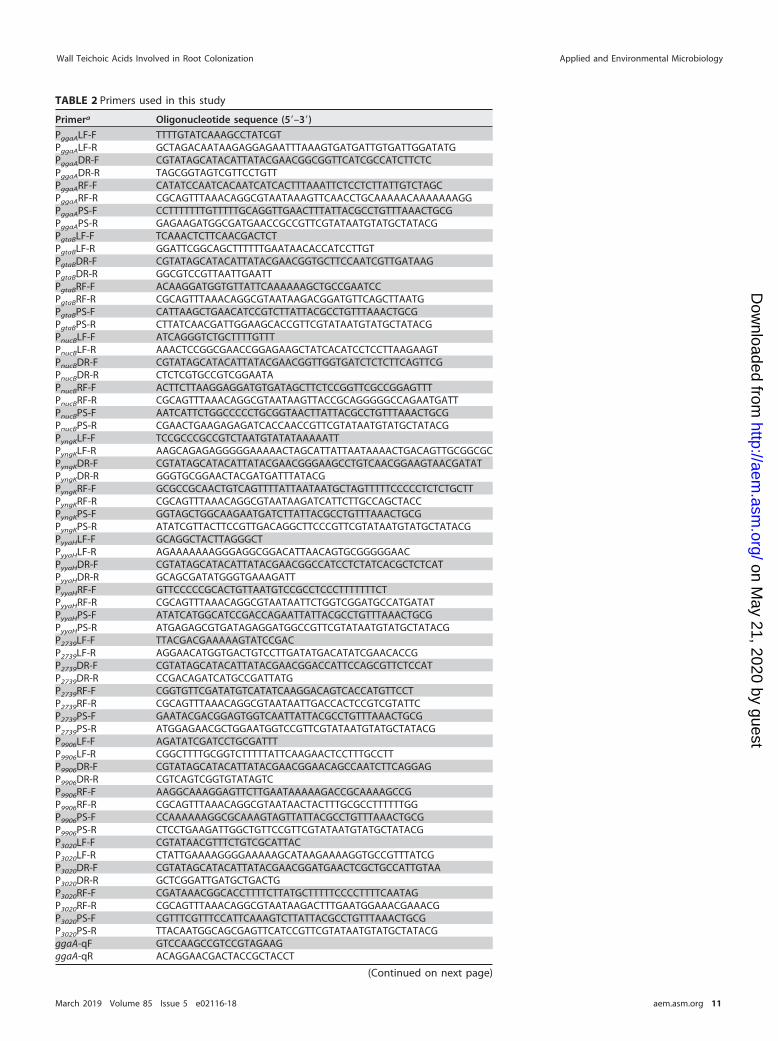
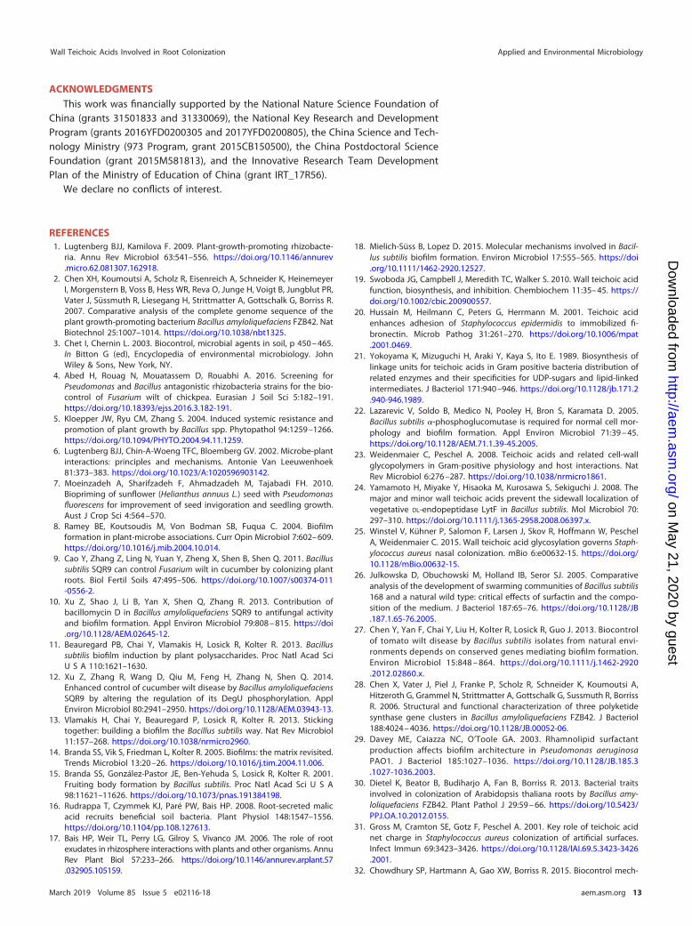
TABLE 2 Primers used in this study
Primera Oligonucleotide sequence (5=–3=)PggaALF-F TTTTGTATCAAAGCCTATCGTPggaALF-R GCTAGACAATAAGAGGAGAATTTAAAGTGATGATTGTGATTGGATATGPggaADR-F CGTATAGCATACATTATACGAACGGCGGTTCATCGCCATCTTCTCPggaADR-R TAGCGGTAGTCGTTCCTGTTPggaARF-F CATATCCAATCACAATCATCACTTTAAATTCTCCTCTTATTGTCTAGCPggaARF-R CGCAGTTTAAACAGGCGTAATAAAGTTCAACCTGCAAAAACAAAAAAAGGPggaAPS-F CCTTTTTTTGTTTTTGCAGGTTGAACTTTATTACGCCTGTTTAAACTGCGPggaAPS-R GAGAAGATGGCGATGAACCGCCGTTCGTATAATGTATGCTATACGPgtaBLF-F TCAAACTCTTCAACGACTCTPgtaBLF-R GGATTCGGCAGCTTTTTTGAATAACACCATCCTTGTPgtaBDR-F CGTATAGCATACATTATACGAACGGTGCTTCCAATCGTTGATAAGPgtaBDR-R GGCGTCCGTTAATTGAATTPgtaBRF-F ACAAGGATGGTGTTATTCAAAAAAGCTGCCGAATCCPgtaBRF-R CGCAGTTTAAACAGGCGTAATAAGACGGATGTTCAGCTTAATGPgtaBPS-F CATTAAGCTGAACATCCGTCTTATTACGCCTGTTTAAACTGCGPgtaBPS-R CTTATCAACGATTGGAAGCACCGTTCGTATAATGTATGCTATACGPnucBLF-F ATCAGGGTCTGCTTTTGTTTPnucBLF-R AAACTCCGGCGAACCGGAGAAGCTATCACATCCTCCTTAAGAAGTPnucBDR-F CGTATAGCATACATTATACGAACGGTTGGTGATCTCTCTTCAGTTCGPnucBDR-R CTCTCGTGCCGTCGGAATAPnucBRF-F ACTTCTTAAGGAGGATGTGATAGCTTCTCCGGTTCGCCGGAGTTTPnucBRF-R CGCAGTTTAAACAGGCGTAATAAGTTACCGCAGGGGGCCAGAATGATTPnucBPS-F AATCATTCTGGCCCCCTGCGGTAACTTATTACGCCTGTTTAAACTGCGPnucBPS-R CGAACTGAAGAGAGATCACCAACCGTTCGTATAATGTATGCTATACGPyngKLF-F TCCGCCCGCCGTCTAATGTATATAAAAATTPyngKLF-R AAGCAGAGAGGGGGAAAAACTAGCATTATTAATAAAACTGACAGTTGCGGCGCPyngKDR-F CGTATAGCATACATTATACGAACGGGAAGCCTGTCAACGGAAGTAACGATATPyngKDR-R GGGTGCGGAACTACGATGATTTATACGPyngKRF-F GCGCCGCAACTGTCAGTTTTATTAATAATGCTAGTTTTTCCCCCTCTCTGCTTPyngKRF-R CGCAGTTTAAACAGGCGTAATAAGATCATTCTTGCCAGCTACCPyngKPS-F GGTAGCTGGCAAGAATGATCTTATTACGCCTGTTTAAACTGCGPyngKPS-R ATATCGTTACTTCCGTTGACAGGCTTCCCGTTCGTATAATGTATGCTATACGPyyaHLF-F GCAGGCTACTTAGGGCTPyyaHLF-R AGAAAAAAAGGGAGGCGGACATTAACAGTGCGGGGGAACPyyaHDR-F CGTATAGCATACATTATACGAACGGCCATCCTCTATCACGCTCTCATPyyaHDR-R GCAGCGATATGGGTGAAAGATTPyyaHRF-F GTTCCCCCGCACTGTTAATGTCCGCCTCCCTTTTTTTCTPyyaHRF-R CGCAGTTTAAACAGGCGTAATAATTCTGGTCGGATGCCATGATATPyyaHPS-F ATATCATGGCATCCGACCAGAATTATTACGCCTGTTTAAACTGCGPyyaHPS-R ATGAGAGCGTGATAGAGGATGGCCGTTCGTATAATGTATGCTATACGP2739LF-F TTACGACGAAAAAGTATCCGACP2739LF-R AGGAACATGGTGACTGTCCTTGATATGACATATCGAACACCGP2739DR-F CGTATAGCATACATTATACGAACGGACCATTCCAGCGTTCTCCATP2739DR-R CCGACAGATCATGCCGATTATGP2739RF-F CGGTGTTCGATATGTCATATCAAGGACAGTCACCATGTTCCTP2739RF-R CGCAGTTTAAACAGGCGTAATAATTGACCACTCCGTCGTATTCP2739PS-F GAATACGACGGAGTGGTCAATTATTACGCCTGTTTAAACTGCGP2739PS-R ATGGAGAACGCTGGAATGGTCCGTTCGTATAATGTATGCTATACGP9906LF-F AGATATCGATCCTGCGATTTP9906LF-R CGGCTTTTGCGGTCTTTTTATTCAAGAACTCCTTTGCCTTP9906DR-F CGTATAGCATACATTATACGAACGGAACAGCCAATCTTCAGGAGP9906DR-R CGTCAGTCGGTGTATAGTCP9906RF-F AAGGCAAAGGAGTTCTTGAATAAAAAGACCGCAAAAGCCGP9906RF-R CGCAGTTTAAACAGGCGTAATAACTACTTTGCGCCTTTTTTGGP9906PS-F CCAAAAAAGGCGCAAAGTAGTTATTACGCCTGTTTAAACTGCGP9906PS-R CTCCTGAAGATTGGCTGTTCCGTTCGTATAATGTATGCTATACGP3020LF-F CGTATAACGTTTCTGTCGCATTACP3020LF-R CTATTGAAAAGGGGAAAAAGCATAAGAAAAGGTGCCGTTTATCGP3020DR-F CGTATAGCATACATTATACGAACGGATGAACTCGCTGCCATTGTAAP3020DR-R GCTCGGATTGATGCTGACTGP3020RF-F CGATAAACGGCACCTTTTCTTATGCTTTTTCCCCTTTTCAATAGP3020RF-R CGCAGTTTAAACAGGCGTAATAAGACTTTGAATGGAAACGAAACGP3020PS-F CGTTTCGTTTCCATTCAAAGTCTTATTACGCCTGTTTAAACTGCGP3020PS-R TTACAATGGCAGCGAGTTCATCCGTTCGTATAATGTATGCTATACGggaA-qF GTCCAAGCCGTCCGTAGAAGggaA-qR ACAGGAACGACTACCGCTACCT
(Continued on next page)
Wall Teichoic Acids Involved in Root Colonization Applied and Environmental Microbiology
March 2019 Volume 85 Issue 5 e02116-18 aem.asm.org 11
on May 21, 2020 by guest
http://aem.asm
.org/D
ownloaded from

pellet was resuspended in 1 ml of tryptone broth (10 g/liter tryptone), and 500 �l of cells was transferredto a new tube. A total of 5 �l of Congo red (CR) stock solution (4 mg/ml CR, filtered) was added to 500 �lcells and 500 �l T-broth as a blank. Subsequently, the tubes were mixed well and shaken vigorously at30°C for 2 h. The OD600 value was measured using the remaining cells (10� dilution, 100 �l cells plus900 �l medium). After 2 h of incubation, samples were centrifuged at 15,000 rpm for 5 min, and theOD490 value of the supernatant was measured. The amount of CR bound to the cells was calculated asfollows: CR (�g/OD600) [(OD490-blank – OD490-sample) � 44.676/OD600] (56).
Bacterial attachment assay. The B. velezensis attachment assay was carried out using the methodsfrom Smit et al. (57) and Dardanelli et al. (58). Strains were incubated at an OD600 of 1.0, pelleted bycentrifugation (6,000 � g, 4°C, 10 min), and then washed three times with phosphate-buffered saline (PBS[pH 7.4]). The pellet was suspended in the same solution to a final OD600 of 0.1. Three sterile cucumberroots were immersed in 50 ml of bacterial suspension for 2 h at room temperature under gentle agitation.Then, the roots were washed 10 times with sterile distilled water and ground up in a sterilized mortar.The number of bound bacteria was quantified by dilution plate counting.
Microscopy. To determine if B. velezensis colonized the cucumber root surface, SQR9-gfp-, ΔggaA-gfpmutant-, and ΔgtaB-gfp mutant-colonized roots were investigated at 2 days after inoculation in hydro-ponics. The roots were washed with distilled water and viewed by a confocal laser scanning microscope(CLSM; model TCS SP2; Leica, Heidelberg, Germany). Images were taken using Leica confocal software,version 2.61 (12).
Quantification of gene transcription by real-time PCR. Total RNA was obtained from biofilmsformed by B. velezensis using the E.Z.N.A. bacterial RNA kit (Toyobo, Japan), according to the instructions.The RNA was detected on a 1% agarose gel, and a NanoDrop ND-2000 spectrophotometer (ThermoFisher Scientific, Wilmington, DE) was used to check the quality and concentration. RNA was reversetranscribed using the PrimeScript RT reagent kit with a genomic DNA (gDNA) eraser (Toyobo). Transcriptlevels of ggaA and gtaB were measured by reverse transcription-quantitative PCR (RT-qPCR) using a SYBRPremix Ex Taq (perfect real time) kit (TaKaRa, Dalian, China). The recA gene was used as an internalcontrol. Reactions were carried out on an ABI 7500 system (Applied Biosystems, USA) under the followingconditions: cDNA was denatured for 10 s at 95°C, followed by 40 cycles consisting of 5 s at 95°C and 34 sat 60°C. The 2�ΔΔCT method was used to analyze the RT-qPCR data (59).
Statistical analysis. Differences among the treatments were calculated and statistically analyzedwith a one-way analysis of variance (ANOVA). Duncan’s multiple-range test was used when one-wayANOVA indicated a significant difference (P � 0.05). All statistical analyses were performed with IBM SPSSStatistics 20. To identify and classify differentially expressed proteins, the Gene Ontology (GO) (www.geneontology.org) and Kyoto Encyclopedia of Genes and Genomes (KEGG) (http://www.genome.jp/kegg/) databases were used.
SUPPLEMENTAL MATERIALSupplemental material for this article may be found at https://doi.org/10.1128/AEM
.02116-18.SUPPLEMENTAL FILE 1, PDF file, 0.8 MB.
TABLE 2 (Continued)
Primera Oligonucleotide sequence (5=–3=)gtaB-qF CGTTGATAAGCCGACAATTCAGgtaB-qR CATTGTCGAAATGGTCTTCTATTGggaALF-F GAGAAGGCGTCGTAAACggaALF-R TAACGGCAGTAAAGAGGTggaACm-F ATTCAAAACCTCTTTACTGCCGTTAGCATAAAGTGTAAAGCCTGGGGggaACm-R CCTTTCGCTGAAAGTAACAAAATGAAATGTGGAATTGGGAACGGAAAggaACG-F TCATTTTGTTACTTTCAGCGggaACG-R ATACAAACCATTCTGCACATAATCAGAACACAGAAAGCTGATTCTggaARF-F TGATTATGTGCAGAATGGTggaARF-R TCTCGATAATATGGTAGGCgtaBLF-F GAGAAGGCGTCGTAAACgtaBLF-R TAACGGCAGTAAAGAGGTgtaBCm-F ATTCAAAACCTCTTTACTGCCGTTAGCATAAAGTGTAAAGCCTGGGGgtaBCm-R AAAACCAGATATCGTTTTGGTCCACAATGTGGAATTGGGAACGGAAAgtaBCG-F GTGGACCAAAACGATATCTgtaBCG-R ATACAAACCATTCTGCACATAATCACTATTCGTCAGCTTCTTCTTgtaBRF-F TGATTATGTGCAGAATGGTgtaBRF-R TCTCGATAATATGGTAGGCaDesignations 2739, 9906, and 3020 refer to the genes with the last four numbers of the GI accessionnumber in the SQR9 genome in the NCBI database (accession no. CP006890), i.e., V529_33780 (https://www.ncbi.nlm.nih.gov/protein/631802739), V529_05450 (https://www.ncbi.nlm.nih.gov/protein/631799906), andV529_36590 (https://www.ncbi.nlm.nih.gov/protein/631803020), respectively.
Xu et al. Applied and Environmental Microbiology
March 2019 Volume 85 Issue 5 e02116-18 aem.asm.org 12
on May 21, 2020 by guest
http://aem.asm
.org/D
ownloaded from
ACKNOWLEDGMENTSThis work was financially supported by the National Nature Science Foundation of
China (grants 31501833 and 31330069), the National Key Research and DevelopmentProgram (grants 2016YFD0200305 and 2017YFD0200805), the China Science and Tech-nology Ministry (973 Program, grant 2015CB150500), the China Postdoctoral ScienceFoundation (grant 2015M581813), and the Innovative Research Team DevelopmentPlan of the Ministry of Education of China (grant IRT_17R56).
We declare no conflicts of interest.
REFERENCES1. Lugtenberg BJJ, Kamilova F. 2009. Plant-growth-promoting rhizobacte-
ria. Annu Rev Microbiol 63:541–556. https://doi.org/10.1146/annurev.micro.62.081307.162918.
2. Chen XH, Koumoutsi A, Scholz R, Eisenreich A, Schneider K, HeinemeyerI, Morgenstern B, Voss B, Hess WR, Reva O, Junge H, Voigt B, Jungblut PR,Vater J, Süssmuth R, Liesegang H, Strittmatter A, Gottschalk G, Borriss R.2007. Comparative analysis of the complete genome sequence of theplant growth-promoting bacterium Bacillus amyloliquefaciens FZB42. NatBiotechnol 25:1007–1014. https://doi.org/10.1038/nbt1325.
3. Chet I, Chernin L. 2003. Biocontrol, microbial agents in soil, p 450 – 465.In Bitton G (ed), Encyclopedia of environmental microbiology. JohnWiley & Sons, New York, NY.
4. Abed H, Rouag N, Mouatassem D, Rouabhi A. 2016. Screening forPseudomonas and Bacillus antagonistic rhizobacteria strains for the bio-control of Fusarium wilt of chickpea. Eurasian J Soil Sci 5:182–191.https://doi.org/10.18393/ejss.2016.3.182-191.
5. Kloepper JW, Ryu CM, Zhang S. 2004. Induced systemic resistance andpromotion of plant growth by Bacillus spp. Phytopathol 94:1259 –1266.https://doi.org/10.1094/PHYTO.2004.94.11.1259.
6. Lugtenberg BJJ, Chin-A-Woeng TFC, Bloemberg GV. 2002. Microbe-plantinteractions: principles and mechanisms. Antonie Van Leeuwenhoek81:373–383. https://doi.org/10.1023/A:1020596903142.
7. Moeinzadeh A, Sharifzadeh F, Ahmadzadeh M, Tajabadi FH. 2010.Biopriming of sunflower (Helianthus annuus L.) seed with Pseudomonasfluorescens for improvement of seed invigoration and seedling growth.Aust J Crop Sci 4:564 –570.
8. Ramey BE, Koutsoudis M, Von Bodman SB, Fuqua C. 2004. Biofilmformation in plant-microbe associations. Curr Opin Microbiol 7:602– 609.https://doi.org/10.1016/j.mib.2004.10.014.
9. Cao Y, Zhang Z, Ling N, Yuan Y, Zheng X, Shen B, Shen Q. 2011. Bacillussubtilis SQR9 can control Fusarium wilt in cucumber by colonizing plantroots. Biol Fertil Soils 47:495–506. https://doi.org/10.1007/s00374-011-0556-2.
10. Xu Z, Shao J, Li B, Yan X, Shen Q, Zhang R. 2013. Contribution ofbacillomycin D in Bacillus amyloliquefaciens SQR9 to antifungal activityand biofilm formation. Appl Environ Microbiol 79:808 – 815. https://doi.org/10.1128/AEM.02645-12.
11. Beauregard PB, Chai Y, Vlamakis H, Losick R, Kolter R. 2013. Bacillussubtilis biofilm induction by plant polysaccharides. Proc Natl Acad SciU S A 110:1621–1630.
12. Xu Z, Zhang R, Wang D, Qiu M, Feng H, Zhang N, Shen Q. 2014.Enhanced control of cucumber wilt disease by Bacillus amyloliquefaciensSQR9 by altering the regulation of its DegU phosphorylation. ApplEnviron Microbiol 80:2941–2950. https://doi.org/10.1128/AEM.03943-13.
13. Vlamakis H, Chai Y, Beauregard P, Losick R, Kolter R. 2013. Stickingtogether: building a biofilm the Bacillus subtilis way. Nat Rev Microbiol11:157–268. https://doi.org/10.1038/nrmicro2960.
14. Branda SS, Vik S, Friedman L, Kolter R. 2005. Biofilms: the matrix revisited.Trends Microbiol 13:20 –26. https://doi.org/10.1016/j.tim.2004.11.006.
15. Branda SS, González-Pastor JE, Ben-Yehuda S, Losick R, Kolter R. 2001.Fruiting body formation by Bacillus subtilis. Proc Natl Acad Sci U S A98:11621–11626. https://doi.org/10.1073/pnas.191384198.
16. Rudrappa T, Czymmek KJ, Paré PW, Bais HP. 2008. Root-secreted malicacid recruits beneficial soil bacteria. Plant Physiol 148:1547–1556.https://doi.org/10.1104/pp.108.127613.
17. Bais HP, Weir TL, Perry LG, Gilroy S, Vivanco JM. 2006. The role of rootexudates in rhizosphere interactions with plants and other organisms. AnnuRev Plant Biol 57:233–266. https://doi.org/10.1146/annurev.arplant.57.032905.105159.
18. Mielich-Süss B, Lopez D. 2015. Molecular mechanisms involved in Bacil-lus subtilis biofilm formation. Environ Microbiol 17:555–565. https://doi.org/10.1111/1462-2920.12527.
19. Swoboda JG, Campbell J, Meredith TC, Walker S. 2010. Wall teichoic acidfunction, biosynthesis, and inhibition. Chembiochem 11:35– 45. https://doi.org/10.1002/cbic.200900557.
20. Hussain M, Heilmann C, Peters G, Herrmann M. 2001. Teichoic acidenhances adhesion of Staphylococcus epidermidis to immobilized fi-bronectin. Microb Pathog 31:261–270. https://doi.org/10.1006/mpat.2001.0469.
21. Yokoyama K, Mizuguchi H, Araki Y, Kaya S, Ito E. 1989. Biosynthesis oflinkage units for teichoic acids in Gram positive bacteria distribution ofrelated enzymes and their specificities for UDP-sugars and lipid-linkedintermediates. J Bacteriol 171:940 –946. https://doi.org/10.1128/jb.171.2.940-946.1989.
22. Lazarevic V, Soldo B, Medico N, Pooley H, Bron S, Karamata D. 2005.Bacillus subtilis �-phosphoglucomutase is required for normal cell mor-phology and biofilm formation. Appl Environ Microbiol 71:39 – 45.https://doi.org/10.1128/AEM.71.1.39-45.2005.
23. Weidenmaier C, Peschel A. 2008. Teichoic acids and related cell-wallglycopolymers in Gram-positive physiology and host interactions. NatRev Microbiol 6:276 –287. https://doi.org/10.1038/nrmicro1861.
24. Yamamoto H, Miyake Y, Hisaoka M, Kurosawa S, Sekiguchi J. 2008. Themajor and minor wall teichoic acids prevent the sidewall localization ofvegetative DL-endopeptidase LytF in Bacillus subtilis. Mol Microbiol 70:297–310. https://doi.org/10.1111/j.1365-2958.2008.06397.x.
25. Winstel V, Kühner P, Salomon F, Larsen J, Skov R, Hoffmann W, PeschelA, Weidenmaier C. 2015. Wall teichoic acid glycosylation governs Staph-ylococcus aureus nasal colonization. mBio 6:e00632-15. https://doi.org/10.1128/mBio.00632-15.
26. Julkowska D, Obuchowski M, Holland IB, Seror SJ. 2005. Comparativeanalysis of the development of swarming communities of Bacillus subtilis168 and a natural wild type: critical effects of surfactin and the compo-sition of the medium. J Bacteriol 187:65–76. https://doi.org/10.1128/JB.187.1.65-76.2005.
27. Chen Y, Yan F, Chai Y, Liu H, Kolter R, Losick R, Guo J. 2013. Biocontrolof tomato wilt disease by Bacillus subtilis isolates from natural envi-ronments depends on conserved genes mediating biofilm formation.Environ Microbiol 15:848 – 864. https://doi.org/10.1111/j.1462-2920.2012.02860.x.
28. Chen X, Vater J, Piel J, Franke P, Scholz R, Schneider K, Koumoutsi A,Hitzeroth G, Grammel N, Strittmatter A, Gottschalk G, Sussmuth R, BorrissR. 2006. Structural and functional characterization of three polyketidesynthase gene clusters in Bacillus amyloliquefaciens FZB42. J Bacteriol188:4024 – 4036. https://doi.org/10.1128/JB.00052-06.
29. Davey ME, Caiazza NC, O’Toole GA. 2003. Rhamnolipid surfactantproduction affects biofilm architecture in Pseudomonas aeruginosaPAO1. J Bacteriol 185:1027–1036. https://doi.org/10.1128/JB.185.3.1027-1036.2003.
30. Dietel K, Beator B, Budiharjo A, Fan B, Borriss R. 2013. Bacterial traitsinvolved in colonization of Arabidopsis thaliana roots by Bacillus amy-loliquefaciens FZB42. Plant Pathol J 29:59 – 66. https://doi.org/10.5423/PPJ.OA.10.2012.0155.
31. Gross M, Cramton SE, Gotz F, Peschel A. 2001. Key role of teichoic acidnet charge in Staphylococcus aureus colonization of artificial surfaces.Infect Immun 69:3423–3426. https://doi.org/10.1128/IAI.69.5.3423-3426.2001.
32. Chowdhury SP, Hartmann A, Gao XW, Borriss R. 2015. Biocontrol mech-
Wall Teichoic Acids Involved in Root Colonization Applied and Environmental Microbiology
March 2019 Volume 85 Issue 5 e02116-18 aem.asm.org 13
on May 21, 2020 by guest
http://aem.asm
.org/D
ownloaded from

anism by root-associated Bacillus amyloliquefaciens FZB42—a review.Front Microbiol 6:780.
33. Compant S, Duffy B, Nowak J, Clément C, Barka EA. 2005. Use of plantgrowth-promoting bacteria for biocontrol of plant diseases: principles,mechanisms of action, and future prospects. Appl Environ Microbiol71:4951– 4959. https://doi.org/10.1128/AEM.71.9.4951-4959.2005.
34. Cascioferro S, Cusimano M, Schillaci D. 2014. Antiadhesion agentsagainst Gram-positive pathogens. Future Microbiol 9:1209 –1220.https://doi.org/10.2217/fmb.14.56.
35. Nakano M, Corbell N, Besson J, Zuber P. 1992. Isolation and character-ization of sfp: a gene that functions in the production of the lipopeptidebiosurfactant, surfactin, in Bacillus subtilis. Mol Gen Genet 232:313–321.
36. Lombardia E, Rovetto A, Arabolaza A, Grau R. 2006. A LuxS-dependentcell-to-cell language regulates social behavior and development in Ba-cillus subtilis. J Bacteriol 188:4442– 4452. https://doi.org/10.1128/JB.00165-06.
37. Zwick J, Noble S, Ellaicy Y, Coe G, Hakey D, King A, Sadauskas A, FaulknerM. 2017. AhpA is a peroxidase expressed during biofilm formation inBacillus subtilis. Microbiologyopen 6:e00403. https://doi.org/10.1002/mbo3.403.
38. Hobley L, Li B, Wood J, Kim S, Naidoo J, Ferreira A, Khomutov M,Khomutov A, Stanley-Wall N, Michael A. 2017. Spermidine promotesBacillus subtilis biofilm formation by activating expression of the matrixregulator slrR. J Biol Chem 292:12041–12053. https://doi.org/10.1074/jbc.M117.789644.
39. Wang H, Gill CJ, Lee SH, Mann P, Zuck P, Meredith TC, Murgolo N, SheX, Kales S, Liang L, Liu J, Wu J, Santa Maria J, Su J, Pan J, Hailey J,Mcguinness D, Tan CM, Flattery A, Walker S, Black T, Roemer T. 2013.Discovery of novel wall teichoic acid inhibitors as effective anti-MRSA�-lactam combination agents. Chem Biol 20:272–284. https://doi.org/10.1016/j.chembiol.2012.11.013.
40. Weidenmaier C, Kokai-Kun JF, Kulauzovic E, Kohler T, Thumm G, Stoll H,Götz F, Peschel A. 2008. Differential roles of sortase-anchored surfaceproteins and wall teichoic acid in Staphylococcus aureus nasal coloniza-tion. Int J Med Microbiol 298:505–513. https://doi.org/10.1016/j.ijmm.2007.11.006.
41. Weidenmaier C, Goerke C, Wolz C. 2012. Staphylococcus aureus determi-nants for nasal colonization. Trends Microbiol 20:243–250. https://doi.org/10.1016/j.tim.2012.03.004.
42. Soldo B, Lazarevic V, Margot P, Karamata D. 1993. Sequencing andanalysis of the divergon comprising gtaB, the structural gene of UDP-glucosepyrophosphorylase of Bacillus subtilis 168. J Gen Microbiol 139:3185–3195. https://doi.org/10.1099/00221287-139-12-3185.
43. Soldo B, Lazarevic V, Pooley HM, Karamata D. 2002. Characterization ofa Bacillus subtilis thermosensitive teichoic acid-deficient mutant: genemnaA (yvyH) encodes the UDP-N- acetylglucosamine 2-epimerase. JBacteriol 184:4316 – 4320. https://doi.org/10.1128/JB.184.15.4316-4320.2002.
44. Chai Y, Beauregard PB, Vlamakis H, Losick R, Kolter R. 2012. Galactosemetabolism plays a crucial role in biofilm formation by Bacillus subtilis.mBio 3:e00184-12. https://doi.org/10.1128/mBio.00184-12.
45. Law A, Gu Y, Marshall V. 2001. Biosynthesis, characterisation, and designof bacterial exopolysaccharides from lactic acid bacteria. Biotechnol Adv19:597– 625. https://doi.org/10.1016/S0734-9750(01)00084-2.
46. Quelas JI, López-García SL, Casabuono A, Althabegoiti MJ, MongiardiniEJ, Pérez-Giménez J, Couto A, Lodeiro AR. 2006. Effects of N-starvation
and C-source on Bradyrhizobium japonicum exopolysaccharide produc-tion and composition, and bacterial infectivity to soybean roots. ArchMicrobiol 186:119 –128. https://doi.org/10.1007/s00203-006-0127-3.
47. Badri DV, Weir TL, van der Lelie D, Vivanco JM. 2009. Rhizospherechemical dialogues: plant-microbe interactions. Curr Opin Biotechnol20:642– 650. https://doi.org/10.1016/j.copbio.2009.09.014.
48. Mark GL, Dow JM, Kiely PD, Higgins H, Haynes J, Baysse C, Abbas A, FoleyT, Franks A, Morrissey J, O’Gara F. 2005. Transcriptome profiling ofbacterial responses to root exudates identifies genes involved inmicrobe-plant interactions. Proc Natl Acad Sci U S A 102:17454 –17459.https://doi.org/10.1073/pnas.0506407102.
49. Zhang N, Yang D, Wang D, Miao YZ, Shao JH, Zhou X, Xu ZH, Li Q, FengHC, Li SQ, Shen QR, Zhang RF. 2015. Whole transcriptomic analysis of theplant-beneficial rhizobacterium Bacillus amyloliquefaciens SQR9 duringenhanced biofilm formation regulated by maize root exudates. BMCGenomics 16:685. https://doi.org/10.1186/s12864-015-1825-5.
50. Kierul K, Voigt B, Albrecht D, Chen XH, Carvalhais LC, Borriss R. 2015.Influence of root exudates on the extracellular proteome of the plantgrowth-promoting bacterium Bacillus amyloliquefaciens FZB42. Microbi-ology 161:131–147. https://doi.org/10.1099/mic.0.083576-0.
51. Laemmli UK. 1970. Cleavage of structural proteins during the assemblyof the head of bacteriophage T4. Nature 227:680 – 685. https://doi.org/10.1038/227680a0.
52. Shevchenko A, Wilm M, Vorm O, Mann M. 1996. Mass spectrometricsequencing of proteins from silver-stained polyacrylamide gels. AnalChem 68:850 – 858. https://doi.org/10.1021/ac950914h.
53. Qiu M, Xu Z, Li X, Li Q, Zhang N, Shen Q, Zhang R. 2014. Comparativeproteomics analysis of Bacillus amyloliquefaciens SQR9 revealed the keyproteins involved in in situ root colonization. J Proteome Res 13:5581–5591. https://doi.org/10.1021/pr500565m.
54. Zhou C, Shi L, Ye B, Feng H, Zhang J, Zhang R, Yan X. 2017. phe*, aneffective host-genotype-independent counter-selectable marker formarker-free chromosome deletion in Bacillus amyloliquefaciens. ApplMicrobiol Biotechnol 101:217–227. https://doi.org/10.1007/s00253-016-7906-9.
55. Hamon M, Lazazzera B. 2001. The sporulation transcription factor Spo0Ais required for biofilm development in Bacillus subtilis. Mol Microbiol42:1199 –1209.
56. Lee VT, Matewish JM, Kessler JL, Hyodo M, Hayakawa Y, Lory S. 2007. Acyclic-di-GMP receptor required for bacterial exopolysaccharide produc-tion. Mol Microbiol 65:1474 –1484. https://doi.org/10.1111/j.1365-2958.2007.05879.x.
57. Smit G, Kijne JW, Lugtenberg BJJ. 1986. Correlation between extracellu-lar fibrils and attachment of Rhizobium leguminosarum to pea root hairtips. J Bacteriol 168:821– 827. https://doi.org/10.1128/jb.168.2.821-827.1986.
58. Dardanelli M, Angelini J, Fabra A. 2003. A calcium-dependent bacterialsurface protein is involved in the attachment of rhizobia to peanut roots.Can J Microbiol 49:399 – 405. https://doi.org/10.1139/w03-054.
59. Livak KJ, Schmittgen TD. 2001. Analysis of relative gene expression datausing real-time quantitative PCR and the 2�ΔΔCT method. Methods25:402– 408. https://doi.org/10.1006/meth.2001.1262.
60. Li B, Li Q, Xu Z, Zhang N, Shen Q, Zhang R. 2014. Responses of beneficialBacillus amyloliquefaciens SQR9 to different soilborne fungal pathogensthrough the alteration of antifungal compounds production. Front Mi-crobiol 5:636. https://doi.org/10.3389/fmicb.2014.00636.
Xu et al. Applied and Environmental Microbiology
March 2019 Volume 85 Issue 5 e02116-18 aem.asm.org 14
on May 21, 2020 by guest
http://aem.asm
.org/D
ownloaded from