Embed Size (px)
Citation preview
Int J Clin Exp Med 2018;11(5):5301-5304www.ijcem.com /ISSN:1940-5901/IJCEM0063634
Case ReportPrimary neuroectodermal tumor: a case report and review of the literature
Zhengde Wen1*, Bicheng Chen2*, Fuxiang Yu1, Weiming Wang1, Zongjing Chen1, Jinjie Li1, Chonglin Tao1, Beilei Zhang1, Shan Luo1, Mengtao Zhou1
1Department of Surgery, The First Affiliated Hospital of Wenzhou Medical University, Wenzhou, Zhejiang Province, P. R. China; 2Wenzhou Key Laboratory of Surgery, Department of Surgery, The First Affiliated Hospital, Wenzhou Medical University, Wenzhou, P. R. China. *Equal contributors and co-first authors.
Received June 22, 2017; Accepted January 25, 2018; Epub May 15, 2018; Published May 30, 2018
Abstract: The primary neuroectodermal tumor (PNET) arising in the colon is extremely rare. We reported a case of 26-year-old male colonic PNET patient whose initial symptoms were just diarrhea and paroxysmal pain. This PNET patient’s journey from onset to death only lasted one and a half months, losing opportunities of surgery, radio-therapy and chemotherapy. We wish to draw the attention of our colleagues to the occurrence of PNET of colorectal origin. At the same time, long-term observation or medical follow-ups may be extremely important for a patient whose cause is not fully understood.
Keywords: Primary neuroectodermal tumor (PNET), colon, colorectum
Background
Primitive neuroectodermal tumors (PNET) are a class of rare neurogenic small round cell tumors with highly malignant potential. According to the literature, it can occur in the brain [1], spine [2], kidney [3] and so on, but rarely in the colon [4] and other colorectal tracts. The incidence of PNET of colorectal origin is extremely low. Up to now, only one case has been reported in the colon [4] and no more than 10 cases in the colorectal tracts. Owing to its rapid develop-ment, lack of effective treatment options and poor prognosis of patients, early detection is particularly critical, so is the experience of admissions doctor. In short, in this study, we reported a case of PNET patient occurring in the colon and reviewed PNET patients occur-ring in the colorectum for the reference to cli- nical workers.
Case presentation
A 26-year-old man came to our hospital for a month of diarrhea and paroxysmal pain, but no fever, chills, emesis or other positive syndrom- es in January 12, 2017. He said he had a con-
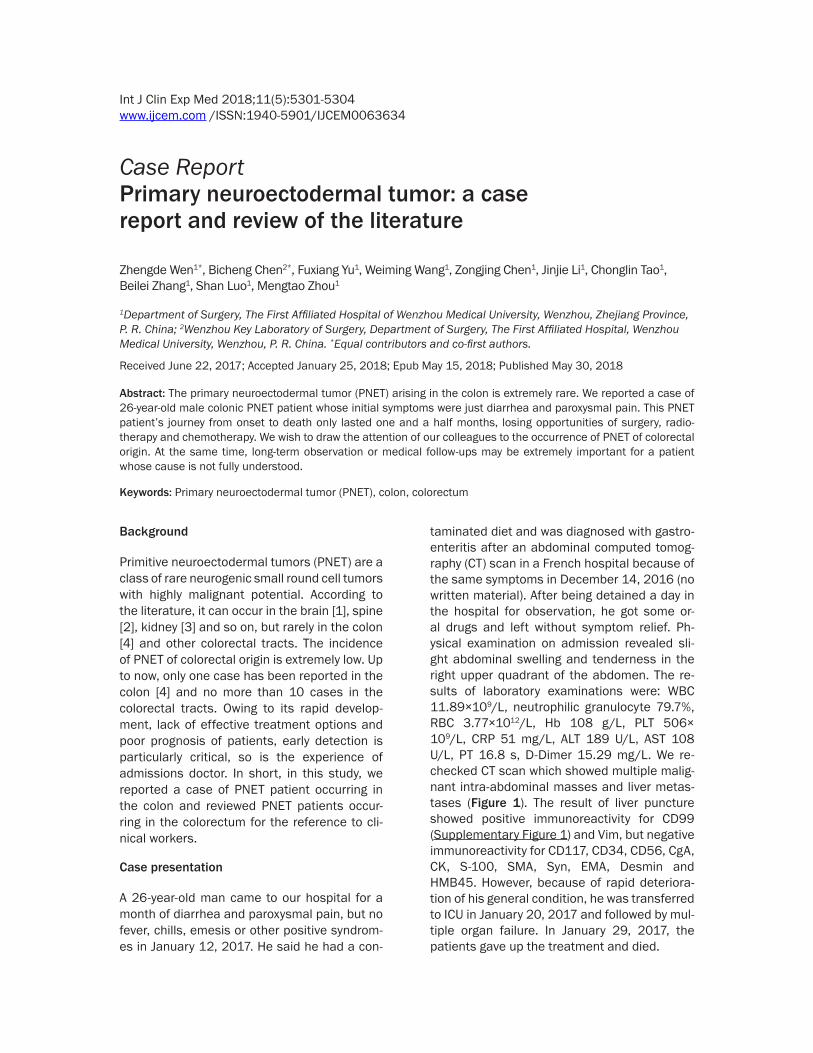
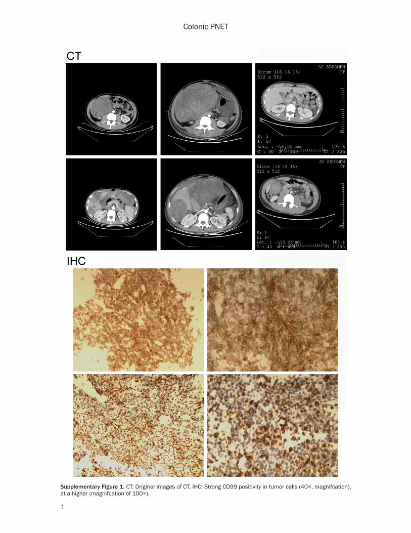
taminated diet and was diagnosed with gastro-enteritis after an abdominal computed tomog-raphy (CT) scan in a French hospital because of the same symptoms in December 14, 2016 (no written material). After being detained a day in the hospital for observation, he got some or- al drugs and left without symptom relief. Ph- ysical examination on admission revealed sli- ght abdominal swelling and tenderness in the right upper quadrant of the abdomen. The re- sults of laboratory examinations were: WBC 11.89×10⁹/L, neutrophilic granulocyte 79.7%, RBC 3.77×10¹²/L, Hb 108 g/L, PLT 506× 109/L, CRP 51 mg/L, ALT 189 U/L, AST 108 U/L, PT 16.8 s, D-Dimer 15.29 mg/L. We re- checked CT scan which showed multiple malig-nant intra-abdominal masses and liver metas-tases (Figure 1). The result of liver puncture showed positive immunoreactivity for CD99 (Supplementary Figure 1) and Vim, but negative immunoreactivity for CD117, CD34, CD56, CgA, CK, S-100, SMA, Syn, EMA, Desmin and HMB45. However, because of rapid deteriora-tion of his general condition, he was transferred to ICU in January 20, 2017 and followed by mul-tiple organ failure. In January 29, 2017, the patients gave up the treatment and died.
Colonic PNET
5302 Int J Clin Exp Med 2018;11(5):5301-5304
Figure 1. Time line of patient’s colonic PNET diagnosis. A: Abdominal CT scan depicting an irregular mass in the colon. B: Enhanced CT revealing increase in heterogeneous mass and occurrence of liver metastases. C: HE stain-ing of the tumor showing tumor nests (40×, magnification), at a higher (magnification of 100×). D: Enhanced CT revealing increase in heterogeneous mass and occurrence of liver metastases. Yellow rectangle near the time line represents this PNET patient’s journey from onset to death.
Table 1. Clinical features of 6 colorectal PENT patients
Authors (Year) Age (yr)/Gender Symptoms Location (Size) Positive
immunomarker Follow-up
Present case 26/M Abdominal pain Colon (9.9*10 cm) CD99, Vin 1.5 mo Died
Peng et al (2014) [9] 36/F Abdominal pain and Abdominal mass Ileocecum (15*15*13 cm) CD99, FLI1, Vim 34 mo Died
Aboumarzouk et al (2009) [10] 34/F Anorectal pain and Rectal bleeding Rectum (10 cm) CD99 7 years DFS
Kuwabara et al (2006) [4] 59/M Mass Colon (10 cm) CD99, CgA, MIB-1 7 mo Died
Vardy et al (2005) [11] 53/M Rectal bleeding Rectum (NM) CD99, Vim 2 years Died
Drut et al (2003) [12] 17/M Intermittent pain and Rectal bleeding Rectum (4.5*4*4 cm) CD99, S100 1 year DFSNM, no mention; DFS, disease-free survival.
A written informed consent for the case report was obtained from the patient. The consent procedure was approved by the Ethics Com- mittee of the First Affiliated Hospital of Wen- zhou Medical University.
Discussion and conclusions
PNET was first reported by hart and Earle to describe a class of neuroectodermal tumors in the brain [5]. With the increase of related lite- ratures, it was found that it can occur in the central nervous system and other peripheral organs. Therefore, it is divided into central and peripheral PNET. However, up to now, the study
of PNET has been more frequently found in case reports, but less in large sample studies.
Due to lack of characteristic clinical manifes- tations, suggestive blood markers and imaging features, it is easy for PNET to be misdiagnos- ed and missed diagnosed [6]. Immunohis- tochemistry results show that Homer-Wright daisy-group and CD99 (+) are specific for diag-nosis in the actual clinical work [7]. Surgery is the preferred treatment, but the results were unsatisfactory according to the current reports. The main reasons are as followed: 1. Patients have liver, lung and other distant metastasis, 2. Extensive resection is difficult for a wide range
Colonic PNET
5303 Int J Clin Exp Med 2018;11(5):5301-5304
of lesion. So the major treatment for PNET patients is the combination of surgery, radio-therapy, and chemotherapy [8].
Only one case of colonic PNET has been report-ed in English literature that the patient died for tumor recurrence in the retroperitoneal metas-tasis after 7 months of his first operation [4]. There are still not enough cases to summarize the clinical features of colonic PNET patients. Our report may be useful. Thus we collected data of PENT patients arising in the colorectum but did not contain mesentery (Table 1). It includes 2 females and 4 males, with an aver-age age of 37.5 years old (17-59 years). The main manifestations are abdominal pain, rec- tal bleeding and symptoms caused by rapidly increased tumors. They are not special, so are the results of diagnostic imaging presentation. Especially the patient in this report, the initial imaging presentation and medical history is confusing, which needs to distinguish from in- tussusception, stromal tumor, enteritis and oth- ers, so the clinical experience is very impor- tant. Due to fast progression of disease and rejection of autopsy, we did not carry out fur-ther examination, but taking the histological results and disease history into account we still considered it is PNET.
In summary, the PNET occurring in the colorec-tum has the characteristic of nonspecific clini-cal manifestations, rapid development, poor prognosis and so on. At the same time, early imaging features and suggestive blood mark-ers are not clear, so it is easy to be misdiag-nosed and missed diagnosed. We hope that clinical workers can learn some experience from this article. For patients with rapidly increasing abdominal mass and rectal bleed-ing, we should take colorectal PNET into ac- count. At the same time, long-term observa- tion or medical follow-ups may be extremely important for a patient whose cause is not fully understood.
Acknowledgements
This project was sponsored by Grants of Zhejiang Provincial Top Key Discipline in Sur- gery and Zhejiang Provincial Program for the Cultivation of High-level Innovative Health Ta- lents.
Disclosure of conflict of interest
None.
Address correspondence to: Mengtao Zhou and Shan Luo, Department of Surgery, The First Affiliated Hospital of Wenzhou Medical University, 2 Fuxue Lane, Wenzhou, Zhejiang Province, P. R. China. Tel: 86-577-55579220; Fax: 86-577-88069555; E-mail: [email protected] (MTZ); [email protected] (SL)
References
[1] dos Santos Rubio EJ, Harhangi BS, Kros JM, Vincent AJ and Dirven CM. A primary extraos-seous Ewing sarcoma in the cerebellopontine angle of a child: review of relevant literature and case report. Neurosurgery 2010; 67: E1852-1856.
[2] Karikari IO, Mehta AI, Nimjee S, Hodges TR, Tibaleka J, Montgomery C, Simpson L, Cum-mings TJ and Bagley CA. Primary intradural ex-traosseous Ewing sarcoma of the spine: case report and literature review. Neurosurgery 2011; 69: E995-999.
[3] Thyavihally YB, Tongaonkar HB, Gupta S, Kurkure PA, Amare P, Muckaden MA and Desai SB. Primitive neuroectodermal tumor of the kidney: a single institute series of 16 patients. Urology 2008; 71: 292-296.
[4] Kuwabara K, Ishida H, Shirakawa K, Yokoyama M, Nakada H, Hayashi Y, Hashimoto D, Miura I, Itoyama S and Heike Y. Primitive neuroectoder-mal tumor arising in the colon: report of a case. Surg Today 2006; 36: 193-197.
[5] Hart MN and Earle KM. Primitive neuroecto-dermal tumors of the brain in children. Cancer 1973; 32: 890-897.
[6] Tan Y, Zhang H, Ma GL, Xiao EH and Wang XC. Peripheral primitive neuroectodermal tumor: dynamic CT, MRI and clinicopathological char-acteristics--analysis of 36 cases and review of the literature. Oncotarget 2014; 5: 12968-12977.
[7] Schmidt D, Herrmann C, Jurgens H and Harms D. Malignant peripheral neuroectodermal tu-mor and its necessary distinction from Ewing’s sarcoma. A report from the Kiel pediatric tu-mor registry. Cancer 1991; 68: 2251-2259.
[8] Packer RJ. Chemotherapy for medulloblasto-ma/primitive neuroectodermal tumors of the posterior fossa. Ann Neurol 1990; 28: 823-828.
[9] Peng L, Yang L, Wu N and Wu BO. Primary prim-itive neuroectodermal tumor arising in the mesentery and ileocecum: a report of three
Colonic PNET
5304 Int J Clin Exp Med 2018;11(5):5301-5304
cases and review of the literature. Exp Ther Med 2015; 9: 1299-1303.
[10] Aboumarzouk OM, Coleman R, Goepel JR and Shorthouse AJ. PNET/Ewing’s sarcoma of the rectum: a case report and review of the litera-ture. BMJ Case Rep 2009; 2009.
[11] Vardy J, Joshua AM, Clarke SJ, Yarrow PM and Lin BP. Small blue cell tumors of the rectum. Case 1. Ewing’s sarcoma of the rectum. J Clin Oncol 2005; 23: 910-912.
[12] Drut R, Drut M, Muller C and Marron A. Rectal primitive neuroectodermal tumor. Pediatr Pathol Mol Med 2003; 22: 391-398.
Colonic PNET
1
Supplementary Figure 1. CT: Original Images of CT, IHC: Strong CD99 positivity in tumor cells (40×, magnifcation), at a higher (magnifcation of 100×).