Embed Size (px)
Citation preview
Primitive Neuroectodermal Tumor of theKidney in a 22-Year-Old Male: A Rare Entity
Veena Gupta, Rajeev Sen, Ashima Batra♦, Promil Jain
Department of Pathology, Pt. B D Sharma PGIMS Rohtak, Haryana, India
Case ReportMiddle East Journal of Cancer; October 2014; 5(4): 233-236
♦Corresponding Author: Ashima Batra, MDSenior Resident, Department ofPathology, Pt. B D SharmaPGIMS Rohtak, Haryana, IndiaTel: +91-9991477127
Email: [email protected]
IntroductionPrimitive neuroectodermal tumor
(PNET) is an uncommon malignancyof bones and soft tissue which rarelyoccurs in the kidneys.1 In the kidneysmost cases occur in the medullary/pelvic region.2 It is a small roundcell tumor that has neuroepithelialdifferentiation, which typically occursin young children and adolescents.1This tumor is a rare entity and itsdiagnosis is usually made athistopathology. A few cases that havebeen reported in the literature reveala variable presentation and aggressivebehavior.3
Case ReportA 22-year-old male presented with
a tender mass in his left loin. Pastmedical history was unremarkable.Routine hematological, biochemicaland urine examinations were withinnormal limits. Computerizedtomography (CT scan) of theabdomen revealed a mass thatinvolved the upper and middle polesof the left kidney without anycapsular invasion or renal vascularinvolvement. There was no evidenceof intra-abdominal or retroperitoneallymphadenopathy or any abdominalorgan involvement. Chest X ray, CTscan of the thorax and bone scan didnot reveal any evidence of lung or
AbstractPrimitive neuroectodermal tumors of the kidney are rare tumors representing about
1% of all sarcomas. Extraskeletal primitive neuroectodermal tumors may be found inthe genitourinary tract, the testes, ovaries, uterus, or pancreas. Renal primitive neuroec-todermal tumors are frequently aggressive and almost 30% of all newly diagnosed casespresent with distant metastases. Surgical intervention, intensive chemotherapeuticdrugs and radiation therapy are the best choices for management.
We discuss the case of a 22-year-old male who presented with a left renal mass. Heunderwent nephrectomy followed by histopathological examination. Microscopic andimmunohistochemical examination revealed a primitive neuroectodermal tumor.
Keywords: Primitive neuroectodermal tumor (PNET), Kidney, Rare
Received: March 17, 2014; Accepted: June 1, 2014
bone involvement. The patient subsequentlyunderwent left radical nephrectomy.
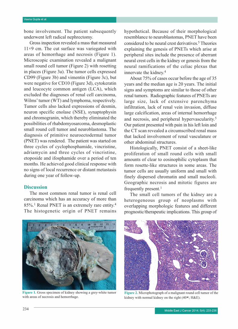
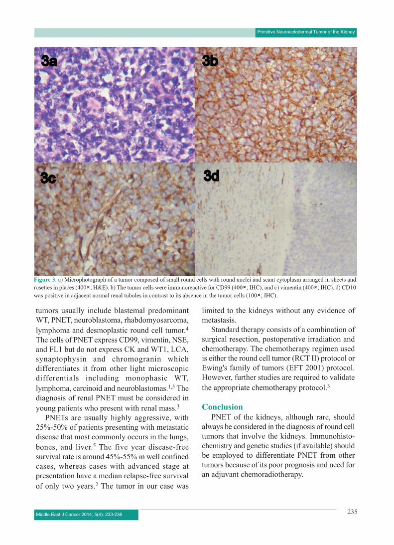
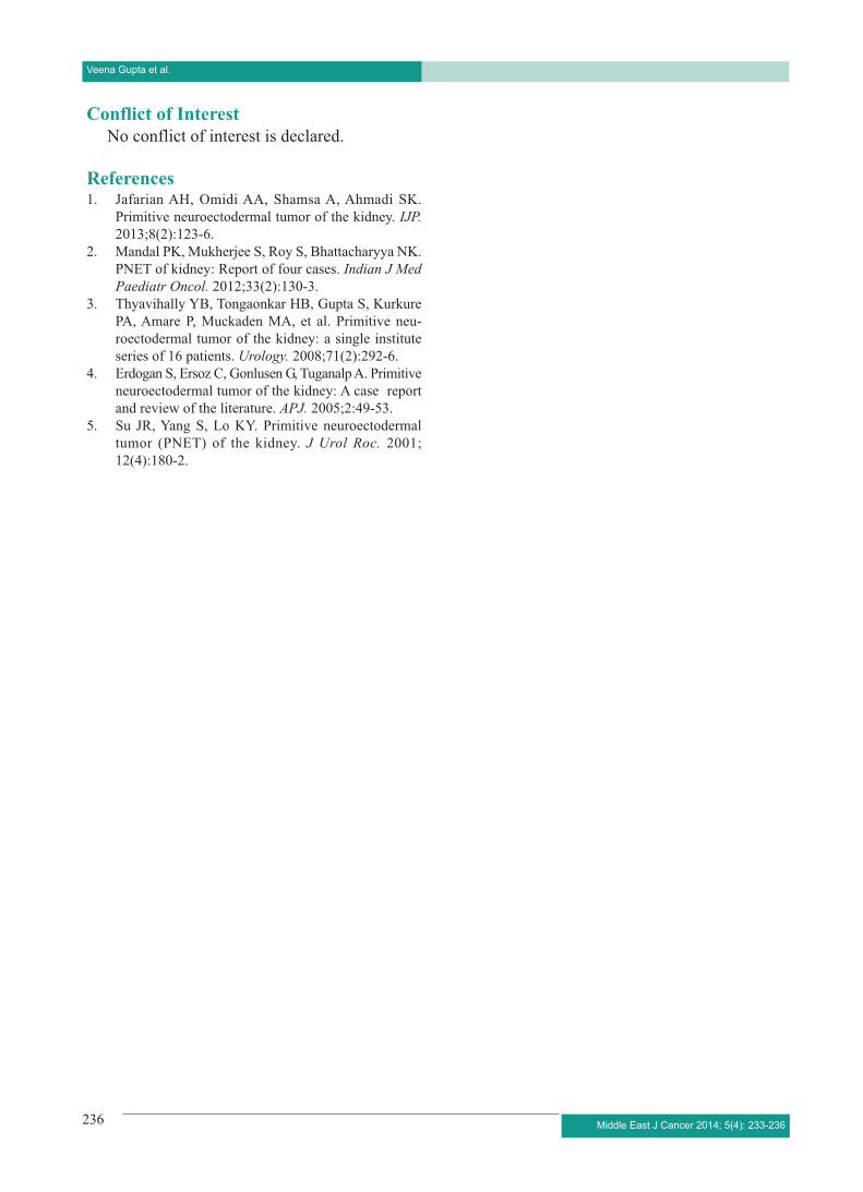
Gross inspection revealed a mass that measured11×9 cm. The cut surface was variegated withareas of hemorrhage and necrosis (Figure 1).Microscopic examination revealed a malignantsmall round cell tumor (Figure 2) with rosettingin places (Figure 3a). The tumor cells expressedCD99 (Figure 3b) and vimentin (Figure 3c), butwere negative for CD10 (Figure 3d), cytokeratinand leucocyte common antigen (LCA), whichexcluded the diagnoses of renal cell carcinoma,Wilms’ tumor (WT) and lymphoma, respectively.Tumor cells also lacked expressions of desmin,neuron specific enolase (NSE), synaptophysinand chromogranin, which thereby eliminated thepossibilities of rhabdomyosarcoma, desmoplasticsmall round cell tumor and neuroblastoma. Thediagnosis of primitive neuroectodermal tumor(PNET) was rendered. The patient was started onthree cycles of cyclophosphamide, vincristine,adriamycin and three cycles of vincristine,etoposide and ifosphamide over a period of tenmonths. He achieved good clinical response withno signs of local recurrence or distant metastasisduring one year of follow-up.
DiscussionThe most common renal tumor is renal cell
carcinoma which has an accuracy of more than85%.1 Renal PNET is an extremely rare entity.4The histogenetic origin of PNET remains
hypothetical. Because of their morphologicalresemblance to neuroblastomas, PNET have beenconsidered to be neural crest derivatives.5 Theoriesexplaining the genesis of PNETs which arise atperipheral sites include the presence of aberrantneural crest cells in the kidney or genesis from theneural ramifications of the celiac plexus thatinnervate the kidney.4
About 75% of cases occur before the age of 35years and the median age is 20 years. The initialsigns and symptoms are similar to those of otherrenal tumors. Radiographic features of PNETs arelarge size, lack of extensive parenchymainfiltration, lack of renal vein invasion, diffuselarge calcification, areas of internal hemorrhageand necrosis, and peripheral hypervascularity.5Our patient presented with pain in his left loin andthe CT scan revealed a circumscribed renal massthat lacked involvement of renal vasculature orother abdominal structures.
Histologically, PNET consist of a sheet-likeproliferation of small round cells with smallamounts of clear to eosinophilic cytoplasm thatform rosette-like structures in some areas. Thetumor cells are usually uniform and small withfinely dispersed chromatin and small nucleoli.Geographic necrosis and mitotic figures arefrequently present.1
The small cell tumors of the kidney are aheterogeneous group of neoplasms withoverlapping morphologic features and differentprognostic/therapeutic implications. This group of
Figure 1. Gross specimen of kidney showing a grey-white tumorwith areas of necrosis and hemorrhage.
234 Middle East J Cancer 2014; 5(4): 233-236
Veena Gupta et al.
Figure 2. Microphotograph of a malignant round cell tumor of thekidney with normal kidney on the right (40×; H&E).
tumors usually include blastemal predominantWT, PNET, neuroblastoma, rhabdomyosarcoma,lymphoma and desmoplastic round cell tumor.4The cells of PNET express CD99, vimentin, NSE,and FL1 but do not express CK and WT1, LCA,synaptophysin and chromogranin whichdifferentiates it from other light microscopicdifferentials including monophasic WT,lymphoma, carcinoid and neuroblastomas.1,5 Thediagnosis of renal PNET must be considered inyoung patients who present with renal mass.3
PNETs are usually highly aggressive, with25%-50% of patients presenting with metastaticdisease that most commonly occurs in the lungs,bones, and liver.5 The five year disease-freesurvival rate is around 45%-55% in well confinedcases, whereas cases with advanced stage atpresentation have a median relapse-free survivalof only two years.2 The tumor in our case was
limited to the kidneys without any evidence ofmetastasis.
Standard therapy consists of a combination ofsurgical resection, postoperative irradiation andchemotherapy. The chemotherapy regimen usedis either the round cell tumor (RCT II) protocol orEwing's family of tumors (EFT 2001) protocol.However, further studies are required to validatethe appropriate chemotherapy protocol.3
ConclusionPNET of the kidneys, although rare, should
always be considered in the diagnosis of round celltumors that involve the kidneys. Immunohisto-chemistry and genetic studies (if available) shouldbe employed to differentiate PNET from othertumors because of its poor prognosis and need foran adjuvant chemoradiotherapy.
235Middle East J Cancer 2014; 5(4): 233-236
Primitive Neuroectodermal Tumor of the Kidney
Figure 3. a) Microphotograph of a tumor composed of small round cells with round nuclei and scant cytoplasm arranged in sheets androsettes in places (400×; H&E). b) The tumor cells were immunoreactive for CD99 (400×; IHC), and c) vimentin (400×; IHC). d) CD10was positive in adjacent normal renal tubules in contrast to its absence in the tumor cells (100×; IHC).
236 Middle East J Cancer 2014; 5(4): 233-236
Conflict of InterestNo conflict of interest is declared.
References1. Jafarian AH, Omidi AA, Shamsa A, Ahmadi SK.
Primitive neuroectodermal tumor of the kidney. IJP.2013;8(2):123-6.
2. Mandal PK, Mukherjee S, Roy S, Bhattacharyya NK.PNET of kidney: Report of four cases. Indian J MedPaediatr Oncol. 2012;33(2):130-3.
3. Thyavihally YB, Tongaonkar HB, Gupta S, KurkurePA, Amare P, Muckaden MA, et al. Primitive neu-roectodermal tumor of the kidney: a single instituteseries of 16 patients. Urology. 2008;71(2):292-6.
4. Erdogan S, Ersoz C, Gonlusen G, Tuganalp A. Primitiveneuroectodermal tumor of the kidney: A case reportand review of the literature. APJ. 2005;2:49-53.
5. Su JR, Yang S, Lo KY. Primitive neuroectodermaltumor (PNET) of the kidney. J Urol Roc. 2001;12(4):180-2.
Veena Gupta et al.