Embed Size (px)
Citation preview
Upadhyay et al.One Stage Excision of Intraspinal Tumour with Immediate Reconstruction of Spine by Laminoplasty – A Case Report,Medical Science, 2016, 20(80), 123-131,www.discoveryjournals.com © 2016 Discovery Publication. All Rights Reserved
Page123
CASE REPORT ARTICLE
Upadhyay PK1҉, Pandey P2, Bora S3, Pandey S4, Gupta U4, Singh R4
1.Head Department of neurosurgery, Institute of Human Behaviour an Allied Sciences (I.H.B.A.S.), Dilshad Garden, New Delhi 110095,India
2.Senior Resident, Department of Neurosurgery, I.H.B.A.S., Dilshad Garden, New Delhi 110095, India3.Senior Resident, Department of Neuroanaesthesia, I.H.B.A.S., Dilshad Garden, New Delhi 110095, India4.Junior Resident, Department of Neurosurgery, I.H.B.A.S., Dilshad Garden, New Delhi 110095, India
҉Corresponding author: Dr P.K. Upadhyay: Type VI Q. no 05 Institute of Human Behaviour and allied sciences, Dilshad Garden, Govtof NCT of Delhi, New Delhi 110095. E-mail: [email protected]
Publication HistoryReceived: 03 April 2016Accepted: 23 April 2016Published: 1 July 2016
CitationUpadhyay PK, Pandey P, Bora S, Pandey S, Gupta U, Singh R. One Stage Excision of Intraspinal Tumour with ImmediateReconstruction of Spine by Laminoplasty – A Case Report. Medical Science, 2016, 20(80), 123-131
Publication License
This work is licensed under a Creative Commons Attribution 4.0 International License.
General NoteArticle is recommended to print in recycled paper.
ABSTRACTLaminoplasty is a technique that indirectly achieves decompression of spinal cord and avoids the complications of fusion (Oyama,1973). It was initially proposed by the Japanese as a treatment of ossified posterior longitudinal ligament in the cervical region. Itsuse has been expanded since then and is widely used in the thoracic and lumbar region (Lonstein, 1976). We present a case report
Medical Science, Vol. 20, No. 80, July 1, 2016 CASE REPORT
Medical Science
One Stage Excision of Intraspinal Tumour with Immediate Reconstructionof Spine by Laminoplasty – A Case Report
ISSN2321–7359
EISSN2321–7367
An International Journal
Upadhyay et al.One Stage Excision of Intraspinal Tumour with Immediate Reconstruction of Spine by Laminoplasty – A Case Report,Medical Science, 2016, 20(80), 123-131,www.discoveryjournals.com © 2016 Discovery Publication. All Rights Reserved
Page124
CASE REPORT ARTICLE
of a 20 year male patient with intramedullary tumour at L2L3 level, who underwent excision and laminoplasty and discuss thesurgical procedure, its merits of the procedure.
Keywords: Laminotomy, Laminoplasty, Laminectomy, Intramedullary tumour
1. INTRODUCTIONLaminotomy has become the standard approach to the spinal canal when bony decompression is not the aim (Goel, 1997;Constantini, 2006). Vascular malformations and tumours of the spinal cord are widely accessed by this procedure (Constantini, 2006).Many reconstructive procedures are prevalent in literature, such as transverse placement laminoplasty, restorative laminoplasty,inverse laminoplasty, en bloc laminoplasty and expansive laminotomy (Mimatsu, 1997; Zheng, 2004).
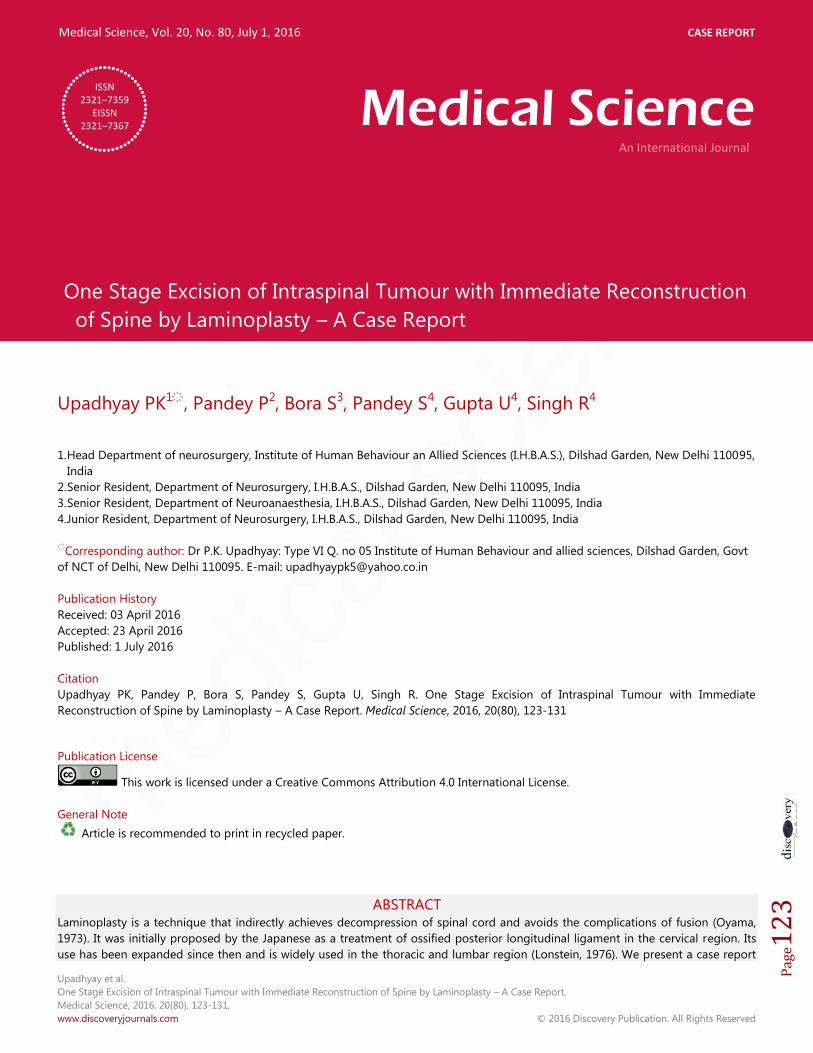
2. CASE HISTORY30 year male presented with non-radiating, moderate to severe low backache of 8 months duration followed by diffuse pain in bothlower limbs of 6 months duration. This was followed by progressive weakness of both lower limbs, loss of sensation below knee inboth lower limbs, diminution in perianal sensation and urinary retention of 2 months duration. General physical examination wasessentially within normal limits. Neurological examination revealed wasting both thigh and calf muscles along with flaccidity of bothlower limbs with power 3/5 to 4/5 bilaterally (Nurick Grade IV). Deep tendon reflexes (DTRs) were absent below L1 level. Planterreflex were not electable bilaterally. All sensory modalities showed 50-75 % loss below L1 dermatome.Preoperative MagneticResonance Imaging (MRI) of lumbosacral spine shows cord expansion at L2 L3 with predominantly T1 weighted hypointense and T2weighted hyperintense, moderately enhancing intramedullary lesion (Figure 1). The patient was provisionally diagnosed to be a L2L3intramedullary dermoid and was taken up for excision.
Figure 1 Preoperative MRI, Sagittal and Axial Section
Upadhyay et al.One Stage Excision of Intraspinal Tumour with Immediate Reconstruction of Spine by Laminoplasty – A Case Report,Medical Science, 2016, 20(80), 123-131,www.discoveryjournals.com © 2016 Discovery Publication. All Rights Reserved
Page125
CASE REPORT ARTICLE
Operative procedure /Steps1. The patient was administered general anaesthesia with endotracheal intubation and
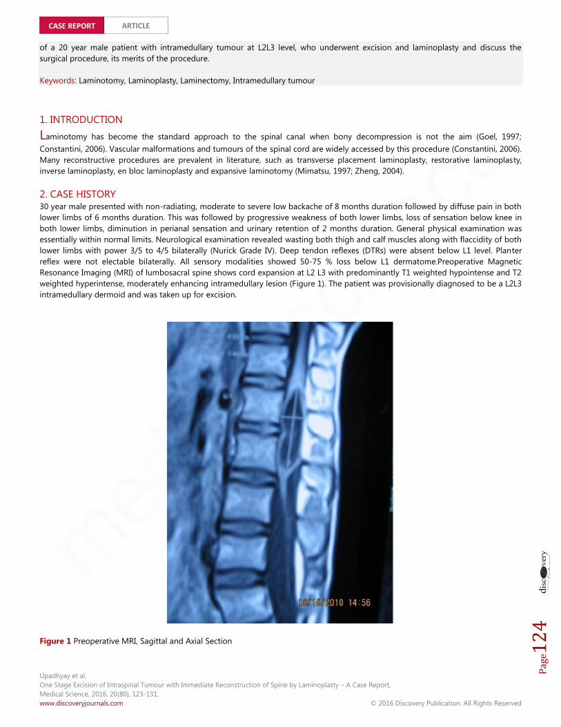
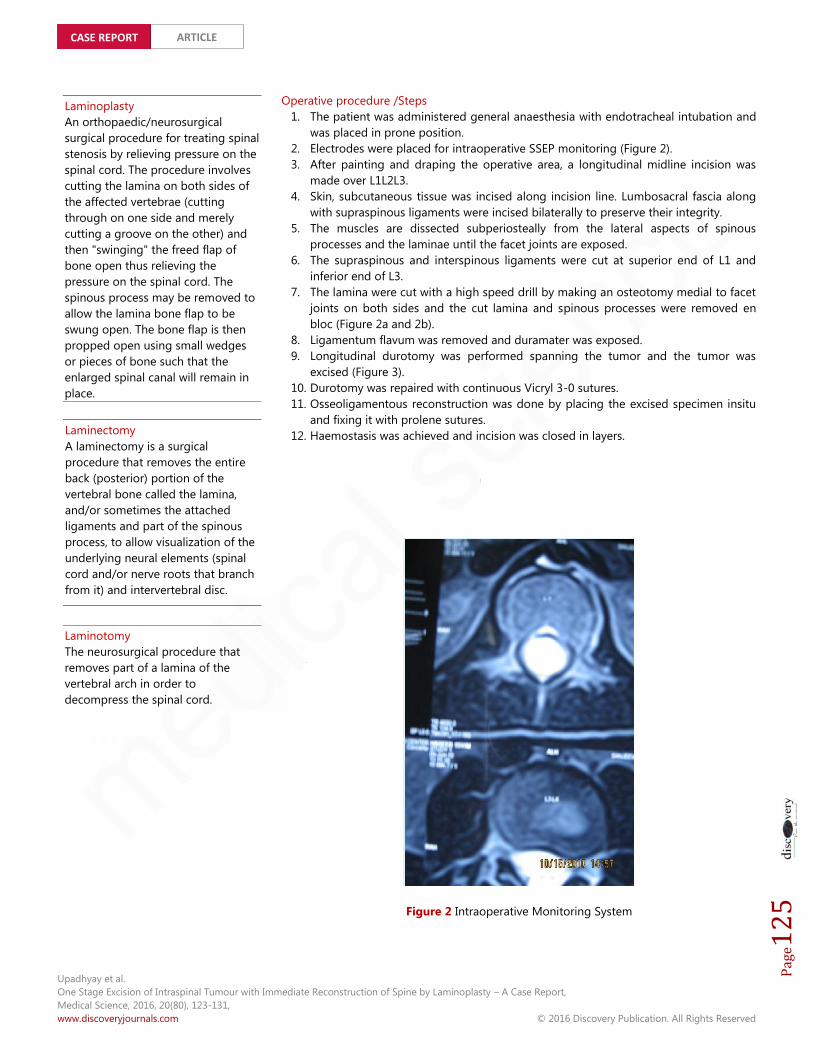
was placed in prone position.2. Electrodes were placed for intraoperative SSEP monitoring (Figure 2).3. After painting and draping the operative area, a longitudinal midline incision was
made over L1L2L3.4. Skin, subcutaneous tissue was incised along incision line. Lumbosacral fascia along
with supraspinous ligaments were incised bilaterally to preserve their integrity.5. The muscles are dissected subperiosteally from the lateral aspects of spinous
processes and the laminae until the facet joints are exposed.6. The supraspinous and interspinous ligaments were cut at superior end of L1 and
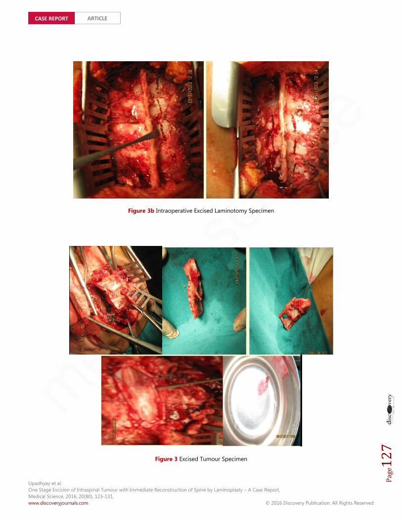
inferior end of L3.7. The lamina were cut with a high speed drill by making an osteotomy medial to facet
joints on both sides and the cut lamina and spinous processes were removed enbloc (Figure 2a and 2b).
8. Ligamentum flavum was removed and duramater was exposed.9. Longitudinal durotomy was performed spanning the tumor and the tumor was
excised (Figure 3).10. Durotomy was repaired with continuous Vicryl 3-0 sutures.11. Osseoligamentous reconstruction was done by placing the excised specimen insitu
and fixing it with prolene sutures.12. Haemostasis was achieved and incision was closed in layers.
Figure 2 Intraoperative Monitoring System
LaminoplastyAn orthopaedic/neurosurgicalsurgical procedure for treating spinalstenosis by relieving pressure on thespinal cord. The procedure involvescutting the lamina on both sides ofthe affected vertebrae (cuttingthrough on one side and merelycutting a groove on the other) andthen "swinging" the freed flap ofbone open thus relieving thepressure on the spinal cord. Thespinous process may be removed toallow the lamina bone flap to beswung open. The bone flap is thenpropped open using small wedgesor pieces of bone such that theenlarged spinal canal will remain inplace.
LaminectomyA laminectomy is a surgicalprocedure that removes the entireback (posterior) portion of thevertebral bone called the lamina,and/or sometimes the attachedligaments and part of the spinousprocess, to allow visualization of theunderlying neural elements (spinalcord and/or nerve roots that branchfrom it) and intervertebral disc.
LaminotomyThe neurosurgical procedure thatremoves part of a lamina of thevertebral arch in order todecompress the spinal cord.
Upadhyay et al.One Stage Excision of Intraspinal Tumour with Immediate Reconstruction of Spine by Laminoplasty – A Case Report,Medical Science, 2016, 20(80), 123-131,www.discoveryjournals.com © 2016 Discovery Publication. All Rights Reserved
Page126
CASE REPORT ARTICLE
Figure 3a Intraoperative Laminotomy
Upadhyay et al.One Stage Excision of Intraspinal Tumour with Immediate Reconstruction of Spine by Laminoplasty – A Case Report,Medical Science, 2016, 20(80), 123-131,www.discoveryjournals.com © 2016 Discovery Publication. All Rights Reserved
Page127
CASE REPORT ARTICLE
Figure 3b Intraoperative Excised Laminotomy Specimen
Figure 3 Excised Tumour Specimen
Upadhyay et al.One Stage Excision of Intraspinal Tumour with Immediate Reconstruction of Spine by Laminoplasty – A Case Report,Medical Science, 2016, 20(80), 123-131,www.discoveryjournals.com © 2016 Discovery Publication. All Rights Reserved
Page128
CASE REPORT ARTICLE
Patient was managed with intravenous antibiotics and steroids in the post-operative stage. No CSF leak was noted in the post-operative period. The patient was weaned off the urinary catheter on postoperative day 5. By day 10 the patient was able to standwithout support and reported complete resolution of pain in lower back.
3. DISCUSSIONLaminotomy was suggested by Sonntag in 2000 (Sonntag, 2000) although confusion prevails regarding the correct terminology.Many studies refer to it as replacement laminotomy, non-expansive laminotomy, osteoplastic laminotomy, laminoplasty, open doorlaminoplasty, non-expansive laminoplasty, recapping laminoplasty (Asazuma, 2003).
Laminoplasty allows the spinal cord and the neural foramen to be decompressed without directly removing anterior pathology.By preserving the dorsal elements of the spine, laminoplasty preserves spine stability and alignment and decreases the risk ofpostlaminectomy kyphosis and instability. Additionally, since fusion is not required, complications such as fixation failure,pseudoarthrosis, loss of motion, and adjacent segment degeneration do not occur. This may allow earlier mobilization andrehabilitation compared to other surgical options. In addition, laminoplasty can avoid graft related complications such as graftextrusion, settling, collapse, dislodgement, and fracture.
With laminectomy, epidural scar formation can form between the duramater and muscle leading to postoperative pain andneurologic compression. However, with laminoplasty, the lamina is preserved and it protects the dura from this “postlaminectomymembrane”. Preserving the lamina also makes revision procedures requiring posterior approaches safer (Ishida, 1989; Steinmetz,2006).
Laminoplasty TechniquesOyama et al. first described cervical laminoplasty in Japanese in 1973 as a treatment for OPLL. Since its initial description by Oyama,laminoplasty techniques have been constantly refined. Most of these changes relate to how the cuts in the lamina or spinousprocess are made and how the laminae are secured in an open position—with wires or heavy sutures, bone anchors or bone blocks,hydroxyapatite blocks, miniplates, local spinous process autograft, and combinations thereof. All variations in laminoplastytechniques maintain the common theme, however, of repositioning the laminae, expanding the canal, and preserving the dorsalelements to maintain stability. In general, none of these technical variations have proven to be any safer or efficacious than theother. Our approach was to use suture for fixation of excised laminae in the described case.
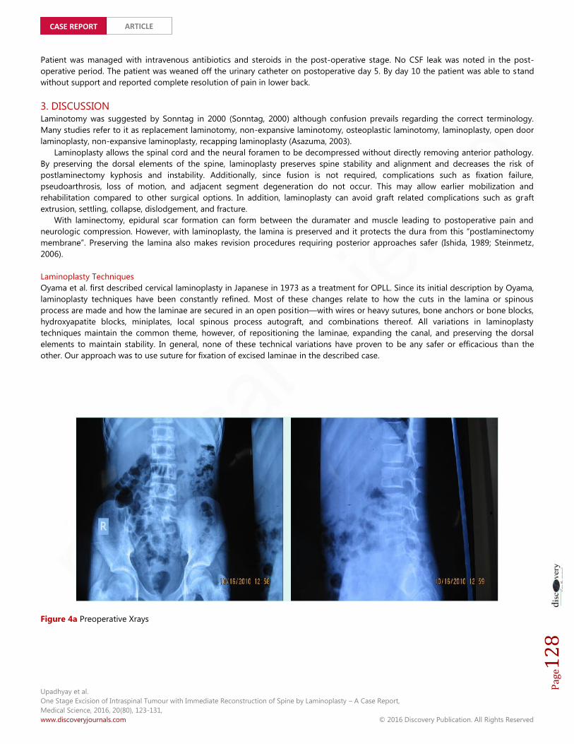
Figure 4a Preoperative Xrays
Upadhyay et al.One Stage Excision of Intraspinal Tumour with Immediate Reconstruction of Spine by Laminoplasty – A Case Report,Medical Science, 2016, 20(80), 123-131,www.discoveryjournals.com © 2016 Discovery Publication. All Rights Reserved
Page129
CASE REPORT ARTICLE
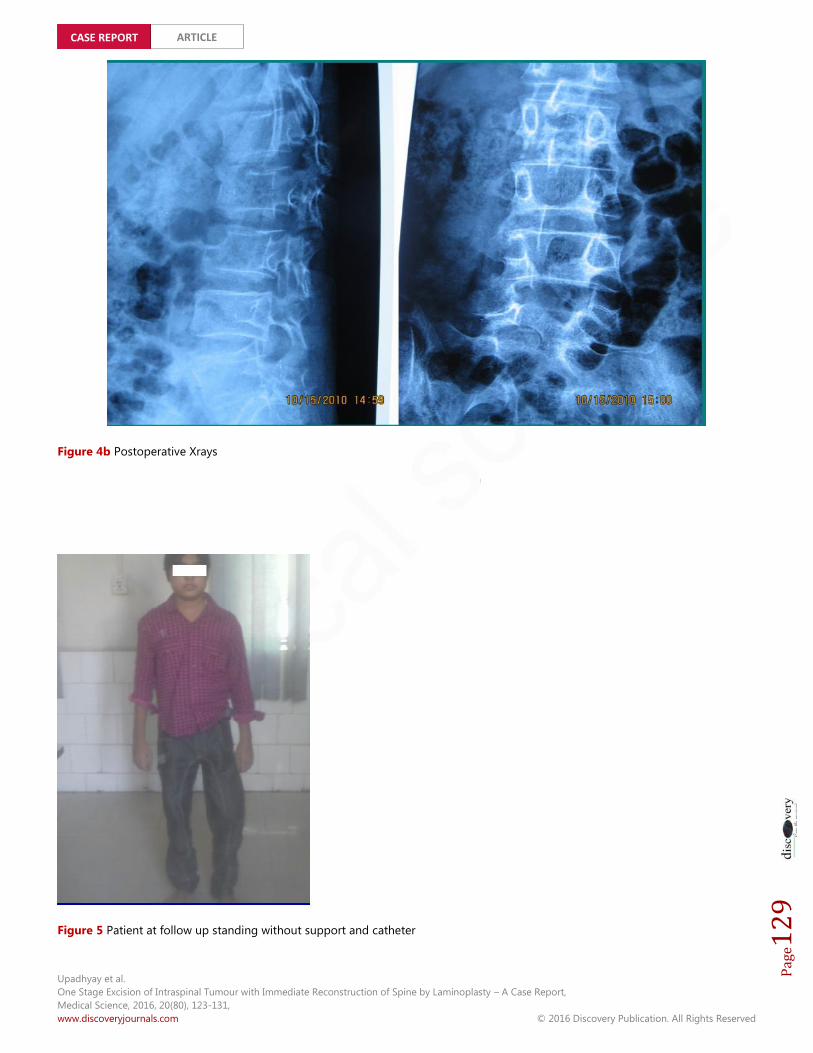
Figure 4b Postoperative Xrays
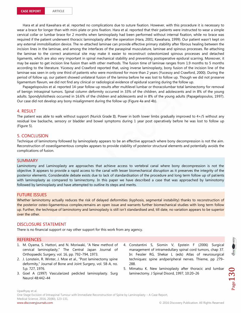
Figure 5 Patient at follow up standing without support and catheter
Upadhyay et al.One Stage Excision of Intraspinal Tumour with Immediate Reconstruction of Spine by Laminoplasty – A Case Report,Medical Science, 2016, 20(80), 123-131,www.discoveryjournals.com © 2016 Discovery Publication. All Rights Reserved
Page130
CASE REPORT ARTICLE
Hara et al and Kawahara et al. reported no complications due to suture fixation. However, with this procedure it is necessary towear a brace for longer than with mini-plate or pins fixation. Hara et al. reported that their patients were instructed to wear a simplecervical collar or lumbar brace for 2 months when laminoplasty had been performed without internal fixation, while no brace wasrequired if the patient underwent thoracic laminoplasty after the operation (Hara, 2001; Kawahara, 1999). Our patient wasn’t kept onany external immobilisation device. The re-attached laminae can provide effective primary stability after fibrous healing between theincision lines in the laminae, and among the interfaces of the paraspinal musculature, laminae and spinous processes. Re-attachingthe laminae to the correct anatomical site may make it easier to reconstruct osteotomized spinous processes and detachedligaments, which are also very important in spinal mechanical stability and preventing postoperative epidural scarring. Moreover, itmay be easier to get incision line fusion than with other methods. The fusion time of laminae ranges from 1.9 months to 5 monthsaccording to the literature. Yücesoy and Crawford reported that, using inverse laminoplasty, bony fusion of the incision line of thelaminae was seen in only one third of patients who were monitored for more than 2 years (Yucesoy and Crawford, 2000). During theperiod of follow up, our patient showed unilateral fusion of the lamina before he was lost to follow up. Though we did not preserveligamentum flavum, we did not find any clinical or radiological evidence of epidural scarring during the follow up.
Papagelopoulos et al. reported 14-year follow-up results after multilevel lumbar or thoracolumbar total laminectomy for removalof benign intraspinal tumors. Spinal column deformity occurred in 33% of the children, and adolescents and in 8% of the youngadults. Spondylolisthesis occurred in 16.6% of the children and adolescents and in 8% of the young adults (Papagelopoulos, 1997).Our case did not develop any bony misalignment during the follow up (Figure 4a and 4b).
4. RESULTThe patient was able to walk without support (Nurick Grade II). Power in both lower limbs gradually improved to 4+/5 without anyresidual low backache, sensory or bladder and bowel symptoms during 1 year post operatively before he was lost to follow up(Figure 5).
5. CONCLUSIONTechnique of laminotomy followed by laminoplasty appears to be an effective approach where bony decompression is not the aim.Reconstruction of osseoligamentous complex appears to provide stability of posterior structural elements and potentially avoids thecomplications of fusion.
SUMMARYLaminotomy and Laminoplasty are approaches that achieve access to vertebral canal where bony decompression is not theobjective. It appears to provide a rapid access to the canal with lesser biomechanical disruption as it preserves the integrity of theposterior elements. Considerable debate exists due to lack of standardisation of the procedure and long term follow up of patientswith laminoplasty as compared to laminectomy. In this paper we have described a case that was approached by laminotomyfollowed by laminoplasty and have attempted to outline its steps and merits.
FUTURE ISSUESWhether laminotomy actually reduces the risk of delayed deformities (kyphosis, segmental instability) thanks to reconstruction ofthe posterior osteo-ligamentous complex,remains an open issue and warrants further biomechanical studies with long term followup. Further, the technique of laminotomy and laminoplasty is still isn’t standardised and, till date, no variation appears to be superiorover the other.
DISCLOSURE STATEMENTThere is no financial support or nay other support for this work from any agency.
REFERENCES1. M. Oyama, S. Hattori, and N. Moriwaki, “A New method of
cervical laminoplasty,” The Central Japan Journal ofOrthopaedic Surgery, vol. 16, pp. 792–794, 1973.
2. J. Lonstein, R. Winter, J. Moe et al., “Post laminectomy spinedeformity,” Journal of Bone and Joint Surgery, vol. 58-A, no.5,p. 727, 1976.
3. Goel A (1997) Vascularized pedicled laminoplasty. SurgNeurol 48:442–44
4. Constantini S, Siomin V, Epstein F (2006) Surgicalmanagement of intramedullary spinal cord tumors, chap 37.In: Fessler RG, Shekar L (eds) Atlas of neurosurgicaltechniques: spine andperipheral nerves. Thieme, pp 279–288.
5. Mimatsu K. New laminoplasty after thoracic and lumbarlaminectomy. J Spinal Disord, 1997, 10:20–26
Upadhyay et al.One Stage Excision of Intraspinal Tumour with Immediate Reconstruction of Spine by Laminoplasty – A Case Report,Medical Science, 2016, 20(80), 123-131,www.discoveryjournals.com © 2016 Discovery Publication. All Rights Reserved
Page131
CASE REPORT ARTICLE
6. Zheng Y, Guan T, Liu X, et al. Thoracic laminoplasty for thetreatment of tumor in spinal canal (Chin). Ji Zhu Wai Ke ZaZhi, 2004, 2: 193–195.
7. Yucesoy K, Sonntag VKH (2000) Terminology confusion inspinal surgery: laminotomy, laminoplasty, laminectomy. JNeurosurg 92:371
8. Asazuma T, Yamagashi M, Sato M, Ichimura S, Fujikawa K(2003) Vertebral arch reconstruction based on 90 degreerotational laminoplasty after removal of spinal cord andcauda equine tumours. Acta Neurochir 145:495–500
9. Y. Ishida, K. Suzuki, K. Ohmori, Y. Kikata, and Y. Hattori,“Critical analysis of extensive cervical laminectomy,”Neurosurgery, vol. 24, no. 2, pp. 215–222, 1989.
10. M. P. Steinmetz and D. K. Resnick, “Cervical laminoplasty,”Spine Journal, vol. 6, no. 6, pp. S274–S281, 2006.
11. Hara M, Takayasu M, Takagi T, et al. En bloc laminoplastyperformed with threadwire saw. Neurosurgery, 2001, 48:235–239.
12. Kawahara N, Tomita K, Shinya Y, et al. Recapping T-sawlaminoplasty for spinal cord tumors. Spine, 1999, 24: 1363–1370.
13. Yücesoy K, Crawford NR. Increase in spinal canal area afterinverse laminoplasty: an anatomical study. Spine, 2000, 25:2771–2776.
14. Papagelopoulos PJ, Peterson HA, Ebersold MJ, et al. Spinalcolumn deformity and instability after lumbar orthoracolumbar laminectomy for intraspinal tumors inchildren and young adults. Spine, 1997, 22: 442–451.