Embed Size (px)
Citation preview

Central Journal of Liver and Clinical Research
Cite this article: Aghajanzadeh M, Alavi A, Jafarnegad A, Tangestaninejad A, Talebi P, et al. (2017) Multiple and Bilaterally Pulmonary Hydatid Cystic Mimick-ing Metastatic Lesions. J Liver Clin Res 4(3): 1040.
*Corresponding authorMahdi Pursafar, Resident of General Surgery, Guilan University of Medical Sciences, Rasht, Iran, Email:
Submitted: 28 September 2017
Accepted: 14 December 2017
Published: 16 December 2017
ISSN: 2379-0830
Copyright© 2017 Pursafar et al.
OPEN ACCESS
Keywords•Hydatid cyst; Echinococcus granulosus; Pulmonary
hydatidosis; Metastasis
Case Report
Multiple and Bilaterally Pulmonary Hydatid Cystic Mimicking Metastatic LesionsManouchehr Aghajanzadeh1, Ali Alavi2, Alirza Jafarnegad2, Azita Tangestaninejad2, PedramTalebi3, Rasool Hassanzadeh2, and Mahdi Pursafar4*1Department of Thoracic Surgery, Guilan University of Medical Sciences, Iran2Department of Pulmonology, Guilan University of Medical Sciences, Iran3Department of Pathology, Guilan University of Medical Sciences, Iran4Resident of General Surgery, Guilan University of Medical Sciences, Iran
Abstract
Hydatid cyst is a parasitic infestation of the humans that caused by echinococcus infection. This infection is considerable significantly aspublic health problem. The most common locations for development of a hydatid cyst are the liver (75%) and the lung tissue (15%). Few reports from the medical literature have presented pulmonary hydatidosis which clinically mimics metastaticmalignant tumors at initial clinical finding. Because of its unusual presentation, the diagnosis may easily be missed with clinical presentations and imaging. Because of this problem, hydatid cyst of lung should be in difrienciated diagnosis of others pulmonary diseases.
Case presentation: A 56 year-Old Iranian female patient presented with a 2-month history of chest pain, loss of appetite, weight loss, and dyspnea was referred to our hospital. Computed tomography of the chest and chest X-Ray shows multiple nodules in both lungs .The open surgery allowed diagnosis. The patient underwent a left anterolateral thoracotomy. When the lesion was incidentally opened, laminated membrane and daughter cysts were seen in three of nodules. The histologically diagnosed also was hydatid cyst. Postoperatively, Albendazol (800mg daily) was administered for three cores of 28 day. The patient was discharged after five day post operatively with well conditions. In six month postoperatively follow up; there was no recurrence of hydatid cyst in the lungs and others organs.
Conclusion: Multiple bilateral pulmonary hydatid cyst is an extremely unusual condition and should be included in the differential diagnosis of multiple bilateral pulmonary mass especially in endemic areas.
BACKGROUNDHydatid disease is a serious health problem in some countries
like Iran, where it is endemic [1,2]. Although it may involve any organ, it most often affects the liver and the lungs tissue [1,3]. Hydatid disease mostly affects the liver (75%) and the lungs tissue (15%), and occurs only 10% in others organ [2-6]. About 60% of pulmonary hydatid cyst occur in the lower lobes of lungs [4,7]. Bilateral pulmonary hydatidosis accounts for 4% to 26.7% in of all cases [1,3] and multiple pulmonary hydatid cysts occur in 30% of cases [3,4]. Complications of pulmonary hydatid cysts include rupture, secondary infection, pneumothorax, and suppuration. Patients may develop sudden onset of chest pain, cough, fever, and hemoptysis after a cyst ruptures, urticaria wheezing and anaphylaxis [2-4]. Other symptoms of pulmonary hydatid cysts include, cough, chest pain, breathlessness, expectoration, fever, hemoptysis, and anaphylactic phenomena [2,4,7]. The most serious complication is a secondary bacterial infection. Infection resulting in difficulty in differentiation from an abscess or neoplasm of lung [2,4,7]. Chest imaging is the principal investigational modality for pulmonary hydatid cyst. Conventional X-ray, computed tomography (CT), and magnetic
resonance imaging (MRI) of the lungs are the various useful modalities in the diagnosis of thoracic hydatid cyst [3,5,7]. The indications for surgery include large cysts that are superficial and likely to rupture, infected cysts, cysts in vital anatomical locations, and cysts exerting substantial mass like effect [2,4-6]. The goal of surgical intervention includes removal of the entire cyst while preserving the lung parenchyma as much as possible and without allowing intra operative spillage [1-4]. The aim of this case study with unusual presentation, was to evaluate the clinical presentation, diagnosis, treatment and outcomes of multiple and bilaterally pulmonary hydatid cyst .Our case was presented with nonspecific chest symptoms and multiple bilateral pulmonary nodules in computed tomography (CT) of chest and radiologist’s report from four center was not hydatid cyst ,but was metastasis lesions. Definitive diagnosis obtained after open biopsy and resection of nodules.
CASE PRESENTATIONA 56 year-Old Iranian female patient presented with a 2-month
history of fever, cough, chest pain, loss of appetite, weight loss, and dyspnea referred to our hospital. There was no importance in his medical history except diabetes type 2 and hypertension.

Central
Pursafar et al. (2017)Email:
J Liver Clin Res 4(3): 1040 (2017) 2/4
Physical examination revealed fever (39 centigrade), blood pressure 140/80 and coarse crackles at middle and lower area of both lungs. There was bilateral multiple nodular lesions at his chest X-ray. AlsoCXR show innumerable nodular lesions in both lungs resembling metastasis (Figure 1). In hospital she take ceftriaxone 2Gr and clindamycin 600mg twice daily .Computed tomography (CT) scan of the chest with IV contrast was obtained after CXR, and the (CT –scan) showed multiple cavitary and solid mass lesions and the size of lesionswere various in diameter .The lesions was located in the central and peripheral zone of both lungs and pericardial effusion was presented in (CT-scan) imaging (Figure 2). Radiologist finding and differential diagnosis was inflammatory lesion as Wegener granuloma, septic emboli, sarcoidosis and pulmonary metastasis. Liver images were normal. Two day after admission, fever dropped and general condition of the patient becomes better. Patient was discharged from hospital on five day after IV antibiotic therapy , with oral antibiotic (cefixim ,clindamycin) for usage in the home .One week later, another computed tomography scan was obtained and show multiple cavitary and solid mass lesions at both lung same as pervious (CT-scan) (Figure 2). Patient readmitted. All lab date was normal except (ESR=40(0–20 mm/h .CRP=22(0–5 mg/L)..,WBC =16000). Fibroptic bronchoscopy was performed. Endobronchial lesion was not observed and bronchial lavage was obtained, Pathology examination of the bronchial lavage was normal, all other biochemical tests (CEA, ACE. RF and rheumatological tests) were normal. With left anterolateral thoracotomy at 4thintercostal space, chest wall was opened .multiple nodules was palpable, the big one was resected as wedge resection, the specimen was opened, the daughter cyst and laminated membrane was exposed (Figure 2). Wedge resection from two other nodules was performed, they shows also the element of hydatid cyst. Chest tube was inserted and chest wall was closed in layers. Second post operativeday, Albendazole was started at a dose of 10 mg/kg/day for three cycle of 28 day with 14 day interval. Pathologist’s repot was hydatid cyst of lung in all three nodules (Figure 2). Patient was discharged in good condition 5 day postoperative .in the six month follow -up time there was no increased the size of both pulmonary nodules (Figures 3,4).
DISCUSSIONHydatid cysts (HC) has a worldwide distribution and is
health problems in endemic area such as Mediterranean region, Asia, Australia, Newzealand, South America, Turkey, Greece, and southern Europe [1,2,7]. It is most prevalent in sheep- and cattle-breeding countries, where the first step in the transmission chain of this infestation occurs [3,5,6]. (HC) is a serious health problem in some countries like Iran where (HC) is endemic [1,7]. Humans may contract the infection either by direct contact with a dog which is the definitive host; or by ingestion of foods or fluids contaminated by the eggs, which are contained in the feces of the dog [2,4] (Figure 5). After ingestion, the eggs loss their coating and larvae penetrate the mucosa of the proximal portion of jejunum and reaching through the venous and lymphatic channels to every region of the body where they transform into small cysts [2,4,7]. Although it may involve any organ, it most often affects the liver and the lungs [1,2,5,7]. (HC) mostly affects the liver (75%) and the lungs tissue (15%), and occurs in only 10% in
Figure 1 CT scan of the chest Show bilateral multiple nodules at both lung.
Figure 2 CT scan of the chest Show bilateral multiple nodules at both lung and cavitation of nodules.
Figure 3 CT scan of the chest after one week, Show bilateral multiple nodules at both lung.
other organs [2,4,5,7]. About 60% of pulmonary hydatid cysts occurs in the lower lobes of both lungs [4,7]. Bilateral pulmonary (HC) accounts for 4% to 26.7% of all cases of pulmonary (HC) [1,3], and multiple cysts in 30% of cases [1,3,4]. The (HC) of organs may remain asymptomatic for a long time [1,2,7]. During the growth and enlarging, the cysts may rupture in the tracheobronchial system or pleural space and patients complain of cough, expectoration of membranes, dyspnea, hemoptysis, and chest wall pain in cases of pulmonary cysts [7,5]. The clinical picture of complicated cysts depends on the nature of the rupture [2,4,5,7]. But in most uncomplicated cases of pulmonary (HC), lung cysts are incidental finding or the patient may presents
Central
Pursafar et al. (2017)Email:
J Liver Clin Res 4(3): 1040 (2017) 3/4
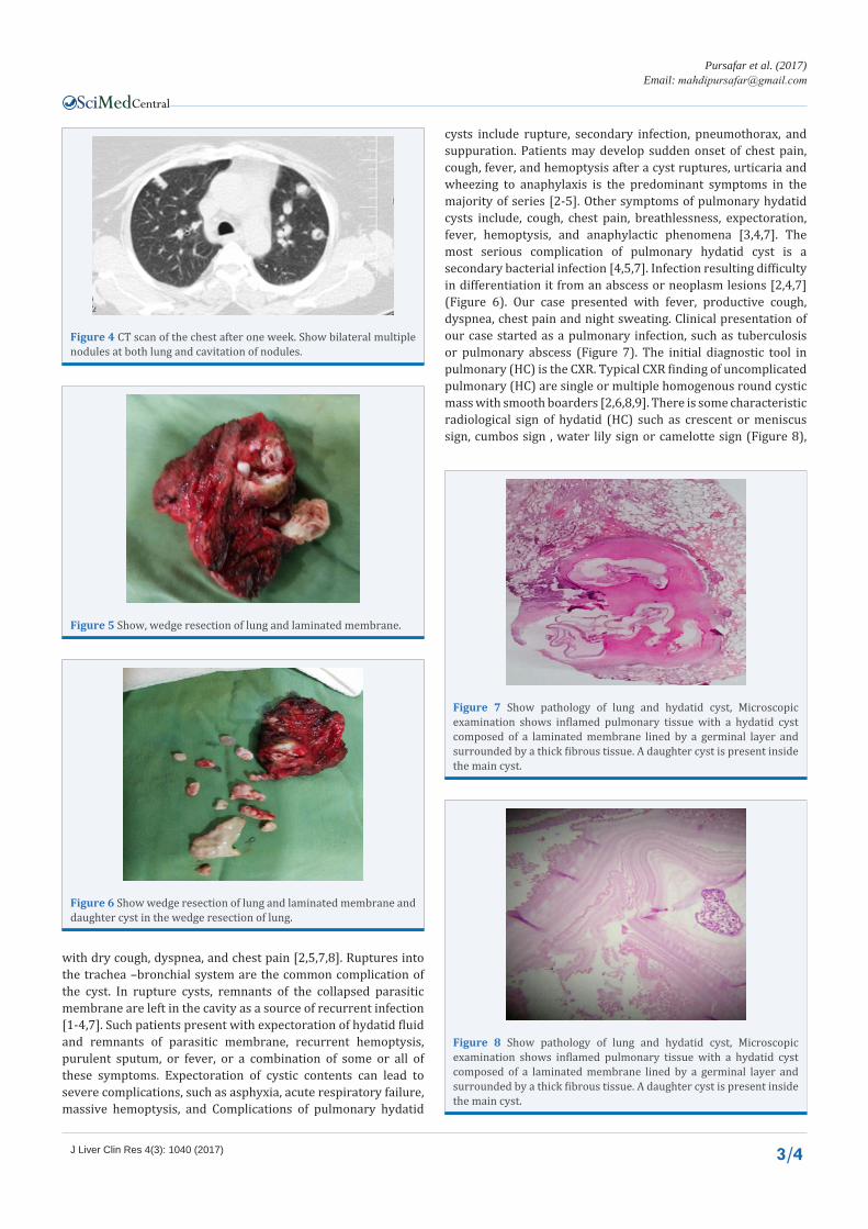
Figure 4 CT scan of the chest after one week. Show bilateral multiple nodules at both lung and cavitation of nodules.
Figure 5 Show, wedge resection of lung and laminated membrane.
Figure 6 Show wedge resection of lung and laminated membrane and daughter cyst in the wedge resection of lung.
Figure 7 Show pathology of lung and hydatid cyst, Microscopic examination shows inflamed pulmonary tissue with a hydatid cyst composed of a laminated membrane lined by a germinal layer and surrounded by a thick fibrous tissue. A daughter cyst is present inside the main cyst.
Figure 8 Show pathology of lung and hydatid cyst, Microscopic examination shows inflamed pulmonary tissue with a hydatid cyst composed of a laminated membrane lined by a germinal layer and surrounded by a thick fibrous tissue. A daughter cyst is present inside the main cyst.
with dry cough, dyspnea, and chest pain [2,5,7,8]. Ruptures into the trachea –bronchial system are the common complication of the cyst. In rupture cysts, remnants of the collapsed parasitic membrane are left in the cavity as a source of recurrent infection [1-4,7]. Such patients present with expectoration of hydatid fluid and remnants of parasitic membrane, recurrent hemoptysis, purulent sputum, or fever, or a combination of some or all of these symptoms. Expectoration of cystic contents can lead to severe complications, such as asphyxia, acute respiratory failure, massive hemoptysis, and Complications of pulmonary hydatid
cysts include rupture, secondary infection, pneumothorax, and suppuration. Patients may develop sudden onset of chest pain, cough, fever, and hemoptysis after a cyst ruptures, urticaria and wheezing to anaphylaxis is the predominant symptoms in the majority of series [2-5]. Other symptoms of pulmonary hydatid cysts include, cough, chest pain, breathlessness, expectoration, fever, hemoptysis, and anaphylactic phenomena [3,4,7]. The most serious complication of pulmonary hydatid cyst is a secondary bacterial infection [4,5,7]. Infection resulting difficulty in differentiation it from an abscess or neoplasm lesions [2,4,7] (Figure 6). Our case presented with fever, productive cough, dyspnea, chest pain and night sweating. Clinical presentation of our case started as a pulmonary infection, such as tuberculosis or pulmonary abscess (Figure 7). The initial diagnostic tool in pulmonary (HC) is the CXR. Typical CXR finding of uncomplicated pulmonary (HC) are single or multiple homogenous round cystic mass with smooth boarders [2,6,8,9]. There is some characteristic radiological sign of hydatid (HC) such as crescent or meniscus sign, cumbos sign , water lily sign or camelotte sign (Figure 8),
Central
Pursafar et al. (2017)Email:
J Liver Clin Res 4(3): 1040 (2017) 4/4
or Monods sign [2,6,8,9]. The contrast-enhanced CT-scan of the chest may show a thin enhancing rim of cyst wall [1,6,9] and diagnosis may be confirmed by the presence of daughter cysts , septation or water lily sign in ruptured cyst [1,2,4,7]. Routine blood tests are usually nonspecific and only 15% patient’s show eosinophilia due to leakage of antigenic material [1,2,8,9]. In our case, serological and immunologic tests were negative. We didn’t use these tests routinely.
The percutaneous aspiration of cyst can establish the diagnosis of (HC) by demonstrating the protoscolices, hooklets or laminated membranes, but it is too risky for routine diagnosis of lung cyst because spillage of content leads to life-threatening anaphylaxis [1,4,5,9]. The percutaneous aspiration for diagnosis of hydatid cyst is not a diagnostic approach in our area. Definitive diagnosis could be made by extraction of laminated membranes or histologically by demonstration of parasite in excised tissue [1-5,9]. Treatment of choice for (HC) is surgical excision or evacuation of the cyst [2,4,8,9]. In our patient we used one lung anesthesia and with complete left lateral positions, fourth intercostal space was opened and exploration was performed ,multiple nodules were seen ,one of that nodules was excised with wedge resection ,the resected tissue was catted, laminated membrane and daughter cyst was presented .Two of the rest nodules has hydatid cyst element. Some scolicidal agents such as hypertonic saline, povidone iodine, formalin, cetrimide, ethanol, and hydrogen peroxide can be used intraoperatively [1,4,5,7],
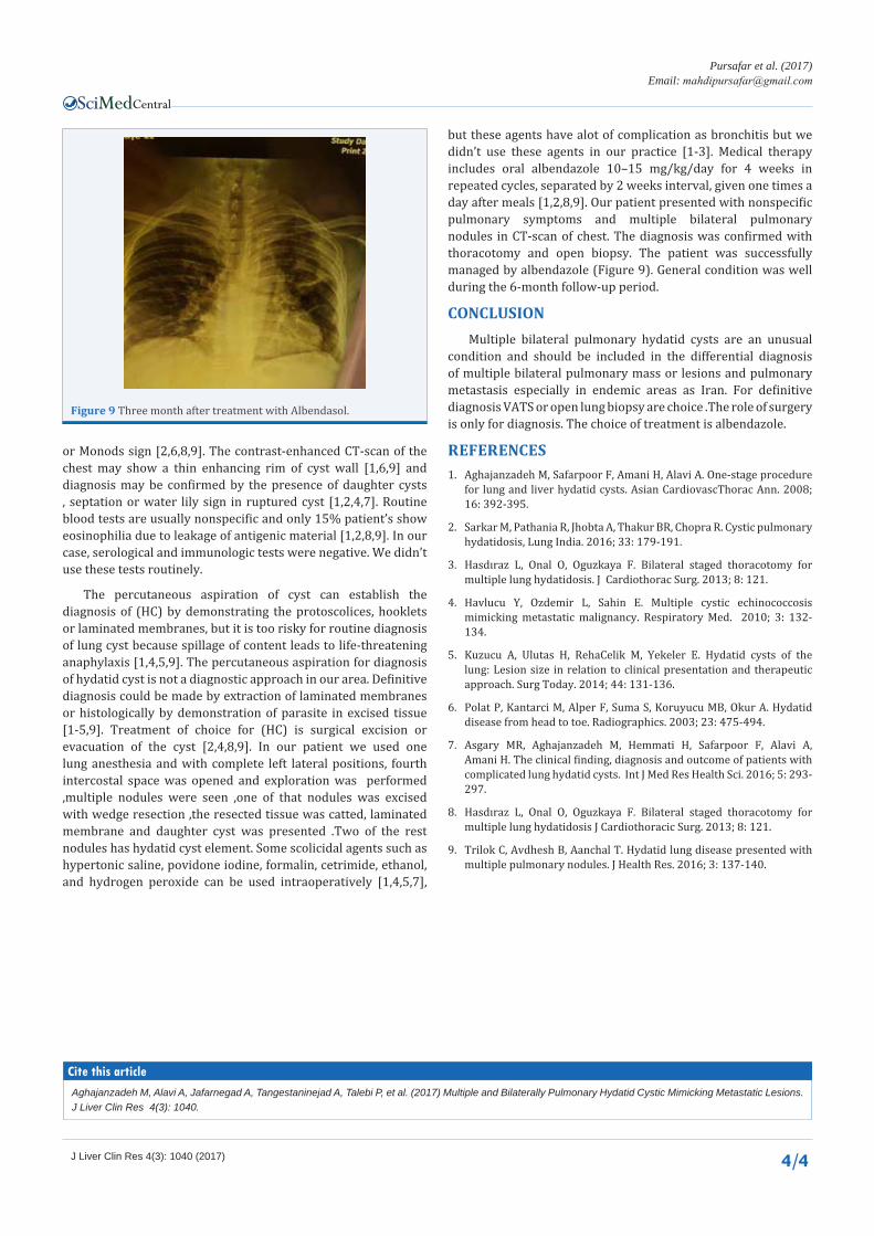
but these agents have alot of complication as bronchitis but we didn’t use these agents in our practice [1-3]. Medical therapy includes oral albendazole 10–15 mg/kg/day for 4 weeks in repeated cycles, separated by 2 weeks interval, given one times a day after meals [1,2,8,9]. Our patient presented with nonspecific pulmonary symptoms and multiple bilateral pulmonary nodules in CT-scan of chest. The diagnosis was confirmed with thoracotomy and open biopsy. The patient was successfully managed by albendazole (Figure 9). General condition was well during the 6-month follow-up period.
CONCLUSIONMultiple bilateral pulmonary hydatid cysts are an unusual
condition and should be included in the differential diagnosis of multiple bilateral pulmonary mass or lesions and pulmonary metastasis especially in endemic areas as Iran. For definitive diagnosis VATS or open lung biopsy are choice .The role of surgery is only for diagnosis. The choice of treatment is albendazole.
REFERENCES1. Aghajanzadeh M, Safarpoor F, Amani H, Alavi A. One-stage procedure
for lung and liver hydatid cysts. Asian CardiovascThorac Ann. 2008; 16: 392-395.
2. Sarkar M, Pathania R, Jhobta A, Thakur BR, Chopra R. Cystic pulmonary hydatidosis, Lung India. 2016; 33: 179-191.
3. Hasdıraz L, Onal O, Oguzkaya F. Bilateral staged thoracotomy for multiple lung hydatidosis. J Cardiothorac Surg. 2013; 8: 121.
4. Havlucu Y, Ozdemir L, Sahin E. Multiple cystic echinococcosis mimicking metastatic malignancy. Respiratory Med. 2010; 3: 132-134.
5. Kuzucu A, Ulutas H, RehaCelik M, Yekeler E. Hydatid cysts of the lung: Lesion size in relation to clinical presentation and therapeutic approach. Surg Today. 2014; 44: 131-136.
6. Polat P, Kantarci M, Alper F, Suma S, Koruyucu MB, Okur A. Hydatid disease from head to toe. Radiographics. 2003; 23: 475-494.
7. Asgary MR, Aghajanzadeh M, Hemmati H, Safarpoor F, Alavi A, Amani H. The clinical finding, diagnosis and outcome of patients with complicated lung hydatid cysts. Int J Med Res Health Sci. 2016; 5: 293-297.
8. Hasdıraz L, Onal O, Oguzkaya F. Bilateral staged thoracotomy for multiple lung hydatidosis J Cardiothoracic Surg. 2013; 8: 121.
9. Trilok C, Avdhesh B, Aanchal T. Hydatid lung disease presented with multiple pulmonary nodules. J Health Res. 2016; 3: 137-140.
Figure 9 Three month after treatment with Albendasol.
Aghajanzadeh M, Alavi A, Jafarnegad A, Tangestaninejad A, Talebi P, et al. (2017) Multiple and Bilaterally Pulmonary Hydatid Cystic Mimicking Metastatic Lesions. J Liver Clin Res 4(3): 1040.
Cite this article