Embed Size (px)
Citation preview

The Cardiovascular System
• Part 1: The Heart and the Cardiovascular System
• Part 2: The Cardiac Cycle and Cardiac Output • Part 3: The Vasculature and Its Control
• Some Basic Ideas: – heart pumps blood into two “circuits” • right side of heart pumps into pulmonary circuit – to and from lungs
• leF side of heart pumps into systemic circuit – to and from enGre body – includes coronary blood flow
The Cardiovascular System

• Some Basic Ideas: – systemic and pulmonary circuits are “in series” • blood coming from the pulmonary circuit enters the systemic circuit • blood from the systemic circuit enters the pulmonary circuit
The Cardiovascular System
The Cardiovascular System
• Some Basic Ideas: – arteries
• carry blood away from heart
• are muscular, can change diameter
• offer resistance to blood flow (uses up blood pressure as blood moves) – “resistance vessels”
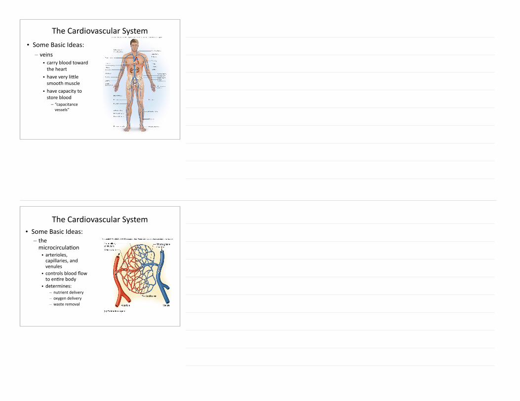
The Cardiovascular System • Some Basic Ideas: – veins • carry blood toward the heart • have very liMle smooth muscle • have capacity to store blood – “capacitance vessels”
The Cardiovascular System • Some Basic Ideas: – the microcirculaGon • arterioles, capillaries, and venules • controls blood flow to enGre body • determines:
– nutrient delivery – oxygen delivery – waste removal

The Pericardium:ProtecGon for the Heart
• fibrous pericardium (layer) – fibrous Gssue of pericardial sac: dense irregular connecGve Gssue
The Pericardium:ProtecGon for the Heart
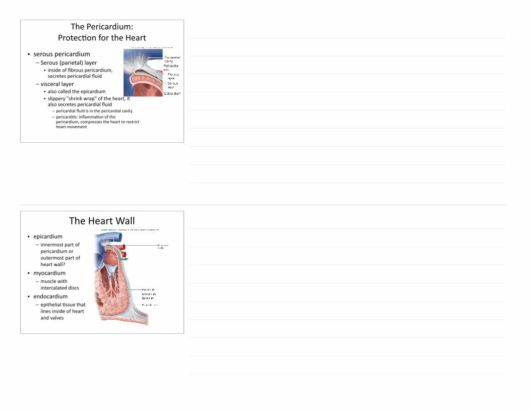
• serous pericardium – Serous (parietal) layer • inside of fibrous pericardium,secretes pericardial fluid
– visceral layer • also called the epicardium • slippery "shrink wrap" of the heart, it also secretes pericardial fluid – pericardial fluid is in the pericardial cavity – pericardiGs: inflammaGon of the pericardium, compresses the heart to restrict heart movement
The Heart Wall• epicardium – innermost part of pericardium or outermost part of heart wall?
• myocardium – muscle with intercalated discs
• endocardium – epithelial Gssue that lines inside of heart and valves
The 4 Chambers of the Heart
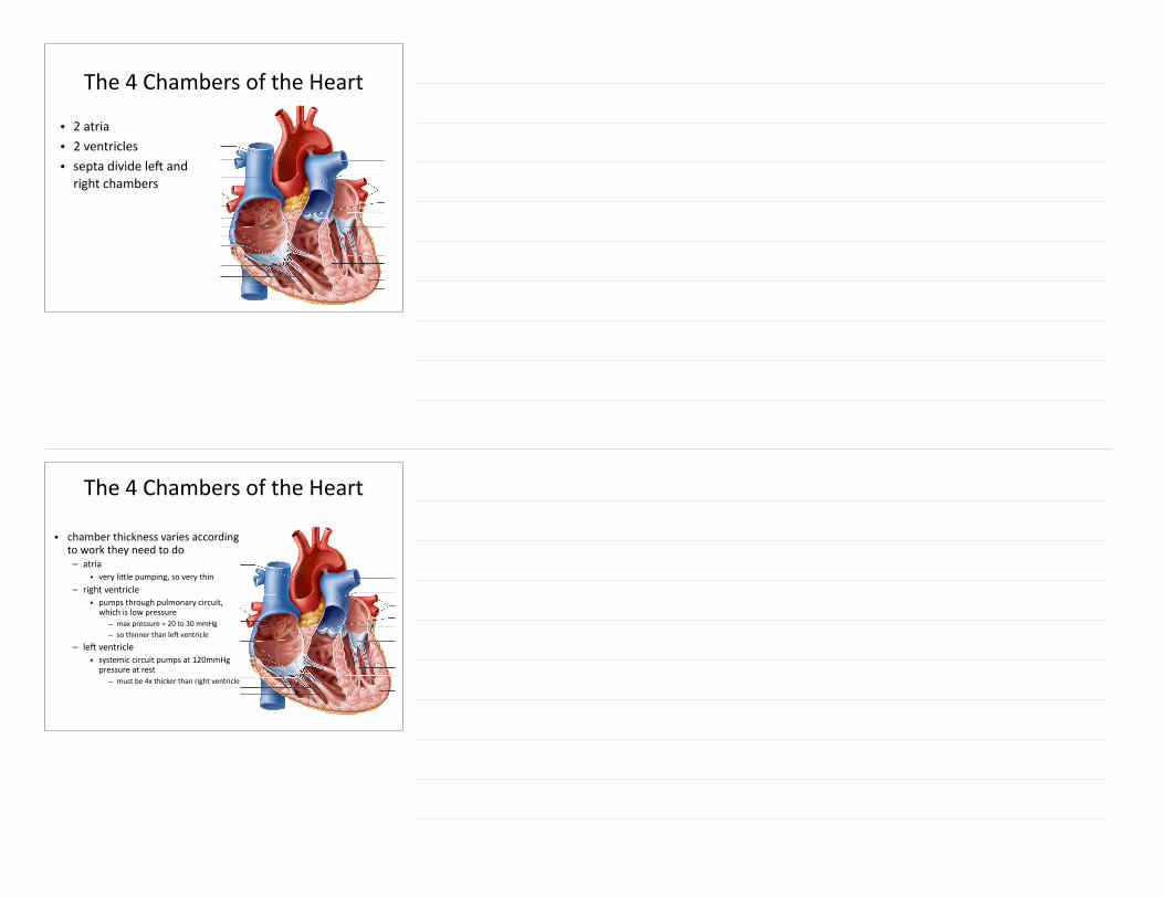
• 2 atria • 2 ventricles • septa divide leF and right chambers
The 4 Chambers of the Heart
• chamber thickness varies according to work they need to do – atria • very liMle pumping, so very thin
– right ventricle • pumps through pulmonary circuit, which is low pressure – max pressure = 20 to 30 mmHg – so thinner than leF ventricle
– leF ventricle • systemic circuit pumps at 120mmHg pressure at rest – must be 4x thicker than right ventricle
The 4 Chambers of the Heart
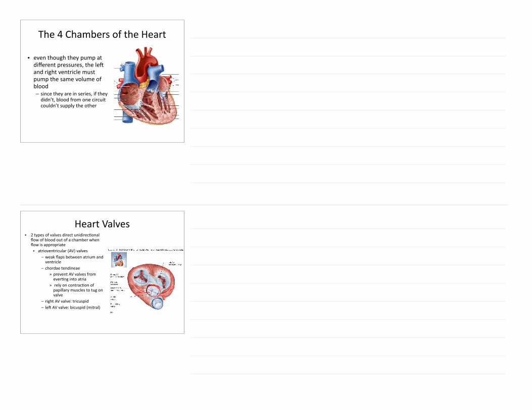
• even though they pump at different pressures, the leF and right ventricle must pump the same volume of blood – since they are in series, if they didn’t, blood from one circuit couldn’t supply the other
Heart Valves• 2 types of valves direct unidirecGonal
flow of blood out of a chamber when flow is appropriate • atrioventricular (AV) valves – weak flaps between atrium and ventricle – chordae tendineae » prevent AV valves from everGng into atria » rely on contracGon of papillary muscles to tug on valve
– right AV valve: tricuspid – leF AV valve: bicuspid (mitral)
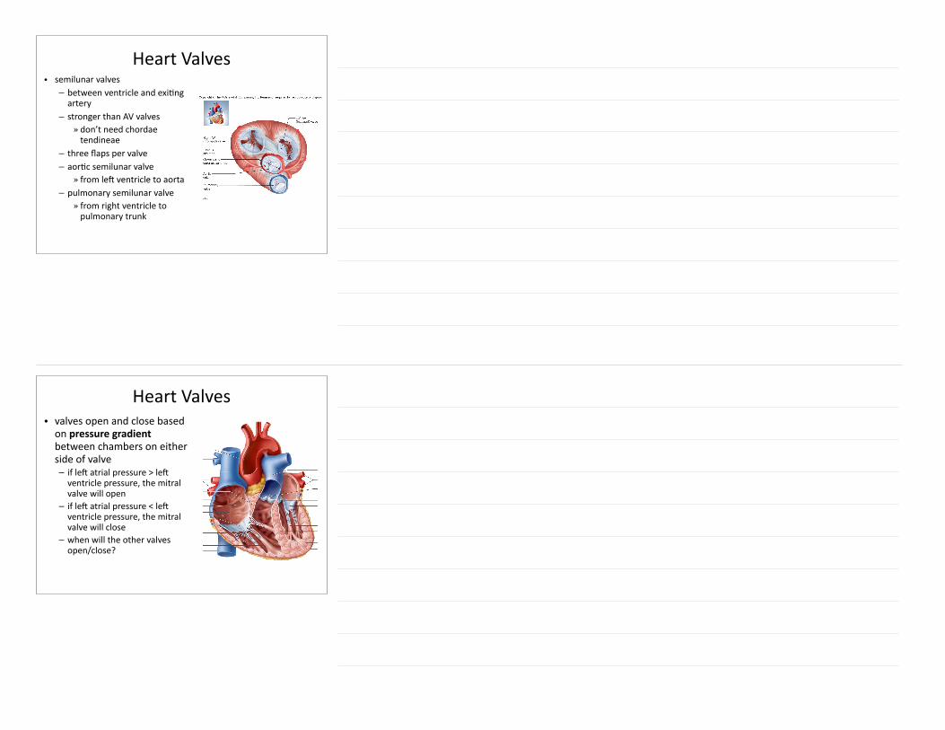
Heart Valves• semilunar valves – between ventricle and exiGng artery – stronger than AV valves » don’t need chordae tendineae
– three flaps per valve – aorGc semilunar valve » from leF ventricle to aorta
– pulmonary semilunar valve » from right ventricle to pulmonary trunk
Heart Valves• valves open and close based on pressure gradient between chambers on either side of valve – if leF atrial pressure > leF ventricle pressure, the mitral valve will open – if leF atrial pressure < leF ventricle pressure, the mitral valve will close – when will the other valves open/close?

Heart Valves• heart sounds
• first heart sound created when AV valves close –“lubb”
• second heart sound created when semilunar valves close –“dub”
• normal sound should be “lubb-‐dub”
Heart Valves
• heart murmur –noise when valves don't close fully and therefore leak • “lubb-‐whoosh-‐dub” would indicate AV valve problem • what would a semilunar valve murmur sound like?

Heart Valves• rheumaGc fever –strep bacterial infecGon • anGbodies damage valves, prevenGng them from closing well • oFen requires valve replacement
Coronary Blood Flow• leF and right coronary arteries come off aorta • coronary sinus collects “deoxygenated”
coronary blood, returns it to right atrium • coronary blood flow occurs when heart muscle
is relaxed (diastole) – heart needs adequate diastolic Gme to
perfuse all heart muscle – ischemia (reduced blood flow) causes
angina pectoralis (chest pain) – myocardial infarcGon (death of heart Gssue)
results from interrupted blood flow to an area of heart muscle
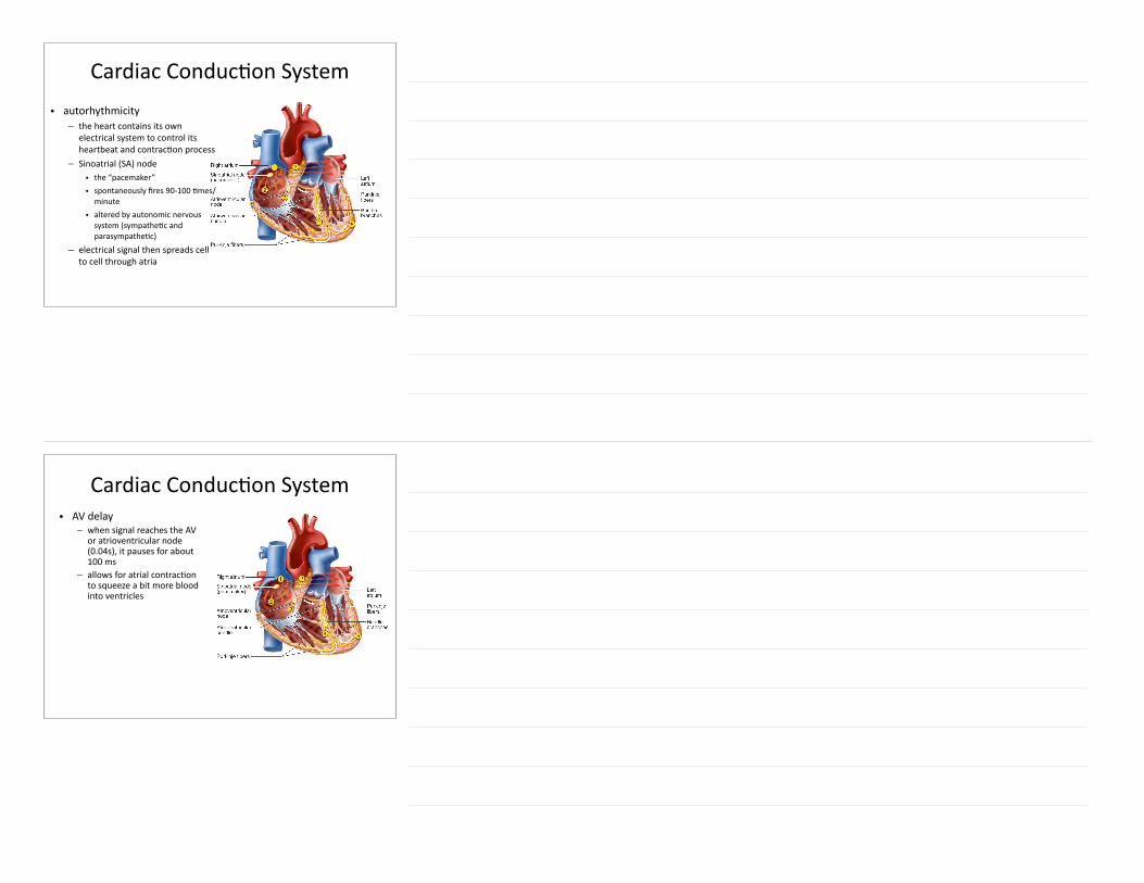
Cardiac ConducGon System• autorhythmicity – the heart contains its own
electrical system to control its heartbeat and contracGon process
– Sinoatrial (SA) node • the “pacemaker” • spontaneously fires 90-‐100 Gmes/minute • altered by autonomic nervous system (sympatheGc and parasympatheGc)
– electrical signal then spreads cell to cell through atria
Cardiac ConducGon System• AV delay – when signal reaches the AV
or atrioventricular node (0.04s), it pauses for about 100 ms
– allows for atrial contracGon to squeeze a bit more blood into ventricles

Cardiac ConducGon System• aFer AV delay, electrical signal
spreads through ventricular conducGon system quickly – AV bundle (Bundle of His) – leF and right bundle branches – Purkinje fibers • reach every ventricle muscle fiber • ensures that every cardiac muscle fiber is sGmulated
Cardiac Muscle AcGon PotenGal
• phase 0: depolarizaGon – due to voltage-‐gated Na+ channels (remember from neural physiology?)
• phase 1: early repolarizaGon – due to opening of voltage-‐gated K+ channels

Cardiac Muscle AcGon PotenGal
• phase 2: the plateau – due to opening of L-‐type calcium (Ca+2)
channels (L = long) • ensures that heart cannot be sGmulated again, before the myocardium completes its contracGon and relaxaGon – prevents cardiac muscle from going into tetanus like skeletal muscle can
• also allows for Ca+2 entry for binding to troponin – cardiac muscle doesn’t have a well-‐developed sarcoplasmic reGculum like skeletal muscle does
Cardiac Muscle AcGon PotenGal
• phase 3: repolarizaGon – due to closing of L-‐type Ca channels, with K+ channels sGll open
• phase 4: relaxaGon – all channels closed
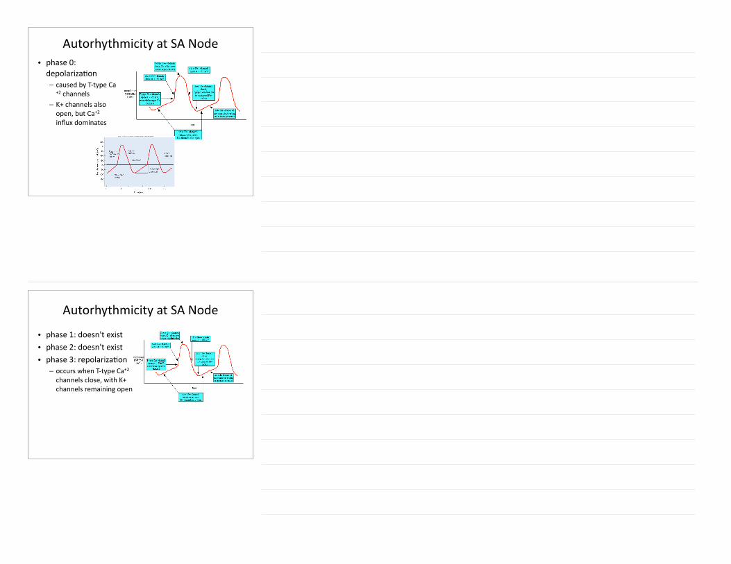
Autorhythmicity at SA Node• phase 0: depolarizaGon – caused by T-‐type Ca
+2 channels – K+ channels also open, but Ca+2 influx dominates
Autorhythmicity at SA Node
• phase 1: doesn't exist • phase 2: doesn't exist • phase 3: repolarizaGon – occurs when T-‐type Ca+2 channels close, with K+ channels remaining open
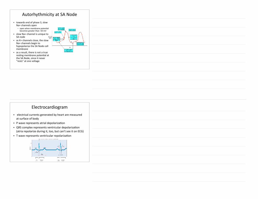
Autorhythmicity at SA Node• towards end of phase 3, slow
Na+ channels open – open when membrane potenGal
becomes greater than -‐50 mV • slow Na+ channel is unique to
SA node • as K+ channels close, the slow
Na+ channels begin to hypopolarize the SA Node cell membrane
• as a result, there is not a true resGng membrane potenGal at the SA Node, since it never “rests” at one voltage
Electrocardiogram• electrical currents generated by heart are measured at surface of body • P wave represents atrial depolarizaGon • QRS complex represents ventricular depolarizaGon (atria repolarize during it, too, but can't see it on ECG) • T wave represents ventricular repolarizaGon
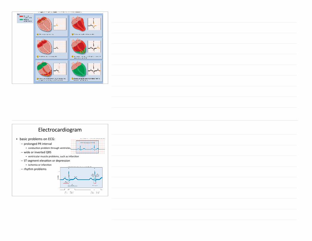
Electrocardiogram• basic problems on ECG: – prolonged PR interval • conducGon problem through ventricles
– wide or inverted QRS • ventricular muscle problems, such as infarcGon
– ST segment elevaGon or depression • ischemia or infarcGon
– rhythm problems