Embed Size (px)
DESCRIPTION
Cardiovascular System Outline. Blood Composition WBC (granular and a granular), RBC, Platelets, Plasma, Serum Heart Anatomy Be able to label the different parts of the heart Blood Flow Be able to trace the flow of blood through the heart Cardiac Conduction System - PowerPoint PPT Presentation
Citation preview
Blood Composition WBC (granular and agranular), RBC, Platelets, Plasma, Serum
Heart Anatomy Be able to label the different parts of the heart
Blood Flow Be able to trace the flow of blood through the heart
Cardiac Conduction System Know the different components of the CCS and how the impulse
flows through the heart SA Node, AV Node, AV Bundle, Purkinje Fibers
ECG What happens during each wave of the electrocardiogram Know the different abnormalities that we discussed in class
Heart Disorders Study the jigsaw fact sheet; specifically know what each disorder is
It seems like a lot, but you’ve got this!!

AB
C
D
E
F
G
HI
J
K
L
M
N
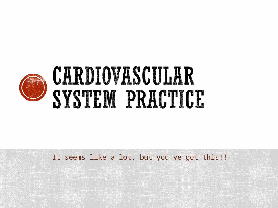
1. _______ Aorta A. Wave that indicates ventricular repolarization
2. _______ Plasma B. Delivers oxygen to every cell in the body
3. _______ Red Blood Cells C. Delay in signal
4. _______ Platelets D. Receives deoxygenated blood from the body
5. _______ White Blood Cells E. Connects atria to ventricles
6. _______ Right Atria F. > 100 beats per minute
7. _______ Left Atria G. Receives oxygen rich blood from the lungs
8. _______ Right Ventricle H. Branches off the heart and then divides into smaller arteries
9. _______ Left Ventricle I. Stops you from bleeding to death by forming clots
10._______ Valve J. Natural pace maker of the heart
11._______AV Node K. Defend the body from infection
12.______SA Node L. Pumps blood to entire body
13._______P Wave M. Wave that indicates atrial contraction
14._______T Wave N. Pumps blood to the lungs
15._______Tachycardia O. Made of mostly water
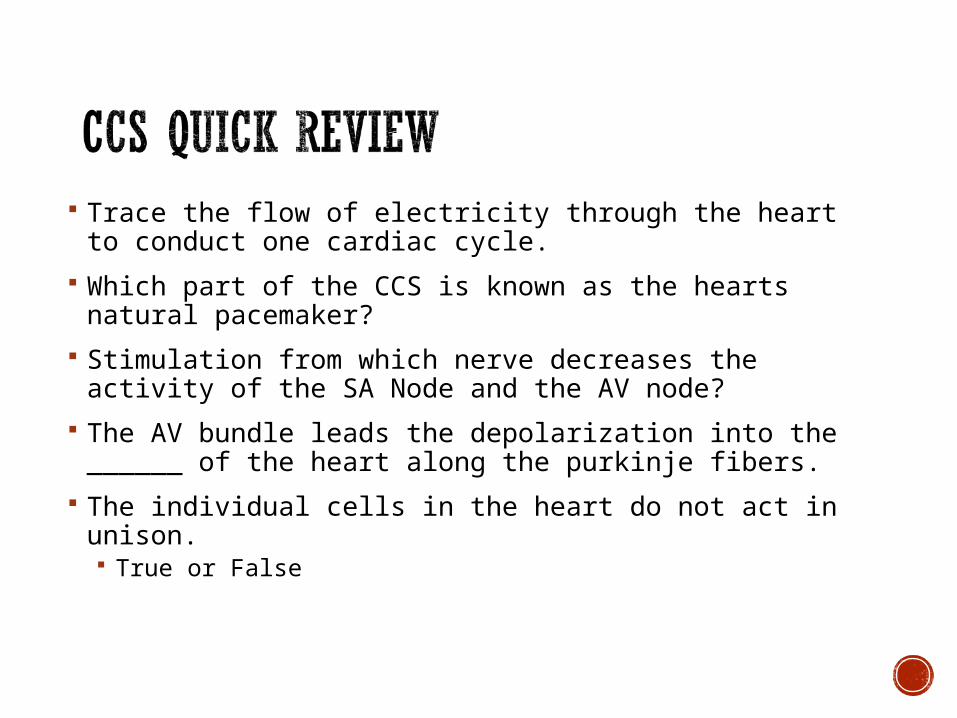
Trace the flow of electricity through the heart to conduct one cardiac cycle.
Which part of the CCS is known as the hearts natural pacemaker?
Stimulation from which nerve decreases the activity of the SA Node and the AV node?
The AV bundle leads the depolarization into the ______ of the heart along the purkinje fibers.
The individual cells in the heart do not act in unison. True or False

Describe what occurs during the T Wave.
Describe what occurs during the P Wave.
Describe what occurs during the QRS Wave.
List and briefly describe two ECG abnormalities.

____1. Accumulation of plaque buildup on walls of arteries
____2. One or both cusps of mitral valve stretches and bulges into left atrium during ventricular contraction.
____3. Can cause the heart muscle to die due to lack of oxygen.
____4. Caused by an obstructed vessel in heart thrombus.
____5. Inflammation of pericardial sac due to viral or bacterial infection.
A. Hypertension
B. Coronary Artery Disease
C. Myocardial Infarction
D. Pericarditis
E. Mitral Valve Prolapse


![Lecture 15 - Cardiovascular - Bio 105.pptx [Read-Only] 15...Chapter 12 Copyright © 2009 Pearson Education, Inc. Outline I. Functions of cardiovascular system ... Cardiovascular System](https://img.pdfslide.us/doc/110x75/5b8a06a07f8b9a5b688edf43/lecture-15-cardiovascular-bio-105pptx-read-only-15chapter-12-copyright.jpg)