Embed Size (px)
Citation preview
Cardiovascular system I.Terminology, ECG basics, heart sounds
Components of cardiovascular system
• Blood
– Liquid medium that is responsible for transport
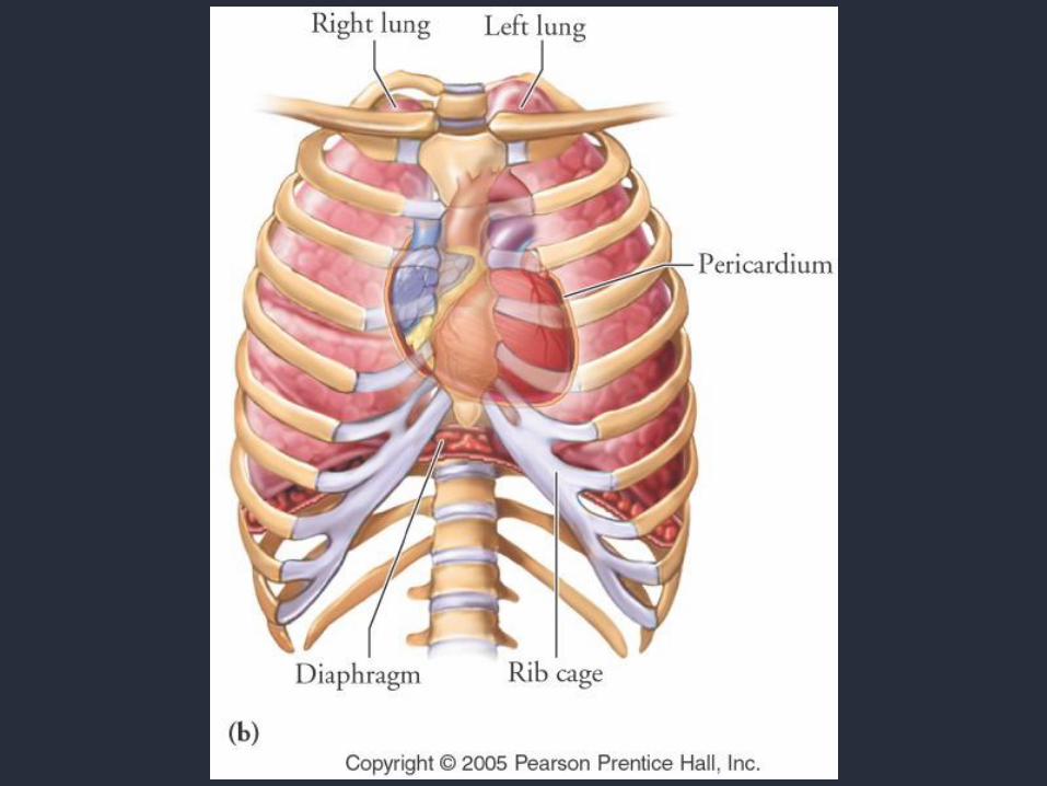
• Heart
– Pump that creates pressure
• Blood vessels
– Distribution cannals/pipes of various lumen
Heart characteristics• Inotropy
– Ability of heart to contract
• Batmotropy– Ability of heart to get excited
• Dromotropy– Ability of heart to conduct impulse
• Chronotropy– Ability of heart to create impulses
• Luzitropy– Ability of heart to relax
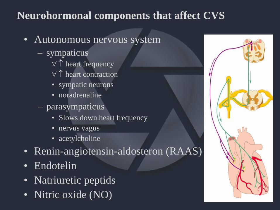
Neurohormonal components that affect CVS
• Autonomous nervous system
– sympaticus
heart frequency
heart contraction
• sympatic neurons
• noradrenaline
– parasympaticus
• Slows down heart frequency
• nervus vagus
• acetylcholine
• Renin-angiotensin-aldosteron (RAAS)
• Endotelin
• Natriuretic peptids
• Nitric oxide (NO)

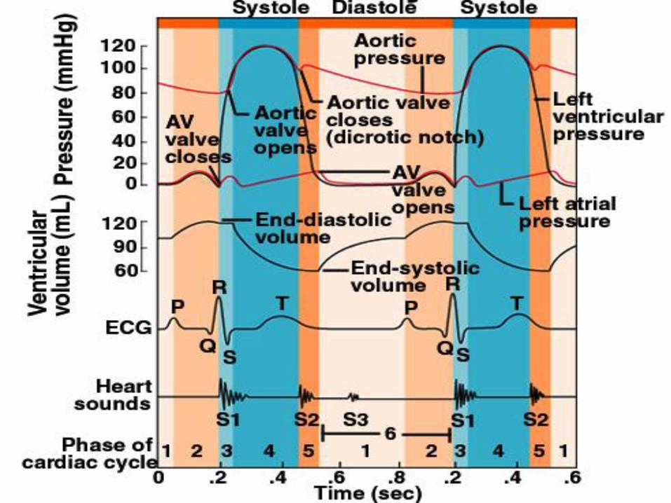
1/3 systole
2/3 diastole
80% ventricle filling ispassive
20% is active by atrium contraction
Heart cycle
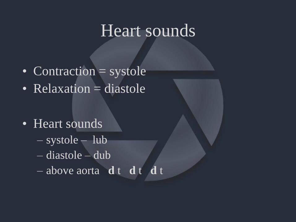
Heart sounds
• Contraction = systole
• Relaxation = diastole
• Heart sounds
– systole – lub
– diastole – dub
– above aorta d t d t d t
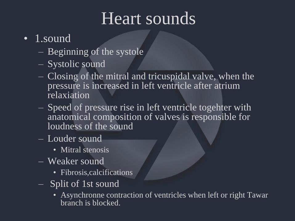
Heart sounds• 1.sound
– Beginning of the systole
– Systolic sound
– Closing of the mitral and tricuspidal valve, when thepressure is increased in left ventricle after atriumrelaxiation
– Speed of pressure rise in left ventricle togehter withanatomical composition of valves is responsible forloudness of the sound
– Louder sound• Mitral stenosis
– Weaker sound• Fibrosis,calcifications
– Split of 1st sound• Asynchronne contraction of ventricles when left or right Tawar
branch is blocked.
Heart sounds
• 2. sound
– Closing of semilunar aortal and pulmonar valve
– Two components
• Pulmonar
• Aortal
– Split of 2nd sound
• enhanced in inspirium
Heart sounds
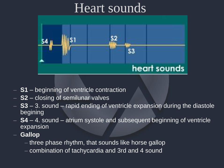
– S1 – beginning of ventricle contraction
– S2 – closing of semilunar valves
– S3 – 3. sound – rapid ending of ventricle expansion during the diastole begining
– S4 – 4. sound – atrium systole and subsequent beginning of ventricleexpansion
– Gallop
– three phase rhythm, that sounds like horse gallop
– combination of tachycardia and 3rd and 4 sound
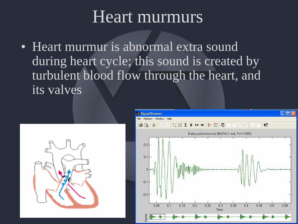
Heart murmurs
• Heart murmur is abnormal extra soundduring heart cycle; this sound is created by turbulent blood flow through the heart, and its valves
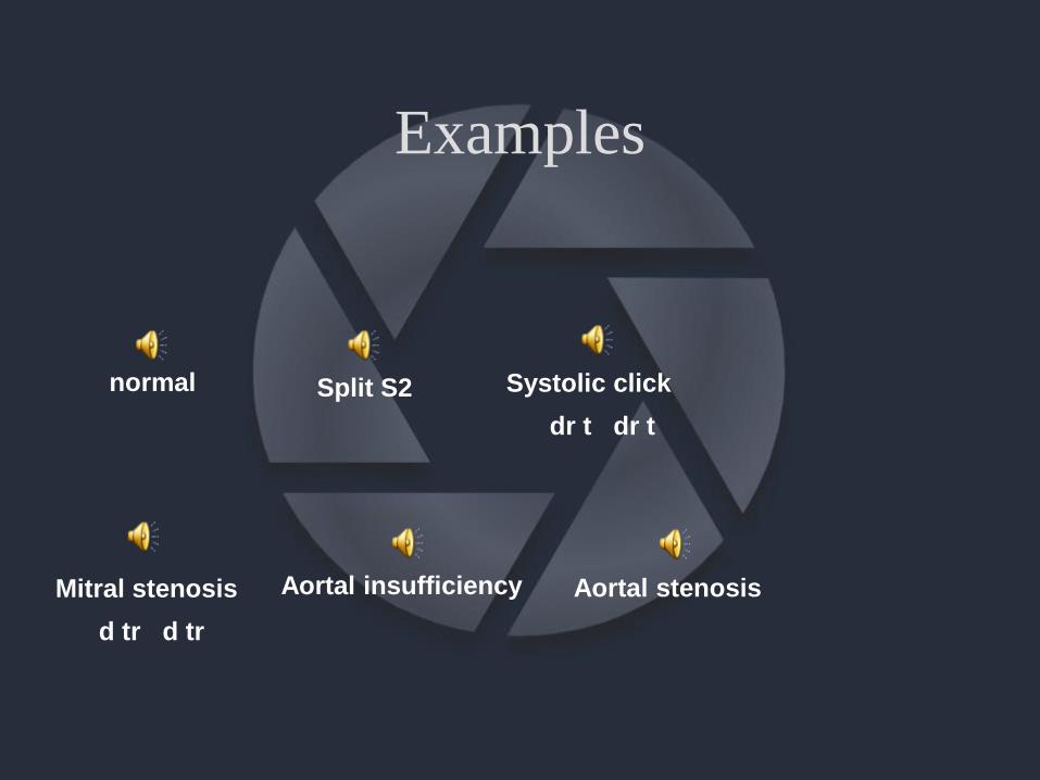
Examples
normal Split S2
Aortal insufficiency Aortal stenosisMitral stenosis
d tr d tr
Systolic click
dr t dr t
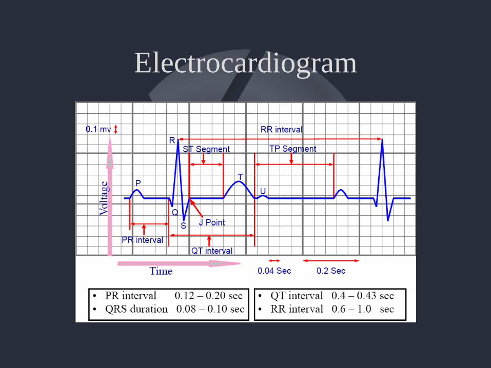
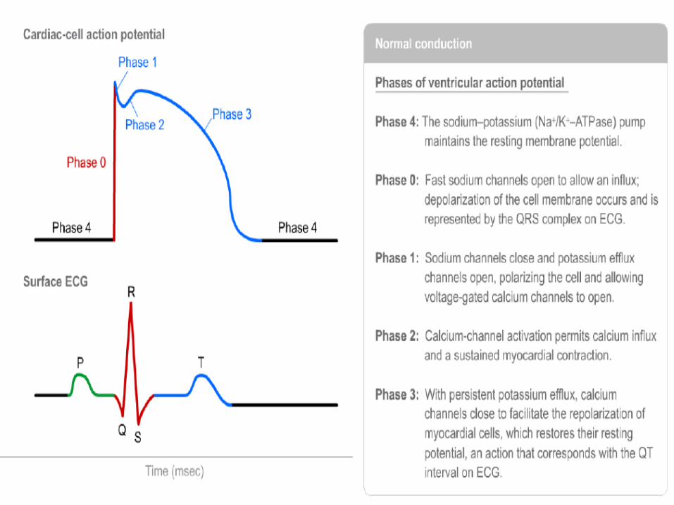
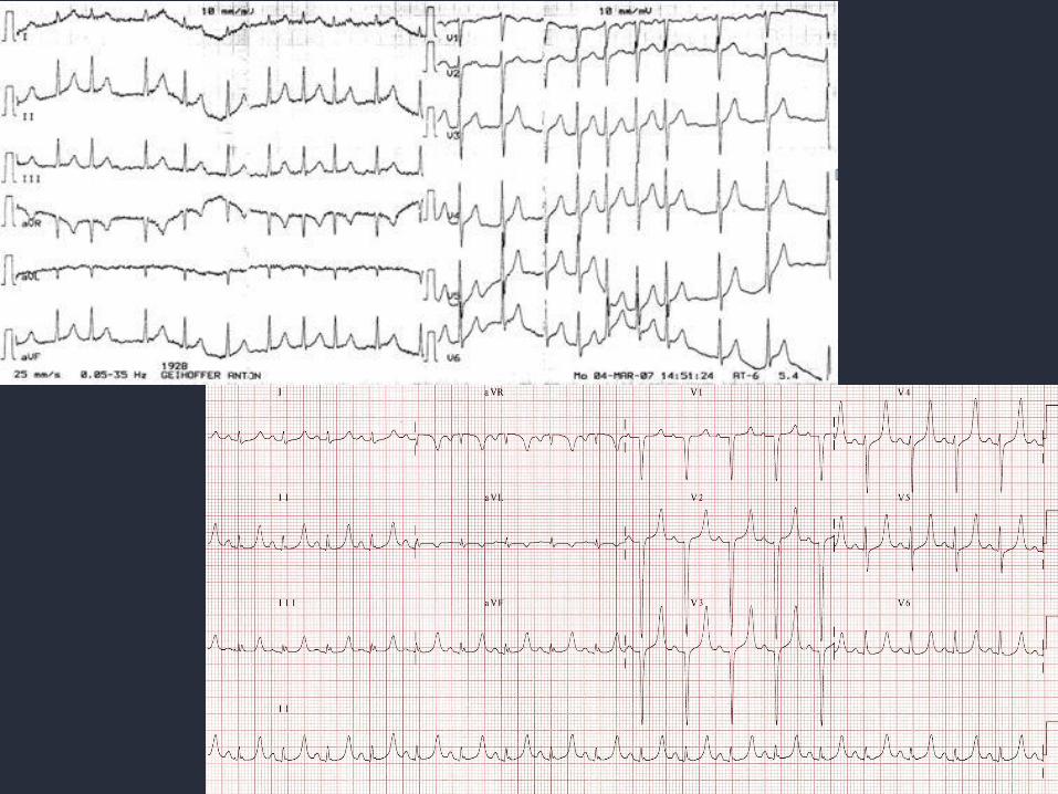
Electrocardiogram
ECG• P wave/PQ interval
– Depolarization of atria/including AV and His bundle
• PQ segment
– Depolarization of AV node and His bundle
• QRS
– Depolarization of ventricles
• ST segment
– Repolarization of ventricles
• Repolarization of atria?
– Hidden in QRS complex
Conduit times
• PQ/PR interval – atrium depolarisation
– 0.12 – 0.20 s
• QRS complex –depolarization of ventricles
– < 0.12 sec
• QT interval - < 0.36 sec
– QTc = (QT/√RR) = <0.44 sec
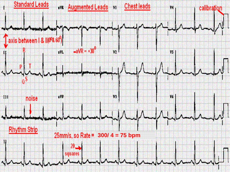
Leads of a 12-lead ECG
• Bipolar
– I, II, III
• Unipolar
– Augmented limb leads
• aVR, aVL, aVF*
– Chest/precordial leads
• V1, V2, V3, V4, V5, V6
• V can be also C
*a = augmented, V = voltage, R = right hand, L = left hand, F = foot
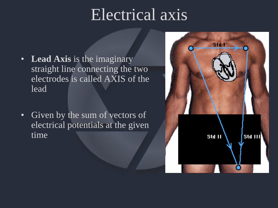
Electrical axis
• Lead Axis is the imaginary straight line connecting the two electrodes is called AXIS of the lead
• Given by the sum of vectors of electrical potentials at the giventime
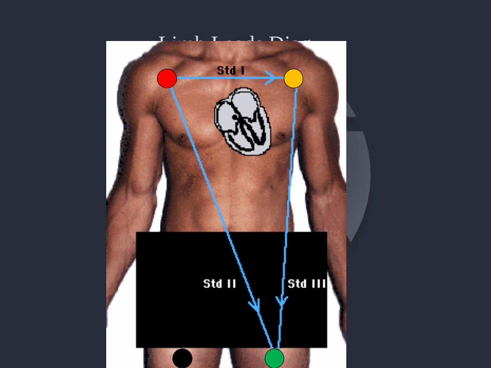
Limb Leads Diag.
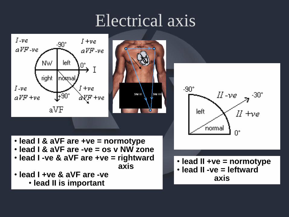
Electrical axis
• lead I & aVF are +ve = normotype• lead I & aVF are -ve = os v NW zone• lead I -ve & aVF are +ve = rightward
axis• lead I +ve & aVF are -ve
• lead II is important
• lead II +ve = normotype• lead II -ve = leftward
axis
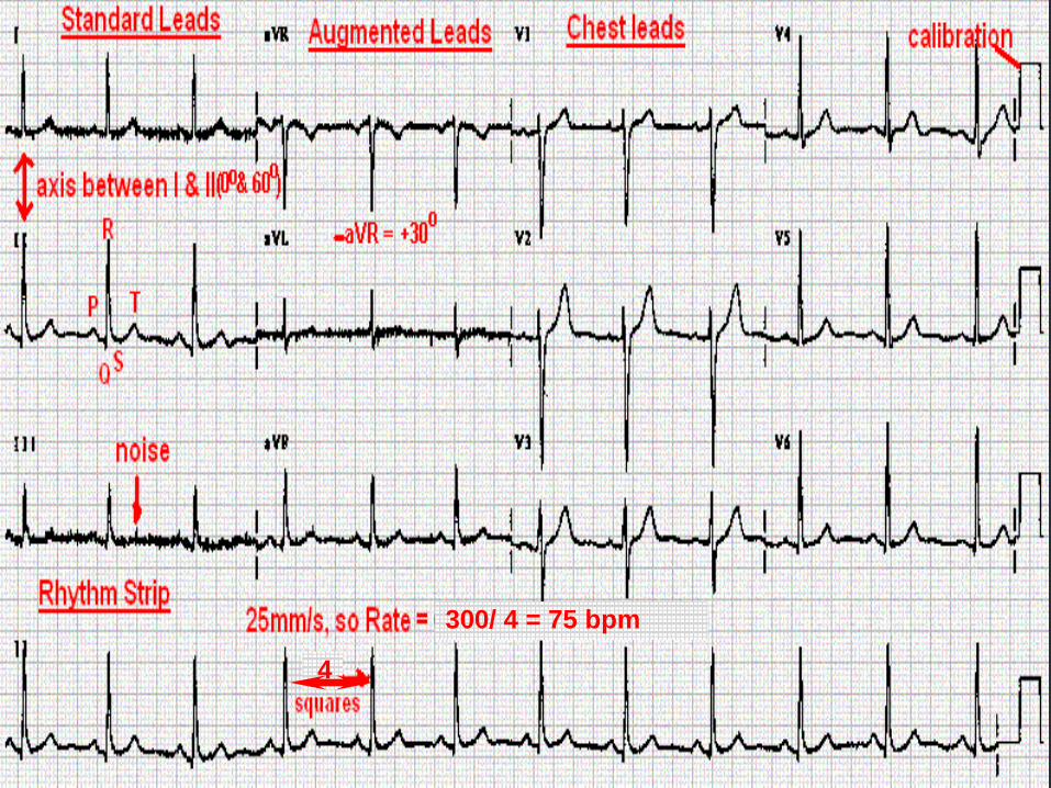
300/ 4 = 75 bpm
4
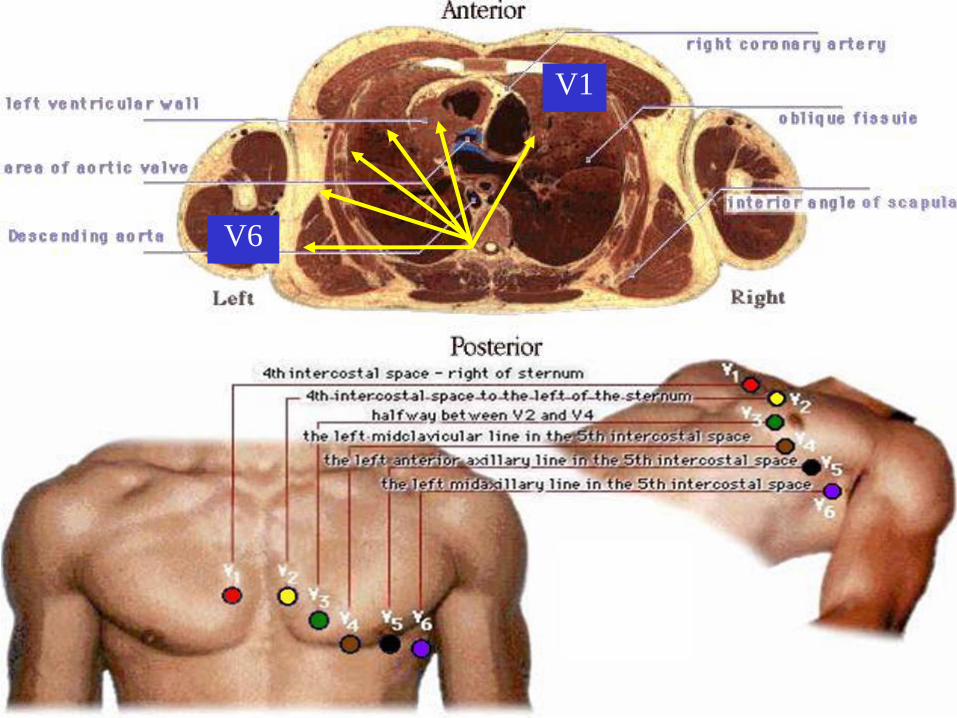
V1
V6
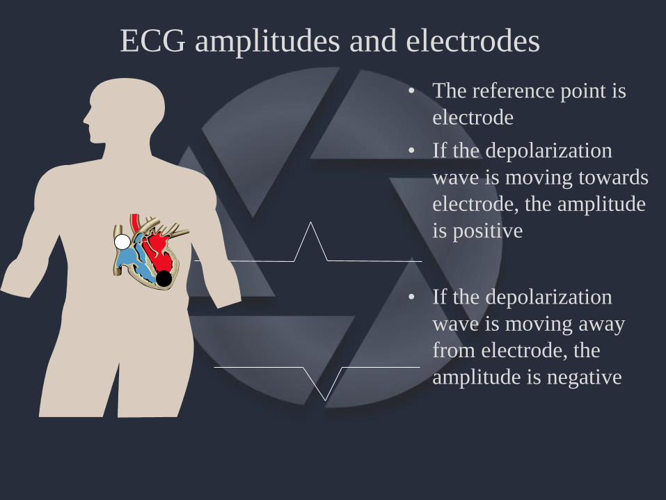
ECG amplitudes and electrodes
• The reference point is
electrode
• If the depolarization
wave is moving towards
electrode, the amplitude
is positive
• If the depolarization
wave is moving away
from electrode, the
amplitude is negative
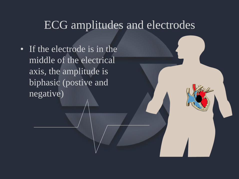
ECG amplitudes and electrodes
• If the electrode is in the
middle of the electrical
axis, the amplitude is
biphasic (postive and
negative)
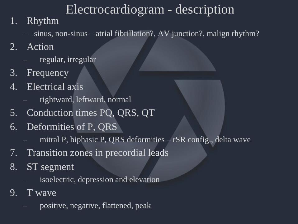
Electrocardiogram - description1. Rhythm
– sinus, non-sinus – atrial fibrillation?, AV junction?, malign rhythm?
2. Action
– regular, irregular
3. Frequency
4. Electrical axis
– rightward, leftward, normal
5. Conduction times PQ, QRS, QT
6. Deformities of P, QRS
– mitral P, biphasic P, QRS deformities – rSR config., delta wave
7. Transition zones in precordial leads
8. ST segment
– isoelectric, depression and elevation
9. T wave
– positive, negative, flattened, peak
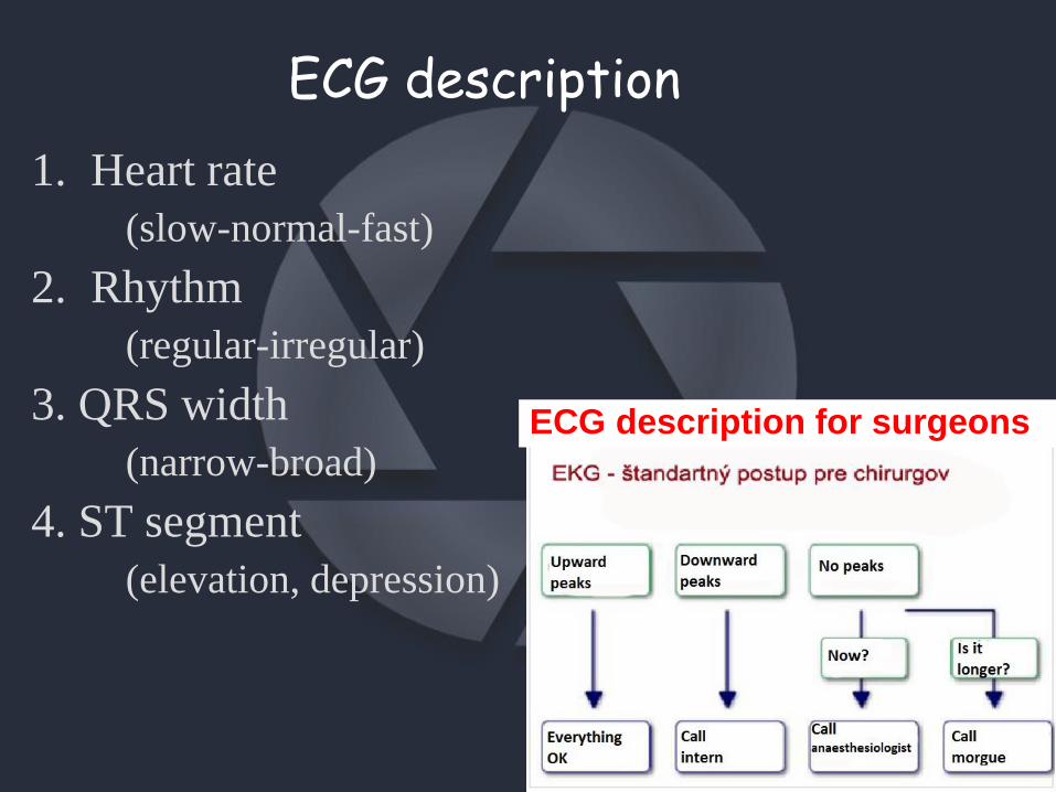
ECG description
1. Heart rate
(slow-normal-fast)
2. Rhythm
(regular-irregular)
3. QRS width
(narrow-broad)
4. ST segment
(elevation, depression)
? ?
ECG description for surgeons
300/ 4 = 75 bpm

Practical tasks
• Auscultation of heart sounds (p. 127)
• Examination of arterial pulse (p.141)
• Experiments on the isolated heart muscle(p.136)