Embed Size (px)
DESCRIPTION
Nicole Weiss, MD Tulane University, December 13, 2012. Cardiac disease Part II. Time Crunch…. Valvular Heart Disease Hypertrophic Cardiomyopathy The Transplanted Heart Congenital Heart Disease Simple Shunts Complex Shunts Antibiotic Prophylaxis Pacemaker Classification. - PowerPoint PPT Presentation
Citation preview
CARDIAC DISEASE PART IINicole Weiss, MD Tulane University, December 13, 2012
Time Crunch… Valvular Heart Disease Hypertrophic Cardiomyopathy The Transplanted Heart Congenital Heart Disease
Simple Shunts Complex Shunts
Antibiotic Prophylaxis Pacemaker Classification
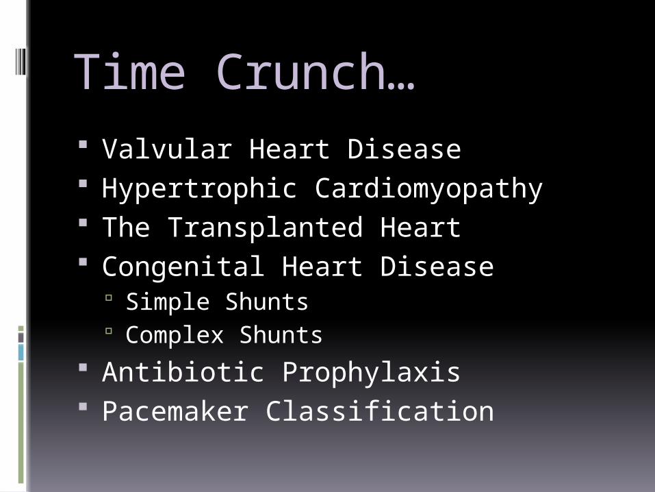
New York Classification of Functional Heart DiseaseClass I: Asymptomatic except during Severe
ExertionClass II: Symptomatic with Moderate ActivityClass III: Symptomatic with Minimal ActivityClass IV: Symptomatic at Rest
Valvular Disease
Mitral Stenosis Most common etiology is rheumatic
disease Symptoms develop 20-30 years later
when mitral valve area decreases from 4-6 cm2 to less than 2cm2
Prone to Pulmonary Hypertension & Pulmonary Edema as Left Atrial Pressures Increase
Anesthetic Goals for Mitral Stenosis Pulmonary Artery Catheter?
Yes, pulmonary artery pressures help guide fluid management
Patients are prone to volume overload and pulmonary edema SVR?
High, flow through the stenotic valve is limited and the heart cannot compensate for decreases in preload
Heart Rate? Normal Sinus Rhythm, Filling is dependent on atrial kick, but
too low and the cardiac output may not be sufficient Supraventricular Tachycardia may cause sudden
hemodynamic collapse
Clinical Correlations Ephedrine or Phenylephrine?
Phenylephrine Ketamine?
Bad Pancuronium?
Bad Neuraxial Anesthesia?
Spinal probably not the best choice Epidurals give us time to stabilize the
hemodynamics
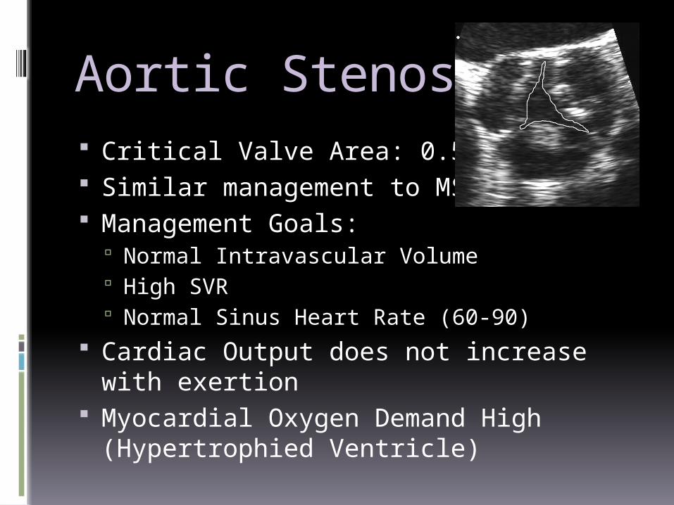
Aortic Stenosis Critical Valve Area: 0.5-0.7 cm2 Similar management to MS Management Goals:
Normal Intravascular Volume High SVR Normal Sinus Heart Rate (60-90)
Cardiac Output does not increase with exertion
Myocardial Oxygen Demand High (Hypertrophied Ventricle)
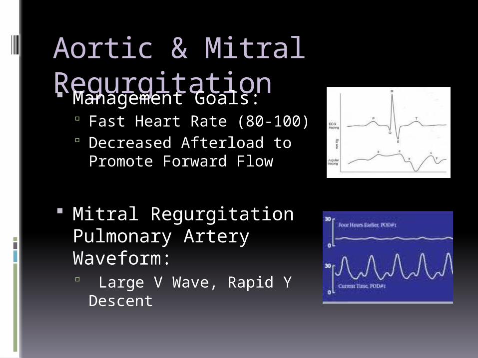
Aortic & Mitral Regurgitation Management Goals:
Fast Heart Rate (80-100) Decreased Afterload to
Promote Forward Flow
Mitral Regurgitation Pulmonary Artery Waveform: Large V Wave, Rapid Y
Descent
A 70 y/o male with severe aortic stenosis has a preinduction HR of 63 and BP of 125/70. Following induction, his HR is 90 and BP is 85/45. The EKG has a new ST Elevation. Drug of Choice?1. Epinephrine2. Isoproterenol3. Calcium Chloride4. Phenylephrine5. Ephedrine
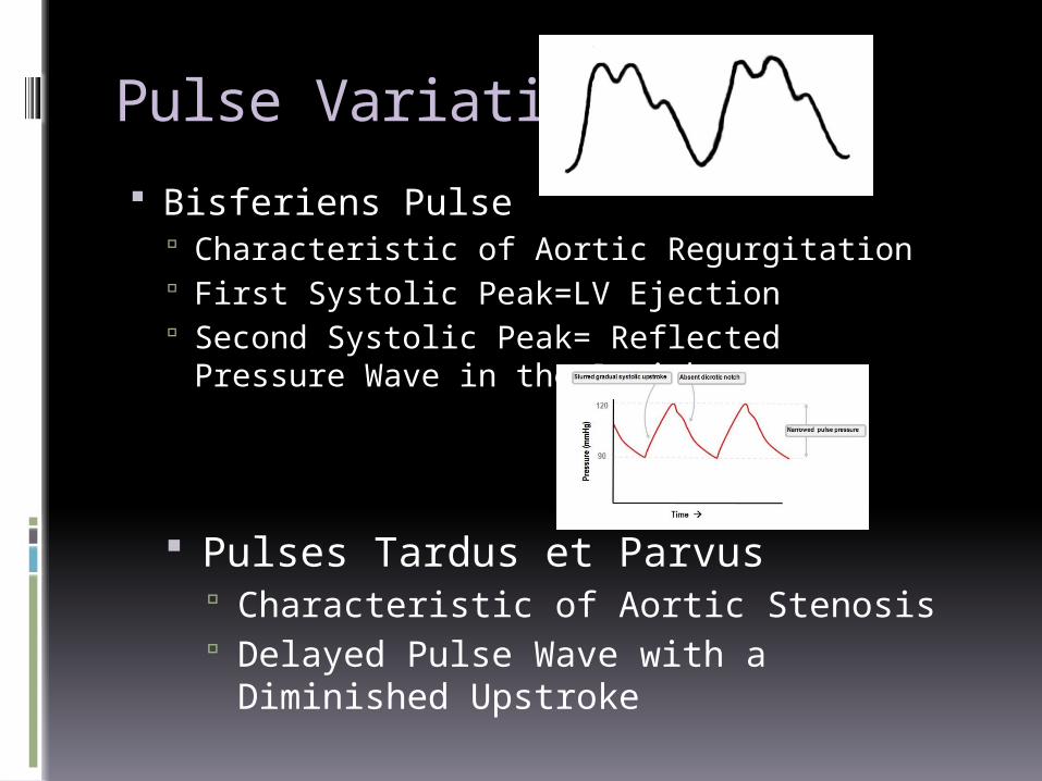
Pulse Variations Bisferiens Pulse
Characteristic of Aortic Regurgitation First Systolic Peak=LV Ejection Second Systolic Peak= Reflected Pressure
Wave in the Periphery
Pulses Tardus et Parvus Characteristic of Aortic Stenosis Delayed Pulse Wave with a
Diminished Upstroke
Hypertrophic Cardiomyopathy
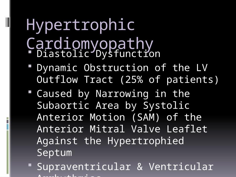
Hypertrophic Cardiomyopathy Diastolic Dysfunction Dynamic Obstruction of the LV
Outflow Tract (25% of patients) Caused by Narrowing in the
Subaortic Area by Systolic Anterior Motion (SAM) of the Anterior Mitral Valve Leaflet Against the Hypertrophied Septum
Supraventricular & Ventricular Arrhythmias
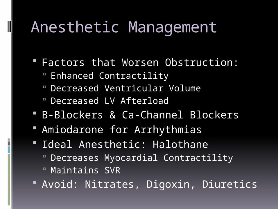
Anesthetic Management Factors that Worsen Obstruction:
Enhanced Contractility Decreased Ventricular Volume Decreased LV Afterload
B-Blockers & Ca-Channel Blockers Amiodarone for Arrhythmias Ideal Anesthetic: Halothane
Decreases Myocardial Contractility Maintains SVR
Avoid: Nitrates, Digoxin, Diuretics
The Transplanted
Heart
The Transplanted Heart Denervated No sympathetic or parasympathetic
input Resting Heart Rate 100-120 (no
vagal) Responsive to catecholamines Low cardiac output, slow to pick up EKG shows two P waves
Pharmacology Direct agents are the best:
Epinephrine & Isoproterenol Indirect vasopressors also work, but are
dependent on catecholamine stores Heart rate is NOT affected by:
Anticholinergics Pancuronium Meperidine Opiods Cholinesterase Inhibitors
A patient has a heart rate of 110 after heart transplant. The most likely etiology is: 1. Altered Barorecepter Sensitivity2. Cardiac Denervation3. Compensation for a fixed Stroke
Volume4. Cyclosporine5. Prednisone

Left to Right (Simple) Shunts
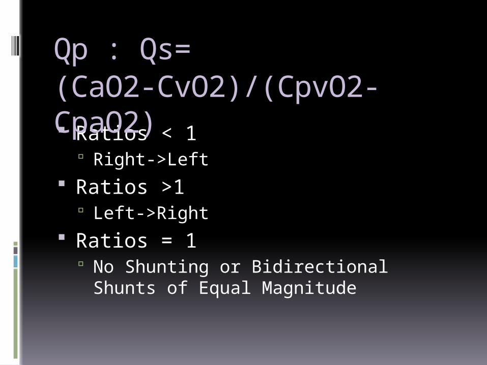
Qp : Qs=
Ratios < 1 Right->Left
Ratios >1 Left->Right
Ratios = 1 No Shunting or Bidirectional Shunts of
Equal Magnitude
(CaO2-CvO2)/(CpvO2-CpaO2)
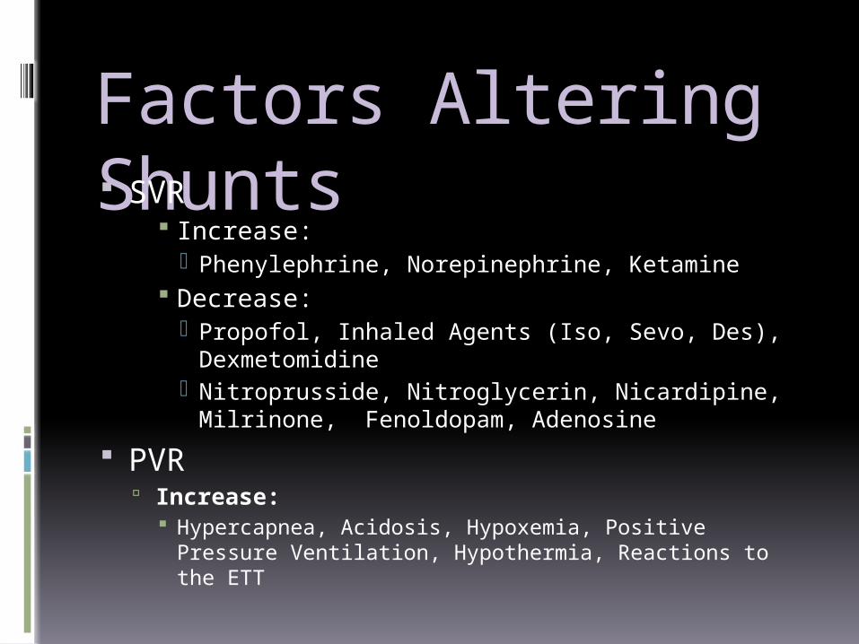
Factors Altering Shunts SVR
Increase: Phenylephrine, Norepinephrine, Ketamine
Decrease: Propofol, Inhaled Agents (Iso, Sevo, Des),
Dexmetomidine Nitroprusside, Nitroglycerin, Nicardipine,
Milrinone, Fenoldopam, Adenosine PVR
Increase: Hypercapnea, Acidosis, Hypoxemia, Positive Pressure
Ventilation, Hypothermia, Reactions to the ETT
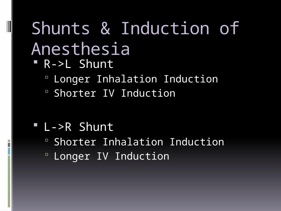
Shunts & Induction of Anesthesia R->L Shunt
Longer Inhalation Induction Shorter IV Induction
L->R Shunt Shorter Inhalation Induction Longer IV Induction
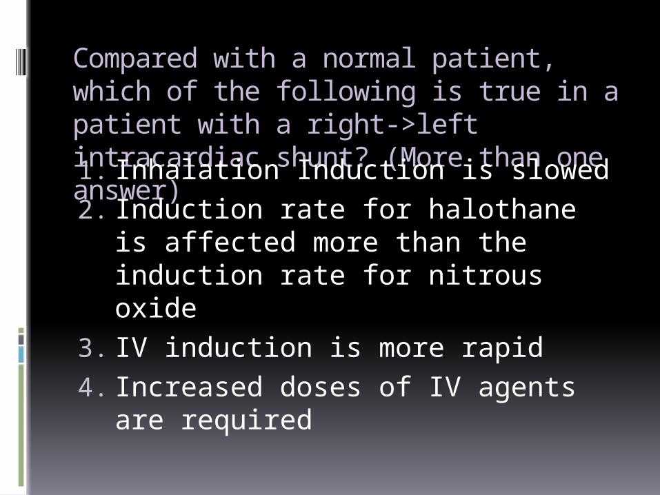
Compared with a normal patient, which of the following is true in a patient with a right->left intracardiac shunt? (More than one answer)1. Inhalation Induction is slowed2. Induction rate for halothane is
affected more than the induction rate for nitrous oxide
3. IV induction is more rapid4. Increased doses of IV agents are
required
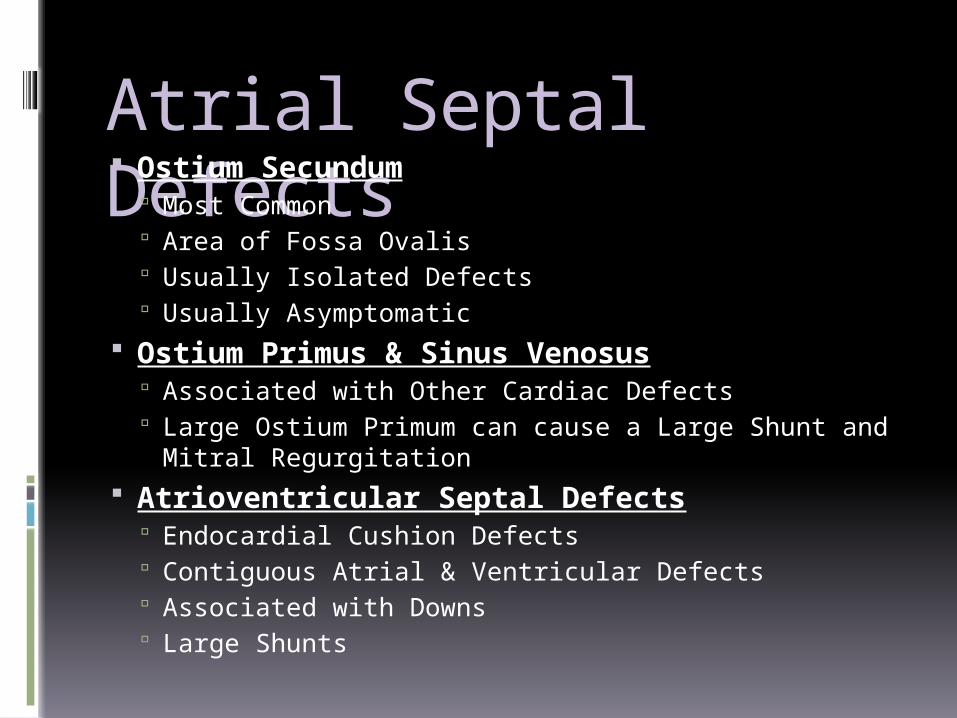
Atrial Septal Defects Ostium Secundum
Most Common Area of Fossa Ovalis Usually Isolated Defects Usually Asymptomatic
Ostium Primus & Sinus Venosus Associated with Other Cardiac Defects Large Ostium Primum can cause a Large Shunt and
Mitral Regurgitation Atrioventricular Septal Defects
Endocardial Cushion Defects Contiguous Atrial & Ventricular Defects Associated with Downs Large Shunts
Ventricular Septal Defects Most common congenital defect Small VSDs often close during
childhood Restrictive are associated with small
L->R Large defects produce large L->R
shunts that vary with SVR and PVR Large VSDs are surgically repaired
before pulmonary disease and Eisenmenger develop
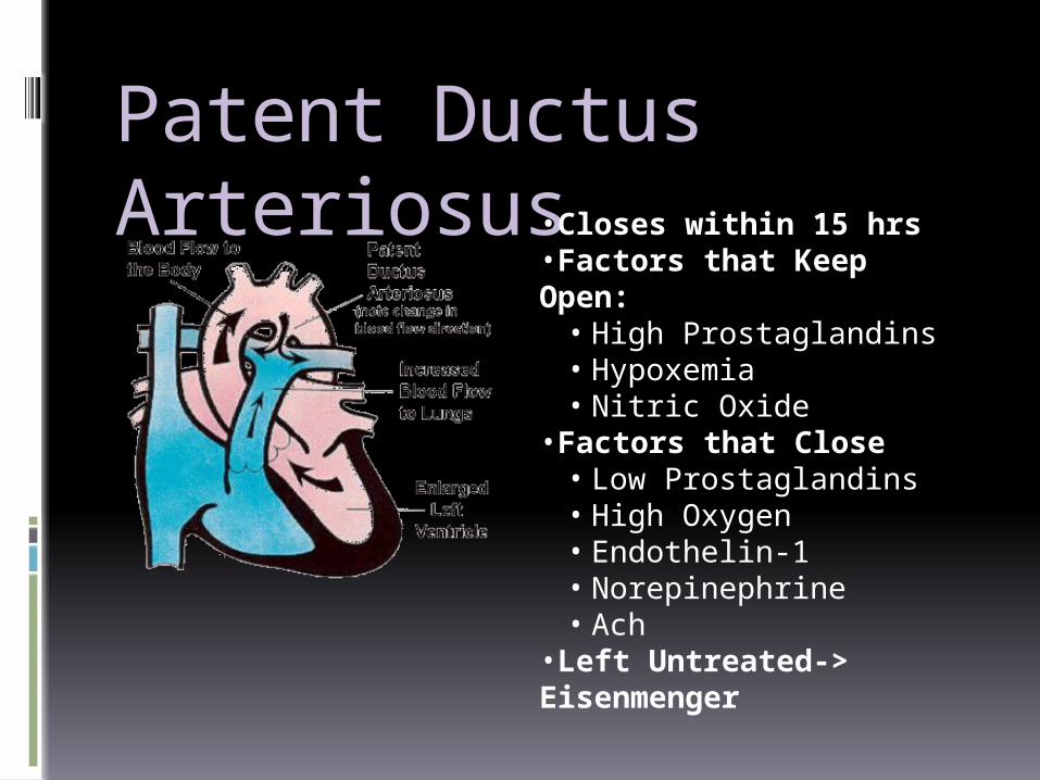
Patent Ductus Arteriosus •Closes within 15 hrs
•Factors that Keep Open:• High Prostaglandins• Hypoxemia• Nitric Oxide
•Factors that Close• Low Prostaglandins• High Oxygen• Endothelin-1• Norepinephrine• Ach
•Left Untreated-> Eisenmenger

Right to Left (Complex)
Shunts
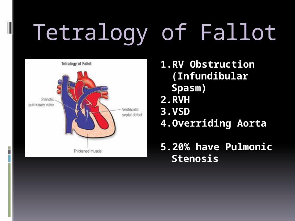
Tetralogy of Fallot1.RV Obstruction
(Infundibular Spasm)
2.RVH3.VSD4.Overriding Aorta
5.20% have Pulmonic Stenosis
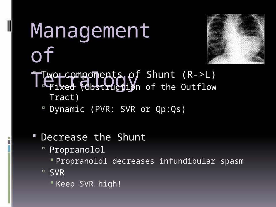
Management of Tetralogy Two components of Shunt (R->L)
Fixed (Obstruction of the Outflow Tract) Dynamic (PVR: SVR or Qp:Qs)
Decrease the Shunt Propranolol
Propranolol decreases infundibular spasm SVR
Keep SVR high!
Tetralogy of Fallot… Four Parts?
RV Outflow Obstruction, RVH, Overriding Aorta, VSD Ketamine?
Maintains SVR Propranolol?
Decreases Infundibular Spasm Prostaglandin E1?
Keeps PDA open Augments Pulmonary Blood Flow in the case of
Right Ventricular Obstruction
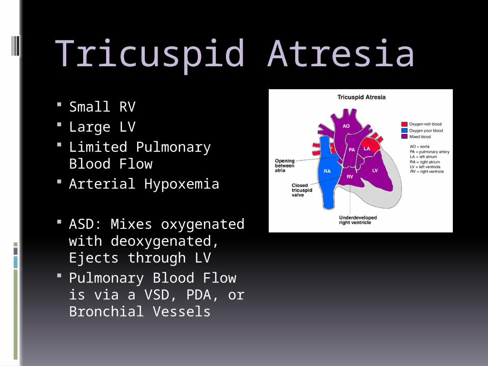
Tricuspid Atresia Small RV Large LV Limited Pulmonary
Blood Flow Arterial Hypoxemia
ASD: Mixes oxygenated with deoxygenated, Ejects through LV
Pulmonary Blood Flow is via a VSD, PDA, or Bronchial Vessels
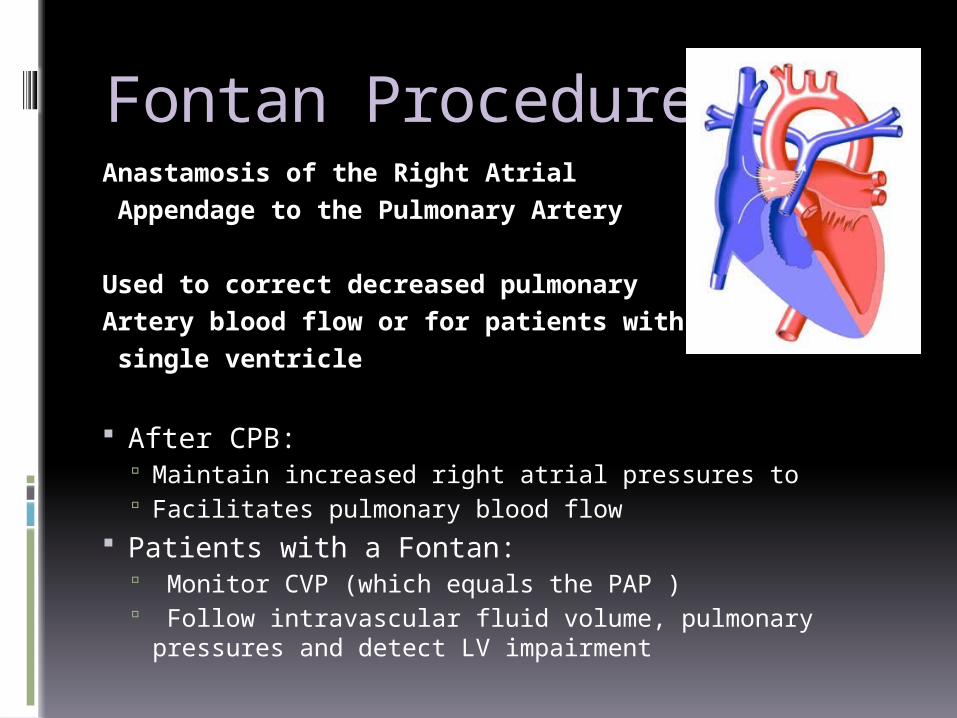
Fontan ProcedureAnastamosis of the Right Atrial Appendage to the Pulmonary Artery
Used to correct decreased pulmonary Artery blood flow or for patients with a single ventricle
After CPB: Maintain increased right atrial pressures to Facilitates pulmonary blood flow
Patients with a Fontan: Monitor CVP (which equals the PAP ) Follow intravascular fluid volume, pulmonary pressures and
detect LV impairment
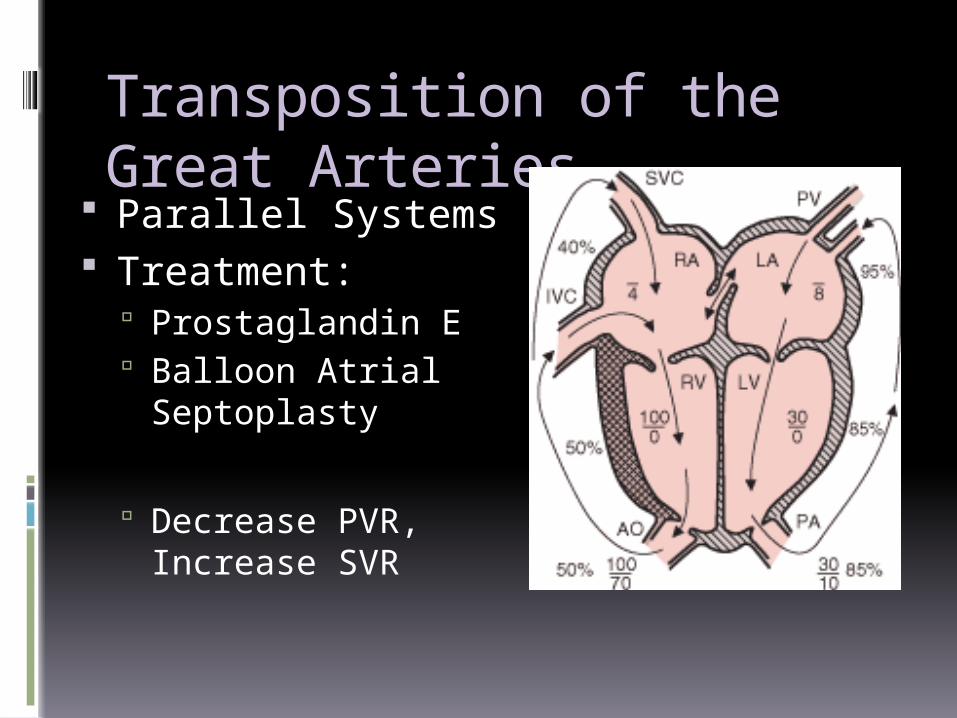
Transposition of the Great Arteries
Parallel Systems Treatment:
Prostaglandin E Balloon Atrial
Septoplasty
Decrease PVR, Increase SVR
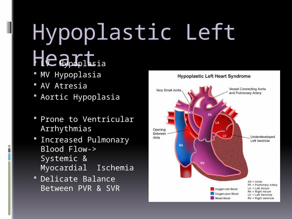
Hypoplastic Left Heart LV Hypoplasia MV Hypoplasia AV Atresia Aortic Hypoplasia
Prone to Ventricular Arrhythmias
Increased Pulmonary Blood Flow-> Systemic & Myocardial Ischemia
Delicate Balance Between PVR & SVR
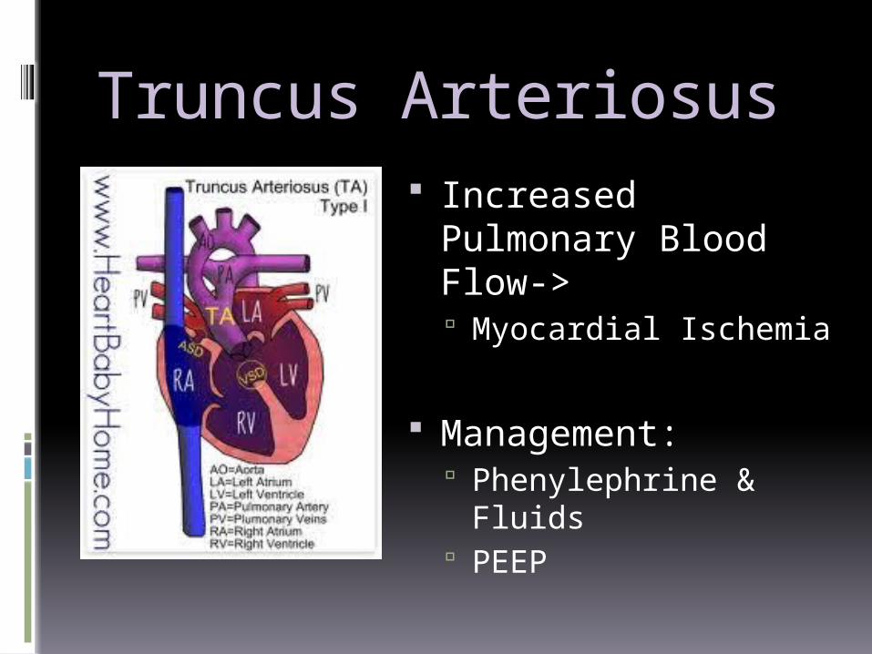
Truncus Arteriosus Increased Pulmonary
Blood Flow-> Myocardial Ischemia
Management: Phenylephrine & Fluids PEEP
Anastamosis of the right atrium to the pulmonary arter (Fontan procedure is useful surgical treatment for each of the following except:1. Tricuspid Atresia2. Hypoplastic Left Heart Syndrome3. Pulmonary Valve Stenosis4. Truncus Arteriosus5. Pulmonary Artery Atresia
Appropriate therapy for “tet spells” include (may be more than one): 1. Propranolol 2. Dobutamine 3. Phenylephrine 4. Ephedrine
Antibiotic Prophylaxis High Risk:
Previous Infective Endocarditis Prosthetic Valves CHD (some) Transplants
Procedure Type None for GI/GU Bronchoscopy- depends Dental Procedures- depends
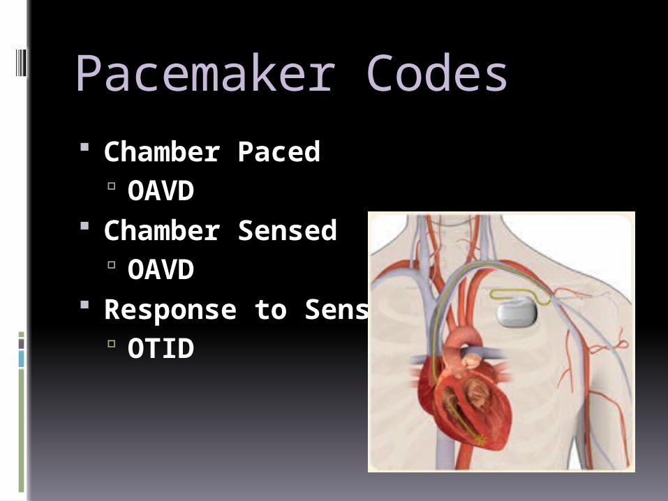
Pacemaker Codes Chamber Paced
OAVD Chamber Sensed
OAVD Response to Sensing
OTID