Embed Size (px)
Citation preview
CARDIAC INFECTIONSRheumatic Heart Disease and
Infective Endocarditis
Jonas D. Del Rosario, MD, FPPS, FPCC
Clinical Associate Professor
UP College of Medicine
RHEUMATIC FEVER
RHEUMATIC HEART DISEASE
RF to RHD
“Rheumatic Fever is a disease the LICKS the joints but BITES heart.”
RHD IS THE MOST SERIOUS COMPLICATION OF RF
Rheumatic Fever
• Most common cause of acquired heart
disease in children & young adults
worldwide
• Diffuse inflammation of connective
tissues of heart, joints, brain, blood
vessels & subcutaneous tissues
• Rheumatic process causes fibrosis of
heart valves leading to RHD
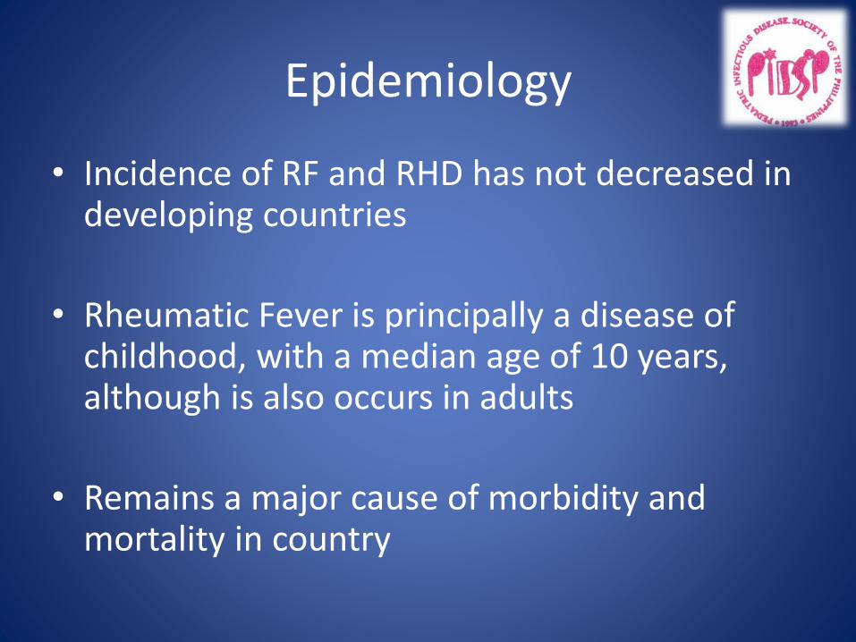
Epidemiology
• Incidence of RF and RHD has not decreased in developing countries
• Rheumatic Fever is principally a disease of childhood, with a median age of 10 years, although is also occurs in adults
• Remains a major cause of morbidity and mortality in country
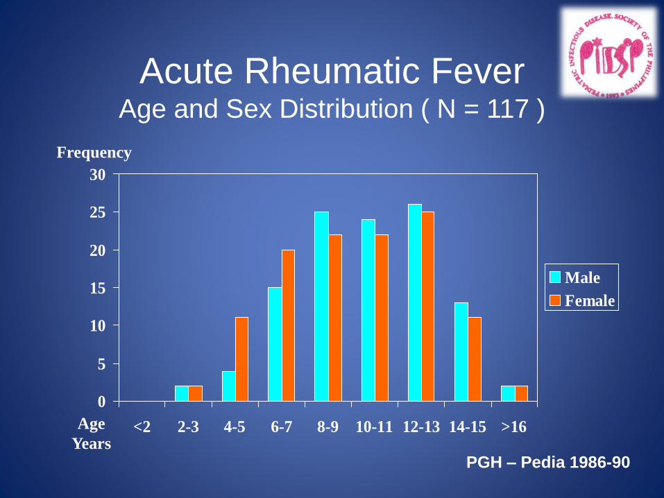
Acute Rheumatic FeverAge and Sex Distribution ( N = 117 )
0
5
10
15
20
25
30
<2 2-3 4-5 6-7 8-9 10-11 12-13 14-15 >16
Male
Female
Age
Years
Frequency
PGH – Pedia 1986-90
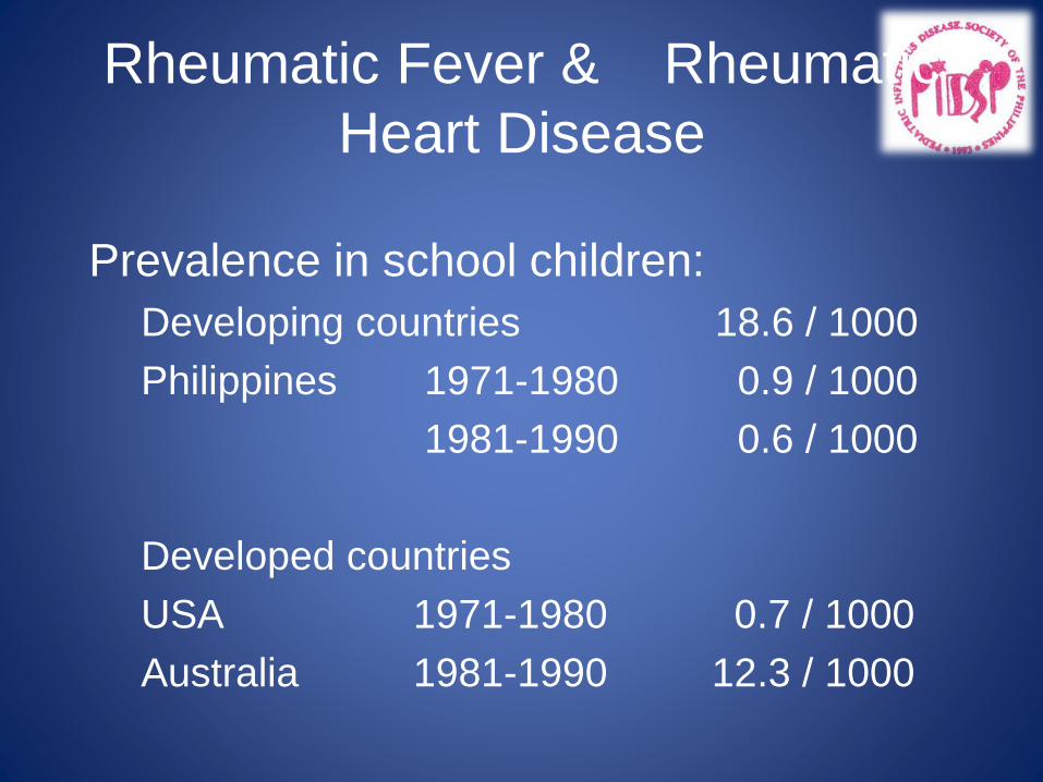
Rheumatic Fever & Rheumatic
Heart Disease
Prevalence in school children:
Developing countries 18.6 / 1000
Philippines 1971-1980 0.9 / 1000
1981-1990 0.6 / 1000
Developed countries
USA 1971-1980 0.7 / 1000
Australia 1981-1990 12.3 / 1000
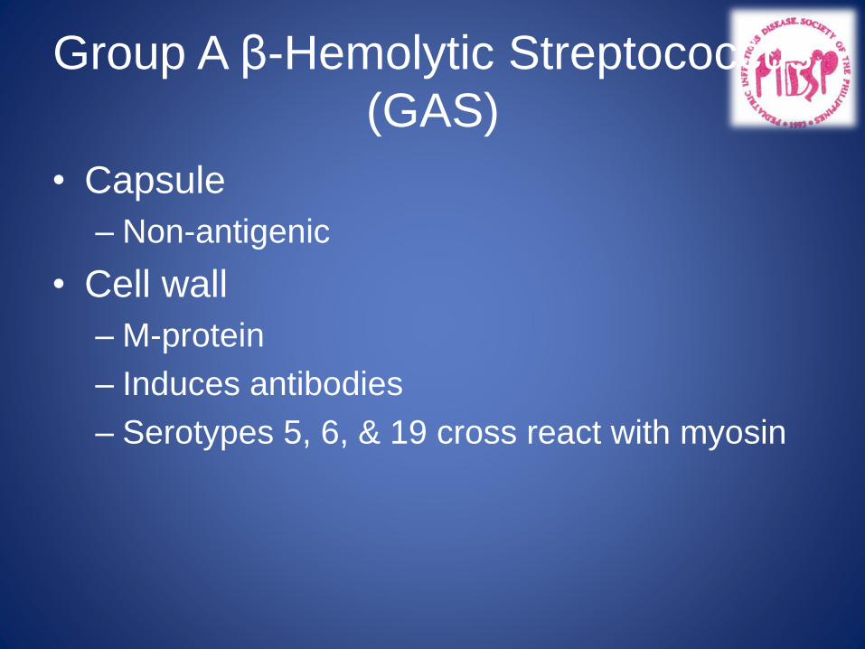
Group A β-Hemolytic Streptococcus
(GAS)
• Capsule
– Non-antigenic
• Cell wall
– M-protein
– Induces antibodies
– Serotypes 5, 6, & 19 cross react with myosin
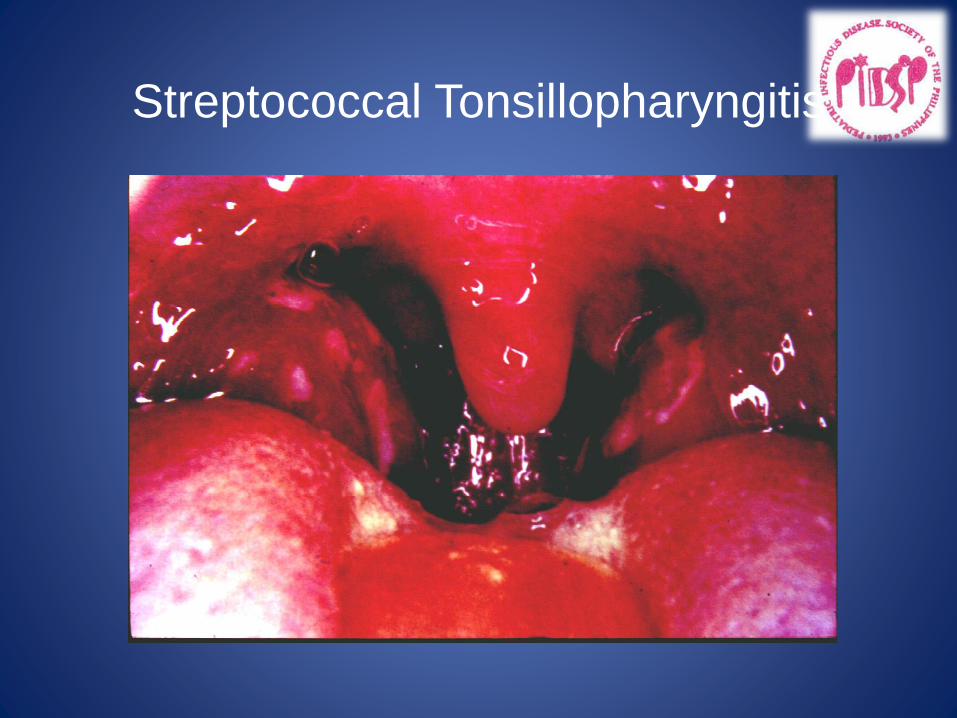
Streptococcal Tonsillopharyngitis
Pathophysiology
• Rheumatic fever develops in some children and adolescents following PHARYNGITIS with a group A beta-hemolytic Streptococcus (Streptococcus pyogenes)
• The organisms attach to the epithelial cells of the upper respiratory tract and produce a battery of enzymes allowing them to damage and invade human tissues
Group A β-Hemolytic Streptococcus
(GAS)
• Enzymes
– Streptolysin
– Deoxyribonuclease
– Fibrinolysin
– Diphosphopyridine nucleotidase
– Hyaluronidase
Rheumatic Fever:
Pathogenesis
• Actual mechanism unknown
• Postulate:
– Autoimmune or hypersensitivity reaction to
GAS produces pathogenic autoantibodies
to cardiac tissues
Pathophysioloy
• In 0.3-3% of cases, streptococcal pharyngitis leads to rheumatic fever several weeks after the sore throat has resolved.
• Only infections of the pharynx initiate or reactivate rheumatic fever.
• Organism spreads by direct contact with oral or respiratory secretions
• Patient remains infected for weeks after symptomatic resolution of pharyngitis
Fever in ARF
• Full manifestations of the disease may be suppressed if patient has previously taken aspirin or other NSAIDs
• Fever above 39° C with no characteristic pattern are initially present in almost every case of acute rheumatic fever
• Fever may be low grade in children with mild carditis or absent in patients with chorea
• Low grade fevers persists for 2-3 weeks
DIAGNOSIS
• A diagnosis of RHD is made after confirming antecedent rheumatic fever
• The JONES criteria require the presence of 2 major or 1 major and 2 minor criteria for the diagnosis of RF
• Additional evidence of previous group A streptococcal pharyngitis is required
Jones Criteria 1992
• Establish initial attack of acute rheumatic fever
• Not intended
– To establish diagnosis of inactive or chronic RHD
– To measure rheumatic activity
– To predict course or severity of disease
• Previous RF or RHD not included as manifestation
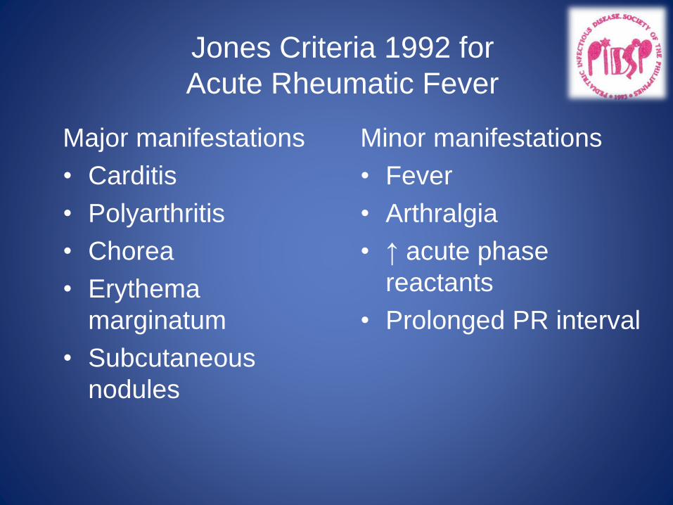
Jones Criteria 1992 for
Acute Rheumatic Fever
Major manifestations
• Carditis
• Polyarthritis
• Chorea
• Erythema
marginatum
• Subcutaneous
nodules
Minor manifestations
• Fever
• Arthralgia
• ↑ acute phase
reactants
• Prolonged PR interval
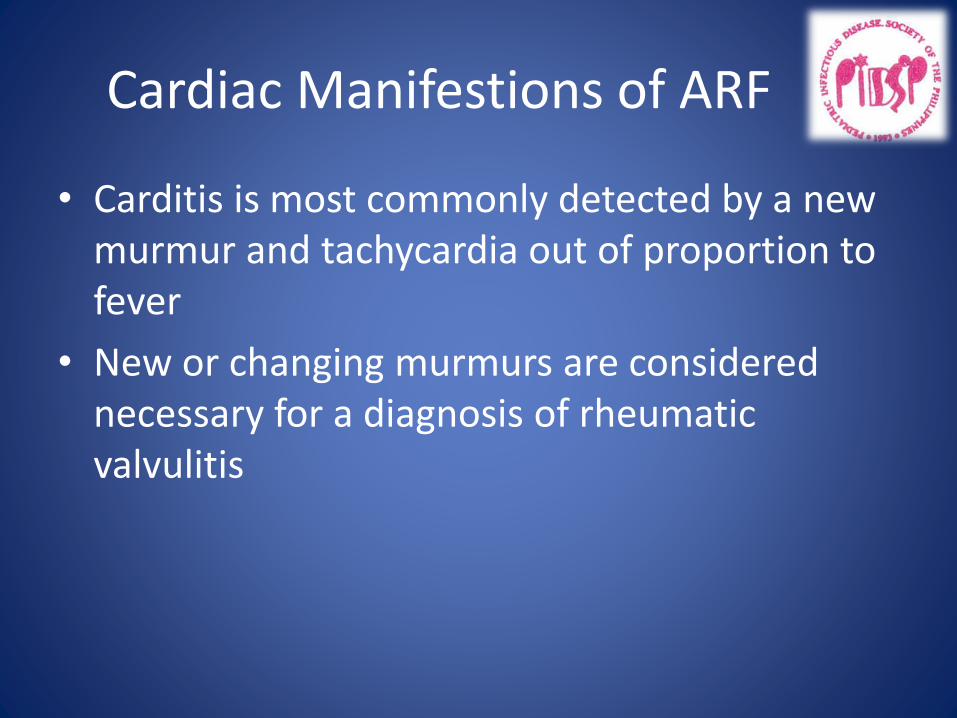
Cardiac Manifestions of ARF
• Carditis is most commonly detected by a new murmur and tachycardia out of proportion to fever
• New or changing murmurs are considered necessary for a diagnosis of rheumatic valvulitis
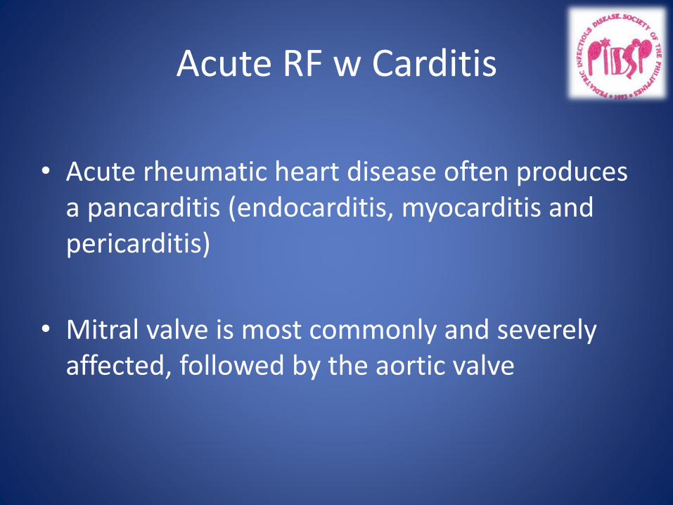
Acute RF w Carditis
• Acute rheumatic heart disease often produces a pancarditis (endocarditis, myocarditis and pericarditis)
• Mitral valve is most commonly and severely affected, followed by the aortic valve
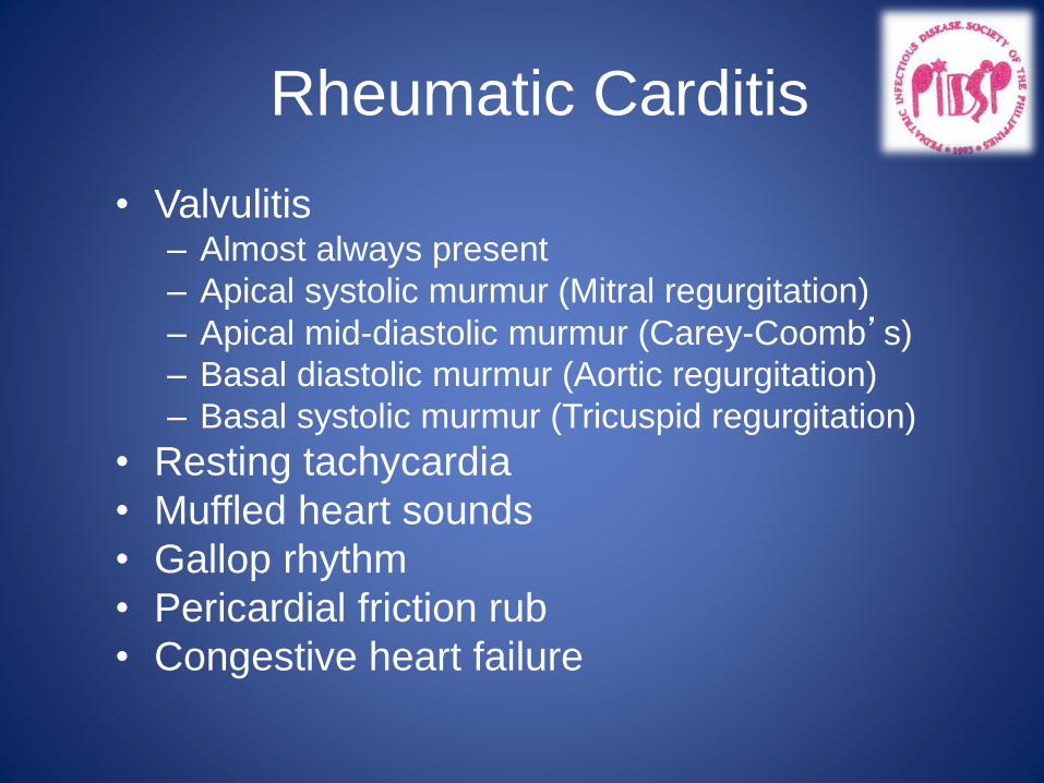
Rheumatic Carditis
• Valvulitis– Almost always present
– Apical systolic murmur (Mitral regurgitation)
– Apical mid-diastolic murmur (Carey-Coomb’s)
– Basal diastolic murmur (Aortic regurgitation)
– Basal systolic murmur (Tricuspid regurgitation)
• Resting tachycardia
• Muffled heart sounds
• Gallop rhythm
• Pericardial friction rub
• Congestive heart failure
“Echocarditis”
• Some cardiologists have proposed that echo-Doppler evidence of mitral insufficiency, particularly in association with aortic insufficiency, may be sufficient for a diagnosis of carditis (even in the absence of accompanying auscultatory findings)
“Echocarditis”
• Echocardiographic findings in acute
rheumatic fever (mitral regurgitation and/or
aortic regurgitation) in the absence of
clinical carditis
• Subclinical carditis
• Included as basis for carditis in modified
Jones criteria?
• Controversial
Jones Criteria 1992
High probability of rheumatic fever :
• 2 major or
• 1 major & 2 minor manifestations
PLUS Evidence of GAS
infection
Evidence of Antecedent Group A
Streptococcal Infections
• ↑ or rising Streptococcal antibody
(ASO) titer
• (+) throat culture or rapid antigen test
Antistreptococcal antibodies
• Clinical features of RF begin at the time antistreptococcal antibodies are at their peak
• Useful for confirming previous group A streptococcal infection
• ASO is the most commonly used
• Sensitivity can be improved by testing for several antibodies (ASO, antiDNase, antihyalarunidase etc.)
Antistreptococcal
Antibody Titers
• Reflect past & not present immunologic events
• No value in the diagnosis of acute pharyngitis
• Valuable for confirmation of previous streptococcal infections in patients suspected of having acute RF or PSGN
• Helpful, in prospective epidemiological studies, for distinguishing patients with acute infection from patients who are carriers
ASO titer
• Peaks 2-3 weeks after the onset of rheumatic fever
• Plateaus for 3-6 months
• Returning to normal levels after 6-12 months
• Antihyaluronidase results are frequently abnormal in RF patients with normal level ASO titer and may rise earlier and persist longer than ASO during RF
Rheumatic Fever
Laboratory Examinations
• CBC – Anemia ; Leukocytosis
• ECG – Sinus tachycardia
– 1o AV block
– No chamber enlargement
– Rarely 2o AV block, low voltages, ST-T wave changes
• CXR– Normal
– Cardiomegaly
– Pulmonary congestion to edema
Acute Phase Reactants
• C reactive protein and ESR
– Elevated in RF due to inflammatory in nature
– Both have a high sensitivity but low specificity
– May be used to monitor the resolution of inflammation, detect relapse when weaning aspirin or identify recurrence of disease
Chest X-ray
• Chest X-ray
– Cardiomegaly may or may not be present
– Pulmonary congestion in congestive heart failure
Rheumatic Carditis
• Mild carditis – No cardiomegaly
• Moderate carditis – mild cardiomegaly
• Severe carditis – cardiomegaly with
severe pulmonary congestion or edema
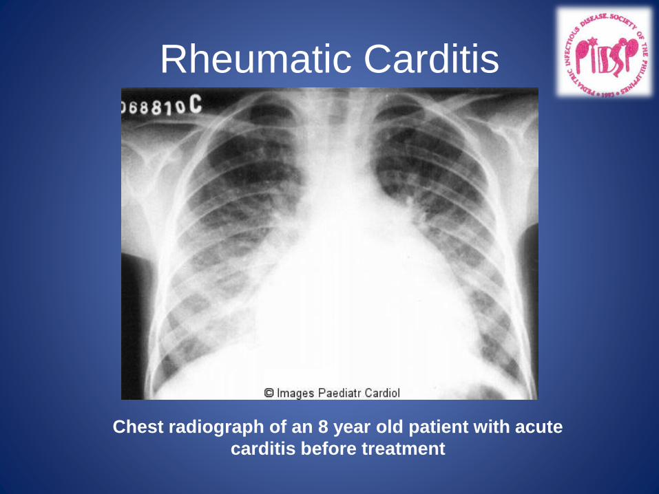
Rheumatic Carditis
Chest radiograph of an 8 year old patient with acute
carditis before treatment
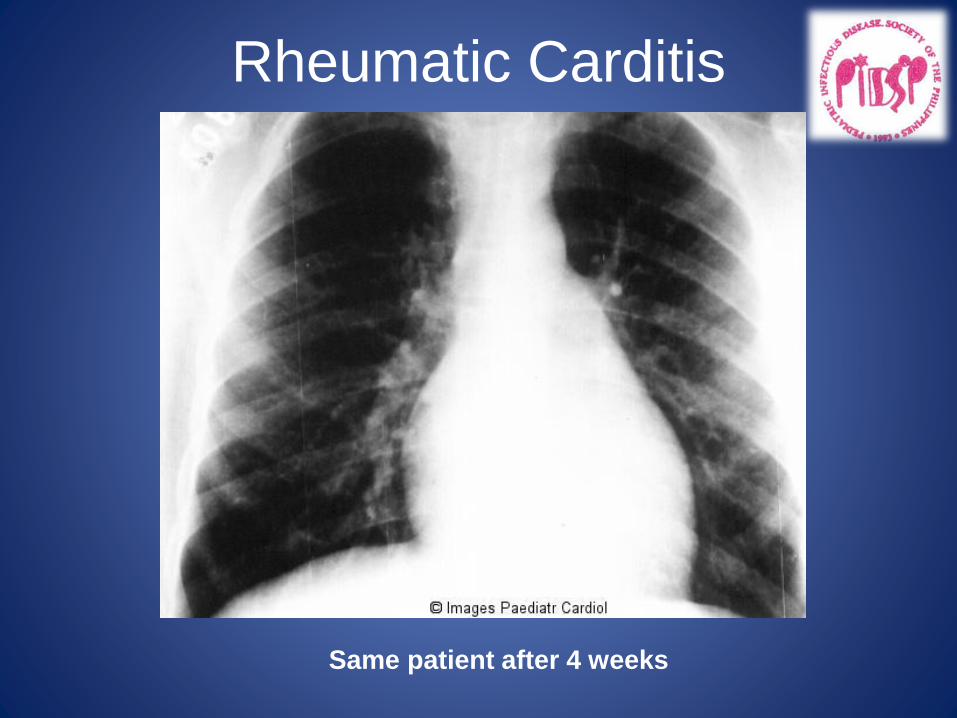
Rheumatic Carditis
Same patient after 4 weeks
ECHOCARDIOGRAPHY
• Is the imaging of choice to detect the presence of rheumatic carditis or rheumatic heart disease
• If the ECHO is normal, diagnosis of RHD is ruled out
Rheumatic Fever :
Echocardiographic Findings
• Focal nodular thickening of valves
• Prolapsed Anterior mitral valve leaflet
• Left chambers enlargement ; normal
function
• Pericardial effusion
• Valvular regurgitation – valves involved &
severity
Ty & Ortiz. Cardiol in the Young 2(3): 229-35, July 1992.
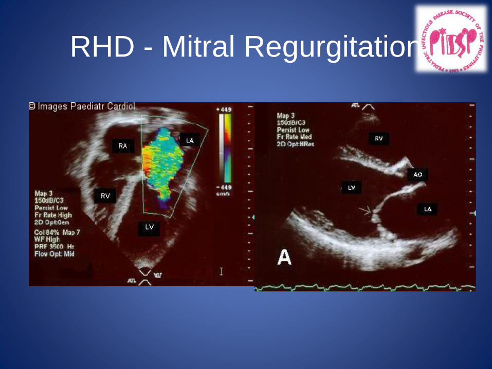
RHD - Mitral Regurgitation
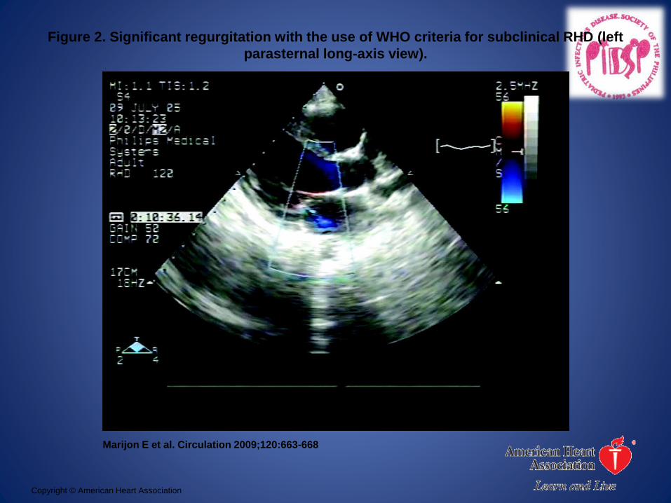
Figure 2. Significant regurgitation with the use of WHO criteria for subclinical RHD (left
parasternal long-axis view).
Marijon E et al. Circulation 2009;120:663-668
Copyright © American Heart Association
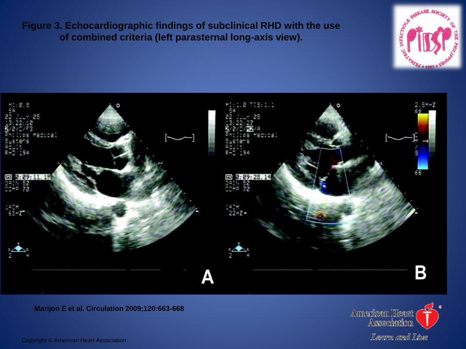
Figure 3. Echocardiographic findings of subclinical RHD with the use
of combined criteria (left parasternal long-axis view).
Marijon E et al. Circulation 2009;120:663-668
Copyright © American Heart Association
Echocardiogram
• Color doppler-echocardiography identifies and quantitates valve insufficiency and ventricular function
• Mild carditis -- mild regurgitation during acute phase but resolves in weeks to months
• Moderate-severe carditis – persistent mitral/aortic regurgitation
Rheumatic Mitral Regurgitation
• The most important echocardiographic features of mitral regurgitation from acute rheumatic valvulitis are annular dilatation, elongation of the chordae to the anterior leaflet, and a posterolaterally directed mitral regurgitation
Echocardiography in RHD
• Acute Rheumatic Fever – Left ventricle is frequently dilated in association with normal or increases fractional shortening
• Chronic rheumatic heart disease – Affected leaflets become diffusely thickened with fusion of commissures and chordae tendinae; calcifications may be seen
Rheumatic Heart Disease
Chronic valvar heart disease as a
sequelae of rheumatic fever
and its recurrences
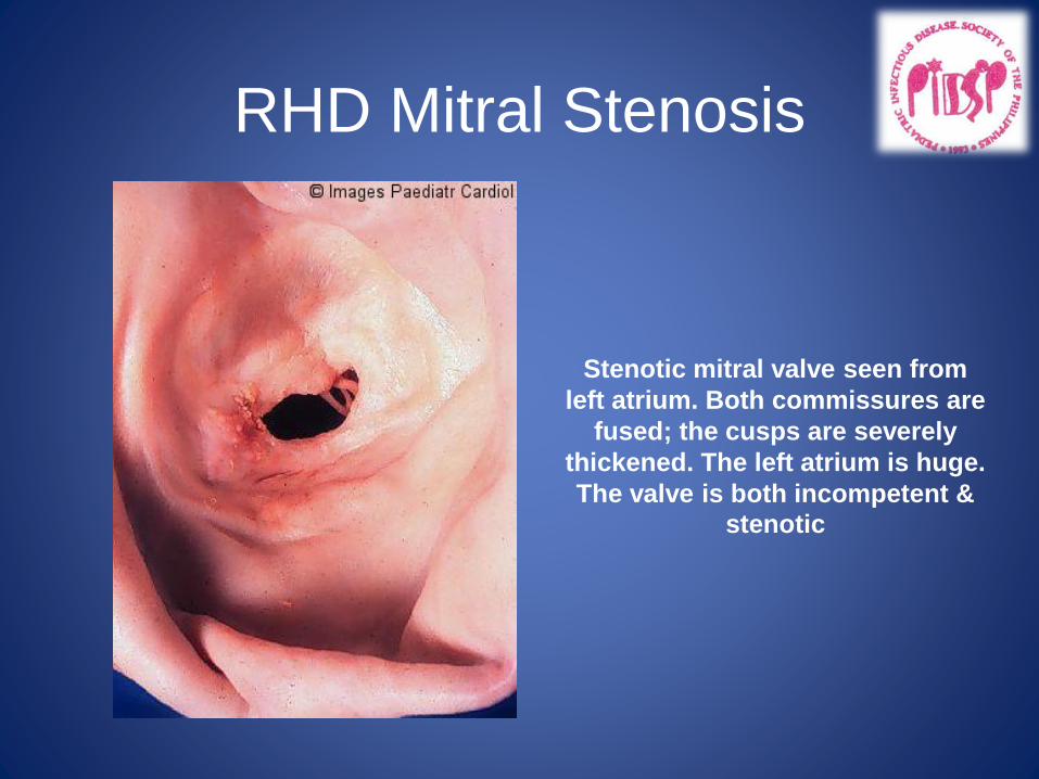
RHD Mitral Stenosis
Stenotic mitral valve seen from
left atrium. Both commissures are
fused; the cusps are severely
thickened. The left atrium is huge.
The valve is both incompetent &
stenotic
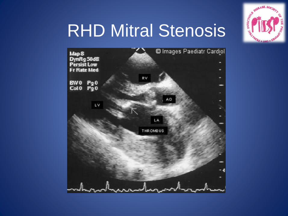
RHD Mitral Stenosis
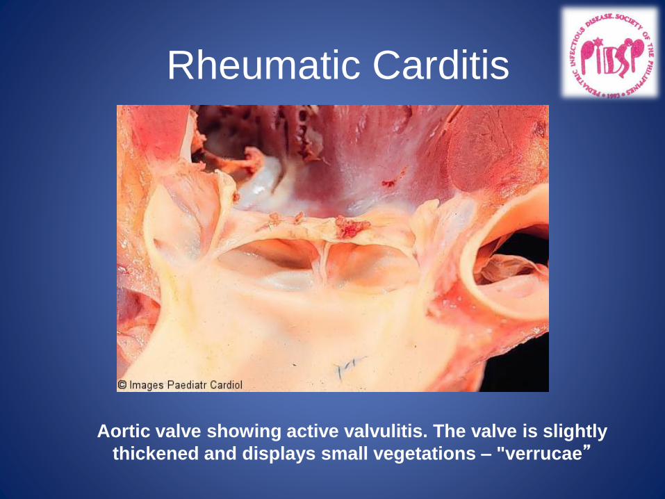
Rheumatic Carditis
Aortic valve showing active valvulitis. The valve is slightly
thickened and displays small vegetations – "verrucae”
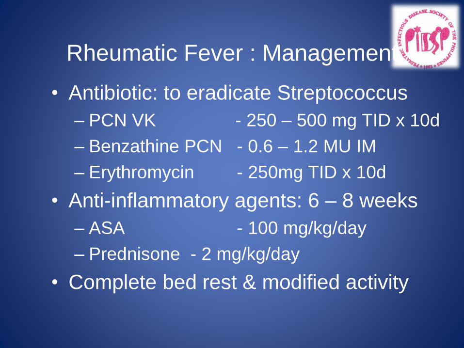
Rheumatic Fever : Management
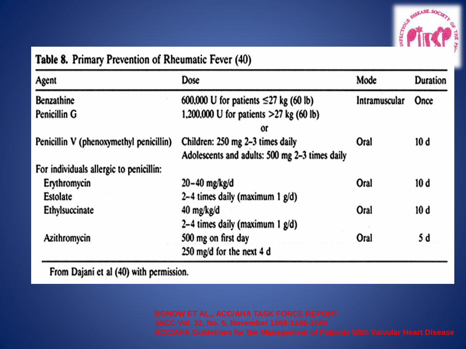
• Antibiotic: to eradicate Streptococcus
– PCN VK - 250 – 500 mg TID x 10d
– Benzathine PCN - 0.6 – 1.2 MU IM
– Erythromycin - 250mg TID x 10d
• Anti-inflammatory agents: 6 – 8 weeks
– ASA - 100 mg/kg/day
– Prednisone - 2 mg/kg/day
• Complete bed rest & modified activity
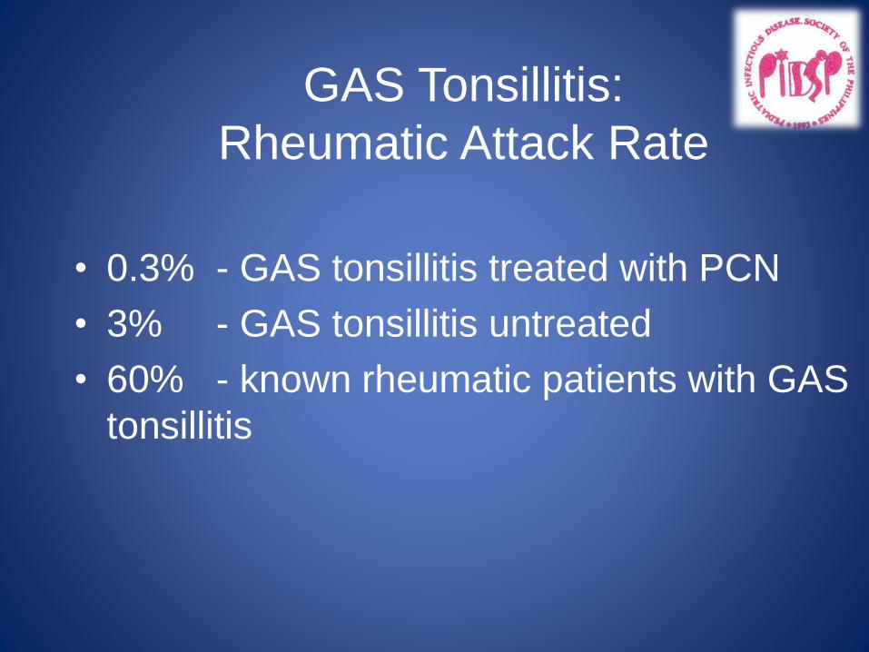
GAS Tonsillitis:
Rheumatic Attack Rate
• 0.3% - GAS tonsillitis treated with PCN
• 3% - GAS tonsillitis untreated
• 60% - known rheumatic patients with GAS
tonsillitis
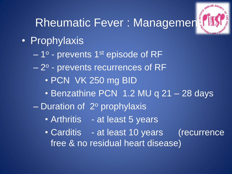
Rheumatic Fever : Management
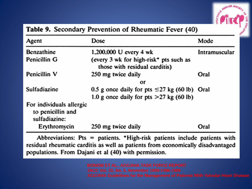
• Prophylaxis
– 1o - prevents 1st episode of RF
– 2o - prevents recurrences of RF
• PCN VK 250 mg BID
• Benzathine PCN 1.2 MU q 21 – 28 days
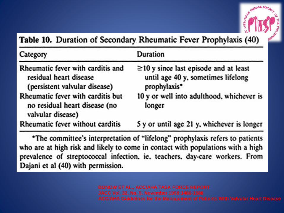
– Duration of 2o prophylaxis
• Arthritis - at least 5 years
• Carditis - at least 10 years (recurrence
free & no residual heart disease)
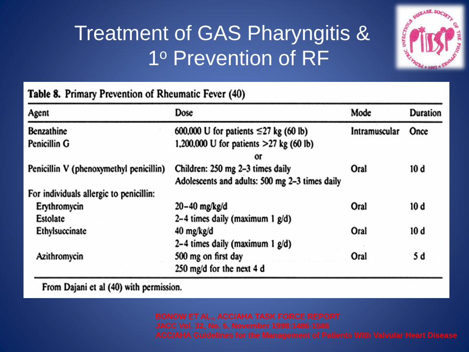
Treatment of GAS Pharyngitis &
1o Prevention of RF
BONOW ET AL., ACC/AHA TASK FORCE REPORT
JACC Vol. 32, No. 5, November 1998:1486-1588
ACC/AHA Guidelines for the Management of Patients With Valvular Heart Disease
BONOW ET AL., ACC/AHA TASK FORCE REPORT
JACC Vol. 32, No. 5, November 1998:1486-1588
ACC/AHA Guidelines for the Management of Patients With Valvular Heart Disease
BONOW ET AL., ACC/AHA TASK FORCE REPORT
JACC Vol. 32, No. 5, November 1998:1486-1588
ACC/AHA Guidelines for the Management of Patients With Valvular Heart Disease
BONOW ET AL., ACC/AHA TASK FORCE REPORT
JACC Vol. 32, No. 5, November 1998:1486-1588
ACC/AHA Guidelines for the Management of Patients With Valvular Heart Disease
Penicillin for 2nd Prophylaxis of Rheumatic
Fever
• Meta-analysis by Manyemba & Mayosi [
Cochrane Library (1) 2003]
• 9 RCT & quasi-RCT with 3008 subjects ;
methodological quality poor
• IM Penicillin more effective than oral penicllin
• 2 weekly or 3 weekly injections more effective
than 4 weekly injections
INFECTIVE ENDOCARDITIS
Infective Endocarditis
• Infection of the endocardium and/or heart valves that involves thrombus formation (vegetation) , which may damage the endocardial tissue and/or valves
• Many aspects of IE are similar in children and adults, but there are some manifestations that are unique to children
Epidemiology
• IE occurs less often in children than in adults and accounts for ≈1 in 1280 pediatric admissions per year
• Frequency of endocarditis among children seems to have increased in recent years.
• This is due in part to improved survival among children who are at risk for endocarditis, such as those with CHD and hospitalized newborn infants.
Epidemiology
• Most children with IE have an identifiable risk factor for disease
• Children with CHD have the highest risk of developing IE.
• The risk is increased in patients with complex cyanotic heart disease, especially those who undergo surgical procedures that introduce prosthetic material (conduits and shunts)
Risk Factors
• Congenital Heart Defects
• Rheumatic Heart Disease
• Intravascular shunts/conduits/prosthesis
• Intravascular catheters
• IV drug abuse
• Prematurity
• Immunocompromise
Pathogenesis
• IE is due to a series of complex interactions among blood borne pathogens, damaged cardiac endothelium, fibrin and platelets that result in the formation of an infected thrombus (vegetation) and damage to endocardium and/or heart valves.
Pathophysiology
1. Turbulent blood flow disrupts the endocardium making it “sticky”
2. Bacteremia delivers the organisms to the endocardial surface
3. Adherence of the organisms to the endocardial surface
4. Eventual invasion of the valvular leaflets
Infecting Organisms
• Common bacteria– S. aureus
– Viridans group streptococci
– Enterococci
• Not so common bacteria– Fungi*
– Coagulase negative staph*
– Pseudomonas
– HACEK
*Common in neonates
Clinical Presentation
• Variable
• Dependent upon the extent of the local cardiac disease
• Degree of involvement of other organs (egembolization)
• Causative agent
Clinical Presentation
• Acute
– Affects normal heart valves
– Rapidly destructive
– Metastatic foci
– Commonly Staph.
– If not treated, usually fatal within 6 weeks
• Subacute
– Often affects damaged heart valves
– Indolent nature
– If not treated, usually fatal by one year
– Usually Viridans strep
Clinical Presentation
• Acute
– High grade fever and chills
– Severely ill
– Hemodynamicallyunstable
• Subacute
– Low grade fever
– Anorexia
– Weight loss
– Exercise intolerance
– Diaphoresis
The onset of symptoms is usually ~2 weeks or less from the initiating bacteremia
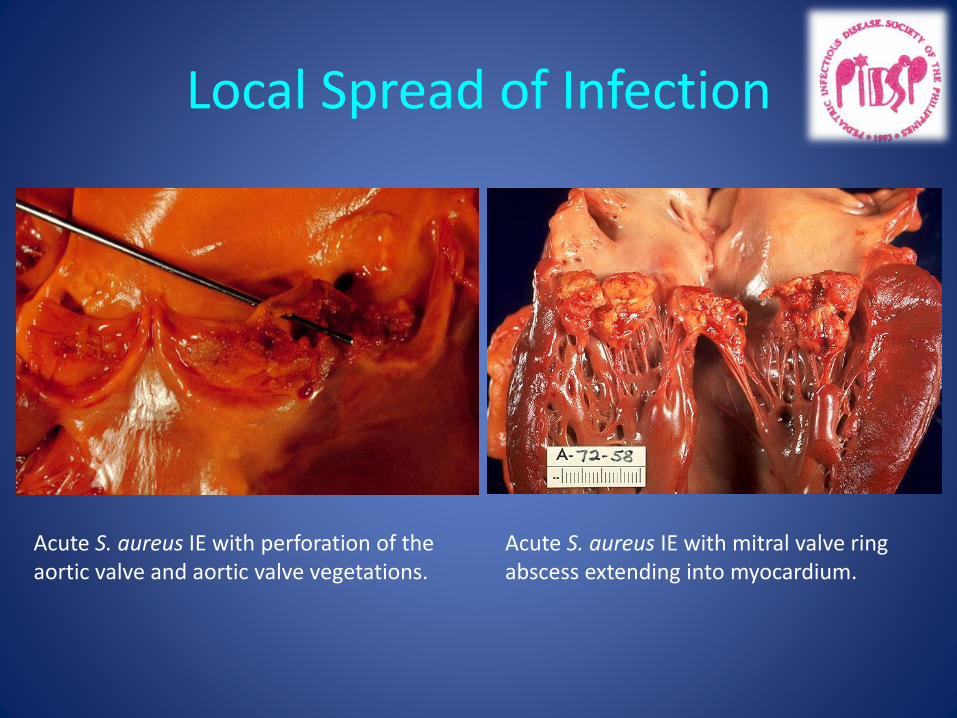
Local Spread of Infection
Acute S. aureus IE with perforation of the aortic valve and aortic valve vegetations.
Acute S. aureus IE with mitral valve ring abscess extending into myocardium.
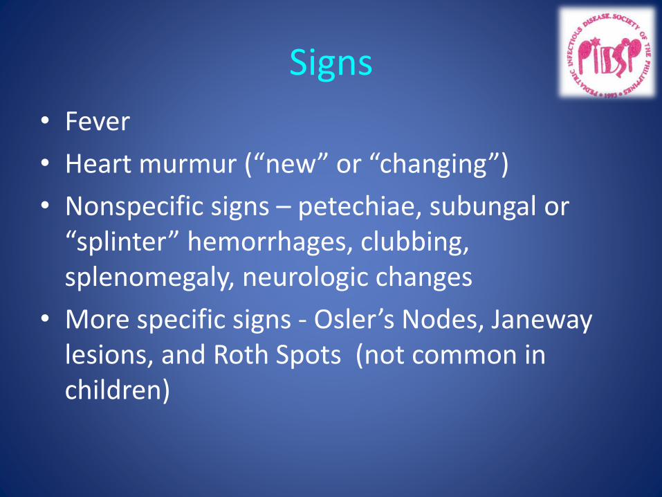
Signs
• Fever
• Heart murmur (“new” or “changing”)
• Nonspecific signs – petechiae, subungal or “splinter” hemorrhages, clubbing, splenomegaly, neurologic changes
• More specific signs - Osler’s Nodes, Janewaylesions, and Roth Spots (not common in children)
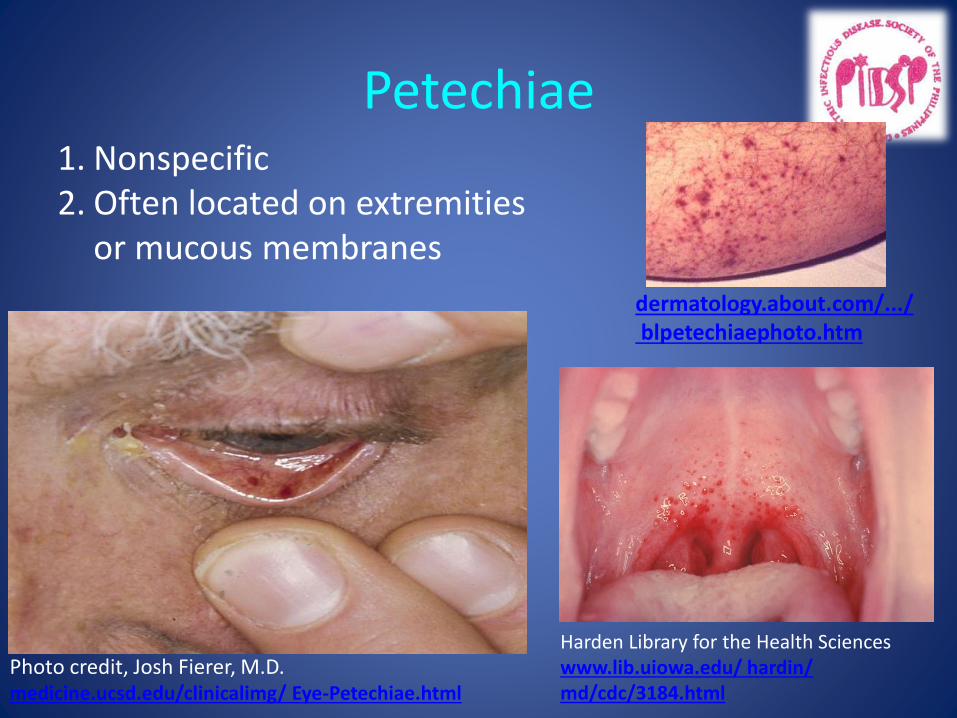
Petechiae
Photo credit, Josh Fierer, M.D. medicine.ucsd.edu/clinicalimg/ Eye-Petechiae.html
Harden Library for the Health Scienceswww.lib.uiowa.edu/ hardin/md/cdc/3184.html
1. Nonspecific2. Often located on extremities
or mucous membranes
dermatology.about.com/.../blpetechiaephoto.htm
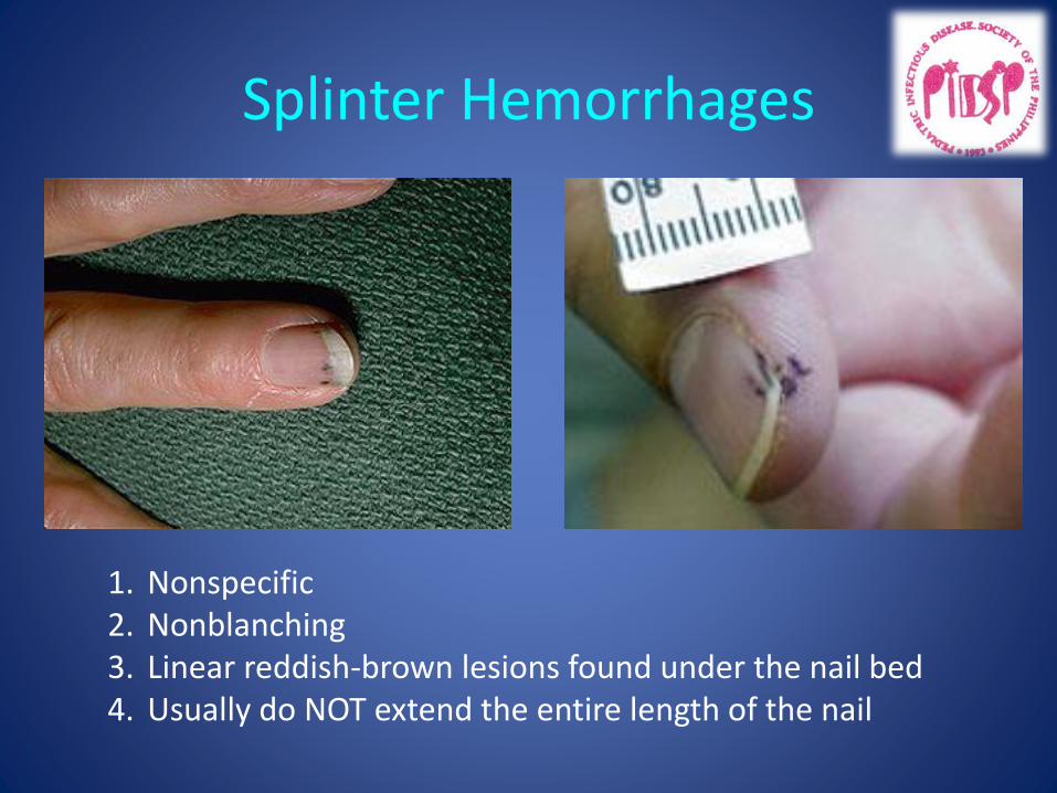
Splinter Hemorrhages
1. Nonspecific2. Nonblanching3. Linear reddish-brown lesions found under the nail bed4. Usually do NOT extend the entire length of the nail
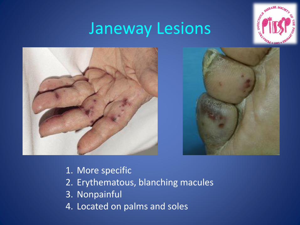
Janeway Lesions
1. More specific2. Erythematous, blanching macules 3. Nonpainful4. Located on palms and soles
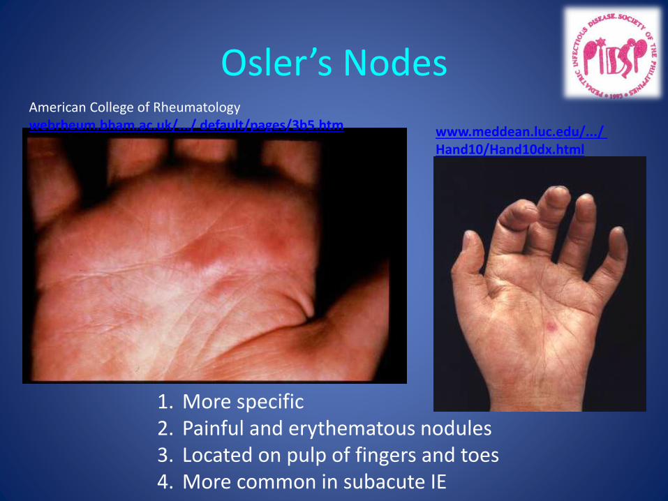
Osler’s Nodes
1. More specific2. Painful and erythematous nodules3. Located on pulp of fingers and toes4. More common in subacute IE
American College of Rheumatologywebrheum.bham.ac.uk/.../ default/pages/3b5.htm www.meddean.luc.edu/.../
Hand10/Hand10dx.html
Making the Diagnosis
• Pelletier and Petersdorf criteria (1977)
– Classification scheme of definite, probable, and possible IE
– Reasonably specific but lacked sensitivity
• Von Reyn criteria (1981)
– Added “rejected” as a category
– Added more clinical criteria
– Improved specificity and clinical utility
• Duke criteria (1994)
– Included the role of echocardiography in diagnosis
– Added IVDA as a “predisposing heart condition”
Modified Duke’s Criteria
The clinical diagnosis of IE is made by fulfilling the modified Duke’s criteria which is based upon physical and echocardiogram
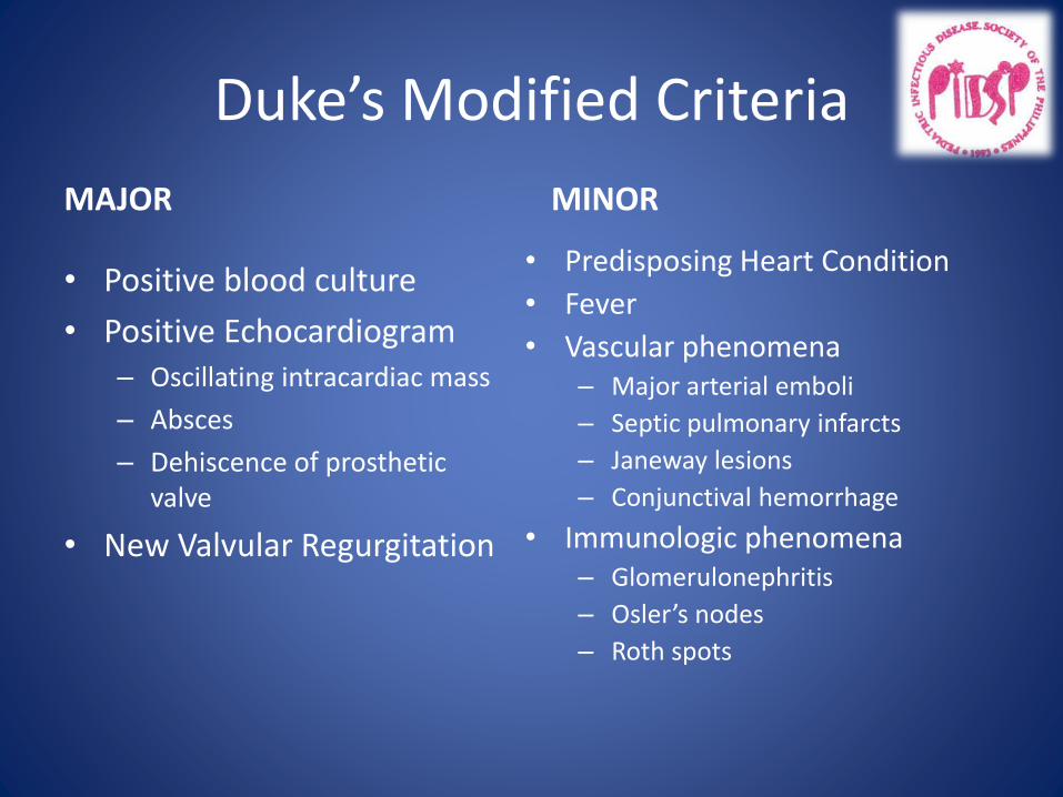
Duke’s Modified Criteria
MAJOR
• Positive blood culture
• Positive Echocardiogram– Oscillating intracardiac mass
– Absces
– Dehiscence of prosthetic valve
• New Valvular Regurgitation
MINOR
• Predisposing Heart Condition
• Fever
• Vascular phenomena– Major arterial emboli
– Septic pulmonary infarcts
– Janeway lesions
– Conjunctival hemorrhage
• Immunologic phenomena– Glomerulonephritis
– Osler’s nodes
– Roth spots
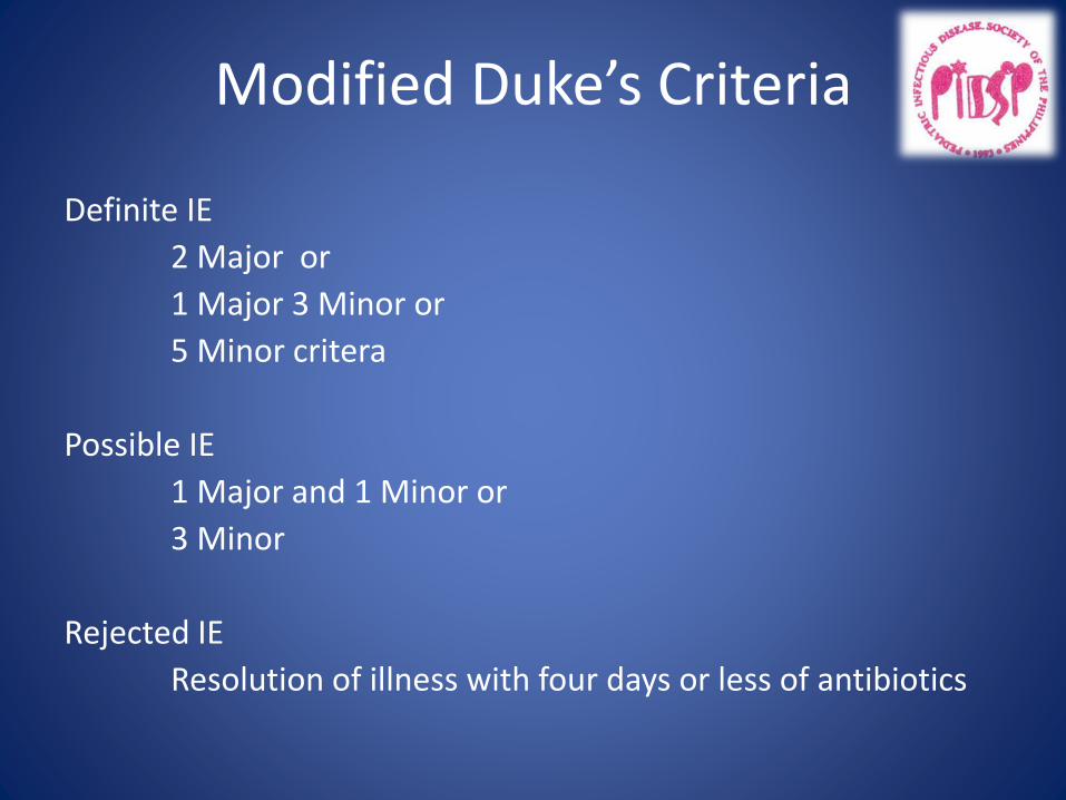
Modified Duke’s Criteria
Definite IE
2 Major or
1 Major 3 Minor or
5 Minor critera
Possible IE
1 Major and 1 Minor or
3 Minor
Rejected IE
Resolution of illness with four days or less of antibiotics
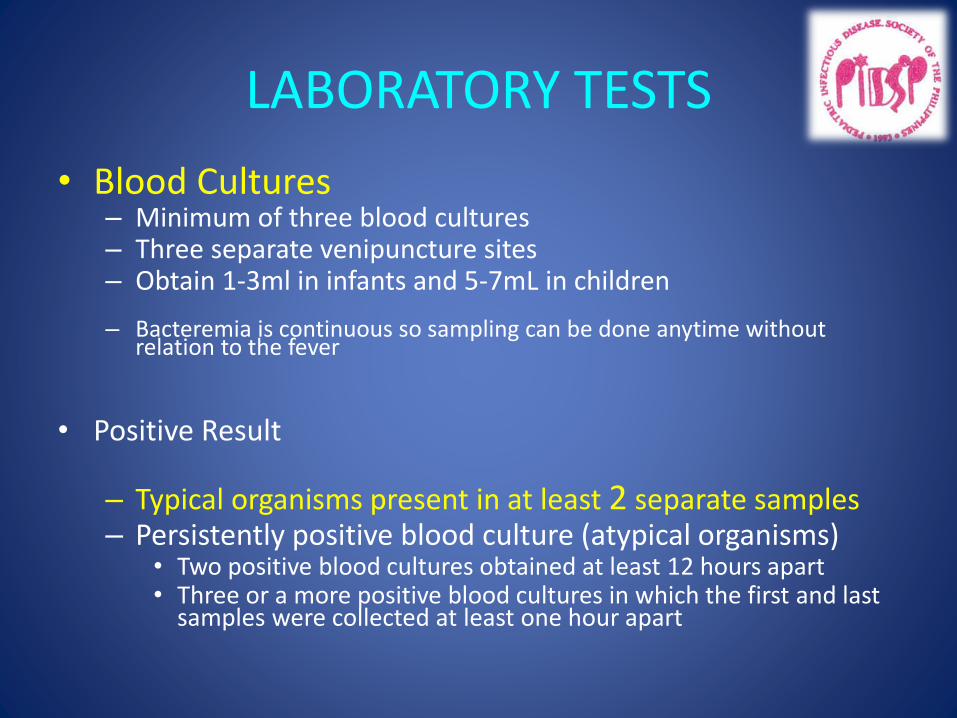
LABORATORY TESTS
• Blood Cultures– Minimum of three blood cultures– Three separate venipuncture sites– Obtain 1-3ml in infants and 5-7mL in children
– Bacteremia is continuous so sampling can be done anytime without relation to the fever
• Positive Result
– Typical organisms present in at least 2 separate samples– Persistently positive blood culture (atypical organisms)
• Two positive blood cultures obtained at least 12 hours apart• Three or a more positive blood cultures in which the first and last
samples were collected at least one hour apart
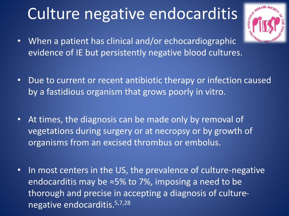
Culture negative endocarditis
• When a patient has clinical and/or echocardiographic evidence of IE but persistently negative blood cultures.
• Due to current or recent antibiotic therapy or infection caused by a fastidious organism that grows poorly in vitro.
• At times, the diagnosis can be made only by removal of vegetations during surgery or at necropsy or by growth of organisms from an excised thrombus or embolus.
• In most centers in the US, the prevalence of culture-negative endocarditis may be ≈5% to 7%, imposing a need to be thorough and precise in accepting a diagnosis of culture-negative endocarditis.5,7,28
Additional Labs
• CBC: Low hemoglobin/hematocrit (ANEMIA)
• Elevated ESR and CRP
• Urinalysis: Hematuria, proteinuria, and red cell casts suggestive of glomerulonephritis
Imaging
• Chest x-ray
– Look for multiple focal infiltrates and calcification of heart valves
• EKG
– Rarely diagnostic
– Look for evidence of ischemia, conduction delay, and arrhythmias
• Echocardiography
ECHOCARDIOGRAPHY
• Should be performed on all patients in whom there is a reasonable suspicion of IE to detect presence of a vegetation
• Main modality to detect endocardial infection
• Can identify the size and location of vegetation , extent of valve damage ,ventricular function and pericardial effusion etc.
Indications for Echocardiography
• Transthoracic echocardiography (TTE)
– First line if suspected IE
– Native valves
• Transesophageal echocardiography (TEE)
– Prosthetic valves
– Intracardiac complications
– Inadequate TTE
– Fungal or S. aureus or bacteremia
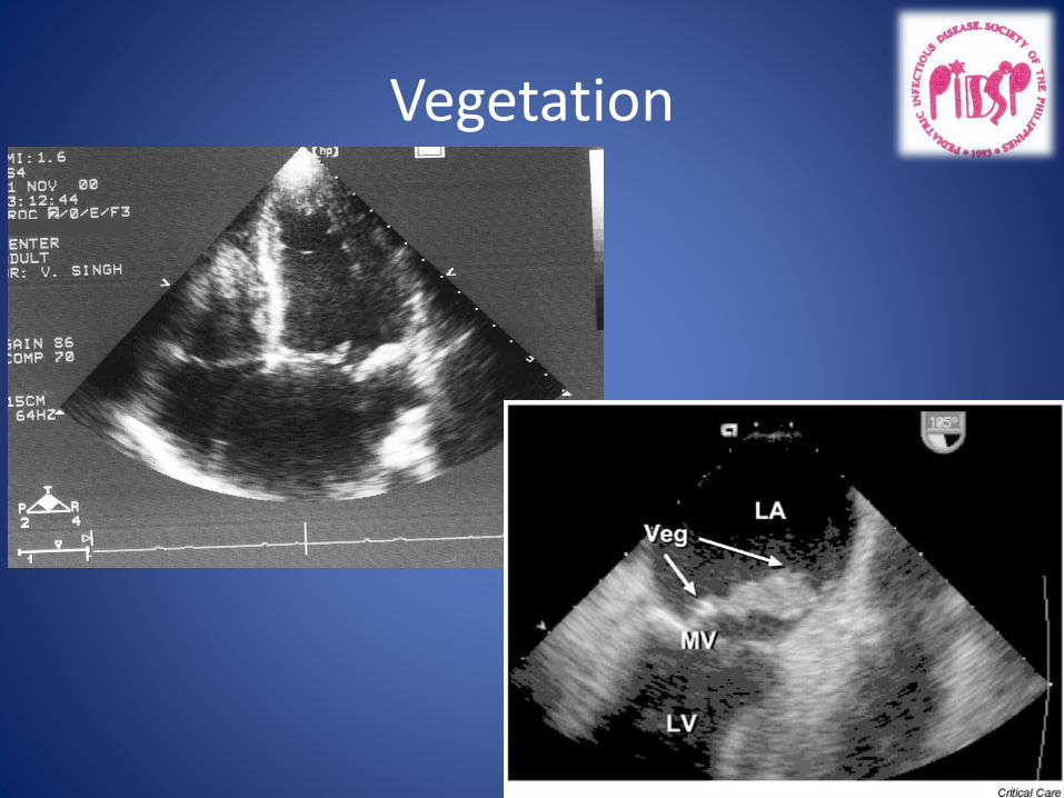
Vegetation
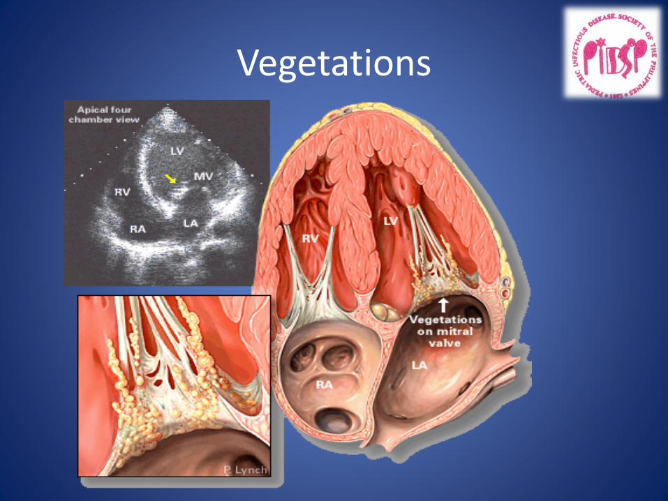
Vegetations
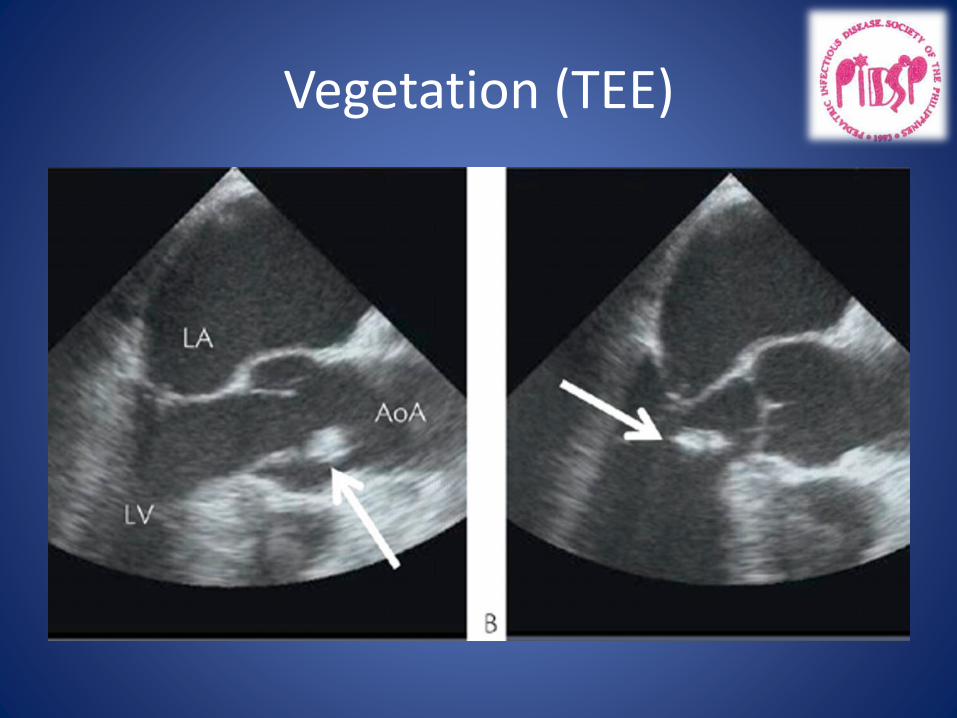
Vegetation (TEE)
Complications
• Heart Failure
• Metastatic Infections due to Septic emboli (Osteomyelitis, Pneumonia, Abscesses)
• Embolic phenomenon (Stroke, MI)
• Glomerulonephritis
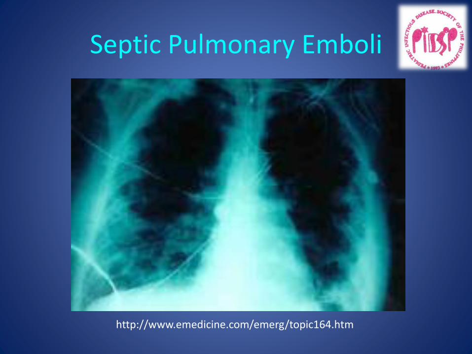
Septic Pulmonary Emboli
http://www.emedicine.com/emerg/topic164.htm
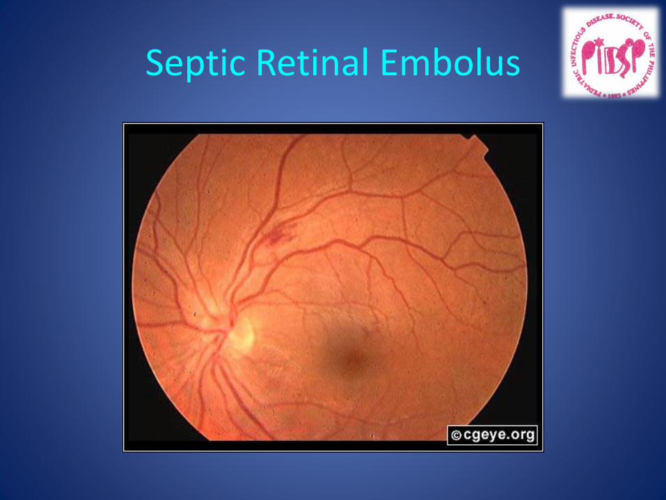
Septic Retinal Embolus
Risk Factors for IE Complications
• Prosthetic cardiac valves
• Left sided involvement
• Staphylococcus areus or fungal IE
• Previous IE
• Prolonged symptoms > 3months
• Cyanotic congenital heart disease
• Systemic to pulmonary shunts
• Poor clinical response to antimicrobial therapy
Treatment
• Antibiotic choice, dosage, and duration of treatment are dependent upon the underlying causative microbial agent
• In acute IE, blood culture should be done as quickly as possible so empiric antibiotic therapy can be started
• Patients who fail medical therapy with persistent vegetations despite antibiotic therapy may be candidates for surgical intervention
Treatment
• Parenteral antibiotics– High serum concentrations to penetrate vegetations
– Prolonged treatment to kill dormant bacteria clustered in vegetations
– Usually 4-8 weeks
– Broad coverage until organism is isolated
• Surgery– Intracardiac complications
• Surveillance blood cultures – During and after treatment
Prophylaxis
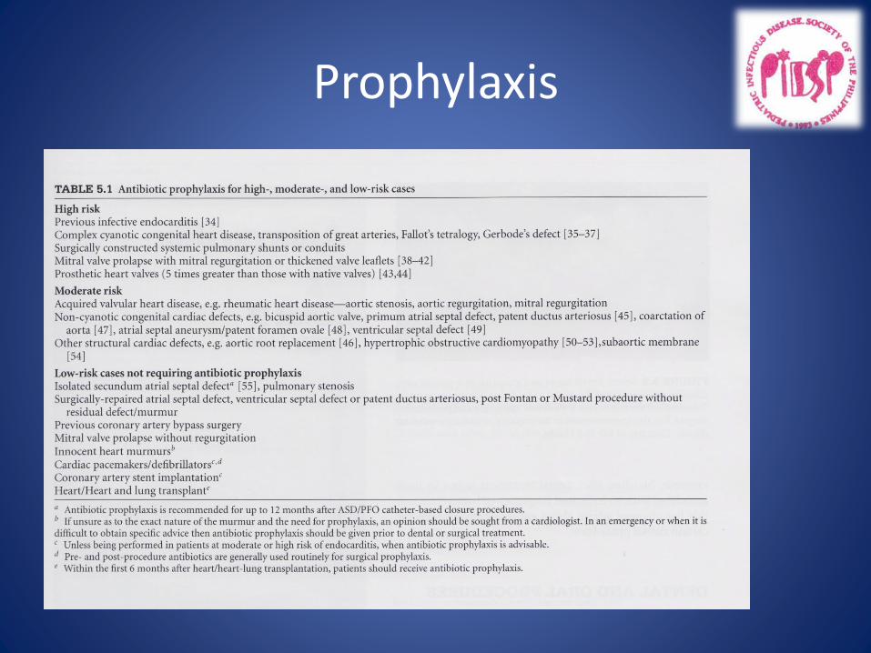
• Revised guidelines from AHA state that antibiotic prophylaxis should be reserved for patients with highest risk of IE
• High Risk Patients:– Prosthetic cardiac valves– Previous IE– Cardiac valvar disease after transplantation– Unrepaired cyanotic CHD– Palliative shunts and conduits– Within 6 months of complete repair with a prosthetic material
or device– Residual defect at the site or adjactent to the site of prosthetic
patch
Prophylaxis
• All dental procedures that involve manipulation of gingival tissues
• Procedures on respiratory tract or infected skin, dermal structures or musculoskeletal tissue
• Any other procedure that is potentially bacteremic
• Focus should shift towards good oral hygiene
Prophylaxis
Summary
• RHD and IE are 2 very important acquired cardiac infections in pediatrics
• Diagnostic criteria have been established to guide us in the treatment of our patients
• Echocardiography play a major role in the initial diagnosis of these conditions
• Prolonged treatment and appropriate prophylaxis are needed to ensure cure and prevent further recurrences