Embed Size (px)
Citation preview
Regulation of skeletal muscle fat oxidationduring exercise in humans
LAWRENCE L. SPRIET
Department of Human Biology & Nutritional Sciences, University of Guelph, Guelph, Ontario, CANADA
ABSTRACT
SPRIET, L. L. Regulation of skeletal muscle fat oxidation during exercise in humans.Med. Sci. Sports Exerc., Vol. 34, No. 9, pp.1477–1484, 2002. Fat and carbohydrate are the major energy substrates during aerobic exercise in well-fed humans. The regulation offat metabolism during exercise has not been as thoroughly studied as carbohydrate metabolism, especially in human skeletal muscle.Traditionally, it was believed that the regulation of skeletal muscle fat metabolism was mainly at the level of the delivery of free fattyacids to the muscle (adipose tissue lipolysis) and transport of the long chain fatty acids into the mitochondria. It is now known thatthe transport of fatty acids into the muscle cell and the regulation of muscle triacylglycerol lipase activity are also important sites ofregulation. New lines of research are currently underway examining the regulation of fat metabolism in skeletal muscle at the level offat transport across the sarcolemmal and mitochondrial membranes and regulation of TG lipase activity in both rodent and humanmodels. A major goal of this research is to determine the regulatory signals that control the up-regulation of fat metabolism during thetransition from rest to low and moderate aerobic exercise (30-65% V˙ O2max) and the down-regulation that occurs when exercising atintense aerobic exercise (~85% V˙ O2max). Although it is expected that the signals that activate carbohydrate metabolism during exercise(Ca2� and free ADP, AMP, and Pi) would also play a role in fat metabolism, this has not been demonstrated to date.Key Words:METABOLIC REGULATION, ADIPOSE TISSUE LIPOLYSIS, MUSCLE MEMBRANE FAT TRANSPORT, MUSCLE TRIAC-YLGLYCEROL LIPASE ACTIVITY, MUSCLE CARNITINE PALMITOYLTRANSFERASE I ACTIVITY
Fat and carbohydrate are the dominant substrates forthe production of ATP (oxidative phosphorylation) inskeletal muscle during aerobic exercise in well-fed
humans. The absolute contribution of fat to the total energyproduction during exercise increases from low power out-puts to a maximum between ~50 and 65% V˙ O2maxand thendecreases as the exercise intensity increases to ~85%V̇O2max and above (18,34,36). Fat is often depicted as theless important of the two fuels, as carbohydrate becomes thedominant substrate during intense aerobic exercise and fatcannot be used to generate ATP via “anaerobic” metabolism(substrate phosphorylation) during sprint exercise. How-ever, it does have some advantages over carbohydrate. Fat isan energy-dense fuel with a high energy yield per unit massand is also stored in large quantities in the body as comparedwith carbohydrate. Therefore, fat can provide a substantialamount of substrate for oxidative phosphorylation duringprolonged exercise at low to moderate intensities. Aerobictraining also increases the absolute rate of energy produc-tion from fat oxidation in skeletal muscle. Increasing fat anddecreasing carbohydrate use during aerobic exercise in-creases the power output and/or time that aerobic exercisecan be maintained before the carbohydrate store in the bodyis consumed. Surprisingly, the regulation of fat metabolismhas not been studied as thoroughly as carbohydrate metab-
olism in skeletal muscle. Although this is changing, there isstill little known regarding the regulation of fat metabolismin human skeletal muscle during exercise.
The purpose of this paper is to briefly review the regulationof fat oxidation in human skeletal muscle during exercise, withan emphasis on the key regulatory sites believed to determinethe rate of fat provision and oxidation. Processes that are notdirectly involved in the oxidation of fat, such as esterificationof triacylglycerol, are not examined. Several detailed reviewsof various aspects of fat metabolism during exercise alreadyexist (8,27,28,39,42,44,50). This article attempts to provide themetabolic bases for the papers that follow in this symposium.
OVERVIEW OF SKELETAL MUSCLE FATMETABOLISM AND REGULATION
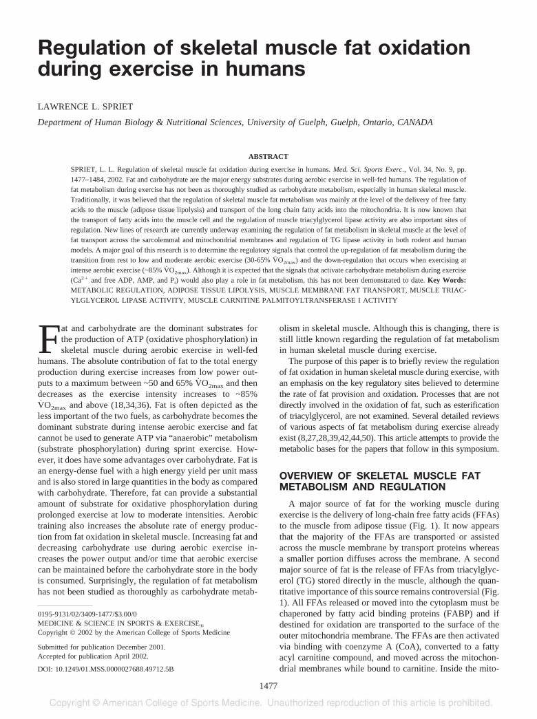
A major source of fat for the working muscle duringexercise is the delivery of long-chain free fatty acids (FFAs)to the muscle from adipose tissue (Fig. 1). It now appearsthat the majority of the FFAs are transported or assistedacross the muscle membrane by transport proteins whereasa smaller portion diffuses across the membrane. A secondmajor source of fat is the release of FFAs from triacylglyc-erol (TG) stored directly in the muscle, although the quan-titative importance of this source remains controversial (Fig.1). All FFAs released or moved into the cytoplasm must bechaperoned by fatty acid binding proteins (FABP) and ifdestined for oxidation are transported to the surface of theouter mitochondria membrane. The FFAs are then activatedvia binding with coenzyme A (CoA), converted to a fattyacyl carnitine compound, and moved across the mitochon-drial membranes while bound to carnitine. Inside the mito-
0195-9131/02/3409-1477/$3.00/0MEDICINE & SCIENCE IN SPORTS & EXERCISE®Copyright © 2002 by the American College of Sports Medicine
Submitted for publication December 2001.Accepted for publication April 2002.
DOI: 10.1249/01.MSS.0000027688.49712.5B
1477
chondria, the carnitine is removed, the CoA is rebound, andthe fatty acyl-CoA molecules are metabolized in the �-ox-idation pathway with the production of reducing equivalents(NADH, FADH2) and acetyl-CoA (Fig. 1). The acetyl-CoAis further metabolized in the tricarboxylic (TCA) pathwaywith the production of additional reducing equivalents. Theelectron transport chain, including oxygen, accepts the re-ducing equivalents to generate the proton motive force,which provides the chemical energy used to synthesize ATPfrom inorganic phosphate (Pi) and ADP in the process ofoxidative phosphorylation.
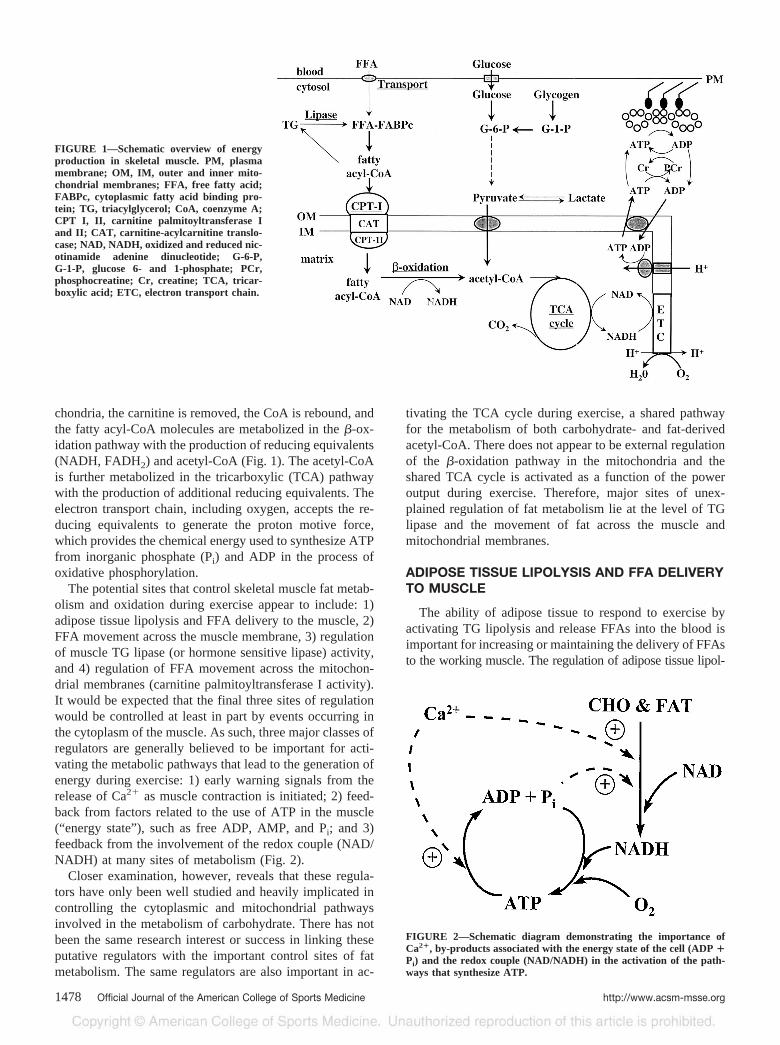
The potential sites that control skeletal muscle fat metab-olism and oxidation during exercise appear to include: 1)adipose tissue lipolysis and FFA delivery to the muscle, 2)FFA movement across the muscle membrane, 3) regulationof muscle TG lipase (or hormone sensitive lipase) activity,and 4) regulation of FFA movement across the mitochon-drial membranes (carnitine palmitoyltransferase I activity).It would be expected that the final three sites of regulationwould be controlled at least in part by events occurring inthe cytoplasm of the muscle. As such, three major classes ofregulators are generally believed to be important for acti-vating the metabolic pathways that lead to the generation ofenergy during exercise: 1) early warning signals from therelease of Ca2� as muscle contraction is initiated; 2) feed-back from factors related to the use of ATP in the muscle(“energy state” ), such as free ADP, AMP, and Pi; and 3)feedback from the involvement of the redox couple (NAD/NADH) at many sites of metabolism (Fig. 2).
Closer examination, however, reveals that these regula-tors have only been well studied and heavily implicated incontrolling the cytoplasmic and mitochondrial pathwaysinvolved in the metabolism of carbohydrate. There has notbeen the same research interest or success in linking theseputative regulators with the important control sites of fatmetabolism. The same regulators are also important in ac-
tivating the TCA cycle during exercise, a shared pathwayfor the metabolism of both carbohydrate- and fat-derivedacetyl-CoA. There does not appear to be external regulationof the �-oxidation pathway in the mitochondria and theshared TCA cycle is activated as a function of the poweroutput during exercise. Therefore, major sites of unex-plained regulation of fat metabolism lie at the level of TGlipase and the movement of fat across the muscle andmitochondrial membranes.
ADIPOSE TISSUE LIPOLYSIS AND FFA DELIVERYTO MUSCLE
The ability of adipose tissue to respond to exercise byactivating TG lipolysis and release FFAs into the blood isimportant for increasing or maintaining the delivery of FFAsto the working muscle. The regulation of adipose tissue lipol-
FIGURE 2—Schematic diagram demonstrating the importance ofCa2�, by-products associated with the energy state of the cell (ADP �Pi) and the redox couple (NAD/NADH) in the activation of the path-ways that synthesize ATP.
FIGURE 1—Schematic overview of energyproduction in skeletal muscle. PM, plasmamembrane; OM, IM, outer and inner mito-chondrial membranes; FFA, free fatty acid;FABPc, cytoplasmic fatty acid binding pro-tein; TG, triacylglycerol; CoA, coenzyme A;CPT I, II, carnitine palmitoyltransferase Iand II; CAT, carnitine-acylcarnitine translo-case; NAD, NADH, oxidized and reduced nic-otinamide adenine dinucleotide; G-6-P,G-1-P, glucose 6- and 1-phosphate; PCr,phosphocreatine; Cr, creatine; TCA, tricar-boxylic acid; ETC, electron transport chain.
1478 Official Journal of the American College of Sports Medicine http://www.acsm-msse.org
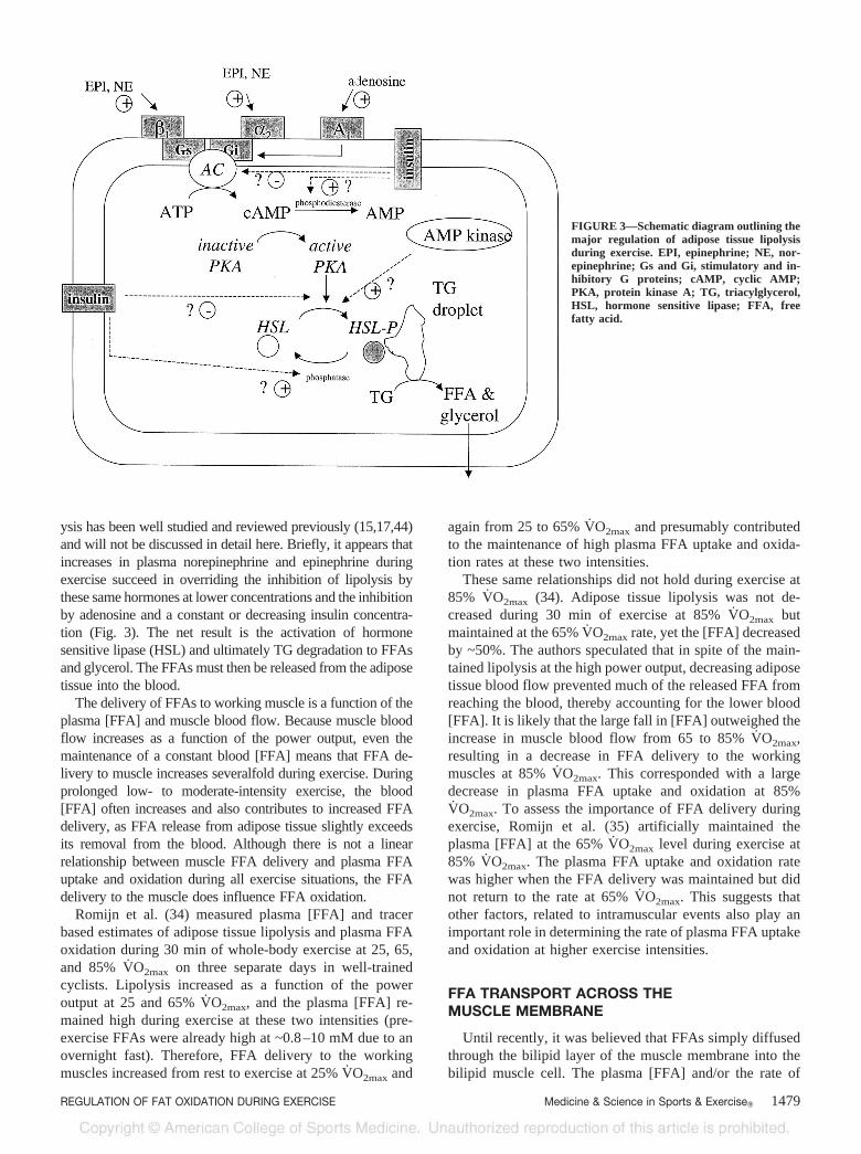
ysis has been well studied and reviewed previously (15,17,44)and will not be discussed in detail here. Briefly, it appears thatincreases in plasma norepinephrine and epinephrine duringexercise succeed in overriding the inhibition of lipolysis bythese same hormones at lower concentrations and the inhibitionby adenosine and a constant or decreasing insulin concentra-tion (Fig. 3). The net result is the activation of hormonesensitive lipase (HSL) and ultimately TG degradation to FFAsand glycerol. The FFAs must then be released from the adiposetissue into the blood.
The delivery of FFAs to working muscle is a function of theplasma [FFA] and muscle blood flow. Because muscle bloodflow increases as a function of the power output, even themaintenance of a constant blood [FFA] means that FFA de-livery to muscle increases severalfold during exercise. Duringprolonged low- to moderate-intensity exercise, the blood[FFA] often increases and also contributes to increased FFAdelivery, as FFA release from adipose tissue slightly exceedsits removal from the blood. Although there is not a linearrelationship between muscle FFA delivery and plasma FFAuptake and oxidation during all exercise situations, the FFAdelivery to the muscle does influence FFA oxidation.
Romijn et al. (34) measured plasma [FFA] and tracerbased estimates of adipose tissue lipolysis and plasma FFAoxidation during 30 min of whole-body exercise at 25, 65,and 85% V̇O2max on three separate days in well-trainedcyclists. Lipolysis increased as a function of the poweroutput at 25 and 65% V̇O2max, and the plasma [FFA] re-mained high during exercise at these two intensities (pre-exercise FFAs were already high at ~0.8–10 mM due to anovernight fast). Therefore, FFA delivery to the workingmuscles increased from rest to exercise at 25% V̇O2max and
again from 25 to 65% V̇O2max and presumably contributedto the maintenance of high plasma FFA uptake and oxida-tion rates at these two intensities.
These same relationships did not hold during exercise at85% V̇O2max (34). Adipose tissue lipolysis was not de-creased during 30 min of exercise at 85% V̇O2max butmaintained at the 65% V̇O2max rate, yet the [FFA] decreasedby ~50%. The authors speculated that in spite of the main-tained lipolysis at the high power output, decreasing adiposetissue blood flow prevented much of the released FFA fromreaching the blood, thereby accounting for the lower blood[FFA]. It is likely that the large fall in [FFA] outweighed theincrease in muscle blood flow from 65 to 85% V̇O2max,resulting in a decrease in FFA delivery to the workingmuscles at 85% V̇O2max. This corresponded with a largedecrease in plasma FFA uptake and oxidation at 85%V̇O2max. To assess the importance of FFA delivery duringexercise, Romijn et al. (35) artificially maintained theplasma [FFA] at the 65% V̇O2max level during exercise at85% V̇O2max. The plasma FFA uptake and oxidation ratewas higher when the FFA delivery was maintained but didnot return to the rate at 65% V̇O2max. This suggests thatother factors, related to intramuscular events also play animportant role in determining the rate of plasma FFA uptakeand oxidation at higher exercise intensities.
FFA TRANSPORT ACROSS THEMUSCLE MEMBRANE
Until recently, it was believed that FFAs simply diffusedthrough the bilipid layer of the muscle membrane into thebilipid muscle cell. The plasma [FFA] and/or the rate of
FIGURE 3—Schematic diagram outlining themajor regulation of adipose tissue lipolysisduring exercise. EPI, epinephrine; NE, nor-epinephrine; Gs and Gi, stimulatory and in-hibitory G proteins; cAMP, cyclic AMP;PKA, protein kinase A; TG, triacylglycerol,HSL, hormone sensitive lipase; FFA, freefatty acid.
REGULATION OF FAT OXIDATION DURING EXERCISE Medicine & Science in Sports & Exercise� 1479
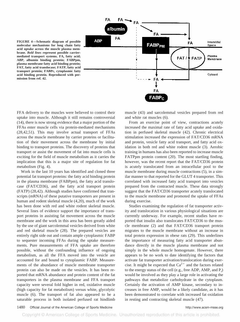
FFA delivery to the muscles were believed to control theiruptake into muscle. Although it still remains controversial(14), there is now strong evidence that a major portion of theFFAs enter muscle cells via protein-mediated mechanisms(28,42,51). This may involve actual transport of FFAsacross the muscle membrane by carrier proteins or facilita-tion of their movement across the membrane by initialbinding to transport proteins. The discovery of proteins thattransport or assist the movement of fat into muscle cells isexciting for the field of muscle metabolism as it carries theimplication that this is a major site of regulation for fatmetabolism (Fig. 4).
Work in the last 10 years has identified and cloned threepotential fat transport proteins: the fatty acid binding proteinin the plasma membrane (FABPpm), the fatty acid translo-case (FAT/CD36), and the fatty acid transport protein(FATP) (28,42). Although studies have confirmed that tran-scripts (mRNA) of these putative transporters are present inhuman and rodent skeletal muscle (4,20), much of the workhas been done with red and white rodent skeletal muscle.Several lines of evidence support the importance of trans-port proteins in assisting fat movement across the musclemembrane and the work in this area has been greatly aidedby the use of giant sarcolemmal vesicles derived from whiteand red skeletal muscle (28). The prepared vesicles areentirely right side out and contain ample cytoplasmic FABPto sequester incoming FFAs during the uptake measure-ments. Pure measurements of FFA uptake are thereforepossible, without the confounding influence of ongoingmetabolism, as all the FFA moved into the vesicle areaccounted for and bound to cytoplasmic FABP. Measure-ments of the abundance of muscle membrane transporterprotein can also be made on the vesicles. It has been re-ported that mRNA abundance and protein content of the fattransporters in the plasma membrane and FFA transportcapacity were several fold higher in red, oxidative muscle(high capacity for fat metabolism) versus white, glycolyticmuscle (6). The transport of fat also appeared to be asaturable process in both isolated perfused rat hindlimb
muscle (43) and sarcolemmal vesicles prepared from redand white rat muscles (6).
From an exercise point of view, contractions acutelyincreased the maximal rate of fatty acid uptake and oxida-tion in perfused skeletal muscle (42). Chronic electricalstimulation increased the expression of FAT/CD36 mRNAand protein, vesicle fatty acid transport, and fatty acid ox-idation in both red and white rodent muscle (3). Aerobictraining in humans has also been reported to increase muscleFATPpm protein content (20). The most startling finding,however, was the recent report that the FAT/CD36 proteinis acutely translocated from an intracellular pool to themuscle membrane during muscle contractions (5), in a sim-ilar manner to that reported for the GLUT 4 transporter. Thiscorrelated with increased fatty acid transport into vesiclesprepared from the contracted muscle. These data stronglysuggest that the FAT/CD36 transporter acutely translocatedto the muscle membrane and promoted the uptake of FFAsduring exercise.
Studies examining the regulation of fat transporter activ-ity and translocation in various physiological situations arecurrently underway. For example, recent studies have re-ported that insulin also translocates FAT/CD36 to the mus-cle membrane (2) and that FAT/CD36 transport proteinmigrates to the muscle membrane without an increase intotal protein expression in obese rats (29). This underlinesthe importance of measuring fatty acid transporter abun-dance directly in the muscle plasma membrane and notsimply in the whole muscle homogenate. However, thereappears to be no work to date identifying the factors thatactivate fat transporter activation/translocation during exer-cise. It might be expected that Ca2� and the factors relatedto the energy status of the cell (e.g., free ADP, AMP, and Pi)would be involved as they play a large role in activating thepathways that metabolize carbohydrate in the cytoplasm.Certainly the activation of AMP kinase, secondary to in-creases in free AMP, would be a likely candidate, as it hasbeen demonstrated to correlate with increased fat oxidationin resting and contracting skeletal muscle (47).
FIGURE 4—Schematic diagram of possiblemolecular mechanisms for long chain fattyacid uptake across the muscle plasma mem-brane. Bold lines represent possible carrier-mediated transport systems. FA, fatty acid;ABP, albumin binding protein; FABPpm,plasma membrane fatty acid binding protein;FAT, fatty acid translocase; FATP, fatty acidtransport protein; FABPc, cytoplasmic fattyacid binding protein. Reproduced with per-mission from ref. 42.
1480 Official Journal of the American College of Sports Medicine http://www.acsm-msse.org

REGULATION OF MUSCLE TRIACYLGLYCEROLLIPASE ACTIVITY
A significant amount of fat is stored in human skeletalmuscle, usually in the range of 20–40 mmol·kg�1 drymuscle (19,21,40,45,46) or enough energy to account for~70–100% of the energy stored as glycogen in a well-fedperson (44). There are many metabolic pathways leading tostorage of FFAs as TG and degradation of TG to FFAs andglycerol. However, the reaction catalyzed by TG lipaseappears to be the only site of external regulation in thesepathways and determines the rate of TG degradation todiacylglycerol and ultimately FFA during exercise. Theadditional enzymes responsible for removing the final twoFFAs are near-equilibrium in nature and continue to degradethe di- and mono-acylglycerol as a function of increasingsubstrate concentrations. TG lipase, or the muscle version ofhormone sensitive lipase (HSL), has been identified in skel-etal muscle and is distinct from the other lipases that exist inmuscle (16). It has a neutral pH optimum and is covalentlyactivated by the action of a kinase that adds a phosphate anddeactivated by a phosphatase that removes a phosphate asdescribed for adipose tissue HSL (Fig. 5).
Very little is known regarding the regulation of thisimportant enzyme as several factors have contributed to ageneral lack of research in this area. The first problem withstudying muscle TG lipase has been the possibility of con-tamination with two other lipases that exist in skeletal mus-cle, a lysosomal lipase with an acidic pH optimum andlipoprotein lipase with an alkaline pH optimum. Lipoproteinlipase is produced in vesicles and secreted to the outside ofthe muscle cell to ultimately reside on the endothelial sur-face of the muscle blood vessels. It is important in regulat-ing the degradation of circulating TGs in the blood duringrest.
A second problem has been the controversy regardingwhether muscle TG is actually utilized during aerobic ex-ercise. Most studies that have measured pre and post exer-cise [TG] in needle muscle biopsy samples have not re-
ported significant decreases (�20% of total TG) after 90–120 min of exercise at 50–65% V̇O2max (13,19,21,40,46).When exercise is prolonged from 4–8 h, studies generallyreported significant decreases (�40% of total TG) in theintramuscular lipid store (7,12). On the other hand, moststudies that have estimated the use of intramuscular TGfrom measurements of the whole-body respiratory exchangeratio and plasma FFA oxidation have concluded that muscleTG is a major contributor of substrate during low- to mod-erate-intensity aerobic exercise (34,36). More recent directmeasurements of muscle TG using a 1H magnetic resonancespectroscopy technique also generally report significantmuscle TG use during aerobic exercise, although absolutecalibrations are difficult (11,24). Although all TG measure-ment techniques suffer from various limitations and as-sumptions, one concern with directly measuring intramus-cular fat use in muscle biopsies over the shorter term is theenergy density of TG. For example, estimations of the fatthat would be required from the muscle TG store duringexercise at 50–65% V̇O2max for 90–120 min is only ~2–4mmol·kg�1 dm or only 10–15% of the total TG store. Asecond concern with the direct measurements of muscle TGis the large variability that has been reported between biop-sies from the same person (20–24%) in untrained subjects(46). These problems suggest that the biopsy technique maynot be sensitive enough to detect the expected magnitude ofchange in many studies, whereas the other measurement/estimation techniques are. However, a recent study reporteda much lower between biopsy variability with aerobicallytrained subjects (12%) and a significant decrease in muscleTG during 120 min of cycling at 55% V̇O2max (45). Takentogether, these recent results and those of the other tech-niques suggest that muscle TG is a significant fuel foroxidation during prolonged moderate-intensity exercise.
A third problem delaying work on the regulation of mus-cle TG has been the lack of a viable analytical technique fortrapping and measuring the activity of the enzyme in theinactive and active fractions during exercise. These tech-niques exist for other covalently regulated enzymes thatmetabolize carbohydrate, such as glycogen phosphorylaseand pyruvate dehydrogenase. Recently, Langfort and col-leagues (23,25–27) reported activities of the inactive andactive fractions of muscle TG lipase in rodent and humanskeletal muscle in a variety of conditions. Their findingsrepresent the first work examining the regulation of thisimportant enzyme. Incubation of rat soleus muscles withepinephrine increased the activity of the enzyme in theactive form (25). The conversion to the active form wasmediated by the B-adrenergic second messenger cyclicAMP, via activation of protein kinase A. Presentation of theantiserum to HSL removed the effects of epinephrine (25).The activity of TG lipase in the active form was alsoincreased in isolated soleus muscles after 1 and 5 min ofelectrical stimulation but returned to control levels after 10and 60 min of contractions (26). Total TG activity wasunaffected during the entire 60 min of stimulation. Neitherprior removal of the sympathoadrenal organs nor the addi-
FIGURE 5—Proposed covalent regulation of muscle triacylglycerollipase activity. The kinase is thought to be activated by cyclic AMP andunknown contraction factors.
REGULATION OF FAT OXIDATION DURING EXERCISE Medicine & Science in Sports & Exercise� 1481
tion of propanolol to the incubation medium impaired thecontraction-induced activation of TG lipase (26).
One study has examined the effect of exercise on theactivity of TG lipase in the active form in human subjects.Control and adrenalectomized (ADR) subjects cycled at~70% V̇O2max for 45 min and at ~86% V̇O2max for 15 min(23). As the ADR subjects had no increase in cat-echolamines during exercise, they repeated the protocol asecond time with the infusion of epinephrine to mimic thesituation in the control subjects. The activation of TG lipaseincreased at the end of the exercise at 70% V̇O2max, with nofurther increase at 86% V̇O2max in the control subjects.However, no activation of TG lipase occurred in the ADRsubjects in the absence of increases in plasma epinephrine,whereas the infusion of epinephrine increased TG lipaseactivation at both power outputs (23).
These experiments demonstrated for the first time thatTG lipase is activated during aerobic exercise in humans.However, the regulatory factors that govern this activa-tion during exercise are unknown. Again, it might bepredicted that increases in Ca2� and free ADP, AMP(AMP kinase) and Pi may be involved, but this has notbeen examined. The results also demonstrate that thepresence of plasma epinephrine was necessary for TGlipase activation in ADR subjects. However, it is notknown whether this is the case for normal subjects withfunctioning adrenal glands and whether events in the cellalone are sufficient to activate TG lipase as demonstratedin the isolated rat soleus preparation.
TRANSPORT ACROSS THEMITOCHONDRIAL MEMBRANES
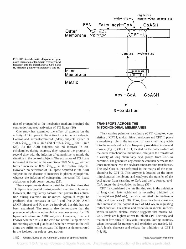
The carnitine palmitoyltransferase (CPT) complex, con-sisting of CPT I, acylcarnitine translocase and CPT II, playsa regulatory role in the transport of long chain fatty acidsinto the mitochondria for subsequent �-oxidation in skeletalmuscle (Fig. 6) (31). CPT I, located on the outer surface ofthe outer mitochondrial membrane, catalyzes the transfer ofa variety of long chain fatty acyl groups from CoA tocarnitine. The generated acylcarnitine can then permeate theinner membrane, via the acylcarnitine/carnitine translocase.The acyl-CoA is then reformed in the matrix of the mito-chondria by CPT II. This enzyme is located on the innermitochondrial membrane and catalyzes the transfer of theacyl group from carnitine to CoA and the re-formed acyl-CoA enters the �-oxidation pathway (31).
CPT I is considered the rate limiting step in the oxidationof long chain fatty acids and is reversibly inhibited bymalonyl-CoA (M-CoA), the first committed intermediate infatty acid synthesis (1,30). Thus, there has been consider-able interest in the potential role of M-CoA in regulatingmitochondrial FFA uptake and oxidation in skeletal muscle.Work in rodent skeletal muscle suggests that muscle M-CoA levels are highest at rest to inhibit CPT I activity andmaintain low rates of fatty acid transport. During exercise,when increased fat transport and oxidation are needed, M-CoA levels decrease and release the inhibition of CPT I(48,49).
FIGURE 6—Schematic diagram of pro-posed regulation of long chain free fatty acidtransport into the mitochondria. CPT I andII, carnitine palmitoyltransferase I and II.
1482 Official Journal of the American College of Sports Medicine http://www.acsm-msse.org
Another line of work also proposed that fat metabolism isregulated by the level of glycolytic activity occurring in themuscle (9,37,38). Increases in glycolytic flux rates as theaerobic exercise intensity increases correlated with decreas-ing rates of fat oxidation. It was proposed that signalsrelated to the increased glycolytic flux down-regulate fatmetabolism inside the muscle cell and that increases in[M-CoA] could be the regulator inhibiting fat uptake andoxidation during intense aerobic exercise.
However, recent studies have demonstrated that M-CoA levels did not change during low to moderate aer-obic exercise of varying durations in human skeletalmuscle despite large increases in fatty acid oxidationrates (10,32,33). In addition, increasing the exercise in-tensity from 65 to 90%V̇O2max was not associated with anincrease in muscle [M-CoA] despite large decreases in fatoxidation (33). Therefore, the conclusion at the presenttime must be that M-CoA is not involved in the up-regulation of fat metabolism from rest to low- and mod-erate-intensity exercise and also not responsible fordown-regulating fat metabolism when aerobic exercisebecomes intense.
These findings in human muscle suggested that theregulation of CPT I activity is more complex than simplechanges in [M-CoA]. Starritt et al. (41) used an isolated,intact mitochondrial preparation from human skeletalmuscle (1) to study CPT I regulation in vitro and deter-mined that CPT I is also inhibited by decreases in pH (7.0to 6.8) but unaffected by other metabolites that accumu-late (acetyl-CoA, acetylcarnitine) or decrease (CoA) dur-ing exercise. This suggests that substrate-enzyme inter-actions, structural changes in the binding of M-CoA toCPT I, and/or the presence of additional regulators maybe important for increased fat transport during exercise.The sensitivity to pH may also explain the decrease in fatmetabolism that occurs when moving from moderate tointense aerobic exercise. An interesting recent develop-ment has been the report that an M-CoA insensitive
CPT I exists in rodent skeletal muscle, suggesting that acontrol system independent of M-CoA may account forthe up-regulation of fat transport and oxidation duringexercise (22). However, no studies to date have examinedwhether CPT I activity is sensitive to increases in cyto-plasmic [Ca2�] or free ADP, AMP (AMP kinase), or Pi,as might be expected for an enzyme that must up-regulateat the onset of exercise. If this type of control did exist,an explanation for how the enzyme becomes less sensi-tive to these signals during intense aerobic exercisewould be needed to explain the decrease in mitochondrialfat transport and oxidation that occurs.
SUMMARY
Fat and carbohydrate are the major fuels metabolized toprovide substrate for the aerobic production of energy dur-ing exercise in well-fed humans. Unlike carbohydrate me-tabolism, the regulation of fat metabolism in human skeletalmuscle during exercise has not been well studied. Tradi-tionally, it was believed that the regulation of fat metabo-lism was mainly at the level of fatty acid provision to themuscle (adipose tissue lipolysis) and transport of long chainfatty acids into the mitochondria (CPT I activity). It is nowknown that the transport of fatty acids into the muscle celland the regulation of muscle triacylglycerol lipase activitywithin the cell are also important sites of regulation. Con-sequently, new lines of research are examining the coordi-nated regulation of skeletal muscle fat metabolism at thelevel of fat transport across the muscle and mitochondrialmembranes, and regulation of TG lipase activity in bothrodent and human models.
The author thanks Dr. D. J. Dyck for critical reading of the manu-script and Figures 3 and 5.
Address for correspondence: Lawrence L. Spriet, Ph.D., Depart-ment of Human Biology & Nutritional Sciences, University of Guelph,Guelph, Ontario, Canada N1G 2W1; E-mail: [email protected].
REFERENCES
1. BERTHON, P. M., R. A. HOWLETT, G. J. F. HEIGENHAUSER, and L. L.SPRIET. Human skeletal muscle carnitine palmitoyltransferase Iactivity determined in isolated intact mitochondria. J. Appl.Physiol. 85:148–153, 1998.
2. BONEN, A. Insulin induces the translocation of the fatty acidtransporter FAT/CD36 to the plasma membrane. Am. J. Physiol.Endocrinol. Metab. 282:E491–E495, 2002.
3. BONEN, A., D. J. DYCK, A. IBRAHIMI, and N. A. ABUMRAD. Musclecontractile activity increases fatty acid metabolism and transportand FAT/CD36. Am. J. Physiol. Endocrinol. Metab. 276:E642–E649, 1999.
4. BONEN, A., D. MISKOVIC, and B. KIENS. Fatty acid transporters(FABPpm, FAT, FATP) in human muscle. Can. J. Appl. Physiol.24:515–523, 1999.
5. BONEN, A., J. J. F. P. LUIKEN, Y. ARUMUGAM, J. F. C. GLATZ, andN. N. TANDON. Acute regulation of fatty acid uptake involves thecellular redistribution of fatty acid translocase. J. Biol. Chem.275:14501–14508, 2000.
6. BONEN, A., J. J. F. P. LUIKEN, S. LIU, et al. Palmitate transport andfatty acid transporters in red and white muscles. Am. J. Physiol.275:E471–E478, 1998.
7. COSTILL, D. L., P. D. GOLLNICK, E. D. JANSSON, B. SALTIN, andE. M. STEIN. Glycogen depletion pattern in human muscle fibersduring distance running. Acta Physiol. Scand. 89:374–383, 1973.
8. COYLE, E. F. Fat oxidation during exercise: Role of lipolysis, FFAavailability, and glycolytic flux. In: Biochemisty of Exercise, X,M. Hargreaves and M. Thompson (Eds.). Champaign, IL: HumanKinetics, 1999, pp. 263–273.
9. COYLE, E. F., A. E. JEUKENDRUP, A. J. M. WAGENMAKERS, andW. H. M. SARIS. Fatty acid oxidation is directly regulated bycarbohydrate metabolism during exercise. Am. J. Physiol. Endo-crinol. Metab. 273:E268–E275, 1997.
10. DEAN, D., J. R. DAUGAARD, M. E. YOUNG, et al. Exercise diminishesthe activity of acetyl-CoA carboxylase in human skeletal muscle.Diabetes 49:1295–1300, 2000.
11. DECOMBAZ, J., B. SCHMITT, M. ITH, et al. Post-exercise fat intakerepletes intramyocellular lipids but no faster in trained than insedentary subjects. Am. J. Physiol. Reg. Integr. Comp. Physiol.281:R760–R769, 2001.
12. FROBERG, S. O., and F. MOSSFELDT. Effect of prolonged strenuousexercise on the concentration of triglycerides, phospholipids andglycogen in muscle of man. Acta Scand. Physiol. 82:167–171, 1971.
REGULATION OF FAT OXIDATION DURING EXERCISE Medicine & Science in Sports & Exercise� 1483
13. GUO, Z., B. BURGUERA, and M. D. JENSEN. Kinetics of intramus-cular triglyceride fatty acids in exercising humans. J. Appl.Physiol. 89:2057–2064, 2000.
14. HAMILTON, J. A., and F. KAMP. How are free fatty acids transportedin membranes? Diabetes 48:2255–2269, 1999.
15. HODGETTS, V., S. W. COPPACK, K. N. FRAYN, and T. D. R. HOCKA-DAY. Factors controlling fat mobilization from human subcutane-ous adipose tissue during exercise. J. Appl. Physiol. 71:445–451,1991.
16. HOLM, C., T. G. KIRCHGESSNER, K. L. SVENSON, et al. Hormone-sensitive lipase: sequence, expression, and chromosomal localiza-tion to 19 c, expression, and chromosomal localization to 19cent-q13.3. Science 241:1503–1506, 1988.
17. HOLM, C., T. OSTERLUND, H. LAURELL, and J. A. CONTERAS. Mo-lecular mechanisms regulating hormone-sensitive lipase and lipol-ysis. Ann. Rev. Nutr. 20:365–393, 2000.
18. HOWLETT, R. A., M. L. PAROLIN, D. J. DYCK, et al. Regulation ofskeletal muscle glycogen phosphorylase and pyruvate dehydroge-nase at varying power outputs. Am. J. Physiol. Reg. Integr. Comp.Physiol. 275:R418–R425, 1998.
19. KIENS, B., B. ESSEN-GUSTAVSSON, N. J. CHRISTENSEN, and B. SALTIN.Skeletal muscle substrate utilization during submaximal exercisein man: effect of endurance training. J. Physiol. 469:459–478,1993.
20. KIENS, B., S. KRISTIANSEN, P. JENSEN, E. A. RICHTER, and L. P.TURCOTTE. Membrane associated fatty acid binding protein (FAB-Ppm) in human skeletal muscle is increased by endurance training.Biochem. Biophys. Res. Commun. 231:463–465, 1997.
21. KIENS, B., and E. A. RICHTER. Utilization of skeletal muscle triac-ylglycerol during postexercise recovery in humans. Am. J. Physiol.Endocrinol. Metab. 275:E332–E337, 1998.
22. KIM, J.-Y., T. R. KOVES, G.-S. YU, et al. Evidence of a malonyl-CoA-insensitive carnitine palmitoyltransferase I activity in redskeletal muscle. Am. J. Physiol. Endocrinol. Metab. 282:E1014–E1022, 2002.
23. KJAER, M., K. HOWLETT, J. LANGFORT, et al. Adrenaline and gly-cogenolysis in skeletal muscle during exercise: a study in adrena-lectomized humans. J. Physiol. 528:2: 371–378, 2000.
24. KRSSAK, M., K. F. PETERSEN, R. BERGERON, et al. Intramuscularglycogen and intramyocellular lipid utilization during prolongedexercise and recovery in man: a 13C and 1H nuclear magneticresonance spectroscopy study. J. Clin. Endocrinol. Metab. 85:748–754, 2000.
25. LANGFORT, J., T. PLOUG, J. IHLEMANN, M. SALDO, C. HOLM, and H.GALBO. Expression of hormone-sensitive lipase and its regulationby adrenaline in skeletal muscle. Biochem. J. 340:459–465, 1999.
26. LANGFORT, J., T. PLOUG, J. IHLEMANN, C. HOLN, and H. GALBO.Stimulation of hormone-sensitive lipase by contractions in ratskeletal muscle. Biochem. J. 351:207–214, 2000.
27. LANGFORT, J., T. PLOUG, J. IHLEMANN, et al. hormone-sensitivelipase (HSL) expression and regulation by in skeletal muscle. In:Skeletal Muscle Metabolism in Exercise and Diabetes, E. A.Richter, B. Kiens, H. Galbo, and B. Saltin (Eds.). New York:Plenum Press, 1998. pp. 219–228.
28. LUIKEN, J. J. F. P., J. F. C. GLATZ, and A. BONEN. Fatty acidtransport proteins facilitate fatty acid uptake in skeletal muscle.Can. J. Appl. Physiol. 25:333–351, 2000.
29. LUIKEN, J. J. F. P., Y. ARUMUGAM, D. J. DYCK, et al. Increased rateof fatty acid uptake and plasmalemmal fatty acid transporters inobese Zucker rats. J. Biol. Chem. 276:10567–10573, 2001.
30. MCGARRY, J. D., S. E. MILLS, C. S. LONG, and D. W. FOSTER.Observations on the affinity for carnitine, and M-CoA sensitivity,of carnitine palmitoyltransferase I in animal and human tissues:demonstration of the presence of malonyl-CoA in non hepatictissues of the rat. Biochem. J. 214:21–28, 1983.
31. MCGARRY, J. D., and N. F. BROWN. The mitochondrial carnitinepalmitoyltransferase system: from concept to molecular analysis.Eur. J. Biochem. 224:1–14, 1997.
32. ODLAND, L. M., G. J. F. HEIGENHAUSER, G. D. LOPASCHUK, and L. L.SPRIET. Human skeletal muscle malonyl-CoA at rest and duringprolonged submaximal exercise. Am. J. Physiol. Endocrinol.Metab. 270:E541–E544, 1996.
33. ODLAND, L. M., R. A. HOWLETT, G. J. F. HEIGENHAUSER, E. HULT-MAN, and L. L. SPRIET. Skeletal muscle malonyl-CoA content at theonset of exercise at varying power outputs in humans. Am. J.Physiol. Endocrinol. Metab. 274:E1080–E1085, 1998.
34. ROMIJN, J. A., E. F. COYLE, L. S. SIDOSSIS, et al. Regulation ofendogenous fat and carbohydrate metabolism in relation to exer-cise intensity and duration. Am. J. Physiol. Endocrinol. Metab.265:E380–E391, 1993.
35. ROMIJN, J. A., E. F. COYLE, L. S. SIDOSSIS, X. J. ZHANG, and R. R.WOLFE. Relationship between fatty acid delivery and fatty acidoxidation during strenuous exercise. J. Appl. Physiol. 79:1939–1945, 1995.
36. ROMIJN, J. A., E. F. COYLE, L. S. SIDOSSIS, J. ROSENBLATT, and R. R.WOLFE. Substrate metabolism during different exercise intensitiesin endurance-trained women. J. Appl. Physiol. 88:1707–1714,2000.
37. SIDOSSIS, L. S., and R. R. WOLFE. Glucose and insulin-inducedinhibition of fatty acid oxidation: the glucose fatty acid cyclereversed. Am. J. Physiol. Endocrinol. Metab. 270:E733–E738,1996.
38. SIDOSSIS, L. S., A. GASTALDELLI, S. KLEIN, and R. R. WOLFE.Regulation of plasma fatty acid oxidation during low- and high-intensity exercise. Am. J. Physiol. Endocrinol. Metab. 272:E1065–E1070, 1997.
39. SPRIET, L. L. Regulation of fat/carbohydrate interaction in humanskeletal muscle during exercise. In. Skeletal Muscle Metabolism inExercise and Diabetes, E. A. Richter, B. Kiens, H. Galbo, and B.Saltin (Eds.). New York: Plenum Press, 1998, pp. 249–262.
40. STARLING, R. D., T. A. TRAPPE, A. C. PARCELL, C. G. KERR, W. J.FINK, and D. L. COSTILL. Effects of diet on muscle triglyceride andendurance performance. J. Appl. Physiol. 82:1185–1189, 1997.
41. STARRITT, E. C., R. A. HOWLETT, G. J. F. HEIGENHAUSER, and L. L.SPRIET. Sensitivity of CPT I activity to malonyl-CoA in trained anduntrained human skeletal muscle. Am. J. Physiol. Endocrinol.Metab. 278:E462–E468, 2000.
42. TURCOTTE, L. P. Muscle fatty acid uptake during exercise: possiblemechanisms. Exerc. Sport Sci. Rev. 28:4–9, 2000.
43. TURCOTTE, B. KIENS, and E. A. RICHTER. Saturation kinetics ofpalmitate uptake in perfused skeletal muscle. FEBS Lett. 279:327–329, 1991.
44. VANDER VUSSE, G. J., and R. S. RENEMAN. Lipid metabolism inmuscle. In: Handbook of Physiology, Section 12, Exercise. Reg-ulation and Integration of Multiple Systems, L. B. Rowell and J. T.Shepherd (Eds.). New York: Oxford University Press, 1996, pp.952–994.
45. WATT, M. J., G. J. F. HEIGENHAUSER, D. J. DYCK, and L. L. SPRIET.Intramuscular triacylglycerol, glycogen and acetyl group metab-olism during 4 hours of moderate exercise. J. Physiol. 541:969–978, 2002.
46. WENDLING, P. S., S. J. PETERS, G. J. F. HEIGENHAUSER, and L. L.SPRIET. Variability of triacylglycerol content in human skeletalmuscle biopsy samples. J. Appl. Physiol. 81:1150–1155, 1996.
47. WINDER, W. W. Energy-sensing and signaling by AMP-activatedprotein kinase in skeletal muscle. J. Appl. Physiol. 91:1017–1028,2001.
48. WINDER, W. W., J. AROGYASAMI, I. M. ELAYAN, and D. CARTMILL.Time course of exercise-induced decline in malonyl-CoA in dif-ferent muscle types. Am. J. Physiol. Endocrinol. Metab. 259:E266–E271, 1990.
49. WINDER, W. W., J. AROGYASAMI, R. J. BARTON, I. M. ELAYAN, andP. R. VEHRS. Muscle malonyl-CoA decreases during exercise.J. Appl. Physiol. 67:2230–2233, 1989.
50. WOLFE, R. R. Fat metabolism during exercise. In: Skeletal MuscleMetabolism in Exercise and Diabetes. Advances in ExperimentalMedicine and Biology, Vol. 441, E. A. Richter, B. Kiens, H,Galbo, and B. Saltin (Eds.). New York: Plenum. 1998, pp. 147–156.
51. ZORZANO, A., C. FANDOS, and M. PALACIN. Role of plasma mem-brane transporters in muscle metabolism. Biochem. J. 349:667–688, 2000.
1484 Official Journal of the American College of Sports Medicine http://www.acsm-msse.org