Embed Size (px)
Citation preview
BioScope ResolveAtomic Force MicroscopeUnrivalled BioAFM for Biomechanics and Highest Resolution Imaging
Atomic Force MicroscopyInnovation with Integrity
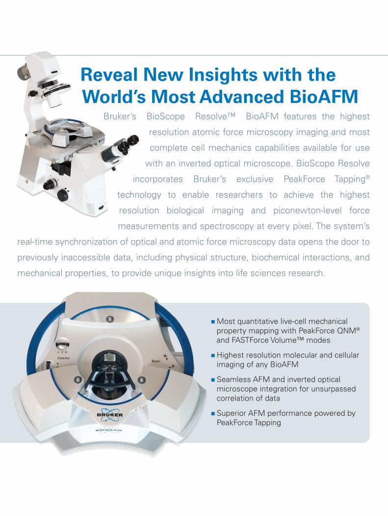
Reveal New Insights with the World’s Most Advanced BioAFM
Bruker’s BioScope Resolve™ BioAFM features the highest
resolution atomic force microscopy imaging and most
complete cell mechanics capabilities available for use
with an inverted optical microscope. BioScope Resolve
incorporates Bruker’s exclusive PeakForce Tapping®
technology to enable researchers to achieve the highest
resolution biological imaging and piconewton-level force
measurements and spectroscopy at every pixel. The system’s
real-time synchronization of optical and atomic force microscopy data opens the door to
previously inaccessible data, including physical structure, biochemical interactions, and
mechanical properties, to provide unique insights into life sciences research.
�Most quantitative live-cell mechanical property mapping with PeakForce QNM® and FASTForce Volume™ modes
�Highest resolution molecular and cellular imaging of any BioAFM
� Seamless AFM and inverted optical microscope integration for unsurpassed correlation of data
� Superior AFM performance powered by PeakForce Tapping
Only Bruker’s exclusive PeakForce Tapping provides biologists a unique combination of the highest resolution imaging, the most quantitative property mapping data, and the easiest to use imaging on the softest of biological samples.
PeakForce QNM provides:
� The fastest and highest resolution mechanical mapping of whole live cells
� Quantitative mapping of mechanical, chemical, and biological interactions
� Sub-molecular AFM imaging with the fastest quantitative mapping of mechanical, chemical, and biological interactions
ScanAsyst-Cell delivers:
� Consistent, expert-quality results for AFM users of all experience levels
�More routine high-resolution molecular and live-cell imaging than any other AFM mode
BioScope Resolve is designed to take full advantage of PeakForce Tapping to deliver a more complete approach to biological investigation, allowing researchers, for the first time, to combine optical and (sub) molecular AFM imaging with the fastest property mapping. Simply put, it’s how bioAFM should be.
PeakForce Tapping — The Most Significant Advancement in BioAFM
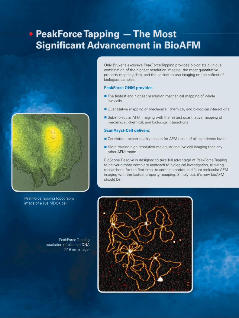
PeakForce Tapping topography image of a live MDCK cell
PeakForce Tapping resolution of plasmid DNA
(419 nm image)
Only Bruker’s exclusive techniques, PeakForce QNM and FASTForce Volume, combine to provide the widest range of ramp frequencies and the most quantitative property mapping for biological samples. Additionally, higher imaging speeds and automated measurement capability, provide more data in less time, leading to faster time to publication.
Unmatched Live-Cell Property Mapping
PeakForce QNM uniquely enables quantitative cell mechanics and imaging of whole, live cells with no artifacts and the fastest cell imaging times.
� High-speed mapping of entire live cells with an unrivalled level of resolution
� Repeatable, robust mechanical property measurements
� Highest resolution image and force curve acquisition with PeakForce Capture™
Quantitative FASTForce Volume
Bruker’s new FASTForce Volume mode compliments PeakForce QNM to provide the widest range of ramp frequencies.
� FASTForce Volume data acquisition, from 0.1 to 1 kHz in liquid or 2 kHz in air
� pN level trigger forces for the most sensitive, highest resolution force distance curves for force spectroscopy
�Widest ramp frequency range, when combined with PeakForce QNM, from 0.1 Hz to 1 kHz in liquid or 2 kHz in air
Superior Force Spectroscopy and Ramp Scripting
BioScope Resolve provides automated scripting and data-collection recipes, making it easy to design extended-time, biological-dynamics studies.
Most Quantitative Cell Mechanics and Molecular Force Spectroscopy Data
“It was previously impossible to resolve the finest structures of a live cell like microvilli, but now with the improved PeakForce Tapping on BioScope Resolve I can image them easily in one hour.”
– Dr. Hermann Schillers of the University of Munster, Germany
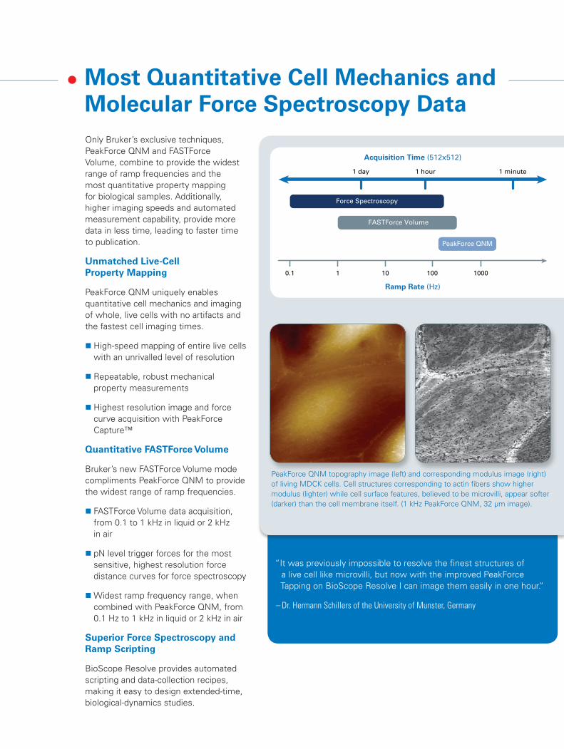
Force Spectroscopy
FASTForce Volume
PeakForce QNM
1 day
0.1 1 10 100 1000
1 hour 1 minute
Acquisition Time (512x512)
Ramp Rate (Hz)
PeakForce QNM topography image (left) and corresponding modulus image (right) of living MDCK cells. Cell structures corresponding to actin fibers show higher modulus (lighter) while cell surface features, believed to be microvilli, appear softer (darker) than the cell membrane itself. (1 kHz PeakForce QNM, 32 µm image).
BioScope Resolve provides the highest resolution imaging for both molecules and live cells, revealing a level of structural detail never imaged before. This capability is achieved through a combination of stable instrument design, PeakForce Tapping, and Bruker’s exclusive high-resolution ScanAsyst-Fluid probes.
BioScope Resolve offers:
� ScanAsyst-Cell™ single-button imaging of whole, live cells with no imaging artifacts
� Imaging of high-resolution, sub-cellular structures on live cells, such as microvilli
� ScanAsyst-Cell single-button imaging for high-resolution molecular investigations
Whether imaging DNA double helices or other biomolecules, BioScope Resolve with ScanAsyst-Cell enables researchers to easily and consistently achieve accurate sub-molecular resolution of biological samples.
Capture the Highest Resolution Molecular and Live-Cell Images Quickly and Easily
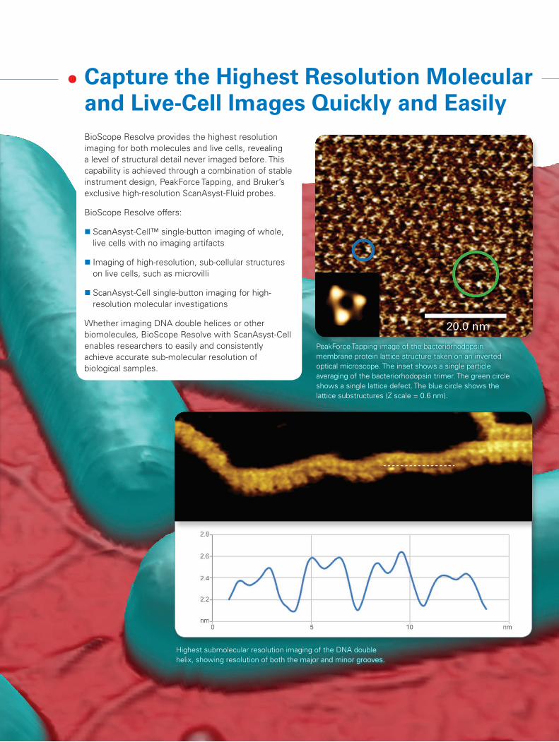
PeakForce Tapping image of the bacteriorhodopsin membrane protein lattice structure taken on an inverted optical microscope. The inset shows a single particle averaging of the bacteriorhodopsin trimer. The green circle shows a single lattice defect. The blue circle shows the lattice substructures (Z scale = 0.6 nm).
Highest submolecular resolution imaging of the DNA double helix, showing resolution of both the major and minor grooves.
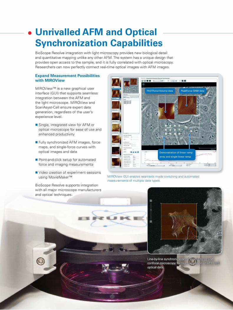
BioScope Resolve integration with light microscopy provides new biological detail and quantitative mapping unlike any other AFM. The system has a unique design that provides open access to the sample, and it is fully correlated with optical microscopy. Researchers can now perfectly connect real-time optical images with AFM images.
Unrivalled AFM and Optical Synchronization Capabilities
Expand Measurement Possibilities with MIROView
MIROView™ is a new graphical user interface (GUI) that supports seamless integration between the AFM and the light microscope. MIROView and ScanAsyst-Cell ensure expert data generation, regardless of the user’s experience level.
� Single, integrated view for AFM or optical microscope for ease of use and enhanced productivity
� Fully synchronized AFM images, force maps, and single-force curves with optical images and data
� Point-and-click setup for automated force and imaging measurements
� Video creation of experiment sessions using MovieMaker™
BioScope Resolve supports integration with all major microscope manufacturers and optical techniques.
Demonstration of linear ramp
array and single linear ramp
FASTForce Volume data PeakForce QNM data
MIROView GUI enables seamless mode switching and automated measurements of multiple data types.
Line-by-line synchronization of atomic force and confocal microscopy for true correlation of AFM and optical data.
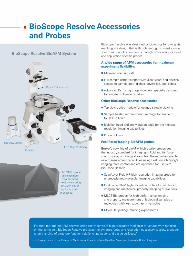
BioScope Resolve Accessories and Probes
Bioscope Resolve was designed by biologists for biologists, resulting in a design that is flexible enough to meet a wide spectrum of application needs through optional accessories and application specific-probes.
A wide range of AFM accessories for maximum experiment flexibility:
�Microvolume fluid cell
� Full sample-carrier support with clear visual and physical access to sample (petri dishes, coverslips, and slides)
� Advanced Perfusing Stage incubator specially designed for long-term, live-cell studies
Other BioScope Resolve accessories:
� Top-view optics module for opaque sample viewing
� Sample heater with temperature range for ambient to 60ºC in liquid
� Isolation hood and anti-vibration table for the highest resolution imaging capabilities
� Probe holders
PeakForce Tapping BioAFM probes:
Bruker’s new line of bioAFM high-quality probes set the industry standard for imaging in fluid and for force spectroscopy of biological samples. These probes enable new measurement capabilities using PeakForce Tapping’s imaging force control and are optimized for use with BioScope Resolve.
� ScanAsyst Fluid-HR high-resolution imaging probe for unprecedented molecular imaging capabilities
� PeakForce QNM high-resolution probes for whole-cell imaging and mechanical property mapping of live cells
�MLCT Bio probes for high performance imaging and property measurement of biological samples or molecules with less topographic variables
�Molecular pulling/unfolding experiments
“For the first time bioAFM analysis can directly correlate high-resolution molecular structures with function on the same cell. BioScope Resolve provides the dynamic range and resolution necessary to allow a deeper understanding of structure-function relationships at cell and tissue surfaces.”
– Dr. Lewis Francis of the College of Medicine and Center of NanoHealth at Swansea University, United Kingdom
EasyAlign™ Station
Top-View Optics
Optical Microscope
Joystick
MLCT-Bio probe on silicon body, manufactured exclusively using Bruker in-house equipment and processes.
BioScope Resolve BioAFM System
Cover ImagesForeground top: PeakForce Tapping image of live E. coli cells. Foreground bottom: PeakForce Tapping topography image of MDCK live cell. Background left: PeakForce QNM image of microvilli on MDCK live cell. Background right: PeakForce Tapping image of DNA double helix.
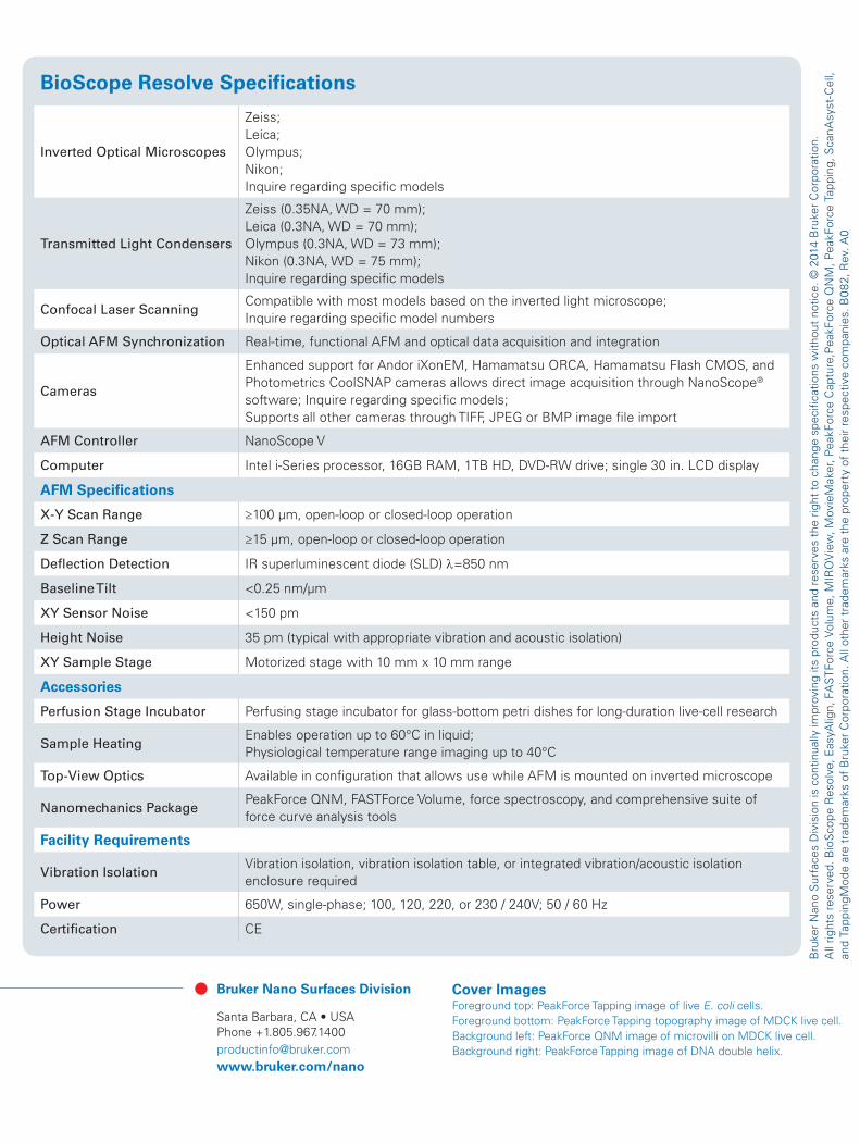
BioScope Resolve Specifications
Inverted Optical Microscopes
Zeiss;Leica;Olympus;Nikon; Inquire regarding specific models
Transmitted Light Condensers
Zeiss (0.35NA, WD = 70 mm);Leica (0.3NA, WD = 70 mm);Olympus (0.3NA, WD = 73 mm);Nikon (0.3NA, WD = 75 mm);Inquire regarding specific models
Confocal Laser ScanningCompatible with most models based on the inverted light microscope; Inquire regarding specific model numbers
Optical AFM Synchronization Real-time, functional AFM and optical data acquisition and integration
Cameras
Enhanced support for Andor iXonEM, Hamamatsu ORCA, Hamamatsu Flash CMOS, and Photometrics CoolSNAP cameras allows direct image acquisition through NanoScope® software; Inquire regarding specific models; Supports all other cameras through TIFF, JPEG or BMP image file import
AFM Controller NanoScope V
Computer Intel i-Series processor, 16GB RAM, 1TB HD, DVD-RW drive; single 30 in. LCD display
AFM Specifications
X-Y Scan Range ≥100 µm, open-loop or closed-loop operation
Z Scan Range ≥15 µm, open-loop or closed-loop operation
Deflection Detection IR superluminescent diode (SLD) λ=850 nm
Baseline Tilt <0.25 nm/µm
XY Sensor Noise <150 pm
Height Noise 35 pm (typical with appropriate vibration and acoustic isolation)
XY Sample Stage Motorized stage with 10 mm x 10 mm range
Accessories
Perfusion Stage Incubator Perfusing stage incubator for glass-bottom petri dishes for long-duration live-cell research
Sample HeatingEnables operation up to 60°C in liquid; Physiological temperature range imaging up to 40°C
Top-View Optics Available in configuration that allows use while AFM is mounted on inverted microscope
Nanomechanics PackagePeakForce QNM, FASTForce Volume, force spectroscopy, and comprehensive suite of force curve analysis tools
Facility Requirements
Vibration IsolationVibration isolation, vibration isolation table, or integrated vibration/acoustic isolation enclosure required
Power 650W, single-phase; 100, 120, 220, or 230 / 240V; 50 / 60 Hz
Certification CE
Bruker Nano Surfaces Division
Santa Barbara, CA • USA Phone +1.805.967.1400 [email protected]/nano
Bru
ker
Nan
o S
urfa
ces
Div
isio
n is
con
tinua
lly im
prov
ing
its p
rodu
cts
and
rese
rves
the
rig
ht t
o ch
ange
spe
cific
atio
ns w
ithou
t no
tice.
© 2
014
Bru
ker
Cor
pora
tion.
A
ll rig
hts
rese
rved
. Bio
Sco
pe R
esol
ve, E
asyA
lign,
FA
STF
orce
Vol
ume,
MIR
OV
iew
, Mov
ieM
aker
, Pea
kFor
ce C
aptu
re,P
eakF
orce
QN
M, P
eakF
orce
Tap
ping
, Sca
nAsy
st-C
ell,
and
Tapp
ingM
ode
are
trad
emar
ks o
f B
ruke
r C
orpo
ratio
n. A
ll ot
her
trad
emar
ks a
re t
he p
rope
rty
of t
heir
resp
ectiv
e co
mpa
nies
. B08
2, R
ev. A
0


![BRITISH BIOSCOPE Co [2] - WordPress.com...BRITISH BIOSCOPE Co [2] AUSTRALIAN VARIETY THEATRE ARCHIVE: RESEARCH NOTES See last page for citation, copyright and last updated details](https://img.pdfslide.us/doc/110x75/5fc3f39876b6f07d2b7a8436/british-bioscope-co-2-british-bioscope-co-2-australian-variety-theatre.jpg)