Embed Size (px)
Citation preview
INTERNATIONAL DICTYOSTELIUMCONFERENCE 2002
Altavilla – Hotel Torre NormannaSeptember 22-27, 2002
Sponsored by:
The University of Turin
We acknowledge support by:
- The University of Turin- Bibby Sterilin- Montepaone SAS
organizers: Salvatore BozzaroAdriano CeccarelliEnrico BraccoBarbara Pergolizzi
IItim
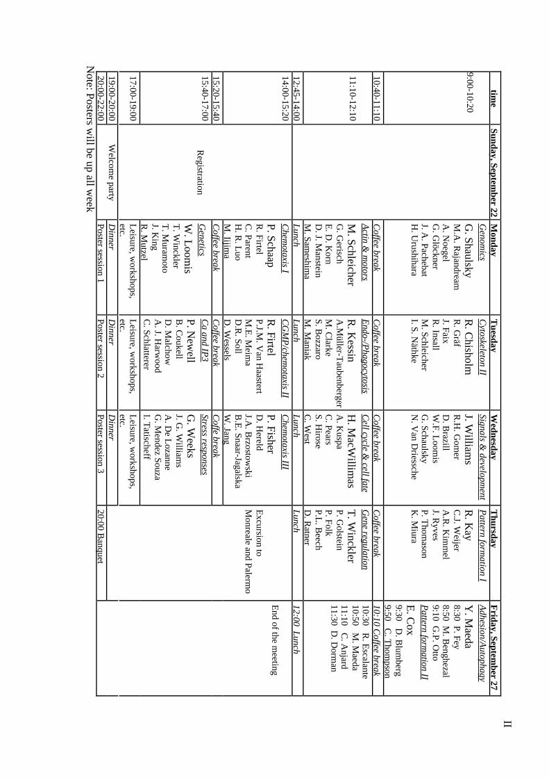
eSunday, Septem
ber 22M
ondayT
uesdayW
ednesdayT
hursdayF
riday, September 27
9:00-10:20
Genom
ics
G. Shaulsky
M.A
. Rajandream
A. N
oegelG
. Glöckner
J. A. Pachebat
H. U
rushihara
Cytoskeleton II
R. C
hisholmR
. Gräf
J. FaixR
. InsallM
. SchleicherI. S. N
äthke
Signals & developm
ent
J. William
sR
.H. G
omer
D. B
razillW
.F. Loom
isG
. SchaulskyN
. Van D
riessche
Pattern form
ation I
R. K
ayC
.J. Weijer
A.R
. Kim
mel
J. Ryves
P. Thom
asonK
. Miura
Adhesion/A
utophagy
Y. M
aeda8:30 P. Fey8:50 M
. Benghezal
9:10 G.P. O
ttoP
attern formation II
E. C
ox9:30 D
. Blum
berg9:50 C
. Thom
pson10:40-11:10
Coffee break
Coffee break
Coffee break
Coffee break
10:10 Coffee break
11:10-12:10
Actin &
motors
M. Schleicher
G. G
erischE
. D. K
ornD
. J. Manstein
M. Sam
eshima
Endo-/P
hagocytosis
R. K
essinA
.Müller-T
aubenbergerM
. Clarke
S. Bozzaro
M. M
aniak
Cell cycle &
cell fate
H. M
acWillim
asA
. Kuspa
C. Pears
S. Hirose
C. W
est
Gene regulation
T. W
incklerP. G
olsteinP. FolkP.L
. Beech
D. R
atner
10:30 R. E
scalante10:50 M
. Maeda
11:10 C. A
njard11:30 D
. Dorm
an
12:45-14:00L
unchL
unchL
unchL
unch12:00 L
unch14:00-15:20
Chem
otaxis I
P. SchaapR
. FirtelC
. ParentH
. R. L
uoM
. Iijima
CG
MP
/chemotaxis II
R. Firtel
P.J.M. V
an Haastert
M.E
. Meim
aD
.R. Soll
D. W
essels
Chem
otaxis III
P. FisherD
. Hereld
J.A. B
rzostowski
B.E
. Snaar-JagalskaW
. Jang15:20-15:40
Coffee break
Coffee break
Coffe break
15:40-17:00G
enetics
W. L
oomis
T. W
incklerT
. Muram
otoJ. K
ingR
. Mutzel
Ca and IP
3
P. New
ellB
. Coukell
D. M
alchowA
. J. Harw
oodC
. Schlatterer
Stress responses
G. W
eeksJ. G
. William
sA
. De L
ozanneG
. Mendez Souza
I. Tatischeff
17:00-19:00
Registration
Leisure, w
orkshops,etc.
Leisure, w
orkshops,etc.
Leisure, w
orkshops,etc.
19:00-20:00D
innerD
innerD
inner
Excursion to
Monreale and Palerm
o
20:00-22:00W
elcome party
Poster session 1Poster session 2
Poster session 320:00 B
anquet
End of the m
eeting
Note: Posters w
ill be up all week

III
POST
ER
SESSIO
N 1
POST
ER
SESSIO
N 2
POST
ER
SESSIO
N 3
70 Abe T
.71 B
eck P.72 H
amlin E
.73 B
arth C.
74 Kuw
ayama H
.75 L
ay S.76 M
oreno N.
77 Szafranski K.
78 Thom
ason P.79 D
e Lozanne A
.80 D
aniels K.
81 Baik M
.82G
räf R.
83 Strassman J.
84 Heuser J.
85 Mai A
.
86 Dondero F.
87 Kriebel W
P.88 L
etourneur F.89 M
anstein D.
90 Mahadeo D
.91 R
ivero F.92 Sasaki K
.93 Steenbergen J.94 U
eda M.
95 Voss E
.96 W
eissenmeyer B
.97 W
essels D.
98 Arigoni M
.99 Z
hang H.
100 Balest A
.101 B
rock D.
102 Dalton E
.
103 Adam
M104 K
ibler K105 C
otter D106 Fukusaw
a M107 Z
hukovskaja N108 O
ohata A109 M
aeda M110 A
lvarez-Curto E
111 Saran S112 Shaw
C113 W
eening K114 W
illiams R
SB115 Y
agura S116 K
atoh M117 B
lumberg D
118 Ikeno D119 Sevcikova
IV
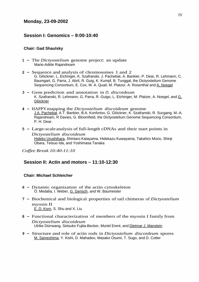
Monday, 23-09-2002
Session I: Genomics – 9:00-10:40
Chair: Gad Shaulsky
1 - The Dictyostelium genome project: an updateMarie-Adèle Rajandream
2 - Sequence and analysis of chromosomes 1 and 2G. Glöckner, L. Eichinger, K. Szafranski, J. Pachebat, A. Bankier, P. Dear, R. Lehmann, C.Baumgart, G. Parra, J. Abril, R. Guig, K. Kumpf, B. Tunggal, the Dictyostelium GenomeSequencing Consortium, E. Cox, M. A. Quail, M. Platzer, A. Rosenthal and A. Noegel
3 - Gene prediction and annotation in D. discoideumK. Szafranski, R. Lehmann, G. Parra, R. Guigo, L. Eichinger, M. Platzer, A. Noegel, and G.Glöckner
4 - HAPPY mapping the Dictyostelium discoideum genomeJ.A. Pachebat, A.T. Bankier, B.A. Konfortov, G. Glöckner, K. Szafranski, R. Sucgang, M.-A.Rajandream, R Davies, G. Bloomfield, the Dictyostelium Genome Sequencing Consortium,P. H. Dear.
5 - Large-scale analysis of full-length cDNAs and their start points inDictyostelium discoideum
Hideko Urushihara, Shintaro Katayama, Hidekazu Kuwayama, Takahiro Morio, ShinjiObara, Tetsuo Ida, and Yoshimasa Tanaka
Coffee Break 10:40-11:10
Session II: Actin and motors – 11:10-12:30
Chair: Michael Schleicher
6 - Dynamic organization of the actin cytoskeletonO. Medalia, I. Weber, G. Gerisch, and W. Baumeister
7 - Biochemical and biological properties of tail chimeras of Dictyosteliummyosin II
E. D. Korn, S. Shu and X. Liu
8 - Functional characterization of members of the myosin I family fromDictyostelium discoideum
Ulrike Dürrwang, Setsuko Fujita-Becker, Muriel Erent, and Dietmar J. Manstein
9 - Structure and role of actin rods in Dictyostelium discoideum sporesM. Sameshima, Y. Kishi, D. Mahadeo, Masako Osumi, T. Sugo, and D. Cotter
V
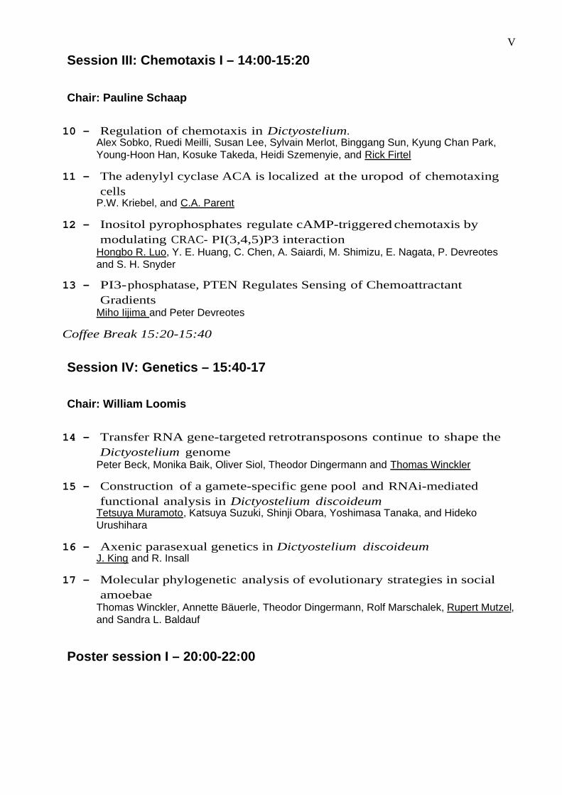
Session III: Chemotaxis I – 14:00-15:20
Chair: Pauline Schaap
10 - Regulation of chemotaxis in Dictyostelium.Alex Sobko, Ruedi Meilli, Susan Lee, Sylvain Merlot, Binggang Sun, Kyung Chan Park,Young-Hoon Han, Kosuke Takeda, Heidi Szemenyie, and Rick Firtel
11 - The adenylyl cyclase ACA is localized at the uropod of chemotaxingcells
P.W. Kriebel, and C.A. Parent
12 - Inositol pyrophosphates regulate cAMP-triggered chemotaxis bymodulating CRAC- PI(3,4,5)P3 interaction
Hongbo R. Luo, Y. E. Huang, C. Chen, A. Saiardi, M. Shimizu, E. Nagata, P. Devreotesand S. H. Snyder
13 - PI3- phosphatase, PTEN Regulates Sensing of ChemoattractantGradients
Miho Iijima and Peter Devreotes
Coffee Break 15:20-15:40
Session IV: Genetics – 15:40-17
Chair: William Loomis
14 - Transfer RNA gene-targeted retrotransposons continue to shape theDictyostelium genome
Peter Beck, Monika Baik, Oliver Siol, Theodor Dingermann and Thomas Winckler
15 - Construction of a gamete-specific gene pool and RNAi-mediatedfunctional analysis in Dictyostelium discoideum
Tetsuya Muramoto, Katsuya Suzuki, Shinji Obara, Yoshimasa Tanaka, and HidekoUrushihara
16 - Axenic parasexual genetics in Dictyostelium discoideumJ. King and R. Insall
17 - Molecular phylogenetic analysis of evolutionary strategies in socialamoebae
Thomas Winckler, Annette Bäuerle, Theodor Dingermann, Rolf Marschalek, Rupert Mutzel,and Sandra L. Baldauf
Poster session I – 20:00-22:00

VI
Tuesday, 24-09-2002
Session V: Cytoskeleton II - 9:00-10:40
Chair: Rex Chisholm
18 - DdCP224 and DdEB1: two Dictyostelium MAPs with a role at thecentrosome and in microtubule plus end dynamics
R. Gräf, A. Hestermann, and M. Rehberg
19 - Signaling molecules as regulators of cytokinesis and cell motilityJan Faix, Igor Weber and Jibi Jacob.
20 - The Arp2/3 complex and the control of actin polymerization inDictyostelium
Simone Blagg, Jason King, Karl Saxe & Robert Insall
21 - Dictyostelium Ste20-like kinases in signalling pathways to thecytoskeleton
Rajesh Arasada, Hyun-Ju Son, Ludwig Eichinger, Michael Schleicher
22 - Cytoskeletal regulation by the Adenomatous Polyposis Coli protein inDictyostelium
Ian P. Newton, Pauline Schaap, Inke S. Näthke
Coffee Break 10:40-11:10
Session VI: Endo-/phagocytosis - 11:10-12:30
Chair: Richard Kessin
23 - Visualizing individual steps during phagocytosisA. Müller-Taubenberger, I. Weber, and G. Gerisch
24 - Dynamics and fusion in the early endocytic pathway ofDictyostelium.
M. Clarke, J. Kohler, J. Heuser, and G. Gerisch
25 - The Dictyostelium homologue of human Nramp1 is required forefficient phagocytosis and resistance to pathogenic bacteria
Barbara Peracino, Carina Skriwan, Alessandra Balest, Alessandra Balbo, BarbaraPergolizzi, Angelika A. Noegel, Michael Steinert and Salvatore Bozzaro
26 - The Dictyostelium LC-FACS protein contributes to fatty acid uptakeand endocytosis
K. von Löhneysen, N. Pawolleck, H. Rühling, and M. Maniak
VII
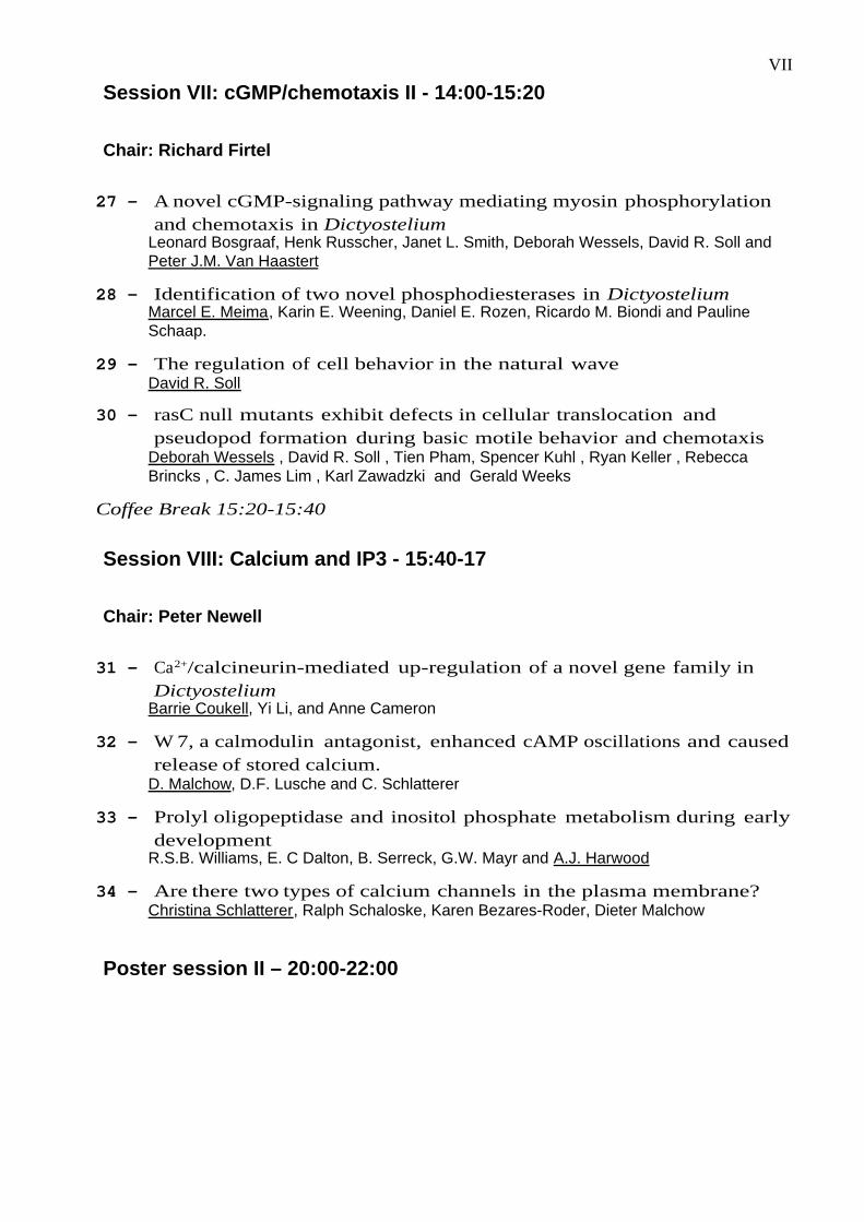
Session VII: cGMP/chemotaxis II - 14:00-15:20
Chair: Richard Firtel
27 - A novel cGMP-signaling pathway mediating myosin phosphorylationand chemotaxis in Dictyostelium
Leonard Bosgraaf, Henk Russcher, Janet L. Smith, Deborah Wessels, David R. Soll andPeter J.M. Van Haastert
28 - Identification of two novel phosphodiesterases in DictyosteliumMarcel E. Meima, Karin E. Weening, Daniel E. Rozen, Ricardo M. Biondi and PaulineSchaap.
29 - The regulation of cell behavior in the natural waveDavid R. Soll
30 - rasC null mutants exhibit defects in cellular translocation andpseudopod formation during basic motile behavior and chemotaxis
Deborah Wessels , David R. Soll , Tien Pham, Spencer Kuhl , Ryan Keller , RebeccaBrincks , C. James Lim , Karl Zawadzki and Gerald Weeks
Coffee Break 15:20-15:40
Session VIII: Calcium and IP3 - 15:40-17
Chair: Peter Newell
31 - Ca2+/calcineurin-mediated up-regulation of a novel gene family inDictyostelium
Barrie Coukell, Yi Li, and Anne Cameron
32 - W 7, a calmodulin antagonist, enhanced cAMP oscillations and causedrelease of stored calcium.
D. Malchow, D.F. Lusche and C. Schlatterer
33 - Prolyl oligopeptidase and inositol phosphate metabolism during earlydevelopment
R.S.B. Williams, E. C Dalton, B. Serreck, G.W. Mayr and A.J. Harwood
34 - Are there two types of calcium channels in the plasma membrane?Christina Schlatterer, Ralph Schaloske, Karen Bezares-Roder, Dieter Malchow
Poster session II – 20:00-22:00

VIII
Wednesday, 25-09-2002
Session IX: Signals and development - 9:00-10:40
Chair: Jeffrey Williams
35 - A single cell-density sensing factor stimulates distinct signaltransduction pathways through two different receptors
William J. Deery, Tong Gao, Robin Ammann, and Richard H. Gomer
36 - A Phospholipase D regulates quorum sensing in Dictyosteliumdiscoideum
Yi Chen, Vanessa Rodrick, Yi Yan, and Derrick Brazill
37 - Genetic modules expressed during early development ofDictyostelium
Negin Iranfar, Danny Fuller, and William F. Loomis with the cooperation of the JapaneseEST Project
38 - Beyond development: transcriptional profiling of the Dictyosteliumcell cycle, spore germination and de-differentiation
Chad Shaw, Nancy Van Driessche,, Miroslava Ibarra, Sujata Sharma, Ezgi Okyay,,Takahiro Morio,, Mariko Katoh, Hideko Urushihara, Yoshimasa Tanaka, Junji Chida, AikoAmagai, Yasuo Maeda, Dana Mahadeo, David Cotter, Adam Kuspa,, and Gad Shaulsky
39 - Microarray phenotyping of the PKA and YakA pathwayNancy Van Driessche, Chad Shaw,, Sujata Sharma, Miroslava Ibarra, Trushar Surang,Adam Kuspa,, and Gad Shaulsky,
Coffee Break 10:40-11:10
Session X: Cell cycle and cell fate - 11:10-12:30
Chair: Harry MacWilliams
40 - Prespore cell-cycle arrest during Dictyostelium developmentGuokai Chen, Gad Shaulsky and Adam Kuspa,
41 - A homologue of Cdk8 required for optimum growth, aggregation andspore cell differentiation.
Hsiu-Hsu Lin, Hao-Jen Huang, Christine Michaelis, Gerry Weeks and Catherine Pears
42 - A transcriptional switch at growth/differentiation transition (GDT) ofDictyostelium cells: cis- and trans- elements of the dia1 and fkbp2genes regulated during the GDT
Shigenori Hirose, Aiko Amagai and Yasuo Maeda
43 - Evolutionary and functional implications of the complexglycosylation of Skp1, a cytoplasmic/nuclear glycoprotein associatedwith polyubiquitination
Christopher M. West, Hanke van der Wel, Suzanne Z. Fisher, Howard Morris, MariaPanico, Thanai Paxton, Anne Dell, Lee Kaplan, and Eric A. Gaucher
IX
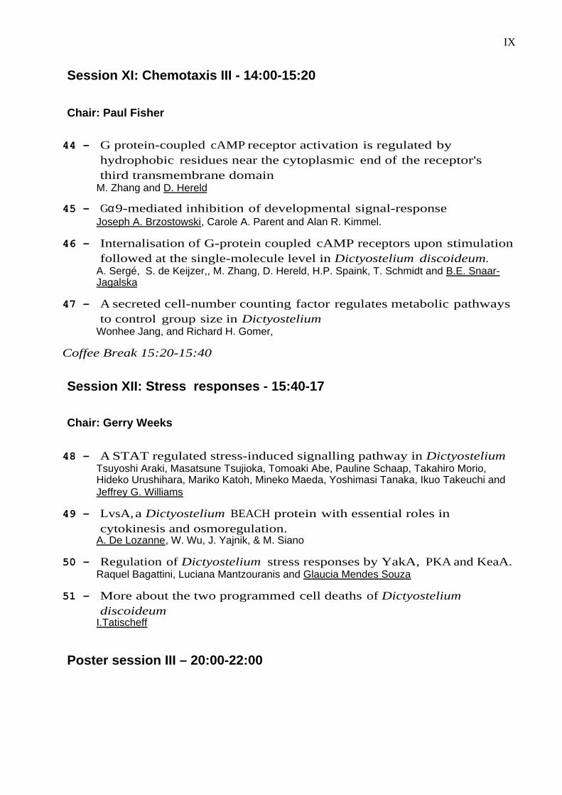
Session XI: Chemotaxis III - 14:00-15:20
Chair: Paul Fisher
44 - G protein-coupled cAMP receptor activation is regulated byhydrophobic residues near the cytoplasmic end of the receptor'sthird transmembrane domain
M. Zhang and D. Hereld
45 - Gα9-mediated inhibition of developmental signal-responseJoseph A. Brzostowski, Carole A. Parent and Alan R. Kimmel.
46 - Internalisation of G-protein coupled cAMP receptors upon stimulationfollowed at the single-molecule level in Dictyostelium discoideum.
A. Sergé, S. de Keijzer,, M. Zhang, D. Hereld, H.P. Spaink, T. Schmidt and B.E. Snaar-Jagalska
47 - A secreted cell-number counting factor regulates metabolic pathwaysto control group size in Dictyostelium
Wonhee Jang, and Richard H. Gomer,
Coffee Break 15:20-15:40
Session XII: Stress responses - 15:40-17
Chair: Gerry Weeks
48 - A STAT regulated stress-induced signalling pathway in DictyosteliumTsuyoshi Araki, Masatsune Tsujioka, Tomoaki Abe, Pauline Schaap, Takahiro Morio,Hideko Urushihara, Mariko Katoh, Mineko Maeda, Yoshimasi Tanaka, Ikuo Takeuchi andJeffrey G. Williams
49 - LvsA, a Dictyostelium BEACH protein with essential roles incytokinesis and osmoregulation.
A. De Lozanne, W. Wu, J. Yajnik, & M. Siano
50 - Regulation of Dictyostelium stress responses by YakA, PKA and KeaA.Raquel Bagattini, Luciana Mantzouranis and Glaucia Mendes Souza
51 - More about the two programmed cell deaths of Dictyosteliumdiscoideum
I.Tatischeff
Poster session III – 20:00-22:00
X
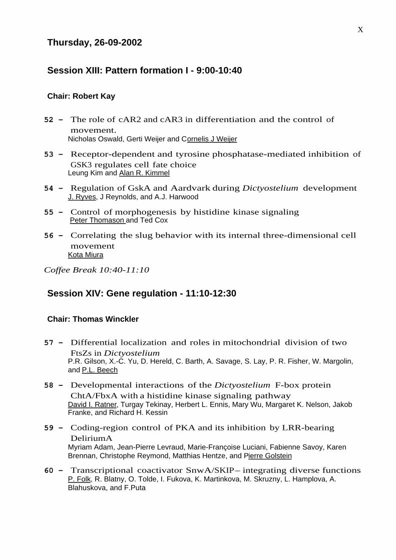
Thursday, 26-09-2002
Session XIII: Pattern formation I - 9:00-10:40
Chair: Robert Kay
52 - The role of cAR2 and cAR3 in differentiation and the control ofmovement.
Nicholas Oswald, Gerti Weijer and Cornelis J Weijer
53 - Receptor-dependent and tyrosine phosphatase-mediated inhibition ofGSK3 regulates cell fate choice
Leung Kim and Alan R. Kimmel
54 - Regulation of GskA and Aardvark during Dictyostelium developmentJ. Ryves, J Reynolds, and A.J. Harwood
55 - Control of morphogenesis by histidine kinase signaling Peter Thomason and Ted Cox
56 - Correlating the slug behavior with its internal three-dimensional cellmovement
Kota Miura
Coffee Break 10:40-11:10
Session XIV: Gene regulation - 11:10-12:30
Chair: Thomas Winckler
57 - Differential localization and roles in mitochondrial division of twoFtsZs in Dictyostelium
P.R. Gilson, X.-C. Yu, D. Hereld, C. Barth, A. Savage, S. Lay, P. R. Fisher, W. Margolin,and P.L. Beech
58 - Developmental interactions of the Dictyostelium F-box proteinChtA/FbxA with a histidine kinase signaling pathway
David I. Ratner, Turgay Tekinay, Herbert L. Ennis, Mary Wu, Margaret K. Nelson, JakobFranke, and Richard H. Kessin
59 - Coding-region control of PKA and its inhibition by LRR-bearingDeliriumA
Myriam Adam, Jean-Pierre Levraud, Marie-Françoise Luciani, Fabienne Savoy, KarenBrennan, Christophe Reymond, Matthias Hentze, and Pierre Golstein
60 - Transcriptional coactivator SnwA/SKIP – integrating diverse functionsP. Folk, R. Blatny, O. Tolde, I. Fukova, K. Martinkova, M. Skruzny, L. Hamplova, A.Blahuskova, and F.Puta
XI
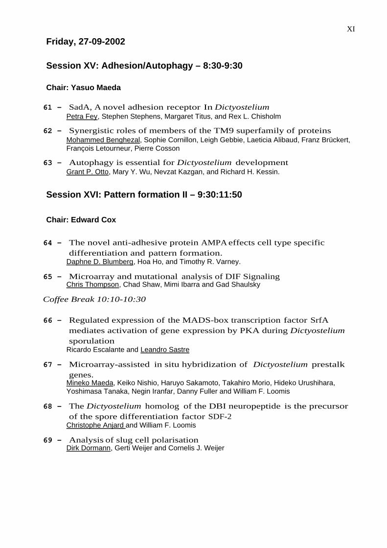
Friday, 27-09-2002
Session XV: Adhesion/Autophagy – 8:30-9:30
Chair: Yasuo Maeda
61 - SadA, A novel adhesion receptor In DictyosteliumPetra Fey, Stephen Stephens, Margaret Titus, and Rex L. Chisholm
62 - Synergistic roles of members of the TM9 superfamily of proteinsMohammed Benghezal, Sophie Cornillon, Leigh Gebbie, Laeticia Alibaud, Franz Brückert,François Letourneur, Pierre Cosson
63 - Autophagy is essential for Dictyostelium developmentGrant P. Otto, Mary Y. Wu, Nevzat Kazgan, and Richard H. Kessin.
Session XVI: Pattern formation II – 9:30:11:50
Chair: Edward Cox
64 - The novel anti-adhesive protein AMPA effects cell type specificdifferentiation and pattern formation.
Daphne D. Blumberg, Hoa Ho, and Timothy R. Varney.
65 - Microarray and mutational analysis of DIF SignalingChris Thompson, Chad Shaw, Mimi Ibarra and Gad Shaulsky
Coffee Break 10:10-10:30
66 - Regulated expression of the MADS-box transcription factor SrfAmediates activation of gene expression by PKA during Dictyosteliumsporulation
Ricardo Escalante and Leandro Sastre
67 - Microarray-assisted in situ hybridization of Dictyostelium prestalkgenes.
Mineko Maeda, Keiko Nishio, Haruyo Sakamoto, Takahiro Morio, Hideko Urushihara,Yoshimasa Tanaka, Negin Iranfar, Danny Fuller and William F. Loomis
68 - The Dictyostelium homolog of the DBI neuropeptide is the precursorof the spore differentiation factor SDF-2
Christophe Anjard and William F. Loomis
69 - Analysis of slug cell polarisationDirk Dormann, Gerti Weijer and Cornelis J. Weijer
XII
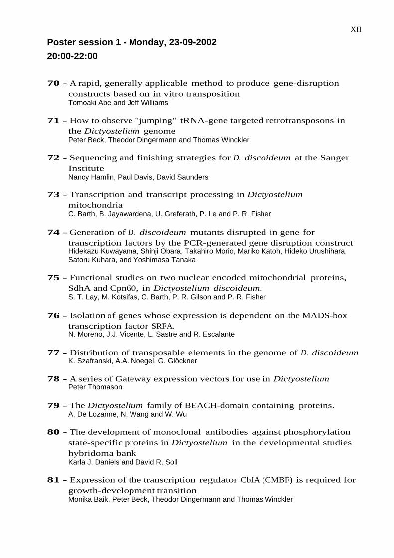
Poster session 1 - Monday, 23-09-2002
20:00-22:00
70 - A rapid, generally applicable method to produce gene-disruptionconstructs based on in vitro transpositionTomoaki Abe and Jeff Williams
71 - How to observe "jumping" tRNA-gene targeted retrotransposons inthe Dictyostelium genomePeter Beck, Theodor Dingermann and Thomas Winckler
72 - Sequencing and finishing strategies for D. discoideum at the SangerInstituteNancy Hamlin, Paul Davis, David Saunders
73 - Transcription and transcript processing in DictyosteliummitochondriaC. Barth, B. Jayawardena, U. Greferath, P. Le and P. R. Fisher
74 - Generation of D. discoideum mutants disrupted in gene fortranscription factors by the PCR-generated gene disruption constructHidekazu Kuwayama, Shinji Obara, Takahiro Morio, Mariko Katoh, Hideko Urushihara,Satoru Kuhara, and Yoshimasa Tanaka
75 - Functional studies on two nuclear encoded mitochondrial proteins,SdhA and Cpn60, in Dictyostelium discoideum.S. T. Lay, M. Kotsifas, C. Barth, P. R. Gilson and P. R. Fisher
76 - Isolation of genes whose expression is dependent on the MADS-boxtranscription factor SRFA.N. Moreno, J.J. Vicente, L. Sastre and R. Escalante
77 - Distribution of transposable elements in the genome of D. discoideumK. Szafranski, A.A. Noegel, G. Glöckner
78 - A series of Gateway expression vectors for use in DictyosteliumPeter Thomason
79 - The Dictyostelium family of BEACH-domain containing proteins.A. De Lozanne, N. Wang and W. Wu
80 - The development of monoclonal antibodies against phosphorylationstate-specific proteins in Dictyostelium in the developmental studieshybridoma bankKarla J. Daniels and David R. Soll
81 - Expression of the transcription regulator CbfA (CMBF) is required forgrowth-development transitionMonika Baik, Peter Beck, Theodor Dingermann and Thomas Winckler

XIII82 - DdNek2, the first non-vertebrate homologue of human Nek2, is
involved in the formation of microtubule-organizing centersR. Gräf
83 - The genetic structure of D. discoideum populationsJoan E. Strassmann, David Queller, Kevin R. Foster, Lorenzo A. Santorelli, AngeloFortunato, Monica Landi
84 - Clathrin coated vesicle formation in Dictyostelium controlled by actindynamicsJohn E. Heuser and Terry O'Halloran
85 - Identification and characterization of a member of importin-β-like
nuclear transport receptors in D. discoideum.Andrea Mai, Enrico Bracco, Alessandra Balbo, Barbara Peracino, Günther Gerisch andSalvatore Bozzaro
XIV
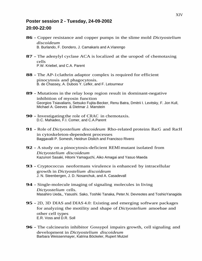
Poster session 2 - Tuesday, 24-09-2002
20:00-22:00
86 - Copper resistance and copper pumps in the slime mold DictyosteliumdiscoideumB. Burlando, F. Dondero, J. Camakaris and A.Viarengo
87 - The adenylyl cyclase ACA is localized at the uropod of chemotaxingcellsP.W. Kriebel, and C.A. Parent
88 - The AP-1 clathrin adaptor complex is required for efficientpinocytosis and phagocytosis.B. de Chassey, A. Dubois Y. Lefkir, and F. Letourneur
89 - Mutations in the relay loop region result in dominant-negativeinhibition of myosin functionGeorgios Tsiavaliaris, Setsuko Fujita-Becker, Renu Batra, Dmitrii I. Levitsky, F. Jon Kull,Michael A. Geeves & Dietmar J. Manstein
90 - Investigating the role of CRAC in chemotaxis.D.C. Mahadeo, F.I. Comer, and C.A.Parent
91 - Role of Dictyostelium discoideum Rho-related proteins RacG and RacHin cytoskeleton-dependent processesBaggavalli P. Somesh, Heidrun Dislich and Francisco Rivero
92 - A study on a pinocytosis-deficient REMI mutant isolated fromDictyostelium discoideumKazunori Sasaki, Hitomi Yamaguchi, Aiko Amagai and Yasuo Maeda
93 - Cryptococcus neoformans virulence is enhanced by intracellulargrowth in Dictyostelium discoideumJ. N. Steenbergen, J. D. Nosanchuk, and A. Casadevall
94 - Single-molecule imaging of signaling molecules in livingDictyostelium cells.Masahiro Ueda,, Yasushi. Sako, Toshiki Tanaka, Peter.N. Devreotes and ToshioYanagida
95 - 2D, 3D DIAS and DIAS 4.0: Existing and emerging software packagesfor analyzing the motility and shape of Dictyostelium amoebae andother cell typesE.R. Voss and D.R. Soll
96 - The calcineurin inhibitor Gossypol impairs growth, cell signaling anddevelopment in Dictyostelium discoideumBarbara Weissenmayer, Katrina Böckeler, Rupert Mutzel

XV97 - Constitutive expression of PKA disrupts the shape of
chemotactically responsive amoebaeDeborah Wessels, Karla Daniels, Hui Zhang, Paul Heid, William F. Loomis and David R.Soll
98 - Ras-GEFM, a novel putative Ras-GEF required for DictyosteliumdevelopmentM.Arigoni, E. Bracco and S. Bozzaro
99 - Basic motility and chemotaxis defects in a mutant with constitutivelyactive PKA activityHui Zhang, Paul Heid, Deborah Wessels, Karla Daniels, Bill F. Loomis and David R. Soll
100 - The NRAMP1 protein in Dictyostelium.Alessandra Balest, Carina Skriwan, Barbara Pergolizzi, Michael Steinert Barbara Peracino& Salvatore Bozzaro
101 - Characterizing the components of CFDebra A. Brock and Richard H. Gomer
102 - The interplay between Aardvark and cell movement.E.C. Dalton, D. Wessels, D.R. Soll, and A.J. Harwood
XVI
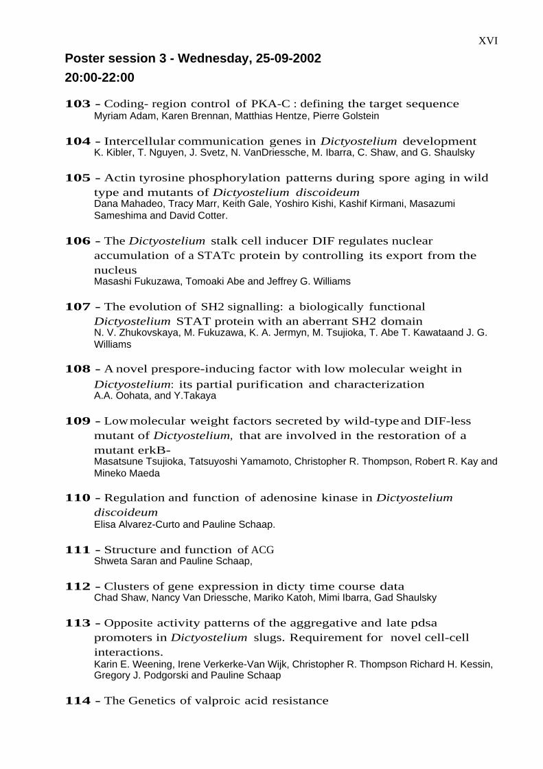
Poster session 3 - Wednesday, 25-09-2002
20:00-22:00
103 - Coding- region control of PKA-C : defining the target sequenceMyriam Adam, Karen Brennan, Matthias Hentze, Pierre Golstein
104 - Intercellular communication genes in Dictyostelium developmentK. Kibler, T. Nguyen, J. Svetz, N. VanDriessche, M. Ibarra, C. Shaw, and G. Shaulsky
105 - Actin tyrosine phosphorylation patterns during spore aging in wildtype and mutants of Dictyostelium discoideumDana Mahadeo, Tracy Marr, Keith Gale, Yoshiro Kishi, Kashif Kirmani, MasazumiSameshima and David Cotter.
106 - The Dictyostelium stalk cell inducer DIF regulates nuclearaccumulation of a STATc protein by controlling its export from thenucleusMasashi Fukuzawa, Tomoaki Abe and Jeffrey G. Williams
107 - The evolution of SH2 signalling: a biologically functionalDictyostelium STAT protein with an aberrant SH2 domainN. V. Zhukovskaya, M. Fukuzawa, K. A. Jermyn, M. Tsujioka, T. Abe T. Kawataand J. G.Williams
108 - A novel prespore-inducing factor with low molecular weight in
Dictyostelium: its partial purification and characterization A.A. Oohata, and Y.Takaya
109 - Low molecular weight factors secreted by wild-type and DIF-lessmutant of Dictyostelium, that are involved in the restoration of amutant erkB-Masatsune Tsujioka, Tatsuyoshi Yamamoto, Christopher R. Thompson, Robert R. Kay andMineko Maeda
110 - Regulation and function of adenosine kinase in DictyosteliumdiscoideumElisa Alvarez-Curto and Pauline Schaap.
111 - Structure and function of ACGShweta Saran and Pauline Schaap,
112 - Clusters of gene expression in dicty time course dataChad Shaw, Nancy Van Driessche, Mariko Katoh, Mimi Ibarra, Gad Shaulsky
113 - Opposite activity patterns of the aggregative and late pdsapromoters in Dictyostelium slugs. Requirement for novel cell-cellinteractions.Karin E. Weening, Irene Verkerke-Van Wijk, Christopher R. Thompson Richard H. Kessin,Gregory J. Podgorski and Pauline Schaap
114 - The Genetics of valproic acid resistance
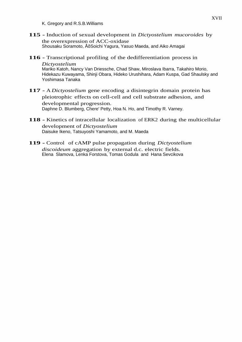
XVIIK. Gregory and R.S.B.Williams
115 - Induction of sexual development in Dictyostelium mucoroides bythe overexpression of ACC-oxidaseShousaku Soramoto, ÅõSoichi Yagura, Yasuo Maeda, and Aiko Amagai
116 - Transcriptional profiling of the dedifferentiation process inDictyosteliumMariko Katoh, Nancy Van Driessche, Chad Shaw, Miroslava Ibarra, Takahiro Morio,Hidekazu Kuwayama, Shinji Obara, Hideko Urushihara, Adam Kuspa, Gad Shaulsky andYoshimasa Tanaka
117 - A Dictyostelium gene encoding a disintegrin domain protein haspleiotrophic effects on cell-cell and cell substrate adhesion, anddevelopmental progression.Daphne D. Blumberg, Chere' Petty, Hoa N. Ho, and Timothy R. Varney.
118 - Kinetics of intracellular localization of ERK2 during the multicellulardevelopment of DictyosteliumDaisuke Ikeno, Tatsuyoshi Yamamoto, and M. Maeda
119 - Control of cAMP pulse propagation during Dictyosteliumdiscoideum aggregation by external d.c. electric fields.Elena Slamova, Lenka Forstova, Tomas Godula and Hana Sevcikova
1
The Dictyostelium genome project: an update
Marie-Adèle Rajandream
Pathogen Sequencing Unit, The Wellcome Trust Sanger Institute, Genome Campus, Hinxton, Cambridge,CB10 1SA, UK
The Dictyostelium genome is one of the most of difficult to sequence because of its biased (high
A+T) base composition and because of the presence of low complexity intergenic regions.
Large highly similar complex repeat structures make up approximately 10% of the genome and
this complicates the process of assembling shotgun reads into large contigs. Nevertheless, the
chr 2 sequence has been published, with about 180 gaps and certain other areas have also been
finished to a high standard. For instance the 1/3 of chr 6 for which the Sanger Institute is
responsible, constitutes 1.46 Mb and currently exists as 75 contigs, the largest of which is 197
Kb. The shotgun phase for the other chromosomes is essentially complete, to an approximate
10-fold coverage, with the exception of chr 3. Combined, this has required 762,528 reads at the
three genome centres (Baylor, Sanger and Jena).
The genome centres are now cooperating closely, with the objective of producing a unified
assembly from these reads. This will be annotated to common standards and should produce a
comprehensive gene catalogue next year.
To this end, an automatic assembly procedure is under development at the Sanger Institute.
Currently this whole genome assembly produces 10,000 contigs on average from the shotgun
reads of the various chromosome-enriched, short-insert libraries. This large number is mainly
due to missed overlaps. The problem can be addressed by making use of read pair information
providing there are sufficient paired reads to work with. An assembly programme which
exploits read pair information is in place at the Sanger Institute. The aim is now to improve the
read data so that the read pair based assembly can become an effective way of obtaining an
automatically produced contig set with a minimum level of redundant or low quality sequence.
In addition to a high percentage of paired shotgun reads, read pairs from large insert clones
(spanning approximately 10-20 kb) are needed, particularly for resolving the larger repeats. This
together with HAPPY and REMI-RFLP mapping information would help resolve the bigger
gaps. Sequence skims from YACs verified versus the HAPPY map are also indispensable in this
process as they can provide coverage for regions not well represented in any of the shotgun or
large insert libraries. This combined approach has previously been successful with the P.
falciparum genome, which presents similar difficulties to the Dictyostelium one.
At present more sequence is being generated by the genome centres to increase the
proportion of paired reads. Efforts are also underway to construct large insert libraries and to
probe the YAC and cYAC libraries to find clones which could span the physical gaps. This is all
underpinned by the HAPPY map from Paul Dear’s team (at the MRC Laboratory of Molecular
Biology, Cambridge) which is complete for chromosomes 2 and 6 and in progress for the
remaining chromosomes.
2
Sequence and analysis of chromosomes 1 and 2
*G. Glöckner, †L. Eichinger, *K. Szafranski, ‡J. Pachebat, ‡A. Bankier, ‡P. Dear, *R. Lehmann, *C.Baumgart, §G. Parra, §J. Abril, §R. Guig_, *K. Kumpf, †B. Tunggal, the Dictyostelium Genome SequencingConsortium, _E. Cox, ¶M. A. Quail, *M. Platzer, #A. Rosenthal and †A. Noegel
*IMB Jena, Dept of Genome Analysis, Beutenbergstr. 11, 07745 Jena, Germany; †Center forBiochemistry, Medical Faculty, University of Cologne, Joseph-Stelzmann-Str. 52, 50931 Köln, Germany,‡PNAC Biotech, Laboratory of Molecular Biology, MRC Centre, Hills Road, Cambridge CB2 2QH, UK;§Grup de Recerca en Informatica Biomedica, Institut Municipal d'Investigació Mèdica, UniversitatPompeu Fabra, Centre de Regulació Genòmica, 08003 Barcelona, Spain; _Princeton University,Princeton, NJ 08544, USA; ¶The Sanger Centre, Wellcome Trust Genome Campus, Hinxton,Cambridgeshire CB10 1SA, UK; #Friedrich Schiller Universität Jena, 07743 Jena, Germany
We report on the sequence of two of the six Dictyostelium discoideum chromosomes.
Chromosome 1 (C1) is close to completion, sequence and analysis of chromosome 2 (C2), the
largest of the six chromosomes, has recently been published (Glöckner et al., Nature 418, 79-85,
2002).
C1 and C2 comprise approximately 5 and 8 megabases (Mb) of sequence representing about 40
% of the genome. Shotgun sequences were assembled by a seed- and BLAST-based cyclic
assembly strategy which, after finishing, resulted in 49 C1 and 152 C2 contigs. Read pair
information, a HAPPY map, previously mapped genes and a circular YAC library were used to
validate the assembly and to order the contigs along the chromosomes.
Despite an A+T content of nearly 80% C1 encodes 1,760 and C2 2,799 predicted protein coding
genes. This gene density, about 1 gene per 2.7 kb, is surpassed only by Saccharomyces
cerevisiae (one per 2 kb) and is similar to that of Schizosaccharomyces pombe (one per 2.5 kb).
If we assume that the other chromosomes have a similar gene density, we expect around 11,000
genes in the D. discoideum genome.
Analysis of the C2 encoded genes showed that a significant number of the genes have higher
similarities to genes of vertebrates than to those of other fully sequenced eukaryotes. This
analysis strengthened the view that the evolutionary position of D. discoideum is located prior to
the branching of metazoa and fungi but after the divergence of the plant kingdom, placing it
close to the base of metazoan evolution.
3
Gene prediction and annotation in D. discoideum
1K. Szafranski, 1R. Lehmann, 2G. Parra, 2R. Guigo, 3L. Eichinger, 1M. Platzer, 3A. Noegel, and 1G.Glöckner
1IMB Jena, Dept of Genome Analysis, Beutenbergstr. 11, 07745 Jena, Germany; 2Grup de Recerca enInformatica Biomedica, Institut Municipal d'Investigació Mèdica, Universitat Pompeu Fabra, Centre deRegulació Genòmica, 08003 Barcelona, Spain; 3Center for Biochemistry, Medical Faculty, University ofCologne, Joseph-Stelzmann-Str. 52, 50931 Köln, Germany
Gene prediction and annotation is the most important step in large scale genomic sequencing
projects. The information which constitutes the basis for a specific organism is hidden in the
sequence. The extraction of genes and their surrounding regions, the most meaningful part of a
genome, is a challenge since the signals determining genes are rather weak.
For ab initio gene finding these prediction programs use features which are also used by the
cellular machinery to identify and correctly transcribe genes. These features include promoters,
splice signals, polyA signals etc.. There are only few signals which are in common for all
eukaryotes. Many features are singular to each species and most likely co-evolved with the parts
of the transcription machinery to fit in its special requirements. Thus, for every species a given
gene prediction program has to be adapted to the genome in question.
For the analysis of chromosomes 1 and 2 we used a version of GeneID, which was trained on
160 previously known Dictyostelium genes. We will present examples of predicted gene models
and discuss the reliability of these predictions in the light of confirmed gene structures.
Classification of predicted genes is the next step in the annotation process. We decided to use the
Gene Ontology system (http://www.geneontology.org/) for a computer-assisted classification.
The obtained results are only preliminary, since in an initial phase the classification process was
done automatically. A comparison of the automated classification with a manual classification
will be presented to show the strengths and weaknesses of our currently available Dictyostelium
annotation database (http://genome.imb-jena.de/dicdi/Dictyosteliuminfo.html)
4
HAPPY mapping the Dictyostelium discoideum genome
‡J.A. Pachebat, ‡A.T. Bankier, ‡B.A. Konfortov, *G. Glöckner, *K. Szafranski, $R. Sucgang, ¶M.-A.Rajandream, ¶R Davies, ¶‡G. Bloomfield, the Dictyostelium Genome Sequencing Consortium, ‡P. H.Dear.
‡PNAC Biotech, Laboratory of Molecular Biology, MRC Centre, Hills Road, Cambridge CB2 2QH, UK;*IMB Jena, Dept of Genome Analysis, Beutenbergstr. 11, 07745 Jena, Germany; $Baylor College ofMedicine, 1 Baylor Place, Houston, TX 77030, USA; ¶The Sanger Centre, Wellcome Trust GenomeCampus, Hinxton, Cambridgeshire CB10 1SA, UK
As part of the Dictyostelium sequencing project an accurate high-resolution HAPPY map of the
D. discoideum AX4 genome is being made. The HAPPY map is an important aid to the
sequencing centres as it defines the order of sequence contigs along each chromosome and aids
gap closure (Glöckner et al., Nature 418, 79-85, 2002).
HAPPY mapping is an in vitro PCR based technique that examines the co-segregation of
sequence-tagged site (STS) markers in a panel of randomly broken genomic DNA. This
technique is accurate and avoids many of the artefacts associated with physical maps.
The HAPPY map of the Dictyostelium genome is being assembled on a chromosome-by-
chromosome basis, using a combination of STS markers supplied from the chromosome specific
sequencing projects and previously mapped genes. On completion the HAPPY map is expected
to consist of over 5000 markers, with an average spacing of 6.8 kb along the 34 Mb genome.
Here we report on the production of high-resolution HAPPY maps for each of the Dictyostelium
chromosomes.
5
Large-scale analysis of full-length cDNAs and their start points inDictyostelium discoideum
Hideko Urushihara1, Shintaro Katayama2, Hidekazu Kuwayama1, Takahiro Morio1, Shinji Obara1, TetsuoIda2, and Yoshimasa Tanaka1
1Institute of Biological Sciences, and 2Institute of Information Sciences and Electronics, University ofTsukuba, Tsukuba-shi Japan
Isolation of complete cDNAs is an essential requirement for precise determination of gene
structures and locations in the genome. To complement the gene identification by large-scale
EST analysis of the Dictyostelium cDNA Project, we have constructed full-length cDNA
libraries by an oligo-capping method from the vegetative, aggregating, migrating and
culminating cells. In this paper, we report about the 28,979 full-length cDNA clones so far
analyzed.
Nucleotide sequences were determined from both ends of each clone (in collaboration with Yuji
Kohara at National Institute of Genetics), yielding altogether 48,001 reads. Comparison of gene
expression profiles among the four libraries demonstrated that the most remarkable change
occurs at the aggregation stage, which is consistent with the results of expression analysis using
DNA microarray (Development 129:1543-1552, 2002). The sequence reads were assembled by
the PHRAP program and clustered into 2,926 independent genes. These numbers indicate high
clone redundancies in the full-length libraries due to the intrinsic bias that only cDNAs reverse-
transcribed up to the 5’ cap site of mRNAs were cloned. Apparently, values of full-length
cDNAs are in determination of the exact gene structure rather than in the gene finding itself.
The forward sequences of full-length libraries without any ambiguous base callings at 5’ ends,
consisting 1,731 cDNA contigs, were used to analyze the transcription initiation sites. The
relevant sequences and contigs will be called TIS-contigs and TIS-sequences, respectively.
When TIS-contigs containing 2 or more TIS-sequences were examined, it was found that the
transcription start sites were not ‘points’ but ‘zones’. Although there is a possibility that the 5’
end of some TIS-sequences had been artificially altered during preparation of the libraries, TISs
do seem to be variable. Detailed examination showed that about one tenth of the TIS-contigs
have “major” TISs where more than half of the TIS-sequences of the relevant contigs start. It
should be noted, however, that despite the considerably large variation in TIS, surprisingly few
TIS-contigs showed alteration in the start point of possible ORF. In addition, stage-dependent
variation of TIS was observed in some TIS-contigs. If above findings are supported by further
experimental analysis, we should regard broader genome regions as regulatory upstream
sequences.

6
Dynamic organization of the actin cytoskeleton
O. Medalia, I. Weber, G. Gerisch, and W. Baumeister
Max-Planck-Institut für Biochemie, Am Klopferspitz 18a, D-82152 Martinsried, Germany
The actin network in the cortex of Dictyostelium cells undergoes rapid reorganization in
chemotaxis, phagocytosis, and cytokinesis. For instance, in dividing cells the actin is
polymerizing at the cell poles, filaments are translocated to the midzone and are disassembled in
the cleavage furrow. GFP-fusions of the Arp2/3 complex or of actin itself reveal the dynamics
of reorganization on the scale of seconds.
Any procedure to preserve the ultrastructure of the membrane-anchored actin network in its
transient functional state should instantly arrest the structure of the cells in order to visualize
macromolecular complexes and cell organelles in their native 3-dimensional organization. We
have used electron tomography of vitrified cells to reconstruct different types of actin networks
together with their connections to the plasma membrane. It is planned to extend cryo-electron
tomography to Dictyostelium mutants altered in specific regulatory proteins.
7
Biochemical and biological properties of tail chimeras ofDictyostelium myosin II
E. D. Korn, S. Shu and X. Liu
National Institutes of Health, Bethesda, MD USA
The actin-activated ATPase activity of myosin II of Dictyostelium discoideum is activated by
phosphorylation of its regulatory light chain whereas myosin II from Acanthamoeba castellanii
is inhibited by phosphorylation of three serines at the tip of the non-helical tailpiece at the end of
the coiled-coil tail of its heavy chain. In previous studies (Liu et al. (2000) PNAS 97, 12553-
12558), we constructed a chimera of the head of Dictyostelium myosin II (Dd-Wt) and the tail of
Acanthamoeba myosin II (Ch-Ac) and, as a control, a chimera of the head of Dd-Wt and the tail
of smooth muscle myosin II (Ch-Sm). Smooth muscle myosin II, like Dd-Wt, is activated by
regulatory light chain phosphorylation. Unexpectedly, we found that both chimeras were about
20-fold more active than Dd-Wt and their actin-activated ATPase activities were essentially
unregulated by either heavy or light chain phosphorylation. In vitro, Ch-Ac formed short,
bipolar filaments similar to Dd-Wt (and Ac-Wt) and Ch-Sm formed very large side-polar
filaments similar to Sm-Wt. In contrast to Dd-Wt, the polymerization of which is regulated by
heavy chain phosphorylation, polymerization of both chimeras was unregulated.
The single myosin II of Dictyostelium discoideum is required for cytokinesis and growth in
suspension culture, for differentiation and development beyond the mound stage, and for capping
of Con A receptors. Myosin II contributes to, but is not essential for, motility and chemotaxis.
In collaboration with T. Q. P. Uyeda, AIST, Japan, and C. Parent, NCI, USA, we have
investigated the ability of Ch-Ac and Ch-Sm to rescue these functions of myosin II when
expressed in myosin II-null cells, using expression of Dd-Wt as a control. Ch-Ac localizes to the
periphery of vegetative cells, like Dd-Wt, but Ch-Sm concentrates mostly in a single cortical
patch. Both chimeras localize properly to the cleavage furrow and support cytokinesis and
growth in suspension culture. Both chimeras support Con A capping and co-localize with the
Con A cap, like wild-type myosin, but Ch-Ac redisperses more slowly than Dd-Wt and Ch-Sm
not at all. Both chimeras partially rescue motility and chemotaxis. Neither chimera, however,
supports full development to fruiting bodies. We conclude: (1) neither regulated ATPase
activity, nor regulated polymerization nor bipolar filaments (if Ch-Sm forms side polar filaments
in vivo as it does in vitro) are required for many functions of myosin II; (2) no specific sequence
may be required for localization of filamentous myosin II to the cleavage furrow; and (3) the
chimeras are unable to replace wild-type myosin at either of the two developmental steps that
require myosin II (Springer et al. (1994) Development 120:2651-2660).
8
Functional characterization of members of the myosin I family fromDictyostelium discoideum
Ulrike Dürrwang, Setsuko Fujita-Becker, Muriel Erent, and Dietmar J. Manstein
Max-Planck Institute for Medical Research, Dept. of Biophysics, Jahnstr. 29, 69120 Heidelberg, Germany
Motor domain constructs of D. discoideum class I myosins were analyzed using steady-state,
stopped-flow, and direct functional assays. Motor domains fused to two α-actinin repeats, acting
as artificial lever arm, produced fast actin-sliding velocities of up to 2 µm/s for MyoB, MyoD
and MyoE at 30°C. The regulation of these class I myosins by heavy-chain phosphorylation was
examined using an Acantamoeba myosin I heavy chain kinase (gift of Dr. E.D. Korn, NIH) togenerate the activated form, λ-protein phosphatase to generate the inactivated form, and
genetically activated or inactivated forms of the myosin I constructs. Phosphorylation or
replacement of the TEDS site serin by a Glu-residue leads to a more than 5-fold increase of
motile activity for each of the constructs examined. In comparison to Dictyostelium myosin II
(KD = 14µM), transient kinetic measurements showed the ADP affinity of MyoB, MyoD, and
MyoE to be increased. KD values are 1 µM for MyoB, 2 µM for MyoD, and 7 µM for MyoE. In
the case of MyoB and MyoD actin binding strongly affects the affinity for ADP, while coupling
between the actin- and nucleotide-binding sites appears to be much weaker for MyoE. KAD
values of 62 µM for MyoB, 104 µM for MyoD, and 23 µM for MyoE were obtained.
Additionally, MyoD displayed marked differences in its interactions with ATP and Mg2+
compared to class II myosins.
9
Structure and role of actin rods in Dictyostelium discoideum spores
M. Sameshima1, Y. Kishi1, D. Mahadeo2, Masako Osumi3, T. Sugo4, and D. Cotter2
1 Electron Microscopy Center, The Tokyo Metropolitan Institute of Medical Science, Tokyo MetropolitanOrganization for Medical Research, Tokyo 113-8613, Japan.; 2 Department of Biological Sciences,University of Windsor, Windsor, Ontario, Canada N9B 3P4.; 3 Department of Chemical and BiologicalSciences, Faculty of Science, Japan Women's University, Tokyo 112-8681, Japan.; 4 Division of Cell andMolecular Medicine, Center for Molecular Medicine, Jichi Medical School, Tochigi 329-0498, Japan
In a recent study, we found in dormant spores of D. discoideum that tyrosine residues on approximately50% of actin molecules are phosphorylated (1,2). The high levels of actin phosphorylation may be
required for maintenance and viability of dormant spores. The rod like structures visible by electron
microscopy that can be stained with fluorescent phalloidin and an anti-actin antibody, appears in dormantspores. Since the rods are densely composed of fibers that are approximately twice as thick as classical
actin filaments, this is a new type of actin rods and may contain novel factors such as actin-modulatingproteins. Here, details of new rod structures were examined by employing quick freeze and high pressure
freeze methods (3). We also found that one of the important enzymes for cellular methylation, S-
adenosyl-L-homocysteine hydrolase (SAHH), may accumulate into the actin rods (4).The rods first appeared in premature spores as bundles composed of straight and parallel fibers cross-
linked at 10 nm intervals on average. On cross-sectional images, the fibers were found to be tubulesapproximately 13.0 nm in diameter arranged hexagonally. Since three electron-dense structures similar to
actin filaments form the tubule wall, we term them actin tubules. Formation of the actin rods beginsduring late culmination stage and proceeds until two days after completion of fruiting bodies. The
physical events occur in the following order; association of several modules of bundles, closely packing
and decrease in diameter of actin tubules, elongation of rods across the nucleus or the cytoplasm. Shortlyafter exposure to conditions for germination, rods were shortened and then disappeared. In shortened
rods, the actin tubules were again unpacked and arranged in a parallel and hexagonal fashion.In spores expressing GFP-actin (linker sequence is DPGGG), only dot or short rod-like fluorescence was
observed. Correspondingly the spore shape was round instead of capsule-like. Moreover, viability of the
spores was reduced 5 days after the end of sporulation. In spores expressing GFP-cofilin, rod-likefluorescence was observed only in the cytoplasm. The rods were fragmented by pressure from the cover
glass indicating that the actin rods may be effective in absorbing physical pressure. Thus the rods seem tobe required for spores to complete the dormant state and to maintain viability.
When supernatants of spore homogenates were incubated under an actin-polymerizing condition,tubular structures approximately 13 nm in diameter, which is similar to that of the actin tubules of intact
spore actin rods were reconstructed. After two additional repeat of the incubation under actin de-
polymerizing and polymerizing conditions, Dictyostelium SAHH was concentrated in the pellets. Rod-like fluorescence of GFP-SAHH was observed both in the nucleus and the cytoplasm of the mature
spores, while it diffusely distributed in the cytoplasm and the nucleus of amoeboid cells. In dormantspores SAHH my be recruited to actin rods, as a result SAHH activity could be restricted and cellular
methylation is maintained to a significantly lower level than amoeboid cells.
10
Regulation of chemotaxis in Dictyostelium.
Alex Sobko, Ruedi Meilli, Susan Lee, Sylvain Merlot, Binggang Sun, Kyung Chan Park, Young-Hoon Han,Kosuke Takeda, Heidi Szemenyie, and Rick Firtel
Section of Cell and Developmental Biology, Center for Molecular Genetics, UCSD, La Jolla, CA 92093-0634
Previously we identified several pathways that regulate the formation of the leading edge and
cell polarity. These include the PI3K pathway, where we have demonstrated that PI3K localizes
to the leading edge, where it is actived downstream from Ras and controls the spatially-restricted
localization of PH domain-containing proteins, including Akt/PKB and PhdA, work from our
lab, and CRAC, work of Carole Parent and Peter Devreotes. These proteins then regulate
different aspects of cell polarity and leading and trailing edge functions. We have also
demonstrated that the tumor suppressor PTEN, a 3’ inositol-PI(3,4,5)P3/PI(3,4)P2-specific
phosphatase negatively regulates this pathway and is required for restricting the formation of
PI(3,4,5)P3/PI(3,4)P2-containing lipid domains, and thus PH domain localization, to the leading
edge. We have further demonstrated that MEK1 and ERK1 localize to the leading edge and are
required for proper chemotaxis. We also showed that the control of MEK1’s subcellular
localization is regulated, in part, via chemotractant-mediated MEK1 SUMOylation. We have
continued to examine the regulation of events controlling regulation at the leading edge in
chemotaxing cells. I will highlight some of our recent findings and the relevance to the control
of chemotaxis and other signaling pathways in vertebrate cells.
Sobko, A., H. Ma, and R A. Firtel (2002). Regulated SUMOylation and ubiquitination of
DdMEK1 is required for proper chemotaxis. Devel. Cell 2:745-756.
Funamoto, S., R. Meili, S. Lee, L. Parry and R.A. Firtel (2002). Spatial and temporal regulation
of 3-phosphoinositides by PI 3-kinase and PTEN mediates chemotaxis. Cell 109:611-623.

11
The adenylyl cyclase ACA is localized at the uropod of chemotaxingcells
P.W. Kriebel, and C.A. Parent
Laboratory of Cellular and Molecular Biology, National Cancer Institute, National Institutes of Health,Bethesda MD 20892-4255
In Dictyostelium discoideum cAMP is important in signal relay and development. Upon
starvation, D. discoideum amoebae secrete cAMP, which is detected by G protein-coupled
receptors (cARs) that specifically bind cAMP. In this system, cAMP acts as a chemoattractant
and the binding of cAMP to cARs activates a multitude of signaling pathways giving rise to
chemotaxis, the synthesis and secretion of additional cAMP via the activation of ACA (signal
relay), and changes in gene expression. In an attempt to gain insight into the role of cAMP in
chemotaxis, we sought to visualize the cellular distribution of ACA in live chemotaxing cells.
We expressed the ACA-YFP fusion protein in aca- cells (ACA-YFP/aca-) and found that it
rescued the developmental defect of the aca- cells and retained wild type biochemical properties.
Surprisingly, microscopic imaging of ACA-YFP/aca- cells revealed plasma membrane labeling
that was highly enriched at the uropod of chemotaxing cells. Moreover, this polarized cellular
distribution was dependent on the actin cytoskeleton since the addition of inhibitors of actin
polymerization led to a uniform distribution of ACA-YFP. Closer examination of ACA-
YFP/aca- cells using confocal imaging, revealed that ACA-YFP also labeled intracellular
vesicles, which appeared to traffic during chemotaxis. To further explore ACA’s role in
chemotaxis, the capacity of aca- cells to respond to chemoattractant gradients was studied. We
found that aca- cells responded well to the gradient but failed to produce streams, which are
generated when cells align in a head to tail fashion. The observations that ACA is essential for
streaming and that ACA-YFP is enriched at the uropod, suggest a mechanism for streaming. In
this model the release of cAMP from the posterior of the cell attracts and orients other cells so
that the cells align head to tail. To further explore this hypothesis, cell mixing experiments were
performed where cells expressing ACA-YFP at the posterior were mixed with aca- cells. We
found that although both ACA-YFP/aca- and aca- cells consistently streamed to the uropods of
cells expressing ACA-YFP, under no circumstances did ACA-YFP/aca- cells align to the back
of aca- cells. These results support the theory that cAMP is released from the uropod during
chemotaxis and streaming. Our findings show that the polarized distribution of adenylyl
cyclases has impact on the biology of D. discoideum development. We propose that a similar
distribution of adenylyl cyclases could be present in other cell types since compartmentalization
of cAMP metabolism has been suggested in a variety of mammalian cells.
12
Inositol pyrophosphates regulate cAMP-triggered chemotaxis bymodulating CRAC-PI(3,4,5)P3 interaction
Hongbo R. Luo1, Y. E. Huang2, C. Chen1, A. Saiardi1, M. Shimizu2, E. Nagata1, P. Devreotes2 and S. H.Snyder1
1Dept. of Neuroscience, 2Dept of Cell Biology, Johns Hopkins Univ. Sch. of Med., 725 N. Wolfe St.,Baltimore, MD 21205
Inositol phosphates possess multiple roles in biology. Best characterized is inositol 1,4,5-
trisphosphate (InsP3), a major second messenger for multiple intracellular signaling pathways. A
variety of higher inositol polyphosphates exist including the recently discovered
diphosphoinositol-pentakisphosphate (InsP7) and bis-diphosphoinositol tetrakisphosphate (InsP8)
which contain energetic pyrophosphate bonds. Inositol polyphosphates have been implicated in
regulation of mRNA transport from the nucleus, vesicular trafficking, apoptosis, DNA
homologous recombination, and nonhomologous DNA end joining (NHEJ). However, their
exact cellular and molecular functions have not been fully clarified. In this study, we used
Dictyostelium discoideum to elucidate the molecular mechanisms of inositol polyphosphates.
The concentration of inositol polyphosphates is extremely high in Dictyostelium, which makes it
easy to label and analyze them. In fact, many inositol polyphosphates, including InsP6, InsP7 and
InsP8, were first identified in this system. The intracellular level of inositol polyphosphates can
also be regulated in Dictyostelium. During development, a 25-fold accumulation of InsP7 and
InsP8 was observed, indicating potential developmental functions for these inositol
pyrophosphates.
After searching the Dictyostelium genomic sequence, We identified 3 genes containing the
unique inositol-binding motif, which is shared by many inositol polyphosphate kinases. We
cloned and identified one of them as InsP6K. The endogenous InsP6K gene was disrupted by
homologous recombination. The InsP6K null cells could no longer make InsP7 and InsP8. Upon
starvation, Dictyostelium cells stop dividing and a portion of the cells secrete cAMP. Cells then
form aggregate using pulses of cAMP as a chemoattractant. Cyclic AMP binds the cAMP
receptor (CAR1) on the cell membrane and triggers downstream signaling pathways leading to
chemotaxis. The InsP6K knockout cells displayed a higher sensitivity to cAMP stimulation,
suggesting InsP7 might be a regulator in the pathway triggered by the cAMP. One of essential
steps in this pathway is the binding of CRAC (cytosolic regulator of adenylyl cyclase) to
PI(3,4,5)P3. It was shown that CRAC binds PI(3,4,5)P3 via its PH domain. We demonstrated
that InsP7 can also bind the same PH domain and compete for its binding with PI(3,4,5)P3 both
in vitro and in vivo. Deletion of InsP6K enhanced the binding between PI(3,4,5)P3 and CRAC,
thus increasing the downstream activity after cAMP stimulation.
13
PI3- phosphatase, PTEN Regulates Sensing of ChemoattractantGradients
Miho Iijima and Peter Devreotes
Department of Cell Biology, Johns Hopkins University School of Medicine, 725 N. Wolfe St., Baltimore,MD 21205, USA.
Mechanisms of gradient sensing and chemotaxis are conserved in the mammalian leukocytes
and Dictyostelium amoebae. Both cells use G protein linked signaling pathways. Although,
chemoattractant receptors and G proteins are evenly distributed along the plasma membrane, PH
domains specific for PtdIns(3,4)P2 and PtdIns(3,4,5)P3 bind to the membrane at the leading edge
of the chemotaxing cell. This suggests that the local production of these phosphoinositides
through regulation of PI3Ks and PTEN phosphatases is a key mechanism for directional sensing.
Translocation of specific PH domain containing proteins may control actin polymerization and
pseudopod formation at the leading edge. However, the connection of the signaling events to
directional movement is still unknown.
To analyze the importance of phosphoinositide, in the signaling events involved in directional
movement, we disrupted a Dictyostelium PTEN homologue and studied its role in chemotaxis.
We found that disruption of PTEN dramatically prolonged the PH domain relocation response in
cells exposed to a uniform increase in chemoattractant. Parallel increases in intracellular cAMP
are augmented, while increases in cGMP are unaffected. Chemoattractant-elicited actin
polymerization responses are greatly increased and prolonged. In exogenous chemotactic
gradients, PH domains cover a broad region at the front of the cell and F-actin filled pseudopodia
are extended from a correspondingly wider region, causing the cells lacking PTEN to follow a
circuitous route towards the attractant. Expressed PTEN-GFP reversed the phenotypes of the
pten- cells and localized to the rear of chemotaxing cells. These results suggest that the
regulation of PTEN is essential for efficient directional movement because specific
phosphoinositides direct physiological responses such as actin polymerization to the leading
edge of the cell.

14
Transfer RNA gene-targeted retrotransposons continue to shape theDictyostelium genome
Peter Beck, Monika Baik, Oliver Siol, Theodor Dingermann and Thomas Winckler
Johann Wolfgang Goethe-Universität (Biozentrum), Institut für Pharmazeutische Biologie, Marie-CurieStrasse 9, D-60439 Frankfurt am Main, Germany
The Dictyostelium genome is highly packed with genes and does not leave much space for
molecular invaders to expand and freely move from one location to another. For mobile elements
such as retrotransposons a useful evolutionary strategy to integrate away from coding sequences
is to selected the vicinity of tRNA genes as integration sites. tRNA genes are superior landmarks
for integration of mobile elements since they are numerous, scattered on all chromosomes, and
usually devoid of genes in their close neighborhood. The D. discoideum cells host seven non-
long terminal repeat (LTR) retrotransposons (TREs) that make up about 4% of the entire
genome. TREs integrate very precisely at genomic loci ca. 50 bp upstream (TRE5) or about 100
bp downstream (TRE3) of tRNA genes. In addition to the TREs the LTR retrotransposon DGLT-
A is frequently found ca. 30 bp upstream of tRNA genes (see figure). The recently finished
chromosome 2 sequence has allowed a close look at the distribution of TREs in the D.
discoideum genome. The TREs have managed to spread all over chromosome 2 and occupy 75%
of the tRNA loci present on this chromosome. Most tRNA genes are associated with more than
one TRE. Although the structures of the TREs and their genomic distributions have been
analysed in detail, the question remained whether the TREs continue to shape the modern D.
discoideum genome by retrotransposing to new genomic locations. A genetic selection protocol
recently developed in our lab allowed us to observe "jumping" TREs in vivo. D. discoideum cells
lacking a functional wildtype UMP synthase gene were equipped with a cloned UMP synthase
gene variant that was tagged with a "bait" tRNA gene. The artificial UMP synthase gene was
frequently disrupted by insertions of TRE5-A and produced mutants resistent to 5-fluorouracil
selection. TRE5-A appears to be the only member of the TRE family that displays significant
tRNA gene-targeted retrotransposition activity in the modern D. discoideum genome. The
relatively high retrotransposition frequency of TRE5-A (about 100 de novo insertions were
isolated from 107 cells) suggests that TRE5-A retrotransposition significantly contributes to the
evolution of the D. discoideum genome.
A B BexB
TRE5 TRE3
tRNA gene
ca. 50 bp
DGLT-A
ca. 100 bpca. 30 bp

15
Construction of a gamete-specific gene pool and RNAi-mediatedfunctional analysis in Dictyostelium discoideum
Tetsuya Muramoto, Katsuya Suzuki, Shinji Obara, Yoshimasa Tanaka, and Hideko Urushihara
Institute of Biological Sciences, University of Tsukuba, Tsukuba-shi 305-8572, Japan
We have been studying the sexual process of Dictyostelium discoideum as a model system for
fertilization. To comprehend the genetic system controlling the sexual maturation and cell
fusion, we are performing the extensive analysis of cDNAs from the fusion-competent cells or
gametes of KAX3. However, the initial analysis of cDNA libraries revealed that a large fraction
of their clones were housekeeping genes such as those for ribosomal proteins and elongation
factors. In the present study, therefore, we constructed a gamete-specific subtraction library (FC-
IC) to enrich the sexuality-related genes, and analyzed the cDNA clones therein.
Nucleotide sequences of all 903 clones in the FC-IC library were determined from both ends (in
collaboration with Yuji Kohara at National Institute of Genetics). The obtained 1,481 sequences
were clustered into 341 non-redundant cDNA fragments derived from 272 independent genes.
By the determination of expression specificities by real-time RT-PCR, we finally collected 68
specific genes to construct a gamete-specific gene pool. Among them, rasG (1,947-fold increase)
and a new Ras family gene (427-fold increase) are the top two in the entire pool. Genes with
possible relevance to the sexual process such as cell adhesion proteins and mating-type related
genes were also found. Although half of the specific genes were functionally annotated and
demonstrated that genes categorized in ‘signal transduction’ and ‘multicellular organization’ are
prevalent in the pool, the rest were completely novel sequences without any hits by homology
search.
One of them, FC-IC0003, was analyzed in detail. It has a region similar to the gamete-specific
hydroproline-rich glycoproteins a2 in Chlamydomonas reinhardtii, and is nearly 30 times more
abundantly expressed in the gametes. Since this gene was found to be mating-type specific as
well, we named it as gmsA (gamete and mating type specific gene A). In addition to the common
proline-rich repeats, gmsA possesses a SCP family of extracellular domain at 5’, and a papain
family cysteine protease domain at 3’ regions, and possibly locates on cell surface to mediate
cell interactions. When the RNAi construct for gmsA was introduced into KAX3 cells, the
resulted transformants showed reduction both in expression level of gmsA and in competency of
sexual cell fusion. These results strongly suggest the involvement of gmsA in the sexual process
of D. discoideum.
16
Axenic parasexual genetics in Dictyostelium discoideum
J. King and R. Insall
School of Biosciences, University of Birmingham, Edgbaston, Birmingham, UK B16 9LJ
We have developed a system whereby diploid strains of Dictyostelium can be readily isolated,
maintained in axenic culture using recessive selectable markers, transformed and segregated to
give recombinant, haploid progeny. In addition this system also allows the manipulation and
replacement of genes which would prove lethal in a haploid cell.
Dictyostelium cells are normally haploid, containing 7 chromosomes in a single nucleus
throughout the G1 phase of the cell cycle. In addition to this normal life cycle, Dictyostelium
also contain elements of a parasexual life cycle in which at low frequency, two parental haploid
cells fuse together to form a single diploid cell with 14 chromosomes contained within a single
nucleus. These diploid cells can then either go through the normal cell cycle, producing diploid
progeny or can revert to the haploid state to produce recombinant progeny.
Using this system we have been able to perform repeated rounds of recombination leading to the
isolation of haploid cells with multiple gene disruptions by combining existing mutants.
In addition, the isolation of diploid strains which can be maintained in axenic culture has enabled
us to disrupt one copy of the arp2 gene (a lethal mutation in a haploid cell) and isolate a haploid
strain wherein the only expressed copy is a tagged, extrachromosomal version.

17
Molecular phylogenetic analysis of evolutionary strategies in socialamoebae
Thomas Winckler1, Annette Bäuerle2, Theodor Dingermann1, Rolf Marschalek1, Rupert Mutzel3, andSandra L. Baldauf4
1 Institut für Pharmazeutische Biologie, Universität Frankfurt/M. (Biozentrum), Marie-Curie-Strasse 9, D-60439 Frankfurt am Main, Germany; 2 Fachbereich Biologie, Universität Konstanz, D-78457 Konstanz,Germany; 3 Fachbereich Biologie, Chemie, Pharmazie, Institut für Biologie - Mikrobiologie, FreieUniversität Berlin, Königin-Luise-Strasse 12-16, D-14195 Berlin, Germany; 4 Department of Biology,University of York, Heslington, York YO105DD, UK
Starving populations of Dictyostelid amoebae aggregate chemotactically into multicellular
mounds that undergo morphogenesis to erect a fruiting body which consists of stalk structures
supporting spheres of spores. Amoebae of species belonging to individual genera construct a
variety of morphologically distinct fruiting bodies. These may or may not sacrifice part of the
initial population in the formation of dead stalk cells, and coordinate chemotactic aggregation by
the generation, reception, and relay of chemically widely diverse signal molecules. In order to
gain insight into the evolutionary constraints behind these adaptive solutions, we haveconstructed phylogenetic trees based on small subunit ribosomal rRNA and protein (EF-1α)
sequences. Our results suggest that early Dictyostelids communicated via a peptide-based
signalling system, while cAMP and folate systems may have evolved later. Formation of cellular
stalks consisting of terminally differentiated, non-viable cells, appears to be a primitive trait
since Acytostelium species are most closely related to Polysphondylium pallidum. The last
common ancestor to modern social amoebae therefore was a multicellular organism coordinating
aggregation via a peptide acrasin, and sacrifying part of the population for the construction of a
cellular stalk.
18
DdCP224 and DdEB1: two Dictyostelium MAPs with a role at thecentrosome and in microtubule plus end dynamics
R. Gräf, A. Hestermann, and M. Rehberg
Adolf-Butenandt-Institut / Zellbiologie, Universität München, Schillerstr. 42, D-80336 München, Germany,
Microtubules interact with huge protein complexes not only with their minus ends but also with
their peripheral plus ends. The centrosome at their minus ends nucleates and organizes the
microtubule cytoskeleton whereas the microtubule plus end complex is thought to mediate
interactions of microtubule tips with cortical actin and membrane proteins. We have
characterized two Dictyostelium microtubule-associated proteins, DdCP224 and DdEB1, with a
dual function since they are both centrosomal components and concentrated at microtubule tips
(Gräf et al. (2000) JCS 113, 1747; Rehberg & Gräf (2002) MBC, in press). DdCP224 is an
essential protein that belongs to the XMAP215-family. It is involved in centrosome duplication
because overexpression provoked formation of supernumerary centrosomes. In order to achieve
underexpression of DdCP224, the endogeous promoter was replaced by the actin-6 promoter and
cells were grown at low densities in Klebsiella suspensions. Underexpression strongly reduced
cell growth and resulted in a striking disappearance of supernumerary centrosomes. Thus,
DdCP224 is essential and the appearance of supernumerary centrosome is dependent of the
protein dosage. Moreover, underexpression, caused nocodazole hypersensitivity and delayed
microtubule regrowth after drug removal. Thus, DdCP224 regulates microtubule plus end
dynamics and is required for microtubule elongation. A further DdCP224 function became
evident in a mutant overexpressing a GFP-fusion protein where the N-terminal half and the C-
terminal fifth of DdCP224 was deleted. These mutants often contained very long, bundled
microtubule that curved along the plasma membrane suggesting that DdCP224 plays a role in the
interaction of microtubule tips with the cell cortex. DdCP224 and DdEB1, the Dictyostelium
EB1homologue, colocalized at the centrosome and MT tips, and were part of the same cytosolic
protein complex suggesting that they act together in their functions. DdEB1 is not only the
largest known EB1-homologue but also the first that is also a genuine centrosomal component.
Microscopic and biochemical analyses of tagged DdEB1 deletion mutants revealed that
microtubule binding requires homo-oligomerization mediated by a coiled-coil domain. A DdEB1
null mutant was viable but retarded in prometaphase progression due to a defect in spindle
formation. As spindle elongation was normal, DdEB1 seems to be required for the initiation of
the outgrowth of spindle microtubules.
19
Signaling molecules as regulators of cytokinesis and cell motility
Jan Faix, Igor Weber* and Jibi Jacob.
A. Butenandt-Institut für Zellbiologie, LMU-Muenchen, Schillerstr. 42, 80336 Muenchen, Germany, and *Max-Planck-Institut für Biochemie, 82152 Martinsried, Germany
Motile processes in eukaryotic organisms during chemotaxis or cytokinesis are controlled by
small GTP-binding proteins, PI-kinases and PH-domain containing proteins, however the
underlying molecular mechanisms are still poorly understood. These proteins are components of
complex signal transduction pathways that integrate and, after activation, relay extracellular and
cell-cycle dependent signals to downstream targets in order to regulate actin cytoskeleton
dynamics. A typical example is the actin-bundling protein cortexillin, a downstream target ofactivated Rac1A (Faix et al., EMBO J. 20, 3705-3715, 2001). Cortexillin belongs to the α-
actinin/spectrin superfamily of actin-binding proteins and its activity is crucial for cytokinesis in
Dictyostelium cells. The IQGAP-related and Rac1-binding protein DGAP1 specifically interacts
with the C-terminal actin-bundling domain of cortexillin I. This C-terminal domain of cortexillin
I was previously shown to be sufficient for targeting to the cleavage furrow and rescue of
cytokinesis of cortexillin I-mutants. Like cortexillin I, DGAP1 is enriched in the cortex of
interphase cells and translocates to the cleavage furrow during cytokinesis. The activated form of
the small GTPase Rac1A recruits DGAP1 into a quaternary complex with cortexillin I and II. In
DGAP1-null mutants, a complex can still be formed with a second IQGAP-related protein,
GAPA. The simultaneous elimination of DGAP1 and GAPA, however, prevents complex
formation and localization of the cortexillins to the cleavage furrow. This leads to a severe defect
in cytokinesis, which is similar to that found in cortexillin I/II double-null mutants. The
observations defined a novel signaling pathway in which activated Rac1A and its IQGAP-related
effector proteins form a quaternary complex with its down-stream target cortexillin.
The wide range of functional roles of small GTPases that are linked to the dynamic
reorganization of the actin cytoskeleton in Dictyostelium cells, however, indicates a complex
hierarchy of molecules regulating cytokinesis. In a search for the upstream components of the
Rac1A/DGAP1/cortexillin pathway we have recently identified a gene that encodes a novel
RacGEF (guanine nucleotide exchange factor). RacGEF-null mutants show severe defects in
cytokinesis and also in cell motility. Complementation of the knock-out mutant with the
complete RacGEF tagged with GFP rescues the phenotype, demonstrating the specificity of this
RacGEF in these processes.
20
The arp2/3 complex and the control of actin polymerization inDictyostelium
Simone Blagg, Jason King, Karl Saxe & Robert Insall
School of Biosciences, University of Birmingham, Birmingham B15 2TT, UK
The seven-membered Arp2/3 complex is thought to control a large proportion of new actin
polymerization during cell movement. In particular, it is thought to connect signalling to actin
dynamics through the adapter proteins Scar1 and WASP. It is still not clear which actin-based
processes do and do not require the Arp2/3 complex. It is also unknown how control of the
Arp2/3 complex is divided between Scar1, WASP and other proteins such as CARMIL.
We have been concentrating on two aspects of the Arp2/3 complex. Firstly, we have been using
multiple methods to generate loss-of-fuction mutants in members of the Arp2/3 complex. This
work is complicated by the clear lethality of simple gene disruptions. Secondly, we have studied
the control of the Arp2/3 complex by Scar1 and signalling. The precise roles of Scar1, and the
way its function is regulated by signalling, are the subject of much current debate. One clear
conclusion is that additional regulatory proteins must be bound to Scar1 during the resting state.
We will describe our progress in identifying these proteins and understanding how they are
regulated.
21
Dictyostelium Ste20-like kinases in signalling pathways to thecytoskeleton
Rajesh Arasada1, Hyun-Ju Son1, Ludwig Eichinger2, Michael Schleicher1
1Adolf-Butenandt-Institute/Cell Biology, Ludwig-Maximilians-Universität, München, FRG; 2Institute ofBiochemistry I, Medical Faculty; Universität zu Köln, Köln, FRG
We cloned and characterised two members of the Dictyostelium discoideum STE20-like kinase
family and named them DST-1 and DST-2 (Dictyostelium STE-20-like kinase). Based on
homology and domain structure both belong to the GCK subfamily of STE20–like kinases.
DST1 displays highest similarity to SOK-1 (STE20/oxidant stress response kinase-1) that is
activated by oxidative stress. Both proteins are 75% identical in their catalytic domains and 31%
identical in their regulatory domains. DST-1 phosphorylates severin, a gelsolin-like F-actin
fragmenting protein (Eichinger et al.,1998, J.Biol.Chem.273:12952-12959).
DST-2 is most homologous to human MST-1 (Mammalian STE20-like kinase 1) with 71%
amino acid identity in the kinase domain. DST-2 was expressed during all stages of D.
discoideum development. In vitro phosphorylation assays with recombinant DST-2 and various
potential substrates showed that DST-2 phosphorylates itself, myelin basic protein, as well as
severin. A series of C-terminal deletions of DST-2 indicated that the regulatory domain of DST-
2 contains elements that inhibit its catalytic activity. Phosphorylation experiments in the
presence or absence of the catalytic subunit of cAMP-dependent protein kinase (PKA) from
bovine heart showed that DST-2 was phosphorylated and activated by PKA in vitro. In vivo,
DST-2 might therefore be a downstream target of PKA and could be involved in the regulation
of the remodelling of the actin cytoskeleton via the actin-fragmenting protein severin.
22
Cytoskeletal regulation by the Adenomatous Polyposis Coli protein inDictyostelium
Ian P. Newton, Pauline Schaap, Inke S. Näthke
Division of Cell & Developmental Biology, University of Dundee, Scotland, UK
Mutations in the Adenomatous Polyposis Coli (APC) gene are responsible for familial and sporadiccolon cancer. A well-characterised function of the APC protein is its ability to bind to a large multi-
protein complex that regulates the degradation of beta-catenin. Many of the truncated forms of APCfound in colonic tumours of patients, are unable to target beta-catenin for degradation and the resulting
increase in the activity of TCF/Lef has been implicated in the transformation that results from such APC
mutations. However, the truncated forms of APC found in tumours also lack other critical domains andthe importance of additional functions of APC is gaining significant recognition. In particular the ability
of APC to regulate the cytoskeleton may be crucial for its role as a tumour suppressor. APC bindsdirectly and indirectly to microtubules and increases their stability. In interphase, APC accumulates at
dynamic microtubule ends to support the formation of parallel microtubule arrays. In mitosis, APClocalises to microtubule ends that are embedded in kinetochores where mitotic spindles are attached to
chromosomes. In addition to regulating microtubules, APC may also affect the actin cytoskeleton. A
number of proteins that bind to APC also interact with actin and APC has been described in f-actin andmicrotubule-associated pools. The distribution of APC between these two pools appears to be finely
tuned, but little is known about the physiological significance of the interactions between APC and f-actin. An intriguing possibility is that APC co-ordinates microtubule and f-actin networks. One
important question that arises from these observations is how the increasingly long list of diverse
functions and protein interactions assigned to APC are co-ordinated. A number of known binding partners for APC, including GSK3 and beta-catenin (= aardvark) are
expressed in Dictyostelium and their importance in differentiation has been well established. In addition,both the Dictyostelium microtubule and f-actin cytoskeleton and their associated proteins are closely
related to those in other organisms and appear to be regulated similarly. These findings suggest thatDictyostelium is likely to contain protein(s) with similar functions to APC. However, only proteins
distantly related to specific domains of APC have been identified in Dictyostelium so far, raising the
possibility that a number of smaller proteins that are related to specific domains of APC perform theindividual tasks of these domains to cumulatively provide the functions that the large APC protein carries
out in other organisms. To determine whether Dictyostelium can serve as an experimental system to study the functions of APC,
in particular its ability to regulate cytoskeletal organisation, we expressed a number of large APC
fragments or the entire APC protein in Dictyostelium and determined the effects on cytoskeletalorganisation, cell migration and development. We found that different domains of APC slowed or
abrogated normal development to varying degrees and preliminary results suggest that changes incytoskeletal organisation may be responsible for these effects. We will present our data to confirm that
Dictyostelium provides a useful experimental system to investigate the function of APC, particularly itsability to regulate cytoskeletal proteins. Furthermore, the high degree of similarity between the APC-
binding site of beta-catenin and the corresponding regions in Dictyostelium aardvark predicts that
aardvark can bind to mammalian APC. Thus, Dictyostelium may enable us to determine how thecytoskeletal functions of APC are related to its role in the regulation of beta-catenin/aardvark.
23
Visualizing individual steps during phagocytosis
A. Müller-Taubenberger, I. Weber, and G. Gerisch
Cell Dynamics Group, Max-Planck-Institute for Biochemistry, Am Klopferspitz 18a, 82152 Martinsried,Germany
Phagocytosis is the uptake of large particles (> 0.5 µm) into cells by a mechanism that is based on thelocal rearrangement of the actin cytoskeleton. In lower unicellular organisms phagocytosis is associatedwith food uptake, while in higher eukaryotic cells like neutrophils and macrophages it contributes to the
immune response and defence against invading pathogens. During phagocytosis at least four stages can bedistinguished: (1) particle adhesion via interaction with a receptor, (2) activation of a signaling cascade
that finally leads to spatial remodeling of the actin cytoskeleton, (3) engulfment of the particle, and (4)removal of the actin-coat and fusion with lysosomes.
In order to gain insight into actin-based processes related to phagocytosis we tagged proteins known to
regulate actin-filament dynamics with GFP. The Arp2/3 complex visualized by GFP-fusions to eitherArp3 or p41-Arc, is recruited within seconds to sites where actin polymerization is induced.
DAip1 (actin interacting protein 1) enhances the activity of the actin-depolymerizing protein cofilin andlocalizes to phagocytic cups. DAip1-null cells are significantly impaired in phagocytosis, supporting the
view that the proper turnover of actin-filaments is necessary for efficient phagocytosis.
Furthermore, we showed that double-null mutants of the ER-resident Ca2+-binding chaperones calreticulinand calnexin are massively disturbed in phagocytosis, suggesting that the Ca2+-storage capacity of
calreticulin and calnexin might directly modulate the actin system during particle uptake. GFP-fusions ofcalnexin and calreticulin revealed a close transient apposition of the endoplasmic reticulum to the nascent
phagocytic cup. This finding is in line with recent data acquired by mouse macrophage proteome analysis,establishing calreticulin and calnexin as constituents of phagosomes.
References:Insall, R., Müller-Taubenberger, A., Machesky, L., Köhler, J., Simmeth, E., Atkinson, S. J., Weber, I.,
and Gerisch, G. (2001). Dynamics of the Dictyostelium Arp2/3 complex in endocytosis, cytokinesis, andchemotaxis. Cell Motil. Cytoskel. 50, 115-128.
Konzok, A., Weber, I., Simmeth, E., Hacker, U. Maniak, M., and Müller-Taubenberger, A. (1999).DAip1, a Dictyostelium homologue of the yeast actin-interacting protein 1, is involved in endocytosis,
cytokinesis, and motility. J. Cell Biol. 146, 453-464.
Müller-Taubenberger, A., Lupas, A. N., Li, H., Ecke, M., Simmeth, E., and Gerisch, G. (2001).Calreticulin and calnexin in the endoplasmic reticulum are important for phagocytosis. EMBO J. 20,
6772-6782.Garin, J., Diez, R., Kieffer, S., Dermine, J.-F., Duclos, S., Gagnon, E., Sadoul, R., Rondeau, C., and
Dejardins, M. (2001).The phagosome proteome: insights into phagosome function. J. Cell Biol. 152, 165-
180.

24
Dynamics and fusion in the early endocytic pathway of Dictyostelium.
M. Clarke1, J. Kohler2, J. Heuser3, and G. Gerisch2
1Program in Molecular and Cell Biology, Oklahoma Medical Research Foundation, Oklahoma City, OK,USA; 2Max-Planck-Institut für Biochemie, D82152 Martinsried, Germany; 3Washington University Schoolof Medicine, St. Louis, MO, USA.
We have examined two aspects of endocytosis in Dictyostelium. First, we have monitored trafficking of
vacuolar proton pumps in the endocytic pathway. We created a fusion between VatM, the 100-kDatransmembrane subunit of the V-ATPase, and GFP. When expressed in Dictyostelium cells, VatM-GFP
was correctly targeted to contractile vacuole and endo-lysosomal membranes, and was competent todirect assembly of the V-ATPase enzyme complex. In cells whose endosomes were pre-labeled with
TRITC-dextran and then fed yeast particles, VatM-GFP was brought to newly formed yeast phagosomesas a component of the membrane of acidic endosomal vesicles, which clustered about the phagosome.
VatM-GFP accumulation in the phagosome membrane and TRITC-dextran entry into the phagosome
occurred with the same time course, suggesting transfer via a direct fusion of endosomes withphagosomes. Several minutes were required before the intensity of the VatM-GFP labeling of new
phagosomes reached the level observed in older phagosomes, suggesting that this fusion process wasprogressive and continuous. VatM-GFP was retrieved from the phagosome membrane prior to exocytosis
of the indigestible remnants of the yeast particle.We have also used fluorescent fluid phase markers and GFP-labeled microtubules to visualize the uptake,
dynamics, and fusion of early endosomes in Dictyostelium. Consecutive labeling with two fluorescent
fluid phase markers demonstrated that within the first few minutes after uptake, new macropinosomesunderwent fusion with pre-existing endosomes. The fusing endosomes, which represent the mixing
compartment, displayed extreme shape changes and rapid transport about the cell in association withmicrotubules. The great plasticity of endosomes at this stage of maturation was also evident by electron
microscopy. The constant undulatory motion of microtubules was implemental in establishing contact
with endosomes. Treatment of cells with agents that selectively disrupted either actin filaments ormicrotubules confirmed that endosome dynamics are microtubule-based. Further maturation of
endosomes led to loss of pleiomorphy in favor of a spherical shape, inability to fuse with newmacropinosomes, and diminished motility.
In summary, pre-existing endosomes deliver both their content and membrane proteins to newphagosomes and macropinosomes. This mechanism allows efficient recycling of the V-ATPase. These
interactions in the early endocytic pathway appear to involve complete fusion rather than a “kiss-and-
run” mechanism.References for this work:
Clarke, M., Köhler, J., Arana, Q., Liu, T., Heuser, J., and Gerisch, G. (2002). Dynamics of the vacuolarH+-ATPase in the contractile vacuole complex and the endosomal pathway of Dictyostelium cells. J. Cell
Sci. 115: 2893-2905.
Clarke, M., Köhler, J., Heuser, J., and Gerisch, G. Endosome fusion and microtubule-based dynamics inthe early endocytic pathway of Dictyostelium. Traffic (submitted).
25
The Dictyostelium homologue of human Nramp1 is required forefficient phagocytosis and resistance to pathogenic bacteria
Barbara Peracino, Carina Skrivan*, Alessandra Balest, Alessandra Balbo, Barbara Pergolizzi, Angelika A.Noegel§, Michael Steinert* and Salvatore Bozzaro#
Dipartimento di Scienze Cliniche e Biologiche, Ospedale S. Luigi, 10043 Orbassano (Italy); *Institut fürMolekulare Infektionsbiologie, Universität Würzburg, Röntgenring 11, 97070 Würzburg (Germany) and§Institut für Biochemie I, Universität zu Köln, Joseph-Stelzmann-Str. 52, 50931-Cologne (Germany)
The Nramp1 gene confers on macrophages resistance to infection by endosymbiontic bacteria of
the genus Legionella, Salmonella, Mycobacteria, and of the protist Leishmania. The Nramp1
gene product is a lysosomal membrane protein with 12 putative transmembrane domains and a
divalent metal transport motif. The protein appears to act as iron and manganese transporter, but
its mechanism of action in pathogen infection is still unclear.
We have cloned the Dictyostelium homologue of human Nramp1. Dictyostelium Nramp1
encodes a protein of 53kDa with 11 putative transmembrane domains. The overall homology
with the human gene is 55%, with 90% in the putative consensus transport signature. The
Nramp1 gene is transcribed during the growth phase and downregulated upon starvation. To
gain insights into its function, GFP fusion proteins with Nramp1 or the vatB subunit of the V-H+
ATPase were expressed in cells. GFP-vatB was inserted in the membranes of all acidic
compartments, in addition to the contractile vacuole system. In contrast, GFP-Nramp1 decorated
membranes of only a subset of acidic vesicles. Late, but not early, endosomes and phagosomes
were decorated with GFP-Nramp1, whereas GFP-vatB labeled both early and late endo- and
phagosomal vesicles. Cells expressing an Nramp1 antisense gene construct displayed reduced
E. coli, L. pneumophila and M. avium phagocytosis, but were better hosts than control cells for
intracellular growth of L. pneumophyla and M. avium.

26
The Dictyostelium LC-FACS protein contributes to fatty acid uptakeand endocytosis
K. von Löhneysen, N. Pawolleck, H. Rühling, and M. Maniak
Abteilung Zellbiologie, Universität Kassel, Heinrich-Plett-Str. 40, 34132 Kassel, Germany
We have produced monoclonal antibodies directed against protein constituents of magnetically
purified phagosomes(1). One antigen that was enriched on phagosomes as compared to total cell
homogenate had over 40% identity to mammalian Long-Chain Fatty-Acyl-CoenzymeA
Synthetases (LC-FACS). These proteins contribute to cellular metabolism in various ways. For
one, they are present in mitochondria or peroxisomes, where they mediate the beta-oxidation of
fatty acids. Secondly, in the endoplasmic reticulum LC-FACS-proteins contribute to the
synthesis of membrane lipids. Third, LC-FACS is involved in the uptake of fatty acids and
therefore assumed to localise to the plasma membrane. Using GFP-fusions targeted to these
compartments e.g.(2) we could not detect an overlap with the distribution of the Dictyostelium
enzyme. Instead, the Dictyostelium LC-FACS associated with endocytic vesicles containing the
vacuolar H+-ATPase(3) minutes after their formation. Subsequently, its amount was decreased on
the surface of late neutral endosomes.
Cells lacking the LC-FACS protein due to gene disruption by homologuous recombination
showed a reduced accumulation of exogeneously supplied fatty acids in intracellular organelles.
When assayed for their kinetic properties in endocytosis, the uptake-rates of particles and fluid
were found to be similar in mutants and wild-type cells. LC-FACS mutants may require a
slightly prolonged time span for transit of endocytosed marker, i.e. the time between
internalisation and exocytosis.
Surprisingly phagocytosis was sensitive to TriacsinC, a potent inhibitor of LC-FACS. Therefore
we considered the possibility that a genetic deficiency in LC-FACS could produce synthetic
phenotypes if combined with known endocytosis mutations. Indeed, a double knockout strain
lacking both LC-FACS and vacuolin, a protein from late neutral endosomes(4,5), showed
endocytosis defects that were not observed in the corresponding single mutants. We therefore
conclude that LC-FACS contributes to endocytosis in addition to its role in fatty acid uptake.
1. Maniak, M., Rauchenberger, R., Albrecht, R., Murphy, J. and Gerisch, G. (1995) Cell, 83,
915-924.
2. Monnat, J., Hacker, U., Geissler, H., Rauchenberger, R., Neuhaus, E., Maniak, M. and Soldati,
T. (1997) FEBS Lett., 418, 357-362.
3. Neuhaus, E. M., Horstmann, H., Almers, W., Maniak, M. and Soldati, T. (1998) J. Struct.
Biol., 121, 326-342.
4. Rauchenberger, R., Hacker, U., Murphy, J., Niewöhner, J. and Maniak, M. (1997) Curr. Biol.,
7, 215-218.
5. Jenne, N., Rauchenberger, R., Hacker, U., Kast, T. and Maniak, M. (1998) J. Cell Sci., 111,
61-70.
27
A novel cGMP-signaling pathway mediating myosin phosphorylationand chemotaxis in Dictyostelium
Leonard Bosgraaf#, Henk Russcher#, Janet L. Smith^, Deborah Wessels*, David R. Soll* and Peter J.M.Van Haastert#
# Department of Biochemistry, University of Groningen, Nijenborgh 4, 9747 AG Groningen, theNetherlands; ^ Boston Biomedical Research Institute, 64 Grove Street, Watertown, Massachusetts02472-2829, U.S.A.; * W.M. Keck Dynamic Image Analysis Facility, Department of Biological Sciences,University of Iowa, Iowa City, IA 52242, U.S.A.
Chemotactic stimulation of Dictyostelium cells results in a transient increase in cGMP levels,
and transient phosphorylation of myosin II heavy and regulatory light chains. In Dictyostelium,
two guanylyl cyclases and four candidate cGMP-binding proteins (GbpA-D) are implicated in
cGMP signaling. GbpA and GbpB are homologous proteins with a Zn2+-hydrolase domain. A
double gbpA/gbpB gene disruption leads to a reduction of cGMP-phosphodiesterase activity and
a ten-fold increase of basal and stimulated cGMP levels. Chemotaxis in gbpA-B- cells is
associated with increased myosin II phosphorylation compared to wild type cells; formation of
lateral pseudopodia is suppressed resulting in enhanced chemotaxis. GbpC is homologous to
GbpD, and contains Ras, MAPKKK and Ras-GEF domains. Inactivation of the gbp genes
indicates that only GbpC harbours high affinity cGMP-binding activity. Myosin
phosphorylation, assembly of myosin in the cytoskeleton as well as chemotaxis are severely
impaired in mutants lacking GbpC and D, or mutants lacking both guanylyl cyclases. Thus a
novel cGMP-signaling cascade is critical for chemotaxis in Dictyostelium, and plays a major role
in myosin II regulation during this process.
28
Identification of two novel phosphodiesterases in Dictyostelium
Marcel E. Meima, Karin E. Weening, Daniel E. Rozen, Ricardo M. Biondi and Pauline Schaap.
School of Life Sciences, University of Dundee, MSI/WTB complex, Dow Street, Dundee DD1 5EH, UK.
We identified two homologous novel genes PdeD and PdeE that each harbour a binuclear Zn2+
binding domain and two cyclic nucleotide binding domains. Knockout and overexpression
studies showed that PdeE encodes a low affinity cAMP phosphodiesterase, while PdeD encodes
a cGMP stimulated cGMP phosphodiesterase. pdeD null mutants displayed extensively
streaming aggregates, prolonged elevation of cGMP levels after chemotactic stimulation and
reduced cGMP-PDE activity. This phenotype is very similar to that of the streamer F mutants,
which are defective in a cGMP stimulated cGMP phosphodiesterase activity. PdeD transcripts
were lacking in stmF mutant NP377, indicating that this mutant carries a PdeD lesion.
Expression of a YFP-PdeD fusion protein in pdeD null cells restored the normal cGMP response.
When purified by immunoprecipitation, the YFP-PdeD fusion protein displayed cGMP-PDE
activity, which was retained in a truncated construct that contained only the binuclear Zn2+
binding domain. The cyclic nucleotide binding domains of PdeD most likely mediate allosteric
activation of phosphodiesterase activity by cGMP.
29
The regulation of cell behavior in the natural wave
David R. Soll
Department of Biological Sciences, University of Iowa, Iowa City, Iowa 52242 USA.
Using a newly developed contextual framework for interpreting the behavioral defects o of
mutants in basic motile behavior and chemotaxis, a set of protocols testing these behaviors, and
computer-assisted 2D and 3D dynamic image reconstruction and analysis systems, a number of
regulatory and cytoskeletal mutants have recently been analyzed. The results of these analyses
suggest parallel dependent pathways emanating from the different phases of the wave, and
distinctions between molecular components playing a role in basic motile behavior and
molecular components playing roles exclusively in upstream regulatory pathways emanating
from the different phases of the wave. A number of characterized mutants will be considered
including regA-, REGA point mutations, pkaR-, scar-, rasC-, Ddmek1-, aar-, torA-, myo I mutants
(myoA-, myoB-, myoC-, myoA-/B-, myoB-/C-, myoF-, myoA-/F-), myosin RLC phosphorylation
mutants, myosin light chain kinase A deletion mutant, myosin heavy chain phosphorylation
mutants, mhc-, cGMP binding proteins, etc. All of these analyses have been done in
collaboration (W.F. Loomis, A. Kuspar, C. Saxe, G. Weeks, R. Firtel, A. Harwood, P. Devreotes,
M. Titus, R. Chisolm, E. Smith, P. van Haastert). The results of these studies suggest
unexpected relationships between particular regulatory pathways and target processes involved
in the basic motile behavior of a cell.

30
rasC null mutants exhibit defects in cellular translocation andpseudopod formation during basic motile behavior and chemotaxis
Deborah Wessels 1*, David R. Soll 1, Tien Pham1, Spencer Kuhl 1, Ryan Keller 1, Rebecca Brincks 1, C.James Lim 2, Karl Zawadzki 2 and Gerald Weeks 2
1Dept. Biological Sciences, University of Iowa, Iowa City, IA 52242 USA and 2Department ofMicrobiology and Immunology, University of British Columbia, Vancouver B.C. V6 TIZ3, Canada
Cellular translocation is a highly complex process that, even in the absence of chemotactic
signals, requires activation of specific pathways that ultimately impinge upon a full repertory of
cytoskeletal elements. Moreover, receptor mediated behaviors are modulated by signals
transduced from the cell surface cAMP receptors to the cytoskeleton, and these pathways are not
necessarily involved in the basic motile behavior. We have designed an experimental protocol to
determine if a particular signal transduction or cytoskeletal molecule functions in basic motile
behavior, and/or the spatial or temporal component of the natural cAMP wave (1). Here we
present work on cells in which the rasC gene has been disrupted (2). Six Dictyostelium proteins
of the Ras subfamily have been previously described, but only one of these, RasC, has been
implicated as the Ras protein required for aggregation and chemotaxis (2). Using 2D-DIAS and
3D DIAS, we have investigated the possible role of RasC in basic motile behavior (i.e., motility
in the absence of an extracellular cAMP signal), as well as in chemotaxis. We determined, first
of all, that this protein is required for normal pseudopod extension and translocation during basic
motile behavior. Furthermore, we have found defects in the rate of cellular translocation and cell
shape even when cAMP pulses are exogenously applied to suspension cultures of rasC null cells.
The role of rasC in receptor-mediated responses including the behavior in spatial gradients of
cAMP and simulated temporal cAMP waves will be described.
1. Zhang, H., Wessels, D., Fey, P., Daniels, K., Chisholm, R. and D.R. Soll. 2002.
Phosphorylation of the myosin regulatory light chain plays a role in motility and polarity during
Dictyostelium chemotaxis. J. Cell Science. 115:1733-1747.
2. Lim, C, J., Spiegelman, G, and Weeks, G. 2001. RasC is required for optimal activation of
adenylyl cyclase and Akt/PKB during aggregation. EMBO. 16: 4490-4499.

31
Ca2+/calcineurin-mediated up-regulation of a novel gene family inDictyostelium
Barrie Coukell, Yi Li, and Anne Cameron
Department of Biology, York University, 4700 Keele St., Toronto, Ontario, CANADA, M3J 1P3
Although free intracellular Ca2+ is an important regulator in eukaryotic cells, elevated levels of
this ion can be toxic. Consequently, cells have evolved a variety of mechanisms to maintain
intracellular Ca2+ homeostasis in the presence of high extracellular Ca2+. Previously, we cloned
and characterized the Dictyostelium gene patA that encodes the P-type Ca2+ ATPase pump,
PAT1. When amoebae are exposed to high external Ca2+, patA expression is up-regulated
dramatically via a calcineurin-dependent mechanism and PAT1 becomes associated with the
contractile vacuolar membrane (Moniakis et al., J. Cell Sci. 112, 405, 1999). To identify other
Ca2+ up-regulated genes in Dictyostelium, we used suppression subtractive hybridization with
RNA extracted from vegetative cells either untreated or treated with 80 mM CaCl2 for 3 h. Of
the 10 differentially expressed clones isolated, 5 detected similar mRNAs on Northern blots that
were elevated ~ 40-fold in the Ca2+-treated cells. Southern blot analysis as well as cDNA and
gDNA databases searches revealed that these transcripts are products of a new gene family with
at least 8 members. These genes have been designated cupA-H ( C a2+ up -regulated) and the
corresponding proteins CUP1-8. Western blot analysis of Ca2+-treated cells using polyclonal
antibodies raised against the N-terminal half of CUP1 detected the up-regulation of 3-5 proteins
with molecular masses of 70-78 kDa. An alignment of the deduced amino acid sequences of the
CUP proteins indicated that they are >80 % identical. CUP proteins are acidic (pI = ~4.7) and
contain two ricin (carbohydrate-binding) domains in the N-terminal half of the molecules.
Results of cell fractionation and immunofluorescence studies suggested that the CUP proteins
are largely soluble and cytosolic. BLAST searches failed to reveal closely related genes or
proteins in other organisms. Induction experiments indicated that cup gene up-regulation by
Ca2+ is quite specific; no up-regulation was detected by other ions (except weakly by Mn2+), heat
shock, osmotic stress or oxidative stress. Moreover, like patA, Ca2+-mediated cup induction is
completely abolished by inhibitors of calcineurin (cyclosporin A or FK506) and protein
synthesis. Together, these results suggest that the CUP proteins are members of a family of
Ca2+-shock proteins in Dictyostelium up-regulated by the same mechanism as PAT1.
Experiments are in progress in an attempt to understand the function(s) of these proteins in
normal and in Ca2+-stressed cells. (Supported by the NSERC of Canada)

32
W 7, a calmodulin antagonist, enhanced cAMP oscillations andcaused release of stored calcium.
D. Malchow, D.F. Lusche and C. Schlatterer
Faculty of Biology, University of Konstanz, D-78457 Konstanz, FRG
To find out whether Ca2+ is involved in the regulation of cAMP oscillations, we applied the
calmodulin antagonist W 7 at the onset of cAMP oscillations to cells in suspension. It turned out
that W 7, in contrast to calmidazolium, another calmodulin antagonist, enhanced cAMP
synthesis. Moreover, W 7 caused alterations in Ca2+- and H+-fluxes that can be explained in part
by the inhibition of the V-type H+ ATPase activity.
Since the W 7-induced release of stored Ca2+ was biphasic we asked whether W 7 acts on both
the acidic Ca2+-store and the endoplasmic reticulum. We used 2-APB, a drug known to block the
store-operated Ca2+ channel ICRAC and to bind non-cooperatively to the InsP3 receptor, thereby
reducing InsP3-mediated Ca2+-release. In D. discoideum we found that 2-APB blocked the
cAMP-induced Ca2+-influx and [Ca2+]i-elevation and it reduced W7-induced Ca2+ release. Since
the action of 2-APB was time dependent and 2-APB caused release of stored Ca2+ in vesicle
suspensions, we conclude that the target of 2-APB is the InsP3-receptor and not the plasma
membrane Ca2+ channel ICRAC. When 2-APB was applied to oscillating cell suspensions in the
presence of W 7 cAMP oscillations stopped.
These results show that W 7 acts on both types of Ca2+ stores and that a functional InsP3-receptor
is required to allow for W 7-enhanced cAMP synthesis.

33
Prolyl oligopeptidase and inositol phosphate metabolism during earlydevelopment
R.S.B. Williams*1, E. C Dalton1, B. Serreck2, G.W. Mayr2 and A.J. Harwood1
1MRC-LMCB, University College London, Gordon St. LONDON, WC1E 6BT, UK and 2Institute forPhysiological Chemistry, University Hospital Eppendorf, University of Hamburg D20246, Germany*Present address: Wolfson Institute for Biomedical Research, University College London, Gower StLondon, WC1 E6BT
Inositol (1,4,5) trisphosphate (InsP3) is produced by hydrolysis of phosphatidyl (4,5)
bisphosphate (PIP2) in the plasma membrane through the action of phospholipase C (PLC). InsP3
however is also generated by dephosphorylation of InsP6 via a pathway present in both
Dictyostelium and mammalian cells. We have previously found that this pathway is up-regulated
after loss of the Dictyostelium prolyl oligopeptidase (dpoA; 1). Such mutants are resistant to the
inositol depeleting drugs lithium and valproic acid (2). DpoA inhibition leads to a rapid increase
in InsP3, suggesting a direct interaction between peptidase activity and InsP metabolism.
Although we can find no evidence for short term regulation of DpoA, we find that dpoA
transcription and enzyme activity is regulated throughout long term development.
To investigate the interaction between DpoA and InsP metabolism, we have used metal dye
detection hplc to compare the InsP content of wild type, dpoA mutant and DpoA overexpressing
cells. We find that changes in DpoA activity lead to gross changes in both absolute mass and
proportion of all InsP species. Analysis of these results suggests that DpoA may act as a “master
regulator” of InsP metabolism.
Lithium and VPA treatment both perturbs InsP metabolism and retards aggregation. To
understand the importance of InsP metabolism during aggregation, we have analysed chemotaxis
and cell movement in lithium treated cells. We find that lithium treatment does not block
movement or chemotaxis, but causes cell rounding and poor persistance when moving to a
localised cAMP signal.
1) Williams, R.S.B., et al (1999) Loss of a prolyl oligopeptidase confers resistance to lithium by
elevation of Inositol (1,4,5) trisphosphate. EMBO. J. 18 2734-2745
2) Williams, R.S.B., et al A common mechanism of action for three mood stabilizing drugs.
Nature 417 292-295
34
Are there two types of calcium channels in the plasma membrane?
Christina Schlatterer, Ralph Schaloske*, Karen Bezares-Roder, Dieter Malchow
Faculty of Biology, University of Konstanz, 78457 Konstanz, FRG; *Dept. of Chemistry & Biochemistry,University of California at San Diego, La Jolla CA 92093-0601, USA
The iplA knockout mutant was observed to form fruiting bodies and displays normal cAMP-
activated cAMP and cGMP elevations in the absence of 45Ca2+-entry or detectable changes in
cytosolic free [Ca2+] [1]. These results challenged the view that Ca2+ signalling is essential for
chemotaxis [2]. It is not clear whether the iplA gene product represents a Ca2+ channel in the
plasma membrane or an IP3-receptor-like channel located in the membrane of the ER. We
analyzed the mutant with respect to its development and its Ca2+-fluxes, both across the plasma
membrane and membranes of stores.
Aggregation of mutant cells was delayed as compared to wild type. Single aggregation strands
broke up leading to formation of multiple, smaller mounds and fruiting bodies. Incubation with
EGTA or Ca2+ affected the aggregation pattern of both wild type and mutant. In the presence of
EGTA, aggregation was further delayed; by contrast, incubation with Ca2+ accelerated the
aggregation of the mutant compared to wild type.
We studied Ca2+-fluxes in cell suspensions with a Ca2+-sensitive electrode and in single cells by
Mn2+-quenching of Fura2-dextran fluorescence. cAMP evoked influx of Ca2+ in cell suspensions.
Entry was small as compared to wild type: it amounted to 2-5% of influx of Ax2. Arachidonic
acid also evoked Ca2+-entry. In suspensions of permeabilized cells, cAMP or arachidonic acid
released stored Ca2+. Similar to electrode recordings, when Mn2+ was used to assess Ca2+-influx
into intact amoebae, both basal and cAMP-stimulated Mn2+-entry occurred, yet higher doses of
Mn2+ were required than in wild type. However, when cells were preincubated with EGTA which
empties intracellular stores, both basal and cAMP-induced Mn2+-entry were strongly activated.
Moreover, entry of Mn2+ was effectively suppressed by preincubating amoebae with the IP3-
receptor antagonist 2-aminophenoxyborane (2-APB) which blocks release of Ca2+ from stores.
Assaying vesicular Ca2+-fluxes, we found similar Ca2+-uptake in the mutant as in wild type.
Addition of both the calmodulin antagonist W7 and of IP3 led to slow sustained release of stored
Ca2+.
From our data we conclude that the iplA mutant possesses an IP3-responsive Ca2+ storage
compartment. cAMP activates release of stored Ca2+ and reduced entry of Ca2+. These Ca2+-
fluxes regulate chemotaxis and aggregation. Either the coupling of the emptied store to the
plasma membrane is disturbed leading to reduced Ca2+-entry or the mutation directly affects a
plasma membrane Ca2+-channel which can be compensated by a second type of channel
permeable to both Ca2+ and Mn2+.
1. Traynor, D., et al. (2000) EMBO J. 19:4846-54
2. Unterweger, N. and C. Schlatterer (1995) Cell Calcium 17:97-110
35
A single cell-density sensing factor stimulates distinct signaltransduction pathways through two different receptors
William J. Deery, Tong Gao, Robin Ammann, and Richard H. Gomer
Howard Hughes Medical Institute, Department of Biochemistry and Cell Biology, MS-140, Rice University,Houston, TX 77005-1892 USA
When Dictyostelium cells starve, they begin secreting a glycoprotein called conditioned medium
factor (CMF). When there is a high density of starved cells, as indicated by a high concentration
of CMF, the cells begin expressing some genes and aggregate using pulses of cAMP as a
chemoattractant. CMF regulates gene expression via a G protein-independent pathway, while
CMF regulates cAMP signal transduction via a G protein-dependent pathway. To elucidate
receptors mediating cell density sensing, we used CMF-sepharose to isolate membrane proteins
that bind CMF. We identified a 50 kD protein, CMFR1, that is sensitive to trypsin treatment of
whole cells. We obtained partial amino acid sequence of CMFR1 and isolated the cDNA
encoding it. The derived amino acid sequence has no significant similarity to known proteins,
and has two or three predicted transmembrane domains. Expression of CMFR1 in insect cells
caused an increase in CMF binding. Repression of CMFR1 in Dictyostelium by gene disruption
resulted in a ~50% decrease of the CMF binding, and a loss of CMF-induced G protein-
independent gene expression. The G protein dependent CMF signal transduction pathways
appear to be functional in cmfr1 cells, suggesting that cells sense the density-sensing factor CMF
using two or more different receptors. Using recombinant fragments of CMF, we find that
stimulation of the IP3 pathway requires amino acids 170 – 180 whereas SP70 accumulation does
not, corroborating a two-receptor model. Cells lacking CMFR1 do not aggregate, due to the lack
of expression of several important early developmentally regulated genes, including gp80.
Although many aspects of early developmental cAMP-stimulated signal transduction are
mediated by CMF, CMFR1 is not essential for cAMP-stimulated cAMP and cGMP production
or Ca++ uptake, suggesting the involvement of a second CMF receptor. Exogenous application of
antibodies against either the region between a first and second or a second and third possible
transmembrane domain of CMFR1 induces SP70 accumulation. Antibody- and CMF-induced
gene expression can be inhibited by recombinant CMFR1 corresponding to the region between
the first and third potential transmembrane domains, indicating that this region is extracellular
and likely contains the CMF binding site. These observations support a model where a one- or
two-transmembrane CMFR1 regulates gene expression and a G protein-coupled CMF receptor
mediates cAR1 signal transduction.
36
A Phospholipase D regulates quorum sensing in Dictyosteliumdiscoideum
Yi Chen, Vanessa Rodrick, Yi Yan, and Derrick Brazill
Department of Biological Sciences, Hunter College, 695 Park Avenue, New York, NY 10021,USA.
The ability of animal cells to sense the density of the cells around them plays an important role in
cellular growth control and differentiation. Without such a quorum sensing ability, a developing
embryo would be unable to properly proportion its cells into different tissue types. However,
studying this phenomenon in animals is difficult due to their complexity and genetic
intractability. Therefore, we examine quorum sensing in Dictyostelium discoideum. Starving
Dictyostelium cells are able to calculate the concentration of starving cells by simultaneously
sensing and secreting a glycoprotein called conditioned medium factor (CMF). When the number
of starving cells is low, the level of CMF is also low. This prevents signaling through the
chemoattractant cAMP receptor cAR1, and aggregation does not occur. When the number of
starving cells is high, the level of CMF is also high. This allows signaling through cAR1 to
proceed, and aggregation ensues. Binding of cAMP to cAR1 activates a heterotrimeric G protein
whose alpha subunit is Gα2. We have shown previously that CMF regulates cAMP signal
transduction in part by decreasing the GTPase activity of Gα2. CMF exerts its effect by
activating a signaling pathway that involves Gα1, phospholipase C and protein kinase C. We
have currently been examining the role of phospholipase D (PLD) in CMF signaling. We have
identified the gene pldB, which has homology to mammalian PLD1. pldB expression can be seen
by 8 hours of development and peaks at 16 hours. PldB protein expression parallels gene
expression. We have disrupted the pldB gene by homologous recombination and find that the
cells are able to chemotax and form fruiting bodies similar to wild type cells. Interestingly, these
cells develop rapidly and generate fruiting bodies within 14 hours, suggesting that pldB may be
involved in controlling the timing of development. In addition, cells lacking pldB aggregate at
very low cell density, arguing that pldB is a negative regulator of quorum sensing.
37
Genetic modules expressed during early development ofDictyostelium
Negin Iranfar, Danny Fuller, and William F. Loomis with the cooperation of the Japanese EST Project
Cell and Developmental Biology, Division of Biology UCSD, La Jolla, CA 92093
We have carried out expression analyses on cells developed either on solid supports or in shaken
suspension. Cy5 labelled probes were prepared from RNA isolated at two hour intervals
throughout development and compared to Cy3 labelled probes from time averaged RNA
prepared by pooling samples from different stages in development as previously described
(Iranfar et al., 2001). The samples were simultaneously hybridized to microarrays carrying 690
previously characterized targets from developmental genes and an additional 5,655 cDNA
targets from the Japanese EST Project (Morio et al., 1998). When wild type AX4 cells in
suspension were given 30 nM pulses of cAMP at 6 minute periods for four hours and then
treated with high concentrations of cAMP, the expression profiles were very similar to those
seen in cells developed on filters up to the culmination stage. We have focused on a non-
redundant set of 125 targets that showed robust developmental signals in pulsed cells.
Nine genes were found to be expressed immediately following the initiation of development in
the presence or absence of cAMP pulses. The products of these genes include ACA, CAR1, Gα2
and RegA, that together establish the oscillatory network that generates cAMP pulses. A second
module of 17 genes was found to be expressed 2 hours later, but only if the cells were pulsed
with cAMP. This module includes lagC, pgmA and genes encoding several calcium binding
proteins. Expression of later genes was dependent on the pulse activated adenylyl cyclase ACA.
These 109 genes will be separated into modules on the basis of their profiles in mutant strains.
Cells lacking both adenylyl cyclases ACA and ACR expressed genes of module 1 in the presence
or absence of cAMP pulses but did not express genes of module 2 even when pulsed. However,
module 2 genes were expressed in the double mutant if the catalytic subunit of PKA was
overexpressed. When developed on filters, these acaA - acrA- pkaCover cells expressed all but 13
developmental genes on the microarrays. Most of these late genes are prespore specific and
included acbA which encodes the signal peptide SDF-2.
38
Beyond development: transcriptional profiling of the Dictyosteliumcell cycle, spore germination and de-differentiation
Chad Shaw1, Nancy Van Driessche1,2, Miroslava Ibarra1, Sujata Sharma1, Ezgi Okyay1,4, TakahiroMorio1,5, Mariko Katoh5, Hideko Urushihara5, Yoshimasa Tanaka5, Junji Chida6, Aiko Amagai6, YasuoMaeda6, Dana Mahadeo7, David Cotter7, Adam Kuspa1,2,3 and Gad Shaulsky1,2,4
1. Department of Molecular and Human Genetics, 2. Graduate Program in Developmental Biology, 3.Department of Biochemistry and Molecular Biology, 4. Graduate Program in Structural & ComputationalBiology and Molecular Biophysics; Baylor College of Medicine, Houston, TX, USA; 5. Institute ofBiological Sciences, University of Tsukuba, Tsukuba, Japan; 6. Department of Developmental Biologyand Neurosciences, Graduate School of Life Sciences, Tohoku University, Aoba, Sendai, Japan; 7.Department of Biological Sciences, University of Windsor, Ontario, Canada.
We have used microarrays to follow the transcriptional profiles of Dictyostelium cells as they
progress through the cell cycle after cold-shock synchronization and as they starve at various
points in the cell cycle. We found a large number of genes that are regulated during the cell
cycle, some of which are similar to classical cell cycle genes from other organisms. We also
found that most of the cell cycle regulated genes are up-regulated after about 12 hours of
development. This finding is consistent with the observation made by G. Chen and A. Kuspa
(see abstract) that G2-phase prespore cells undergo mitosis and exhibit G1 phase DNA content at
that period.
We followed the transcriptional profiles of Dictyostelium during spore germination after
activation by heat activation or by DMSO treatment. We found several groups of genes with
peaks of expression during the process. Three of the major peaks correspond to the three
morphological stages in germination: activation, swelling and emergence of amoebae. The
remaining groups suggest the existence of yet unidentified stages in spore germination. The
most striking observation we made is a large change in the level of gene expression immediately
after the activation event, several hours prior to spore swelling and the emergence of amoebae.
We followed the transcriptional profiles of Dictyostelium cells during de-differentiation (erasure)
from the aggregation stage, the finger stage and the Mexican hat stage of development. We
found a large number of genes that were expressed with specific erasure patterns, regardless of
the developmental stage, and that many of the erasure-induced genes were regulated during
normal development as well. These results indicate that the process of erasure is regulated by a
dedicated pathway. The expression of erasure genes during development may reflect an
evolutionary adaptation to the possibility that developing cells may become disaggregated prior
to culmination.
39
Microarray phenotyping of the PKA and YakA pathway
Nancy Van Driessche1,2,*, Chad Shaw1,*, Sujata Sharma1, Miroslava Ibarra1, Trushar Surang1, AdamKuspa1,2,3 and Gad Shaulsky1,2* equal contribution
1. Department of Molecular and Human Genetics, 2. Graduate Program in Developmental Biology, 3.Department of Biochemistry and Molecular Biology; Baylor College of Medicine, Houston, TX, USA
Development in Dictyostelium is characterized by a series of highly regular sequential steps that
occur with nearly invariant timing and eventually give rise to a fruiting body. We tested whether
expression patterns can reflect the developmental physiology of Dictyostelium development and
found that the temporal relationship between the RNA samples, isolated from developing cells at
different developmental stages is conserved, and that transition from unicellularity to
multicellularity is accompanied by the biggest change in the expression of the genes in the
genome (Van Driessche N. et al., 2002). More recently, we defined six expression modes that
characterize Dictyostelium development, representing events such as cAMP-mediated
aggregation, culmination and sporulation. We are defining modes that reflect the dependent
sequence of Dictyostelium development.
Different mutants may have the same morphology or other common characteristics. To test the
idea that expression patterns can be used as a high resolution phenotype, we analyzed several
mutants from the PKA and YakA pathway. Cells were collected at two-hour intervals during the
24 hours of the development, RNA was isolated and tested with the micro-array. We found that
agg- mutants like pkaC-, acaA- and yakA- do not down-regulate hundreds of genes that are
expressed during the growth phase and fail to induce developmental genes. By analyzing the
pkaR-, regA- and pufA- mutants we found that physiological parameters such as cAMP-dependent
or independent aggregation and precocious spore formation can be deduced from the array data.
Preliminary results also show that transcriptional profiles reflect epistatic relations between
genes. We generated several double mutants such as yakA- pufA-,; pkaR- regA-; and pufA- pkaR-,
and compared their transcriptional profiles to those of the single mutants. We found that in
general, expression patterns are sufficient to reconstruct epistatic relations.
Van Driessche N. et al. (2002) A Transcriptional Profile of Multicellular Development in
Dictyostelium discoideum. Development 129, 1543-1552.

40
Prespore cell-cycle arrest during Dictyostelium development
Guokai Chen1, Gad Shaulsky2 and Adam Kuspa
Departments of 1Biochemistry and Molecular Biology, and 2Molecular and HumanGenetics, BaylorCollege of Medicine, One Baylor Plaza, Houston TX 77030.
To clarify the cell cycle state of developing Dictyostelium cells we followed cell division while
monitoring new DNA synthesis and the DNA content of Ax4 cells during development. In the
first 6 hours, cell numbers increased ~20%. This increase could be accounted for by the
resolution of multi-nucleated cells into mono-nucleated cells by cytokinesis, as the nucleus/cell
ratio decreased from 1.28 to 1.08 in the first 6 hours. Between 12 to 20 hours of development the
total number of cells increased ~60%, consistent with what has been reported for the axenic
strain Ax2 (1). We monitored the synthesis of chromosomal DNA, rDNA and mtDNA by
monitoring BrdU incorporation. Cells were allowed to develop on filters layered over cellulose
pads saturated with starvation buffer containing BrdU. High molecular weight DNA was
harvested from filter-developed cells and from purified spore or stalk cells. DNA synthesis was
detected by resolving the three major cellular DNAs by pulsed-field gel electrophoresis and
BrdUspecific antibody staining after Southern transfer. Our results are consistent previous BrdU
incorporation studies where it was observed that the mtDNA is the only cellular DNA that
incorporates BrdU during development (2). The lack of significant incorporation of BrdU into
chromosomal DNA or rDNA indicates that cells do not synthesize nuclear DNA at anytime
during development. These results suggest that more than half of the cells undergo mitosis
between 12-16 hours of development without initiating a new round of DNA synthesis and
therefore must complete development as G1-arrested cells. We examined DNA content by flow
cytometry on whole cells since our analyses of nuclei led to unreliable and aberrant DNA content
profiles. Between 6-20 hours the average whole-cell DNA content decreased ~40%, while rDNA
and mtDNA did not decrease during this same time interval as determined by quantitative
Southern blots. Spores were found to have ~48% the DNA content of growing cells. We then
monitored the DNA content in prespore and prestalk cells by FACS and flow cytometry with the
cell-specific reporters cotB/GFP and ecmA/GFP. The DNA content of cotB/GFP-positive
prespore cells decreased from 12-20 hours of development while that of ecmA/GFPpositive
prestalk cells did not decrease. To confirm that prespore cells undergo differentiation into spores
while in G1, we monitored BrdU incorporation in germinating spores. We observed significant
BrdU incorporation into chromosomal DNA after germination but prior to the first cell division
indicating that chromosomal synthesis precedes mitosis as emerging spores initiate vegetative
growth. Our results indicate that prespore cells divide and arrest in the G1 phase of the cell cycle
prior to terminal differentiation.
1. Zada-Hames, I. M. and J. M. Ashworth (1978). The cell cycle and its relationship to
development in Dictyostelium discoideum. Dev. Biol. 63:307-320.
2. Shaulsky, G. and W. F. Loomis (1995). Mitochondrial DNA replication but no nuclear DNA
replication during development of Dictyostelium.Proc. Natl. Acad. Sci. USA
92:5660-5663.
41
A homologue of Cdk8 required for optimum growth, aggregation andspore cell differentiation.
Hsiu-Hsu Lin, Hao-Jen Huang3, Christine Michaelis2, Gerry Weeks2 and Catherine Pears
Biochemistry Department, Oxford University, South Parks Road, Oxford OX1 3QU, England; 2Department of Microbiology and Immunology, University of British Columbia, 300-6174 UniversityBoulevard, Vancouver, British Columbia, Canada V6T 1Z3;3 Current address:- Department of Biology, National Cheng-Kung University, 701, Tainan, Taiwan.
The Cdk8 proteins are kinases which can phosphorylate the carboxy terminal domain (CTD) of
RNA polymerase II and are, therefore, involved in the regulation of transcription. We have
characterised a Cdk8 homologue from Dictyostelium. Like its mammalian counterpart, the
protein is part of a high molecular weight complex localised in the nucleus and, when
immunoprecipitated from Dictyostelium cells, is capable of phosphorylating a CTD peptide.
Analysis of the null strain revealed that DdCdk8 activity plays a role during both growth and
development. Ddcdk8- cells grew slowly and failed to form aggregates on starvation in
conditions where parental cells aggregated readily. This was accompanied by altered expression
patterns for some, but not all, genes normally expressed in response to starvation. When plated at
high cell density, especially after being pulsed with cAMP in starved suspension, Ddcdk8- cells
formed some aggregates and some of these aggregates went on to form abnormal terminally
differentiated structures. Within these structures the Ddcdk8- cells did not differentiate into spore
cells, although stalk cell differentiation appeared unaffected. Examination of the fate of Ddcdk8-
cells in chimaeric structures with parental cells confirmed a stalk cell preference. Altered
expression of both prestalk and prespore marker genes was apparent. These results suggest that
modulation of DdCdk8 activity is used to regulate the changes in gene expression which
accompany the transition from growth to aggregation and the determination of cell type.
42
A transcriptional switch at growth/differentiation transition (GDT) ofDictyostelium cells: cis- and trans- elements of the dia1 and fkbp2
genes regulated during the GDT
Shigenori Hirose, Aiko Amagai and Yasuo Maeda
Department of Developmental Biology and Neurosciences, Graduate School of Life Sciences, TohokuUniversity, Aoba, Sendai, 980-8578, Japan
The precise mechanism of growth/differentiation transition (GDT) is a crucial issue to be solved
in cellular and developmental biology. We have already demonstrated that dia1, one of genes
specifically expressed in response to differentiation from the PS-point (a growth/differentiation
checkpoint in the cell cycle), negatively regulates the GDT by suppressing cAMP signaling
cascades (Hirose et al., 2000).
Recently, the Dictyostelium Genome Sequencing Project has revealed the 5’ flanking region of
dia1. By search of this region, we found that there is another gene upstream of dia1, of which the
ORF is located in the reverse direction of the dia1 ORF. After BLAST search, the gene (termed
fkbp2 by Tapparo et al., 1999) encodes a homologue of FK506 binding proteins. The fkbp2 gene
is specifically expressed at the vegetative stage, and the expression is rapidly eliminated in
response to deprivation of nutrients. Thus, the expression profile of fkbp2 is inversely co-related
to that of dia1. In other words, both of the genes hold the promoter region in common, possibly
as reciprocally cis-acting elements. Analysis of the promoter region must offer us valuable
information about how a set of genes involved in the GDT are up-regulated or down-regulated
The intergenic promoter region between the dia1 and fkbp2 ORFs is 605 bp long and highly AT-
rich. The proximal part of dia1 has two AGCTCGA sequences with a 67-bases insertion, while
that of fkbp2 has two similar sequences, GT(G/C)A(C/G)TC. DNA retardant assay using the
promoter sequence showed the presence of a different set of DNA-binding proteins between
vegetative cells and differentiating cells.
We are also attempting to establish a system to visualize the GDT of Dictyostelium cells with
GFP and RFP.
Hirose et al. (2000) Development, 127:3263-270.
Tapparo et al. (1999) Biochimie, 81:943-54.
43
Evolutionary and functional implications of the complex glycosylationof Skp1, a cytoplasmic/nuclear glycoprotein associated with
polyubiquitination
Christopher M. Westa, Hanke van der Wela, Suzanne Z. Fishera, Howard Morrisb, Maria Panicob,ThanaiPaxtonb, Anne Dellb, Lee Kaplana, and Eric A. Gaucherc
aDept. of Anatomy and Cell Biology, University of Florida College of Medicine, Gainesville, FL32610-0235; bDept. of Biochemistry, Imperial College of Science, Technology and Medicine, LondonSW7 2AY,UK, and cDept. of Chemistry, University of Florida, Gainesville, FL 32611-7200, USA
Protein degradation is regulatory for the cell cycle, gene transcription and other physiological
pathways. A critical step in this highly dynamic and selective process is the initial marking of the
target protein, resulting in recognition for polyubiquitination by one of a large number of E3-
ubiquitin ligases. Both target marking and E3-ubiquitin ligase activity are associated with
common as well as unusual posttranslational modifications. Hydroxylation of specific
Proresidues marks mammalian HIF-α for rapid polyubiquitination, and also occurs in Skp1, a
subunit of the SCF-class of E3-ubiquitin ligases, from Dictyostelium. In Dictyostelium Skp1, the
HyPro is further modified by a chain of five sugars. These sugars are added sequentially by
enzymes that reside in the cytoplasmic compartment rather than in the secretory pathway. Two
of the glycosyltransferases appear to be positioned in ancient evolutionary lineages that bridge
prokaryotes and eukaryotes. The first glycosyltransferase, which attaches GlcNAc to HyPro, is
related in sequence and function to animal Golgi-associated mucin-type polypeptide
GalNActransferases. GlcNAc is extended by a bifunctional glycosyltransferase that mediates theordered addition of β−1,3-linked Gal and α−1,2-linked Fuc. The architecture of this enzyme
resembles that of certain 2-domain prokaryotic glycosyltransferases. Mutational and
pharmacological perturbation of glycosylation alters the subcellular localization of Skp1, growth
properties of cells, and dominant negative effects of Skp1 overexpression on terminal
morphogenesis associated with a checkpoint for spore coat formation. Complex O-glycosylation
provides the cell with new options for epigenetic regulation in its cytoplasmic and nuclear
compartments.
44
G protein-coupled cAMP receptor activation is regulated byhydrophobic residues near the cytoplasmic end of the receptor's third
transmembrane domain
M. Zhang and D. Hereld
Department of Microbiology & Molecular Genetics, University of Texas Health Science Center, Houston,TX77030, USA
cAMP receptor 1 (cAR1) of Dictyostelium discoideum, a seven-transmembrane-spanning G
protein-coupled receptor, is required during aggregation for chemotaxis, cAMP wave
propagation, and cAMP-induced gene expression. In a genetic screen, we previously identified
two dominant negative cAR1 mutants that block aggregation. Interestingly, both mutant
receptors appear to be constitutively activated because they are constitutively phosphorylated
and have elevated affinities for cAMP. Other evidence suggests that these mutant receptors block
development by constantly activating normal adaptation mechanisms. Both mutations (L100H
and I104N) alter neighboring hydrophobic residues at the cytoplasmic end of the third
transmembrane helix (TM3), suggesting that this region of cAR1 plays an important role in
regulating the receptor's activation.
To better understand this region's role in cAR1 activation, we mutated isoleucine-104 to all other
possible amino acids and expressed this set of mutants independently in wild-type AX3 cells.
These substitutions had varying effects on the receptors' constitutive phosphorylation, dominant
negative activity, and cAMP affinity which were all positively correlated with the polarity of the
substituting amino acid. In terms of constitutive phosphorylation, which was quantified as the
percentage of cAR1 in the low mobility, ~43- kDa form in the absence of cAMP, the I104
mutants were broadly distributed, ranging from a few percent for the wild-type receptor and
those with conservative substitutions to over 75% for highly polar substitutions such as I104K
and I104R. In terms of their dominant negative activity, the mutant receptors with constitutive
phosphorylation in excess of 50% (roughly one-third of the mutants) blocked aggregation,
whereas the remainder had little, if any, impact on development. Taken together, these
observations suggest that substitution of I104 yielded mutant receptors with varying degrees of
constitutive activity but only those exceeding a specific threshold can dictate the outcome of
development. This threshold is presumably determined by the competing activities of the
expressed mutant and the endogenous wild-type cAR1.
Lastly, systematic I104 substitution had a dramatic impact on receptor affinity. With the
exception of a few highly conservative mutations, the majority of I104 substitutions effected 10-
to 100-fold elevations in cAMP affinity. The hyperbolic relationship between the apparent Kd's
of the I104 mutants and their more broadly distributed extents of constitutive phosphorylation is
consistent with a simple two-state model for cAR1 activation in which mutation of I104 merely
affects the equilibrium between the receptor's inactive (R) and active (R*) states.
45
Gα9-mediated inhibition of developmental signal-response
Joseph A. Brzostowski, Carole A. Parent* and Alan R. Kimmel.
LCDB, NIDDK (Bldg. 50/3351), *LCMB, NCI, National Institutes of Health, Bethesda, MD 20892-8028
Seven-transmembrane receptor (7-TMR)-G protein networks cycle through activated (sensitized)
and inhibited (de-sensitized) states, and, while many of the molecular components for signal
activation have been described, inhibitory mechanisms are not well characterized. In
Dictyostelium, 7-TM cAMP receptors direct chemotaxis and development, but also regulate the
periodic synthesis of their own ligand, the chemoattractant/morphogen cAMP. We now
demonstrate through loss-of-function, gain-of-function studies that the novel heterotrimeric Gα9
protein subunit regulates an inhibitory pathway during early development for cAMP signal-
response.
ga9-null cells form more cAMP signaling centers, are more resistant to compounds that inhibit
cAMP signaling, and complete aggregation sooner and at lower cell densities than wild-type.
These phenotypes are consistent with the loss of an inhibitory signaling pathway duringdevelopment of Gα9-null cells. Cells expressing constitutively activated Gα9 are defective in
cAMP signal center formation and development at low cell density and display an increased
sensitivity to cAMP signal inhibition that is characteristic of enhanced suppression of cAMP
signal-response. In addition, when Gα9-null cells are co-developed with a majority of wild-type
cells, Gα9-null cells primarily establish cAMP signaling centers and are able to non-
autonomously direct wild-type cells to adopt a Gα9-null-like phenotype. We have analyzed the
biochemical response of CAR1-mediated pathways including CRAC translocation, the
production of cGMP and cAMP, actin polymerization and chemotaxis in a variety of Gα9-
mutant cell lines. The results from these experiments suggest that, mechanistically, Gα9
mediates the adaptation/de-adaptation of the cAMP response in cells. We propose that
Gα9functions in an inhibitory-feedback pathway that regulates cAMP signal center formation
and propagation. Gα9may be part of the mechanism that regulates lateral signal inhibition or that
modulates receptor de-sensitization.
46
Internalisation of G-protein coupled cAMP receptors upon stimulationfollowed at the single-molecule level in Dictyostelium discoideum.
A. Sergé1, S. de Keijzer1,2, M. Zhang3, D. Hereld3 , H.P. Spaink2 , T. Schmidt1 and B.E. Snaar-Jagalska2
1 Department of Biophysics, 2 Institute of Molecular Plant Science, Leiden University, Wassenaarseweg64, 2333 AL Leiden, The Netherlands and 3 Department of Microbiology and Molecular Genetics,University of Texas, Houston, TX 77030, USA.
Single molecule microscopy is a new approach to monitor the behaviour of single molecules in
vivo. Our experimental set-up consists of an inverted microscope, laser excitation between 450
and 640 nanometers, and image detection on an ultra-sensitive CCD-camera. This set-up allows
images of individual fluorophores within a time-frame of a few milliseconds at a signal-to-
background ratio of 30. The technique, as being developed for artificial systems, has recently
been extended by us to the auto-fluorescent proteins. This will enable a multitude of cell
biological systems to be analysed at the single molecule level.
Here we applied this technique to the chemotactic model of Dictyostelium discoideum.
It is the general assumption that G-protein coupled receptors are internalised upon persistent
stimulation. In Dictyostelium discoideum, for the cAMP receptors, this has not been visualised.
We tested the internalisation hypothesis by measuring the behaviour of cAR1 receptors fused to
YFP in vivo at the single molecule level.
We followed individual cAR1 in real time, after stimulation with 10_M cAMP. The numbers of
receptors at the plasma membrane and in the cytosol were simultaneously determined, by
focusing into the middle of the cell. We could show that the proportion of cytosolic receptors
increases with time after stimulation. For the phosphorylation deficient mutant cm1234-YFP, the
proportion of cytosolic receptors remained constant after stimulation, leading to the conclusion
that phosphorylation is required for this process. Further, we found that the actin cytoskeleton is
not involved in this process. Experiments are under way to determine if receptors are recycled or
degraded, and if they are internalised in the ligand-bound state.
47
A secreted cell-number counting factor regulates metabolic pathwaysto control group size in Dictyostelium
Wonhee Jang*, and Richard H. Gomer*,+
+Howard Hughes Medical Institute and *Department of Biochemistry and Cell Biology, Rice University,Houston, TX 77005-1892
It is unclear how an organism can regulate the size of its components during development.
Dictyostelium cells form relatively evenly sized groups of about 2x10⁴ cells during aggregation.
A secreted 450 kDa protein complex called counting factor (CF) helps to regulate group size by
inducing stream breakup when there are too many cells in a stream. Stream breakup is induced
by repressing cell-cell adhesion and myosin polymerization, and increasing cAMP-stimulated
cAMP production, actin polymerization, and cell motility. Countin is a component of CF, and
recombinant countin appears to have the same activity as purified CF, with both inducing the
formation of small fruiting bodies.
Garrod and Ashworth (1972) showed that wild-type cells grown with a high concentration (86
mM) of glucose formed larger fruiting bodies than control cells. We previously found that CF
regulates group size in part by repressing internal glucose levels, which in turn regulate adhesion
and motility. We have found that a one-minute exposure of cells to recombinant countin
significantly reduces the levels of internal glucose. The addition of glucose does not change CF
secretion, and there is not a significant amount of glucose secreted into conditioned media.
Adding 1 mM exogenous glucose negates the effect of high levels of extracellular CF and
mimics the effect of depleting CF on glucose levels, gp24 expression, cell-cell adhesion, cAMP
pulse size, actin polymerization, myosin assembly, and motility. Addition of phosphodiesterase
or pulsing cells with cAMP did not have any effect on internal glucose levels, suggesting that
glucose is upstream of cAMP with respect to group size. Using computer simulations to model
stream breakup, we observed that although changing adhesion alone or motility alone by these
amounts only slightly increased group size, changing both adhesion and motility by these
amounts increased group size by roughly the amount observed with glucose. In cells, some
enzymes that make glucose are glycogen phosphorylase and amylase, which break down
glycogen, and fructose 1,6-bisphophatase, which is a key enzyme in gluconegenesis. Using
smlA- cells (which oversecrete CF), and countin- cells (which have very little detectable CF
activity), we found that in addition to glucose the levels of glycogen, pyruvate, ATP, and oxygen
consumption are negatively regulated by CF. CF also repressed the activity of fructose 1,6-
bisphosphatase, and increased the activity of glycogen phosphorylase, and had no effect on the
activity of amylase. These results suggest that CF uses a fast signal transduction pathway to
regulate metabolism which in turn, possibly through glucose levels, regulates group size.

48
A STAT regulated stress-induced signalling pathway in Dictyostelium
Tsuyoshi Araki, Masatsune Tsujioka, Tomoaki Abe, Pauline Schaap, Takahiro Morio+, HidekoUrushihara+, Mariko Katoh+, Mineko Maeda^, Yoshimasi Tanaka+, Ikuo Takeuchi@ and Jeffrey G.WilliamsTsuyoshi Araki and Masatsune Tsujioka contributed equally to this work
School of Life SciencesUniversity of DundeeWellcome Trust BiocentreDow StreetDUNDEE, DD1 5EH,UK; + "Institute of Biological Sciences, University of Tsukuba,Tsukuba, Ibaraki 305-8572, JAPAN";^Department of Biology,Osaka University,Machikaneyama 1-16, Toyonaka,Osaka 560-0043 JAPAN;@Novartis Foundation (Japan) for thePromotion of Science, Roppongi, Minato-ku, Tokyo 106-0032,JAPAN
The Dictyostelium stalk cell inducer DIF directs tyrosine phosphorylation and nuclear
accumulation of Dd-STATc. We show that hyper-osmotic shock, and several other stress
factors, also activate Dd-STATc. Hyper-osmotic stress elevates intracellular cGMP levels and
the membrane permeant analogue 8-bromo-cGMP activates Dd-STATc with the same rapid
kinetics as osmotic shock or DIF. 8-bromo-cAMP is a less effective inducer than 8-bromo-
cGMP and analysis of mutants in components in the cAMP-mediated stress-response pathway
also argue against cAMP involvement. Micro-array analysis identified two genes genes that are
induced by hyper-osmotic stress. Neither gene is inducible by DIF. Osmotic stress induction is,
however, entirely dependent upon Dd-STATc and both genes are inducible with 8-bromo-
cGMP. These data suggest that, in addition to its role as a DIF-regulated repressor of
developmental gene expression, Dd-STATc functions as a transcriptional activator in a stress
response pathway.
49
LvsA, a Dictyostelium BEACH protein with essential roles incytokinesis and osmoregulation.
A. De Lozanne, W. Wu, J. Yajnik, & M. Siano
Department of Molecular Cell & Developmental Biology, University of Texas, Austin TX 78712
BEACH-domain containing proteins are a novel family of proteins with important but poorly
understood roles in membrane traffic and signal transduction. We have identified six members
of this family in Dictyostelium (see related poster) and have begun to dissect their functions. Of
those, LvsA is a protein that we identified in a screen for proteins essential for cytokinesis. To
better understand the function of this 408-kDa protein we tagged LvsA with GFP using
recombination techniques. GFP-LvsA is primarily associated with the membranes of the
contractile vacuole system and it also has a punctate distribution in the cytoplasm. Two markers
of the Dictyostelium contractile vacuole, the vacuolar proton pump and calmodulin, show
extensive colocalization with GFP-LvsA on contractile vacuole membranes. Interestingly, the
association of lvsA with contractile vacuole membranes occurs only during the discharge phase
of the vacuole. In LvsA mutants the contractile vacuole becomes disorganized and calmodulin
dissociates from the contractile vacuole membranes. Consequently, the contractile vacuole is
unable to function normally, it can swell but seems unable to discharge and the LvsA mutants
become osmosensitive. These results demonstrate that LvsA can associate transiently with the
contractile vacuole membrane compartment and that this association is necessary for the function
of the contractile vacuole during osmoregulation.
We are currently mapping the functional contribution of different domains in LvsA to its
function in vivo. We have also initiated a genetic screen for suppressors of LvsA mutants using
D. Robinson’s expression library. Interestingly, we have recently isolated an LvsA mutant strain
that has been suppressed for its cytokinesis defect but not for its osmoregulation defect. This
result indicates that the function of LvsA in these two processes is distinct and separable.
50
Regulation of Dictyostelium stress responses by YakA, PKA andKeaA.
Raquel Bagattini, Luciana Mantzouranis and Glaucia Mendes Souza.
Instituto de Química, Departamento de Bioquímica, University of São [email protected]
The Dictyostelium protein kinase YakA is required for the growth to development transition.
During growth YakA controls the cell cycle, regulating the intervals between cell divisions.
When starved for nutrients Dictyostelium cells arrest growth and undergo changes in gene
expression, decreasing vegetative mRNAs and inducing the expression of pkaC. YakA is an
effector of these changes, being necessary for the decrease of vegetative mRNA expression and
the increase of PKA activity that will ultimately regulate expression of adenylyl cyclase, cAMP
synthesis and the induction of development. An additional role for this kinase includes the
regulation of nitrosoative and oxidative stress responses. Hydrogen peroxide and sodium
nitroprusside arrest the growth of cells and trigger cAMP synthesis and activation of PKA in a
manner similar to the well established response to nutrient starvation. We have found that yakA
null cells are hyper sensitive to nitrosoative/oxidative stress and that a second-site mutation in
pkaC suppresses this sensitivity. Likewise, a second-site mutation in keaA, supresses the death
induced by SNP. A similar gene has been implicated in mice in the regulation of the defenses
against reactive oxygen species. In Dictyostelium we have found that KeaA is also a regulator of
aggregation, since keaA null cells are deficient in this process.
51
More about the two programmed cell deaths of Dictyosteliumdiscoideum
I.Tatischeff
Laboratoire de Physicochimie Biomoléculaire et Cellulaire, CNRS UMR 7033, Université Pierre et MarieCurie, 4, pl. Jussieu, case 138, F- 75252 Paris Cedex 05, France
Two programmed cell deaths (PCD) processes have been shown in Dictyostelium cells: a
unicellular–specific PCD, mimicking mammalian apoptosis (1,2) and a developmental PCD occuringduring the starvation–induced differentiation (3). The unicellular-specific PCD is aimed to eliminate the
abnormal amoebae, which are unable to aggregate and further differentiate, whereas the multicellular-
specific PCD shapes the whole differentiated organism.To study the inter-relationship between these two PCDs, the HMX44 mutant used for in vitro studying of
the developmental PCD was compared with the Ax-2 axenic strain used for showing Dictyosteliumapoptosis. HMX44 cells were exposed to both experimental conditions for which an apoptotic death was
observed with Ax-2 cells, i.e. aging in culture and starvation- induced death in a t22 conditioned buffer.
Each cell population (HMX44 or Ax-2) was also induced to die in a t22 conditioned buffer prepared froma previous starvation of the other cell population, Ax-2 or HMX44 respectively. Thus, it was possible to
observe both Dictyostelium PCDs together. This suggests that the type of PCD is not programmed by thecells themselves but that it is imposed on to them from extracellular signals found in their environment.
On the other hand, the last step of mammalian apoptosis corresponds to the clearing of apoptotic cells.This process is important for the in vivo protection of tissues from the toxic contents of dying cells. It is
performed by both professional phagocytes, such as macrophages, and by neighbouring cells, which are
known as "amateur" phagocytes. For Dictyostelium, such clearing of apoptotic cells does also occur. Afew neighbouring cells, which are apparently more resistant to induced apoptosis, efficiently engulf their
apoptotic congeners. The whole life cycle of the Dictyostelium phagocytes (growth and starvation-induced development) is sustained by eating remnants of the dead Dictyostelium cells. Thus, apoptotic
death of most cells of an Ax-2 cloned Dictyostelium population might be a "germ of life" for the few
emerging phagocytes surviving the stress of death. Those Dictyostelium phagocytes have been isolatedand they keep their specific recognition of the same Dictyostelium cells although apoptotic. These
phagocytes have been compared with the normal cells of the cloned Ax-2 population with regard togrowth, aggregation, differentiation and death. Other experiments aimed to specify the nature of the
phagocytic death recognition mechanism (s) are in progress. Thus, Dictyostelium might be of some helpfor further studying the less well known step of mammalian apoptosis.
REFERENCES
1. Tatischeff, I., Petit, P. X., Grodet, A., Tissier, J.P., Duband-Goulet, I., Ameisen, J. C. (2001): Inhibitionof multicellular development switches cell death of Dictyostelium discoideum towards mammalian-like
unicellular apoptosis. Eur. J. Cell Biol. 80, (6), 428-441.2. D. Arnoult, I. Tatischeff, J. Estaquier, M. Girard, F. Sureau, J.P. Tissier, A. Grodet, M.
Dellinger, F. Traincard, A. Kahn, J.-C. Ameisen, and P.X. Petit (2001): On the evolutionary conservation
of the cell death pathways: Mitochondrial release of an Apoptosis Inducing Factor during Dictyosteliumdiscoideum cell death (2001), Mol. Biol. Cell.12 (10), 3016-3030.
3. Cornillon, S., Foa, C., Davoust, J., Buonavista, N., Gross, J. D., Golstein P. (1994): Programmed celldeath in Dictyostelium. J. Cell Sci. 107, 2691-2704.
52
The role of cAR2 and cAR3 in differentiation and the control ofmovement.
Nicholas Oswald, Gerti Weijer and Cornelis J Weijer
Wellcome Trust Biocentre, University of Dundee, Dundee DD1 5EH
We are investigating the role of the various cAMP receptors in later development to better
understand their role in the reception and transduction of cAMP signals to cell movement and
cell differentiation. We have made GFP tagged versions of the cAR2 and cAR3 receptors and
show that these constructs are fully functional. We have over expressed these receptors in
various wild type and receptor null backgrounds under the control of generic A15 or prespore
(psA) and prestalk (ecmA) specific promoters. We show that cAR3 is effectively downregulated
in prestalk A cells at the protein level via and endocytic process. We also show that car3 does not
affect the initial cell type proportioning, but plays a major role in the regulation of cell types in
the slug stage. Over expression of cAR3 in prespore cells results in an increase of pstO cells and
a concomitant decrease in prespore cells. This increase in pstO cells, at the expense of prespore
cells is not found in the DIF methylase null mutant, suggesting that cAR3 over-expression results
in and increased DIF synthesis in prespore cells resulting in their re-differentiation in pstO cells
in the slug stage. Furthermore we are re-analysing the role of cAR2 in the control of cell
movement and cell movement, since we find that in Ax2 and AX3 backgrounds deletion of
cAR2 does not result in a mound arrest phenotype.
53
Receptor-dependent and tyrosine phosphatase-mediated inhibition ofGSK3 regulates cell fate choice
Leung Kim and Alan R. Kimmel
Laboratory of Cellular and Developmental Biology (Bldg. 50/3351), NIDDK, National Institutes of Health,Bethesda, MD 20892-8028, USA
Asymmetric body axis formation is central to metazoan development. Dictyostelium establishes
an anterior/posterior axis utilizing 7-TM cAMP morphogen receptors (CARs) and GSK3-
mediated signal transductions that has a parallel with metazoan Wnt/Frizzled-GSK3 pathways.
In Dictyostelium, GSK3 promotes posterior cell patterning but inhibits anterior cell
differentiation. Tyrosine kinase ZAK1 mediates GSK3 activation. We now show that CAR4
regulates a tyrosine phosphatase that inhibits GSK3 activity. We have also identified essential
phospho-tyrosines in GSK3, confirmed their role in activated/de-activated regulation and cellfate decisions, and relate them to the predicted 3D-structure of GSK3β. CARs differentially
regulate GSK3 activity by selectively activating a tyrosine phosphatase or kinase for pattern
formation. The findings may provide a comparative understanding of CAR-GSK3 and
Wnt/Frizzled-GSK3 pathways.
54
Regulation of GskA and Aardvark during Dictyostelium development
J. Ryves, J Reynolds, and A.J. Harwood
MRC-LMCB, University College London, Gordon St. LONDON, WC1E 6BT, UK
The protein kinase GSK-3 and the cytoskeletal protein β-catenin are central components of the
“canonical” Wnt signalling. In this signal transduction pathway, Wnt stimulation inhibits GSK-3
phosphorylation of β-catenin leading to its stablisation. Increased concentrations of β-catenin
lead to changes in gene expression through its binding to members of the TCF/LEF transcription
factor family.
We have isolated homologues of GSK-3 and β-catenin from Dictyostelium, known as gskA and
aardvark (aar) . These genes interact to regulate expression of the prepore gene, psA (1). We
will discuss new results concerning the regulation of GskA and Aar.
1. Grimson, M., et al. (2000) Adherens junctions and b-catenin mediated cell signalling in a non-
metazoan organism. Nature 408 727-731.
55
Control of morphogenesis by histidine kinase signaling
Peter Thomason and Ted Cox
Molecular Biology Dept., Princeton University, Princeton, NJ 08544 USA
The first step in Dictyostelium culmination is morphogenesis, during which the slug begins to
change shape and reorganize itself prior to forming the fruiting body. The front of the slug stops
moving, allowing the rear to catch up, resulting in a shortened, fattened slug. The tip of the slug
then rises off the substratum and the anterior-posterior axis of the organism changes from
horizontal to vertical. This mound-like structure then compacts further to form the Mexican Hat,
which normally precedes stalk elongation and culmination. We have identified a histidine kinase
gene necessary for Mexican Hat formation, which we call Sombrero (SmbA). Through the use of
time-lapse microscopy we find that smbA- mutants form slugs that behave normally until
morphogenesis. Slugs migrate and then compact as expected, but can neither form a Mexican
Hat nor extend their tip upwards from the compact mound. With no other choice, they switch
their axis from vertical back to horizontal, and re-form a migrating slug. The slug tries several
times to undergo morphogenesis but, unable, either arrests as a rounded structure or else
culminates horizontally on the substratum. The reduced ability of smbA to culminate is to some
extent dependent on the growth conditions of cells, with axenically grown cells having the most
difficulty culminating, and cells grown in association with Klebsiella having the least trouble.
The developmental phenotype of the smbA mutant is reflected in its expression of cell-type
specific genes. The mutant shows greatly prolonged expression of the pre-spore gene PsA and
conversely a very delayed expression of the spore gene SpiA. Spore production during
development is also delayed by several hours.
SmbA encodes a histidine protein kinase predicted to have multiple transmembrane domains and
it is likely that it is a receptor for a factor that regulates morphogenesis (and as a consequence,
culmination and sporulation). We are currently using an in vitro sporulation assay to explore
whether SmbA is required for the response of cells to spore differentiation factors (SDFs) and
other activities present in wild-type Dictyostelium, which regulate morphogenesis and
culmination in normal development.

56
Correlating the slug behavior with its internal three-dimensional cellmovement
Kota Miura 1, 2
1. JSPS fellow, Dept. of Biology, Faculty of Science, Osaka Univ., Japan; 2. Cell Biology & Biophysics,EMBL Heidelberg, Germany
Study of the phototactic behavior of D. discoideum slugs led to hypotheses that: a) Bending of
the slug body causes the phototactic turning of slug. b) Changes in the relative position of cells
bend the slug body (the Asymmetric Cell Accumulation hypothesis). c) Light induced secretion
of cAMP affects the cell movement pattern and causes the asymmetric cell accumulation (*1).
The second conjecture is currently under examination to know exactly how the cell movement
contributes to the slug morphological changes / movement. Analysis of phototaxis mutants with
a phenotype of aberrant phototaxis directionality showed that defects in the cell movement
pattern does affect the slug movement. Since cells are moving three-dimensionally within slug,
cell movement must be followed in the three-dimension to correlate the cell movement to slug
turning. Global analysis of the multicellular movement was previously done using 2D video
sequences (xy-t *2, 3) by a gradient method (the vector field method). This algorithm was
extended to the three-dimension to enable the processing of 3D (xyz-t) sequences. The program
was written using IgorPro programming language (Wavemetrics). The program was first tested
using artificially generated animations with dots moving in three-dimensional space. Three-
dimensional cell movement during slug turning was analyzed using this program. In particular,
the correlation between the slug bending movement and cell movement will be discussed in the
presentation.
*1 Miura & Siegert (2000) PNAS (97) pp2111-2116
*2 Siegert, Weijer, Nomura & Miike (1994) J. Cell Sci. (107) pp97-104
*3 Nomura, Miike & Koga (1991) Pattern Recog. Letters (12) pp183-190

57
Differential localization and roles in mitochondrial division of twoFtsZs in Dictyostelium
P.R. Gilson1, X.-C. Yu2, D. Hereld2, C. Barth3, A. Savage1, S. Lay3, P. R. Fisher3, W. Margolin2, and P.L.Beech1
1Centre for Cellular and Molecular Biology, School of Biological and Chemical Sciences, DeakinUniversity, 221 Burwood Hwy, Melbourne, 3125, Australia; 2Department of Microbiology and MolecularGenetics, University of Texas- Medical School, 6431 Fannin, Houston, TX 77030, USA; 3Department ofMicrobiology, La Trobe University, Bundoora, 3083, Australia
In bacteria, FtsZ is the principle component of the Z-ring which constricts the cell at division.
Though all mitochondria probably arose through a single, ancient endosymbiosis of an alpha-
proteobacterium, the mitochondria of only certain protists appear to have retained FtsZ. FtsZ is
absent from fungi, animals and higher-plants, organisms that use dynamin-like proteins to
regulate division of their outer mitochondrial membrane. We showed that mitochondria of the
chromophyte alga, Mallomonas splendens, contain FtsZ (Beech et al. 2000, Science 287:1276-
1279) and have now investigated the role FtsZ plays in mitochondrial division in the genetically-
tractable Dictyostelium discoideum. Dictyostelium amoebae have two nuclear-encoded FtsZs,
FszA and FszB, which, when fused to the green fluorescent protein, are targeted to mitochondria.
FszA-GFP (and endogenous FszA) forms puncta or belts at presumptive mitochondrial division
sites. FszB-GFP, in constrast, usually localises to a subdomain within mitochondria at only one
end of the organelles. Null mutations of fszA and/or fszB do not prevent mitochondrial division,
though they do reduce its efficiency, producing slightly enlarged mitochondria. It is surprising
that these FtsZs are not required for mitochondrial division in Dictyostelium, because FszA
localizes to likely sites of mitochondrial division, and other FtsZs are essential for the division of
bacteria and chloroplasts. It may be that dynamin-like proteins (Wienke et al. 1999, Mol. Biol.
Cell 10: 225-243) are the dominant mitochondrial division effectors in Dictyostelium, and that
this organism illustrates a transition from a primitive to an advanced type of mitochondrial
division.

58
Developmental interactions of the Dictyostelium F-box proteinChtA/FbxA with a histidine kinase signaling pathway
David I. Ratner1,3, Turgay Tekinay3, Herbert L. Ennis, Mary Wu, Margaret K. Nelson2, Jakob Franke, andRichard H. Kessin
Department of Anatomy and Cell Biology, Columbia University, 630 W. 168th St New York, NY, 10032; 1,Department of Biology, Amherst College, Amherst, MA, 01002; and 2, Department of Biology, AlleghenyCollege, Meadville, PA 16335; 3, these authors contributed equally.
Dictyostelium amoebae altered in a gene called chtA/fbxA, which is thought to encode a
component of an SCF E3 ubiquitin ligase, have defective regulation of cell-type proportionality.
In chimeras with wild-type cells, the mutant amoebae (chtA/fbxA) form mainly spores, leaving
the construction of stalks to wild-type cells. We have recovered the promoter of chtA/fbxA and
shown that, when fused to the structural gene, it complements the mutation. The promoter, when
fused to the gene coding for green fluorescent protein (GFP), shows a pattern of early expression
resembling that of a prestalk-specific gene, but late in development GFP is also expressed in
developing spore cells.
Because chtA/fbxA cells are developmentally deficient in pure culture, we were able to select
suppressor mutations that promote sporulation of the original mutant. One suppressor mutation
resides within the gene regA, which encodes a cAMP phosphodiesterase linked to an activating
“response regulator” domain. RegA appears precociously and accumulates to a high level in
chtA/fbxA mutants. In another suppressor, there has been a disruption of dhkA, a gene encoding a
“two-component” histidine kinase known to influence Dictyostelium development. RegA
appears precociously but is restored to wild-type levels in the chtA/fbxA mutant suppressed by a
mutation in dhkA. The tendency of the chtA/fbxA mutant to “cheat,” that is to form mainly
spores in mixture with wild-type amoebae, is also suppressed by the regA and dhkA mutations.

59
Coding-region control of PKA and its inhibition by LRR-bearingDeliriumA
Myriam Adam,1 Jean-Pierre Levraud,1 Marie-Françoise Luciani,1 Fabienne Savoy,2 Karen Brennan,3
Christophe Reymond,2 Matthias Hentze,3 and Pierre Golstein1
1Centre d'Immunologie de Marseille-Luminy, INSERM-CNRS-Univ.Med., Campus de Luminy, Case 906,13288 Marseille Cedex 9, France; 2Universite de Lausanne,IBCM, Rue du Bugnon 9, Lausanne CH-1005, Switzerland; 3EMBL, Meyerhofstrasse 1, D-69117 Heidelberg, Germany
Expression of important biological molecules is tightly regulated in particular at the mRNA
level. Protein kinase A (PKA) is a major signal transducer in animal and other cells, such as in
Dictyostelium where it plays an essential role in controlling development. Regulation of PKA-C
( the catalytic subunit of PKA ) expression and activity is therefore crucial and achieved at
several levels in mammalian and Dictyostelium cells.
Insertional mutagenesis and functional screening in Dictyostelium led to the identification of a
leucine-rich-repeat (LRR)-bearing gene we named DeliriumA ( DlrA ). Inactivation of DlrA
resulted in block of development at an early stage. This was traced to a DlrA requirement for
expression of PKA-C mRNA, which involved two components. First, in the absence of DlrA,
expression of PKA-C mRNA was negatively controlled by its own coding region. Transfection
of exogenous constructs showed that a nucleotide stretch within the PKA-C coding region could
down-regulate to undetectable levels the mRNA expression of a cis-linked reporter sequence.
Second, in the presence of DlrA, e.g. in wild type conditions, PKA-C mRNA as well as mRNA
of an experimentally linked reporter sequence were normally expressed. Thus, expression of
DlrA counteracted the down-regulation by the PKA-C coding region. DlrA may be a first
example of a molecule counteracting, in an apparently specific manner, coding-region-mediated
down-regulation of a given mRNA. Band shift experiments provided an independent
demonstration of a link between DlrA and PKA mRNA, and specified a binding oligonucleotide.
These results demonstrate coding-region control of mRNA expression in Dictyostelium, beyond
the small number of essential molecules and of organisms where it had previously been
described ; show that coding-region control in this case can be counteracted by an LRR-bearing
molecule, DlrA ; and reveal a powerful, previously unsuspected, LRR-driven, PKA regulation
system.
60
Transcriptional coactivator SnwA/SKIP – integrating diverse functions
P. Folk, R. Blatny, O. Tolde, I. Fukova, K. Martinkova, M. Skruzny, L. Hamplova, A. Blahuskova, andF.Puta
Department of Physiology and Developmental Biology, Faculty of Sciences, Charles University, Vinicna 7,128 00 Praha 2, Czech Republic; [email protected]
Coregulators mediate the coupling between transcription initiation and later phases of mRNA
production by modifying nucleosomes, interacting with splicing complexes, or targeting
nucleoskeletal components. All these features seem to be indispensable for explaining the
structural dependence of gene expression regulation in the nuclei of eukaryotic cells (Elgin &
Workman, 2001).
We discovered in Dictyostelium discoideum and later characterized in other lower eukaryotes
(Schizosaccharomyces pombe and Saccharomyces cerevisiae) a novel type of coregulator - the
SNW/SKIP protein. We found the SNW proteins from D. discoideum and S. pombe to sequester
a hitherto unknown cyclophilin (CypE/Cyp2), suggesting that these coregulators may aid
conformational changes of the complexes involved in transcription regulation (Skruzny et al.,
2001). Human SNW modulates the activity of vitally important signaling pathways, such as
those of steroid receptors, Notch receptor, or TGFbeta receptor, its mode of action is however,
unknown. Human SNW was found enriched in interchromatin granule clusters (Mintz et al.,
1999) or reconstituted spliceosomes (Neubauer et al., 1998) and was implicated in nuclear
targeting of CBF1 in HeLa cells (Zhou & Haward, 2001). We found the S. pombe SNW in
contact with the auxiliary splicing factor U2AF35 and also showed that in S. pombe SNW is an
essential gene (Ambrozkova et al., 2001), which, together with other information from lower
eukaryotes, suggests that SNW has a role that is ancestral to its functioning as a coregulator.
Here we characterized the intracellular distribution of SNW proteins in vivo and their binding
properties in vitro . D. discoideum GFP-SNW was accumulated in distinct, actinomycin D-
sensitive foci, which were associated with nucleoli. The accumulation into nuclear foci, which
was also apparent in the yeast cells, was in contrast to the reported distribution of the human
GFP-SNW, which was found dispersed among multiple interchromatin granule clusters (IGCs;
Neubauer et al., 1998). Biochemical analysis of D. discoideum SNW revealed its strong
association with nuclear matrix fractions. We succeeded in isolating the GFP-SNW-enriched foci
as protein complexes amenable for proteomic analyses. The SNW complexes may be exploited
to address the differences in nuclear organization between D. discoideum and higher eukaryotes.
We acknowledge the financial support of GACR (204/02/ 1512) and FRVS (2318/ 2002) .
Ambrozkova M. et al. (2001) Biochem Biophys Res Commun 284: 1148-1154
Elgin S.C.R., Workman J.L. (eds.) (2001) Chromatin and Transcription: Merging Package and
Process, Oxford University Press
Mintz P.J. et al. (1999) EMBO J 18: 4308-4320
Neubauer G et al. (1998) Nat Genet 20: 46-50
Skruzny M. et al. (2001) Biochim Biophys Acta 1521: 146-151
Zhou S., Hayward S.D. (2001) Mol Cell Biol 21(18):6222-32
61
SadA, a novel adhesion receptor In Dictyostelium
Petra Fey1, Stephen Stephens2, Margaret Titus2, and Rex L. Chisholm1
1 Northwestern University, Feinberg School of Medicine, Dept. of Cell & Molecular Biology, 303 EChicago Ave, Ward 11-100, Chicago, IL 60611; 2University of Minnesota, Department of Genetics, CellBiology and Development, 321 Church St. S.E., 6-160 Jackson Hall, Minneapolis, MN 55455.
Little is known about cell-substrate adhesion and how motile and adhesive forces work together
in moving cells. The ability to rapidly screen a large number of insertional mutants prompted us
to perform a genetic screen in Dictyostelium to isolate adhesion deficient mutants. The resulting
sad (substrate adhesion deficient) mutants grew in plastic dishes without attaching to the
substrate. One of these mutants, sadA-, completely lacked substrate adhesion in growth medium.
The sadA- mutant also showed a slightly impaired cytokinesis, an increased growth rate, and a
strong phagocytosis defect. Deletion of the sadA gene by homologous recombination resulted in
the recreation of the original mutant phenotype. Expression of sadA-GFP in sadA-null cells
restored the wild-type phenotype. In sadA-GFP rescued mutant cells, sadA-GFP localized to the
cell surface, appropriate for an adhesion molecule. These data combined suggest that sadA is a
novel substrate adhesion receptor in Dictyostelium.
SadA contains nine putative transmembrane domains and three conserved EGF-like repeats in a
predicted extracellular domain. The EGF repeats are similar to corresponding regions in proteins
known to be involved in adhesion, such as tenascins and integrins. Outside this region, sadA has
little similarity to any other known proteins. In general, EGF repeats are found in the
extracellular domains of membrane - or secreted proteins, and in some cases they have been
shown to be directly responsible for protein – protein interactions. We hypothesize that sadA’s
EGF repeats might be involved in specific extracellular interactions that mediate substrate
adhesion, and that one or several of the intracellular domains might link the sadA receptor to the
cytoskeleton. Studies to investigate these hypotheses are currently under way.
62
Synergistic roles of members of the TM9 superfamily of proteins
Mohammed Benghezal1, Sophie Cornillon1, Leigh Gebbie1, Laeticia Alibaud, Franz Brückert2, FrançoisLetourneur3, Pierre Cosson1
1 Université de Genève, Centre Médical Universitaire, Département de Morphologie, 1 rue Michel Servet,CH-1211 Genève 4, Switzerland; 2 Laboratoire de Biochimie et Biophysique des Systèmes Intégrés,UMR314, CNRS, CEA, Grenoble, France; 3 Institut de Biologie et de Chimie des Protéines, UMR 5086CNRS, Université Lyon I, 7 passage du Vercors, 69367 Lyon Cedex 07, France.
The transmembrane protein 9 superfamily (TM9SF) comprises numerous members in
eukaryotes. Whereas their function remains essentially unknown in higher eukaryotes, the
Dictyostelium discoideum Phg1a TM9SF protein has recently been reported to be essential for
cellular adhesion and phagocytosis.
To gain insight into the function of the TM9 family of proteins we analyzed the TM9 family in
Dictyostelium discoideum. Analysis of the phenotype of phg1a, phg1b and phg1a/phg1b mutants
revealed that Phg1a and Phg1b proteins play synergistic roles in controling cellular adehsion,
phagocytosis and development. However complementation experiments are not compatible with
two distinct adhesion receptors operating in parallel. Rather they favor a model where Phg1
proteins would act as regulators of cellular adhesion. Consistent with this view, in phg1 mutants
the pattern of cell surface protein expression is altered, suggesting that Phg1 proteins might
regulate cellular adhesion by controling the composition of the cell surface.
63
Autophagy is essential for Dictyostelium development
Grant P. Otto, Mary Y. Wu, Nevzat Kazgan, and Richard H. Kessin.
Department of Anatomy and Cell Biology, Columbia University.
Autophagy is a mechanism employed by eukaryotic cells to recycle non-essential cellular
components during starvation. Double membrane vesicles enclose cytosol non-selectively, and
are targeted to the vacuole/lysosome for degradation. Autophagy is a universal process,
occurring in yeast, plants, animals and Dictyostelium. Despite its universality, autophagy is a
poorly understood process. A number of autophagy mutants have been isolated in yeast, and
they define a set of genes required for this process. A number of autophagy genes are required
for two conjugation reactions related to ubiquitination that are essential for this process: Apg12p
conjugation to Apg5p, and Apg8p conjugation to the lipid phosphatidylethanolamine. These
reactions require the action of the E1-like enzyme Apg7p, and the E2-like enzymes, Apg3p and
Apg10p.
We have identified Dictyostelium orthologues of most yeast autophagy genes from the
Dictyostelium Genome Sequencing Consortium. To study the function of autophagy in
Dictyostelium, we generated null mutations in two genes involved in the conjugation reactions,
Apg5 and Apg7. Apg5 is the target for Apg12 conjugation, whereas Apg7 is an E1-like enzyme
required for the activation of both Apg8 and Apg12 prior to E2-mediated conjugation to their
targets. apg5 and apg7 do not affect growth, but survival of mutant cells in amino acid-free
medium is reduced. These autophagy mutants have a protein turnover defect, and do not show
the reduction in whole cell protein levels observed in wild-type cells upon starvation and
subsequent development. Both mutants have defective development, forming multi-tipped
aggregates that result in aborted fruiting bodies that contain no viable spores. To further
characterise autophagy in Dictyostelium, we constructed GFP fusions of Apg5, Apg7, Apg8,
Apg9 and Apg12, and examined their subcellular distribution.
Autophagy has been implicated in the intracellular multiplication of bacterial pathogens such as
Brucella abortus, Legionella pneumophila and Porphyromonas gingivalis, in macrophages. We
demonstrate that L. pneumophila intracellular replication in Dictyostelium does not require
autophagy. The apg5 and apg7 mutants support Legionella infections, and kill intracellular
multiplication mutants of L. pneumophila, in a manner similar to wild-type. These results
suggest that the presumed role of the autophagolysosomal pathway in L. pneumophila
multiplication must be reevaluated.
64
The novel anti-adhesive protein AMPA effects cell type specificdifferentiation and pattern formation.
Daphne D. Blumberg, Hoa Ho, and Timothy R. Varney.
Department of Biological Sciences, University of Maryland, Baltimore County, 1000 Hilltop CircleBaltimore, Maryland 21250
The ampA gene encodes a novel protein that modulates cell-cell and cell-substrate adhesions
during development. Developmental expression of the ampA gene is localized to the anterior
like cells. In mounds, these ampA expressing cells are largely localized to the mound periphery
and base where they differentiate to form basal disc and lower cup structures. A few ampA
expressing cells are scattered within the mound and localize to the upper cup. Here we
demonstrate that insertional inactivation of the ampA gene results in patterning defects that
define two distinct roles for the ampA gene product during development. Analysis of reporter
gene expression during development of ampA null strains and chimeras formed with varying
percentages of wild type cells mixed with the ampA null cells indicates that the AmpA protein is
necessary in a non-cell autonomous manner to prevent expression of a prespore gene marker in
cells at the mound periphery which will ultimately differentiate into prestalk cells. The use of
synthetic oligopeptides has identified a 9 amino acid domain of AmpA that functions to reduce
the number of cells that assume the prespore fate when added back to ampA null cells. A factor
that induces cells to assume a prespore fate accumulates in ampA null cells. A model is
presented suggesting that extracellular AmpA protein secreted from anterior like cells at the
mound periphery functions to inactivate a prespore inducing factor (PIF) allowing cells in at the
mound periphery to assume a prestalk fate. A second cell autonomous function of ampA is
needed to reduce cell-cell adhesion and to enable a subset of ampA expressing cells to localize
to the interior of the mound. A model is presented for two distinct ampA functions during
development.
65
Microarray and mutational analysis of DIF Signaling
Chris Thompson*, Chad Shaw, Mimi Ibarra and Gad Shaulsky
Department of Molecular and Human Genetics, Baylor College of Medicine, Houston, Texas, USA 77030
Although DIF signaling has been studied for many years, the isolation of a mutant specifically defective
in DIF biosynthesis (DmtA-) has enhanced our understanding of its basic function in vivo, leading to theproposal that DIF acts as the prestalkO cell inducer. Despite this, little is known about the signaling
pathway utilized by DIF in vivo. One problem is that the DIF signaling pathway is not amenable to asimple genetic analysis where one might assume DIF signaling mutants would resemble DmtA-, since the
phenotype is practically impossible to distinguish from wild type. Furthermore, there need not necessarilybe a simple relationship between DIF and its signaling pathway(s) (e.g. some genes may play other roles),
so DIF signaling mutants could actually appear more or less severe than DmtA-.
We have attempted to address this in two ways: (1) To define the DmtA - microarray phenotype (2) Toisolate mutants in DIF signaling.
(1) Microarrays provide a powerful method to assess the expression profile of thousands of genessimultaneously. These profiles can be regarded as the phenotype resulting from the input of many
signaling cascades. We therefore sought to determine the microarray phenotype of DmtA- to generate a“DIF signaling signature”. mRNA was extracted from DmtA-, DmtA-+DIF, Ax2 and Ax2+DIF at various
developmental stages and hybridized to a 8000 gene microarray. Analysis revealed overall gene
expression trajectories to be similar between samples. However, a subset of genes show markedlydifferent levels of expression. These genes fall into smaller groups according to their timing of expression
and therefore provide insights into the timing of DIF action in vivo . This analysis not only provides avaluable dataset to which potential DIF signaling mutants can be compared but also reveals a set of in
vivo DIF dependent genes, the nature of which will be discussed.
(2) We devised a screen to identify mutants unable to respond to DIF based on the simple idea that cells(either pkaR - or Ax4) incubated at low density in buffered salts with cAMP (pkaR-) or 8-Br-cAMP (Ax4)
will normally be diverted away from the spore cell fate (detergent resistant/alive) by DIF to remainamoeboid (detergent sensitive) or become stalk cells (dead). However DIF insensitive mutants (DIMs) or
DIF undersensitive mutants (DUMs) will remain as viable spores. We found that this resulted in an~1000x enrichment for such mutants. From ~10,000 mutants generated by REMI in Ax4 or pkaR-, 1 DIM
was isolated in each background. Southern blots revealed these to be different alleles of the same
mutation (DIMa). In addition, we have isolated 3 DUMs from the Ax4 screen. Although Br-cAMPtreatment results in levels of spore induction indistinguishable to Ax4, when 100nM DIF-1 is added,
DIMa shows no response whatsoever whereas DUMa, b and c exhibit <25% response compared to Ax4.Strikingly, the morphological phenotype of DIMa is indistinguishable from DmtA-, supporting the idea
that it represents the first key regulator of DIF signaling in vivo to be identified. Although DUMa-c do not
phenocopy DmtA-, they also exhibit stalked slug migration, raising the possibility of partially overlappingfunction or that they may act in specific elements of the DIF response. The gene disrupted in DIMa has
been cloned out. Sequencing reveals it is likely to encode a previously uncharacterized transcriptionfactor of the basic region leucine zipper family. It is developmentally regulated and expressed at highest
levels in prestalk cells after the finger stage of development. The implications of these findings will bediscussed with reference to previously identified DIF responsive sequences.
66
Regulated expression of the MADS-box transcription factor SrfAmediates activation of gene expression by PKA during Dictyostelium
sporulation
Ricardo Escalante and Leandro Sastre
Instituto de Investigaciones Biomédicas C.S.I.C/U.A.M. C/Arturo Duperier, 4, 28029Madrid, Spain.
Cell differentiation and morphogenesis are tightly regulated during sporulation in the lower
eukaryote Dictyostelium discoideum. The control of the cAMP-dependent protein kinase (PKA)
is essential to coordinate this process. Several signal transduction pathways are being recognized
that lead to the regulation of intracellular cAMP levels. However, very little is known about the
events laying downstream of PKA that are essential to activate late gene expression and terminal
differentiation of the spores. We have studied the relationship between PKA and the MADS-box
transcription factor SrfA, essential for spore differentiation. The constitutive activation of PKA
was not able to rescue sporulation in a strain that lack srfA suggesting the possibility that srfA
function downstream of PKA in a signal transduction pathway leading to spore maturation. A
distal promoter region regulates the induction of srfA expression in the prespore region during
culmination. We found that this promoter can be induced precociously by activating PKA with
8-Br-cAMP suggesting a transcriptional regulation by PKA. Moreover, precocious sporulation
and expression of the spore marker spiA in the strain that over-expresses PKA correlates with a
precocious induction of srfA expression. The temporal and spatial pattern of expression was also
studied in a mutant strain lacking the main adenylyl cyclase that functions during culmination,
ACR. This strain is expected to have lower PKA activity and consistently, the srfA level of
expression was reduced. Moreover, the temporal induction of srfA in the prespore region was
also delayed during culmination. Our results strongly suggest that PKA activation during
culmination lead to the induction of the expression of srfA. SrfA is, in turn, essential for the
expression of a set of spore specific genes that have been identified by a differential screening.
The correct temporal and spatial pattern of srfA expression appears to be part of a mechanism
that ensures the addecuate coordination of gene expression and morphogenesis.
67
Microarray-assisted in situ hybridization of Dictyostelium prestalkgenes.
Mineko Maeda1, Keiko Nishio1, Haruyo Sakamoto1, Takahiro Morio2, Hideko Urushihara2, YoshimasaTanaka2, Negin Iranfar3, Danny Fuller3 and William F. Loomis3
1 Osaka University, Osaka, Japan; 2 University of Tsukuba, Tsukuba, Japan; 3 University of CaliforniaSan Diego, La Jolla, CA,USA
Gene expression analyses of prespore and prestalk cells separated on percol gradients have been
carried out using genome-wide cDNA microarrays (Iranfar et al. 2001; Van Driessche et al.,
2002; our unpublished data). mRNAs from several hundred genes were found to be more than
two fold enriched in prestalk cells. However, this population of prestalk cells is heterogeneous
and can be subdivided into PstA, PstO ,and PstAB cells based on position within slugs.
Therefore, we have used in situ hybridization to localize mRNA abundance of a large number of
prestalk genes at various stages of development.
We were able to show strong anterior (prestalk) localization at the slug stage for 29 out of 30
genes that gave at least 5 fold higher signals with prestalk RNA than with prespore RNA on the
microarrays. Among 90 genes that were recognized by microarray analyses to be enriched 2.5 to
5 fold, 57 showed anterior localization. Only 11 of the 30 genes apparently enriched 2.25 to 2.5
fold in prestalk cells were found to give strong anterior specific in situ hybridization. These
results help establish the reliability of microarray analyses.
The spatial patterns of in situ hybridization were determined for prestalk genes at the tipped
mound, slug, Mexican hat, and culmination stages. At the slug stage we found 56 genes
expressed throughout the anterior domain (PST-AO) or preferentially in the most anterior cells
(PST-A); 38 genes expressed only at the back of the anterior domain (PST-O); and 13 genes
expressed in a funnel of axial cells at the anterior (PST-AB). At later stages, these genes were
found to be expressed at the positions expected from their cell type specificity.
A few genes were found to be expressed in all cell types at the slug stage but preferentially
expressed in prestalk cells at the Mexican Hat stage and during culmination. Recognizing such
dynamic changes in gene expression is essential for a holistic understanding of development in
Dictyostelium.
Primary sequence homology and structural inferences will be used for putative functional
annotation of these prestalk genes and posted in a publically available database. Molecular
genetic approaches will be used to verify these predictions.
68
The Dictyostelium homolog of the DBI neuropeptide is the precursorof the spore differentiation factor SDF-2
Christophe Anjard and William F. Loomis
Center for Molecular Genetics, Cell and Developmental Biology, Division of Biology, UCSD. La Jolla, CA92093
Prespore and prestalk cells differentiate and sort out from one another during the mound stage of
development in Dictyostelium but do not form spores or stalk cells until culmination about10
hours later. Terminal differentiation is temporally coordinated among the cell types by a signal
peptide, SDF-2, that induces rapid encapsulation of prespore cells. We have previously shown
that release of SDF-2 is dependent on the prestalk specific ABC transporter TagC and that
responses to SDF-2 are dependent on the receptor histidine kinase, DhkA. Phenotypic and
epistatic analyses suggest that DhkA inhibits RegA phosphodiesterase such that cAMP can
accumulate in the cell and lead to an increase in PKA activity. In prestalk cells PKA activity
leads to further release of SDF-2 while in presore cells it leads to rapid encapsulation.
During gene expression analyses using microarrays we noticed that a prespore specific gene,
acbA, which is expressed late in development, encodes a homolog of the mammalian
neuropeptide DBI. DBI is cleaved to generate various peptides one of which counteracts the
effects of benzodiazapims (such as Valium) in the brain by affecting the GABA B receptor.
Others are involved in cell differentiation and mitochondrial function. The neuroactive peptides
are predicted to have many of the biochemical properties expected for SDF-2 such as low pI and
a high content of hydrophobic residues. We synthesized two peptides expected to be generated
by trypsin cleavage of the product of acbA and found that they mimicked SDF-2 and were able
to induce rapid encapsulation of sporogenous cells of the K-P strain when added at
concentrations as low a 0.1 pM. The human counterpart of these peptides was also able to induce
encapsulation but required concentrations greater than 10 nM.
We found that strains in which we had disrupted acbA have reduced spore viability and form
long stalks reminiscent of the phenotype of dhkA- strains which lack the SDF-2 receptor. Spore
viability of acbA- cells was rescued when they were developed synergistically with 10% wild-
type cells as expected for mutants unable to generate an intercellular signal. Measurements of
SDF-2 using specific antibodies raised to the peptides will be reported.
69
Analysis of slug cell polarisation
Dirk Dormann, Gerti Weijer and Cornelis J. Weijer
Wellcome Trust Biocentre, University of Dundee, Dundee DD1 5EH, UK
Cell movement in slugs is coordinated by periodic cAMP signals, which emanate from the slug
tip and propagate backwards through the entire slug in a wave-like fashion. We have previously
shown that the GFP tagged CRAC PH domain, which is required for the activation of adenylyl
cyclase ACA and cAMP relay, translocates transiently to the leading edge of aggregating cells in
response to cAMP induced activation of PI3-Kinases. However cells in slugs are constantly
polarised with the strongest binding of CRAC-PH-GFP at the leading edge, without signs of
periodic translocation. CRAC-PH-GFP localisation does not show adaptation to high levels of
cAMP as occurs in the aggregation and mound stages. Furthermore dissociated slug cells do not
show CRAC-PH-GFP membrane localisation, while slug cells in direct contact with other cells
do show localised CRAC- PH binding at the site of cell-cell contact implying that cell-cell
contact might activate PI3-Kinase signalling. Using various cAMP receptor and cell adhesion
mutants that express PH-domain and Actin binding-domain GFP fusion proteins we are currently
investigating the molecular basis of cell polarisation in slugs.
70
A rapid, generally applicable method to produce gene-disruptionconstructs based on in vitro transposition
Tomoaki Abe and Jeff Williams
Wellcome Trust Biocentre, University of Dundee, Dundee DD1 5EH
Production of gene-disruption constructs with traditional ‘cut and insert’ methods involving
restriction digests and ligations can often be troublesome and laborious. We have therefore
employed an in vitro transposon reaction in order to rapidly produce disruption constructs. The
target gene is first isolated from genomic DNA via PCR and inserted into a "topo" vector, with
amp and kan resistance cassettes, in a simple one-step cloning reaction. Then a transposon
containing the bacterial tetr resistance cassettes and Dictyostelium Actin15-bsr cassette, linked in
tandem, is randomly inserted into the topo plasmid containing the target gene in an in vitro
reaction. Transformants are selected with tetracycline, ampicillin and kanamycin. Bacteria that
carry plasmids lacking the transposon insertion and bacteria that contain plasmids with the
transposon inserted in a ‘wrong’ location, (e.g., in the selection marker-resistance cassettes or the
origin of replication) are eliminated by the selection process. In our hands, roughly 1 in 4 of
clones selected in the process carry the transposon in the target gene and these clones are suitable
to use for Dictyostelium gene-disruption. We describe the use of this technique to search for the
tyrosine kinases that are responsible for activating Dd STATs.
71
How to observe "jumping" tRNA-gene targeted retrotransposons inthe Dictyostelium genome
Peter Beck, Theodor Dingermann and Thomas Winckler
Johann Wolfgang Goethe-Universität (Biozentrum), Institut für Pharmazeutische Biologie, Marie-CurieStrasse 9, D-60439 Frankfurt am Main, Germany
About 10% of the D. discoideum genome is composed of mobile elements. The TREs (tRNA
gene-targeted retroelements) are a family of seven retrotransposons that integrate at precise
positions upstream or downstream of tRNA genes. Are the TREs of the modern D. discoideum
genome still actively moving? To address this question we developed a genetic screening
protocol that allowed us to isolate new integration events upstream of a "bait" tRNA gene with
known flanking sequences. We positioned a tRNA gene within an artificial intron that was
placed into the D. discoideum UMP synthase gene. This construct was inserted into the D.
discoideum genome and presented as a potential site of de novo insertions of mobile TREs. We
found that the tRNA gene-tagged UMP synthase gene was frequently disrupted by de novo
insertions of endogenous TRE5-A copies, resulting in mutants that survived 5-fluoro orotic acid
(5-FOA) selection. Approximately 96% of all isolated 5-FOA-resistent clones contained TRE5-
A insertions, whereas the remaining 4% resulted from transposition-independent mutations. The
newly inserted TRE5-As showed complex structural variations and were found about 50 bp
upstream of the reporter tRNA gene similar to previously analysed genomic copies of TRE5-A.
tRNA gene-targeted retrotransposition in modern D. discoideum cells may be limited to TRE5-A
since no integrations into the UMP synthase reporter gene by other members of the TRE family
were observed.
72
Sequencing and finishing strategies for D. discoideum at the SangerInstitute
Nancy Hamlin, Paul Davis, David Saunders
Pathogen Sequencing Unit, The Wellcome Trust Sanger Institute, Genome Campus, Hinxton, Cambridge,CB10 1SA, UK
The Pathogen Sequencing Unit (PSU) at the Wellcome Trust Sanger Institute is one of the
members of an international collaboration set up to sequence the genome of the social amoeba
Dictyostelium discoideum. In addition to providing clones and library resources, the PSU has
performed shotgun sequencing on chromosomes 4/5 and 6. The finishing of chromosome 6 is
divided amongst several of the members of the EC funded EUDICT consortium and Baylor
College of Medicine, Houston, USA. The PSU is responsible for finishing nearly all of a 1.5Mb
section on the right hand end of chromosome 6 and all of chromosome 5. The latter is being
done with funding from the MRC. The unique way in which these D. discoideum chromosomes
have been sequenced has shaped the approach to finishing them, whilst also drawing on
experience gained in finishing other genomes of similar composition (e.g. Plasmodium
falciparum chromosomes 1, 3-9, 13). Additional mapping information in the form of Happy
Mapping, RFLP information and YAC skims were instrumental in piecing together the
chromosomes. Key stages of the finishing process and specific problems encountered will be
covered.
73
Transcription and transcript processing in Dictyosteliummitochondria
C. Barth1, B. Jayawardena1, U. Greferath2, P. Le1 and P. R. Fisher1
1Department of Microbiology, La Trobe University, Melbourne, Victoria, Australia; 2Department ofAnatomy and Cell Biology, University of Melbourne, Melbourne, Victoria, Australia.
The circular mitochondrial genome of Dictyostelium discoideum is transcribed into eight major,
polycistronic transcripts encoding polypeptides, ribosomal RNAs and interspersed transfer
RNAs. Most of these polycistronic transcripts are subsequently processed into smaller mono-, di-
or tricistronic RNAs. The processing mechanisms appear to be similar to those reported in
mammalian mitochondria, where the maturation of the polycistronic transcripts involves
cleavage using tRNA as excision signals. A comparison of the cDNA sequence for the small
ribosomal subunit RNA (rns) with the mitochondrial DNA sequence of the rns gene revealed C-
to-U substitutional editing of the rns ribosomal RNA at a single site. It is suggested that the
editing may be required for normal pseudoknot formation in the 530 loop of the RNA and thus
important for efficient, accurate translation in the mitochondria.
The 5’ ends of the polycistronic transcripts have been mapped in primer extension experiments.
Based on sequence alignments of these potential transcription start sites a short oligonucleotide
consensus initiation sequence (GAGGNTKA) has been identified which did not reveal any
significant sequence homologies to known promoter regions from other organisms. To verify and
to further characterize the putative promoter regions in foot printing experiments, the nuclear
gene of the Dictyostelium mitochondrial RNA polymerase has been cloned in order to express
the gene in E. coli cells and to purify the protein. The gene exhibits a high sequence homology to
RNA polymerases of T3/T7 bacteriophages and to mitochondrial RNA polymerases of
Chenopodium album, Arabidopsis thaliana and of various yeasts. A helical wheel plot of the first
18 amino acids of the RNA polymerase sequence suggests the presence of an N-terminal
mitochondrial targeting sequence and the targeting of the protein to the mitochondria.

74
Generation of D. discoideum mutants disrupted in gene fortranscription factors by the PCR-generated gene disruption construct
Hidekazu Kuwayama1, Shinji Obara1, Takahiro Morio1, Mariko Katoh1, Hideko Urushihara1, SatoruKuhara2, and Yoshimasa Tanaka1
1. Institute of Biological Sciences, University of Tsukuba, Tsukuba, Ibaraki 305-8572, Japan. and 2.Graduate School of Genetic Resources Technology, Kyushu University, Fukuoka 812-8581, Japan
A large-scale analysis of gene-expression in Dictyostelium development revealed that many
developmentally regulated genes are classified into several groups judged by their expression
pattern. This observation indicates that genes showing a similar expression pattern are governed
by a common transcription factor. Furthermore, orchestration of the regulatory mechanisms of
the factors is considered to play a central role in regulating transcriptional profile in
development. In order to understand which transcription factor concerns the developmental
gene-expression of a gene group, how it regulates the developmental program and, thus to draw
the genetic network for the developmental gene-expression by DNA array technology, we
identified a number of genes potentially encoding transcription factors from our cDNA and
Dictyostelium genome database and tried to make all KO transformants of the factors.
The strategy for the identification of transcription-regulating genes from the cDNA database was
1) to obtain amino acid sequences categorized in ‘transcriptional control’ or ‘transcription factor’
from the databases, TRANSFAC, MIPS for Arabidopsis thaliana and MIPS for Saccharomyces
cerevisiae, 2) to run blastx homology search with the cDNA contigs against the amino acid
sequences, and then 3) to sort out the contigs which gave e-value>0.01 and significant sequence
homology in important domains for the function of transcription. On the other hand, from the
genome database, we 1) obtained amino acid sequences of conserved domains for transcription
factors from the CDD (Conserved Domain Database and Search Service), using the key-words,
‘transcription factor’, and ‘DNA-bind’. 2) run the tblastn homology search with the amino acid
sequences against the presumed 8,017 ORF sequences discovered by Dr. Loomis group. 3)
selected ORFs which gave e-value>0.001 and significant sequence homology in key domains for
the function of transcription.
By careful examination of these contigs and ORFs, we have identified 40 clones and 64 ORFs
and obtained 54 independent genes as transcription factor candidates. Generation of gene
disruption constructs for almost all these candidates was performed speedily by the PCR-based
preparation of gene disruption construct, which we have presented in the last international
Dictyostelium conference. Subsequently, effective isolation of some KO transformants was
succeeded. The latest data including phenotypic characterization of the mutants will be
presented.
75
Functional studies on two nuclear encoded mitochondrial proteins,SdhA and Cpn60, in Dictyostelium discoideum.
S. T. Lay1, M. Kotsifas1, C. Barth1, P. R. Gilson2 and P. R. Fisher1
1 Department of Microbiology, La Trobe University, Bundoora, VIC, Australia , 3086; 2 Centre for Cellularand Molecular Biology, School of Biological and Chemical Sciences, Deakin University, VIC, Australia,3125.
Here we describe the cloning, sequencing and functional characterization of the Dictyostelium discoideumgenes for the nuclear encoded mitochondrial proteins, the flavoprotein subunit (SdhA) of succinate
dehydrogenase and chaperonin 60 (HspA). Succinate dehydrogenase is an essential enzyme of thetricarboxylic acid (TCA) cycle that is responsible for the oxidation of succinate to fumarate. It is a
heterotetrameric enzyme composed of four subunits, a flavoprotein subunit containing the active site of
the enzyme, an iron sulfur protein and two hydrophobic membrane anchoring subunits. Together theyform Complex II of the electron transport chain which is located in the inner mitochondrial membrane of
eukaryotes. Chaperonin 60 (Cpn60), together with chaperonin 10 (Cpn10), forms a protein foldingmachine that is ubiquitous in eubacteria and in the mitochondria and chloroplasts of eukaryotic cells
where it assists in the folding (or refolding) of nascent (or denatured) proteins.Sequence comparisons of the hspA and sdhA genes revealed that both gene sequences have been highly
conserved throughout evolution. Transcription of both genes is down regulated during early Dictyostelium
development in response to starvation, while the levels of the chaperonin 60 protein and flavoproteinsubunit remained constant throughout the life cycle. The hspA gene contains two introns and, unusually
for a gene belonging to one of the major heat shock gene families, is not stress-inducible in response toheat, cold or cadmium ions. The sdhA gene contains no introns and encodes a protein of about 64 kDa
that is targeted to the mitochondria. This localization was supported by the expression and observation of
SdhA-GFP fusion protein in D. dicoideum cells. In an attempt to understand how mitochondrialdysfunction affects phototactic signal transduction, targeted disruption, gene knockout and antisense
inhibition experiments were performed using the cloned gene sequences.Consistent with the essential role of chaperonin 60 in mitochondrial biogenesis and succinate
dehydrogenase in cellular energetics, we were unable to isolate mutants in which the hspA gene or thesdhA gene had been disrupted. Transformants were however isolated that exhibited differing levels of
antisense inhibition of chaperonin 60 expression, depending upon the number of copies of the antisense-
expressing plasmid in the genome. Orientation in phototaxis (and thermotaxis) was severely impaired inhspA antisense transformants, while growth and morphogenesis were markedly defective only in
transformants with higher levels of antisense inhibition. In contrast, transformants containing an antisenseRNA-expressing plasmid did not show decreased levels of sdhA mRNA or protein, nor was any apparent
effect on phototactic accuracy or growth observed. To investigate the possible impact of decreased SdhA
activity in cells, an sdhA construct carrying a point mutation (His77Ser) at a conserved histidine residuewas introduced. His77 is necessary for covalent binding of FAD and enzyme activity of Complex II.
Northern and Western blot analysis and measurement of the energy status of resultant transformants werethen conducted.
76
Isolation of genes whose expression is dependent on the MADS-boxtranscription factor SRFA.
N. Moreno, J.J. Vicente, L. Sastre and R. Escalante
Instituto de Investigaciones Biomedicas CSIC/UAM. Arturo Duperier, 4. 28029-Madrid. Spain
The MADS-box transcription factor SRFA is involved in terminal spore differentiation andmorphogenesis. Strains where the srfA gene has been interrupted present developmental delay, defects in
slug migration and rounded spores that are not resistant to heat or detergent treatments (Escalante andSastre, 1998; Escalante et al. 2001). Since SRFA is a transcription factor, this phenotype must be the
consequence of alteration in the expression of SRFA-dependent genes in these strains. Therefore, we are
interested in the isolation of SRFA-dependent genes to study their function in spore differentiation andmorphogenesis. A differential screening, looking for genes expressed in terminal structures of Wild Type,
but not in the same strain with the srfA gene interrupted, has allowed the isolation of four SRFA-dependent genes. One of these genes codes for the homologue of the NADP-dependent malic enzyme.
This gene is expressed in vegetative cells and throughout development although a large induction isobserved after 20 h of development. The induction is significantly impaired in srfA- strains. The malic
enzyme mRNA is greatly enriched in the sorus of culminant structures.
The second gene isolated codes for a protein homologue to the hMP70 family of human proteins, ofunknown function. These proteins present a putative signal peptide and nine predicted transmembrane
fragments. This gene is transcribed into two different mRNAs, originated by differential splicing of twointernal exons, that are only present in the larger mRNA. The exclusion of the two exons originates a
protein with a 173 aminoacid long deletion, including one predicted transmembrane fragment, which
could drastically affect the functionality of the protein. The larger mRNA is induced after 20 h ofdevelopment in wild type but not in srfA- strains. Another of the isolated genes codes for a 445
aminoacids long protein with more than 25 % identity with D. discoideum Spore Coat proteins SP87,SP70 and SP60. The corresponding mRNA is only expressed at late culmination (24h) in the sorus of
Wild Type but not srfA- strains.The fourth gene isolated codes for the previously described GP63 protein. This gene is only expressed
after 20 h of development in the sorus of Wild Type strains, but not in srfA- strains. Louhgran et al (2000)
have shown that GP63 expression is dependent on the presence of the GATA transcription factor Stalky.The expression of other Stalkydependent gene, that codes for a RNP homologue, is also absent in srfA-
strains. These results could indicate partial overlapping in the regulation of gene expression by SRFA andStalky transcription factors at late developmental stages. The expression of the mRNA coding for the
RNP homologue can be induced by 8-Br-cAMP treatment in srfA- strains, as previously described for
spiA (Escalante and Sastre, 1998). However, the expression of the other four SRFA-dependent genes cannot be induced by 8-Br-cAMP in srfA- strains, although it is induced in Wild Type strains. These data
could indicate that the expression of some genes can be regulated by PKA through SRFA-dependent and -independent pathways. Other genes, however, seems to require SRFA for their expression Escalante, R.
and Sastre, L. (1998) Development, 125, 3801-3808.Escalante, R., Vicente, J.J.., Moreno, N. and Sastre, L. Dev. Biol., 235, 314-329. Louhgran, G., Pinter, K.,
Newell, P.C. and Gross, J.D., (2000) Differentiation, 66, 71-80.
77
Distribution of transposable elements in the genome of D. discoideum
K. Szafranski1, A.A. Noegel2, G. Glöckner1
1 IMB Jena, Dept of Genome Analysis, Beutenbergstr. 11, 07745 Jena, Germany; 2 Center forBiochemistry, Medical Faculty, University of Cologne, Joseph-Stelzmann-Str. 52, 50931 Köln, Germany
A proportion of 10 % of the genome of D. discoideum is contributed by transposon (Tn)
sequences. A total of 18 different Tn species grouping to 7 separate families has been identified
in a systematic, genome-wide search for Tns (1). Tn copies are inhomogenously distributed over
the D. discoideum genome. This is partly due to the Tn's insertion behaviour. A well-defined
family of non-LTR retrotransposons is found strictly associated with tRNA genes, therefore
named "tRNA-associated retroelements" (TREs) (2). A similar insertion-site specificity is found
for an LTR-retrotransposon, DGLT-A.
Current studies in our group focus on the genomic distribution of the remaining Tns. According
to results of large-scale sequencing of particular Tn loci and on statistical analysis of Tn
sequence association, these Tn species form large copy clusters in the vicinity of the
centromeres. But, since the spatial relations of the Tn copies in these clusters does not follow
detectable rules, this picture of distribution cannot be explained by an hypothesized sequence-
specific insertion mechanism of all these Tn species. We postulate global, phenotype-mediated
selection as a shaping force for the distribution pattern.
Futher analysis of these phenomena should help to understand D. discoideum's genome
organisation and fluctuation.
(1) G. Glöckner et al. Genome Res. 11, 585-594 (2001).
(2) K. Szafranski et al. Mol. Gen. Genet 262, 772-780 (1999).

78
A series of Gateway expression vectors for use in Dictyostelium
Peter Thomason
Molecular Biology Dept., Princeton University, Princeton, NJ 08544 USA
The Gateway cloning technology developed by Invitrogen is based on bacteriophage λrecombination, and allows for restriction enzyme-free cloning of PCR products into any vector
bearing the appropriate phage λ recombination sites. The usefulness of this system is that any
gene region of interest (coding sequence, promoter, combinations thereof, or anything else) can
be easily cloned using proof-reading PCR into a so-called entry vector, and from there
recombined into any suitable expression vector of choice. Expression vectors must contain phageλ attR1 and attR2 sites, and I have converted several useful Dictyostelium vectors so that they
contain these recombination sites. Since all cloning is carried out without restriction enzymes,
there are no limitations imposed by the nucleotide sequence of either the insert of interest or the
expression vector to be used. Furthermore, both the 5’-3’ directionality of the insert and its
translational reading frame are set by the initial PCR cloning reaction, and are compatible with
all of the expression vectors. This allows all expression systems to be tried with all inserts,
literally without any restrictions. Two examples of the use of this new system are:
1) A coding region of interest can be PCR cloned and then recombined into Dictyostelium
expression vectors that will drive the expression of the coding region from the Actin15 promoter.
Vectors are available that will express just the native protein, or a fusion of the protein with GFP
at either the N- or C-terminus. It is not necessary to choose which vector is most appropriate –
they can all be tried at the same time.
2) A promoter of interest (or just as easily, a complete gene including promoter and coding
region) can be recombined into expression vectors that will express reporter genes (GFP or β-
gal) under the control of the inserted promoter. This is ideal for the analysis of novel promoters,
and may offer a technique that is much more sensitive than in situ hybridization, but with
equivalent fidelity. For expression vectors including GFP, there is also the potential to
investigate subcellular localization of a protein by driving its expression (from its own promoter)
with a C-terminal GFP fusion.
I have tested several of these vectors using the promoters for known cell type marker genes and
verified their use. I am now using them
79
The Dictyostelium family of BEACH-domain containing proteins.
A. De Lozanne, N. Wang and W. Wu
Department of Molecular Cell & Developmental Biology, University of Texas, Austin TX 78712
The BEACH-domain containing proteins constitute a new family of proteins found in all
eukaryotes. The function of these proteins, which include the Chediak-Higashi syndrome
protein, Neurobeachin, LvsA and FAN, is still poorly understood. To understand the diversity of
this novel protein family, we analyzed a large array of BEACH-family protein sequences from
several organisms. Comparison of all these sequences suggests that they can be classified into
five distinct groups that may represent five distinct functional classes. In Dictyostelium we
identified six proteins in this family, named LvsA-F, which belong to four of those classes. To
test the function of these proteins in Dictyostelium we created disruption mutants in each of the
lvs genes. Phenotypic analyses of these mutants indicate that LvsA is required for cytokinesis
and osmoregulation and LvsB functions in lysosomal traffic. The LvsC-F proteins are not
required for these or other processes such as growth and development. These results strongly
support the concept that BEACH proteins from different classes have distinct cellular functions.
Having six distinct BEACH proteins, Dictyostelium should be an excellent model system to
dissect the molecular function of this interesting family of proteins.

80
The development of monoclonal antibodies against phosphorylationstate-specific proteins in Dictyostelium in the developmental studies
hybridoma bank
Karla J. Daniels and David R. Soll
Developmental Studies Hybridoma Bank, Department of Biological Sciences, University of Iowa, IowaCity, Iowa 52242 USA.
The Developmental Studies Hybridoma Bank at Iowa is run under the auspices of the Institutes of ChildHealth and Human Development at NIH. The bank now has a collection of close to 500 hybridoma lines
and filled close to 6000 orders in 2001. Collection and sales have expanded at approximately 20% per
year for the past 6 years. The only developmental system not represented in the bank is Dictyostelium.The Bank has now begun to make monoclonals against Dictyostelium molecules involved in motility and
chemotaxis. Monoclonals can play a major role in defining changes in the localization of molecules,especially those changes due to phosphorylation. The first monoclonals are now being developed against
components of the RegA pathway and the phosphorylated and unphosphorylated states of the myosin II
heavy chain (MHC) and the myosin II regulatory light chain (RLC). Phosphorylation of these lattermolecules has been implicated in the basic motile behavior of a cell and in chemotaxis. The
phosphorylation sites in myosin II have been mapped to a single serine residue (Ser13) at the N-terminusof RLC and three threonine residues in the tail region of MHC. These identified sequences were the
targets for generation of phosphorylation-specific reporter monoclonal antibodies. Amino acid sequenceswere synthesized as peptides (unphosphorylated) and phosphopeptides with a cysteine residue added to
the C-terminal end to permit conjugation to a carrier protein. The target peptide for the RLC
(STKRRLNREESSVVLGEEC) extends from serine 3 at the N-terminus. Ser14 is not phosphorylated byMLCK and was not modified in the phosphopeptide. The target sequence for MHC was a linear 18-mer
peptide containing Thr1823 and Thr1833 (DLKDTKYKLN DEAATKTQ). Inclusion of Thr1823 andThr1833 as a single peptide was chosen because of the close proximity to each other and prospect of
generation of contiguous epitopes. Peptides were prepared by solid phase peptide synthesis using 9-
fluorenyl-methoxycarbonyl (FMOC) protected amino acids. Phosphoserine residues were incorporated asFMOC-Ser (PO3HBzl)-OH and phosphothreonine residues as FMOC-Thr- (PO (OBzl) OH)-OH. These
modified phosphoamino acids are protected from phosphatase activity. Four sets of peptides wereprepared (phosphorylated and non-phosphorylated MRLC peptides and phosphorylated and non-
phosphorylated MHC peptides). The peptides and phosphopeptides conjugated to KLH and 50 µg ofimmunostimulatory unmethylated CpG oligodeoxynucleotides were injected into Balb/c mice. Following
standard protocols for boosting immunization and peptide blot analysis of tail bleeds, hybridomas wereformed by fusion of splenocytes with myeloma cells. Positive hybridomas were identified in a series of
selections including HAT media, peptide dot blots and immunostaining of fixed cells. Myosin IIphosphorylation-deficient strains S13A (MRLC mutant) and 3XALA (MHC mutant) were used as
negative controls for staining. The staining pattern will be described of phosphorylated andnonphosphorylated MHC and MRLC during cellular translocation in buffer, in a spatial gradient of
cAMP, and in the different phases of a simulated temporal wave.

81
Expression of the transcription regulator CbfA (CMBF) is required forgrowth-development transition
Monika Baik, Peter Beck, Theodor Dingermann and Thomas Winckler
Johann Wolfgang Goethe-Universität (Biozentrum), Institut für Pharmazeutische Biologie, Marie-CurieStrasse 9, D-60439 Frankfurt am Main, Germany
C-module-binding factor (CMBF) is a DNA-binding activity specific for a DNA sequence within
the D. discoideum retrotransposon TRE5-A (the C-module). The CMBF-encoding gene cbfA
predicts a 115 kDa protein that is detectable in whole-cell and nuclear extracts by means of
monoclonal antibodies. By comparison of amino acid motifs the CMBF protein can be divided
into three functional domains. A jumonji-C (JmjC) domain is located at the amino-terminus of
CMBF. JmjC domains are found in proteins from a wide range of organisms and are discussed to
possess metalloenzyme functions in chromatin remodelling complexes. The central part of the
CMBF protein contains three zinc finger-like motifs that may either serve as interaction
interfaces for other proteins or function in DNA binding. A 25 kDa carboxy-terminal domain,
which is separated from the zinc fingers by a ca. 200 amino acids asparagine-rich spacer,
contains an AT-hook that we showed to mediate the binding of CMBF to A+T-rich DNA in
vitro. Searching the Dictyostelium genome databases has identified a second JmjC-containing
Dictyostelium protein that is 23% identical with CMBF in the JmjC and zinc finger regions. Due
to the apparent common origin of the two proteins we propose to use cbfB as the name for the
newly identified gene (genef01955), whereas CMBF will be renamed CbfA.
The cbfA gene could not be inactivated by homologous recombination, suggesting that it may be
essential for growth. We therefore produced viable mutant cells that expressed <5% of wildtype
CbfA levels by introducing an amber (TAG) translation stop codon into the chromosomal cbfA
copy and expressing a suppressor tRNA gene in the same cell. The resulting mutant cells grew
slowly on bacterial lawns prepared on nutrient-rich (SM) agar plates and formed aberrant
aggregates that did not proceed to multicellular development. When plated on nutrient-free agar
only a minor fraction of mutant cells managed to enter multicellular development after a
prolonged period of starvation (72 hours). CbfA-depleted cells plated on buffer-supported filter
pads failed to activate genes required for cyclic AMP pulsing and aggregation, e.g. carA, acaA,
and csaA. Addition of pulsatile nanomolar cAMP to mutant cells developing in shaking culture
partially rescued the expression of cAMP pulse-induced genes and speeded up multicellular
development of the mutant cells. Mutant cells expressed discoidin in a cell density-dependent
manner similar to wildtype cells, demonstrating normal expression of prestarvation factor (PSF).
Thus CbfA-depleted cells may be blocked in a pathway downstream of PSF that mediates the
onset of cAMP pulsing at the growth-to-development transition.
82
DdNek2, the first non-vertebrate homologue of human Nek2, isinvolved in the formation of microtubule-organizing centers
R. Gräf
Adolf-Butenandt-Institut / Zellbiologie, Universität München, Schillerstr. 42, D-80336 München, Germany,Email: [email protected]
Dictyostelium Nek2 (DdNek2) is the first structural and functional non-vertebrate homologue of
human Nek2, a NIMA-related serine/threonine kinase required for centrosome splitting in early
mitosis. DdNek2 shares 43% overall amino-acid identity with its human counterpart and even
54% within the catalytic domain. Both proteins can be subdivided in an N-terminal catalytic
domain, a leucine zipper, and a C-terminal domain. Kinase assays with bacterially expressed
DdNek2 and C-terminal deletion mutants revealed that catalytic activity requires the presence of
the leucine zipper and that autophosphorylation occurs at the C-terminus. Microscopic analyses
with DdNek2 antibodies and expression of a GFP-DdNek2 fusion protein in Dictyostelium
showed that DdNek2 is a permanent centrosomal resident and suggested that it is a component of
the centrosome core. The GFP-DdNek2 overexpressing mutants frequently exhibit
supernumerary microtubule-organizing centers (MTOCs) most likely representing bona fide
centrosomes. This phenotype did not require catalytic activity because it was also observed in
cells expressing inactive GFP-K33R. However, it was caused by overexpression of the C-
terminal domain since it also occurred in GFP-mutants expressing only the C-terminus or a
leucine zipper/C-terminus construct but not in those mutants expressing only the catalytic
domain or a catalytic domain/leucine zipper construct. These results suggest that DdNek2 is
involved in the formation of MTOCs. Furthermore, the localization of the GFP-fusion proteins
revealed two independent centrosomal targeting domains of DdNek2, one within the catalytic or
leucine zipper domain and one in the C-terminal domain.
83
The genetic structure of D. discoideum populations
Joan E. Strassmann, David Queller, Kevin R. Foster, Lorenzo A. Santorelli, Angelo Fortunato, MonicaLandi
Department of Ecology and Evolutionary Biology, MS 170, Rice University, 6100 Main St., Houston TX77005-1892 USA
D. discoideum has great potential as a model system for social evolution. This promise is based
on the extensive work already done on this species as a model system for development; many
genes that influence aggregation and specialization within resulting slug and fruiting body are
known. In one crucial respect, D. discoideum fruiting resembles sociality more than
development: the sorocarp forms by aggregation of numerous independent individuals. Thus,
aggregations may not be clonal, and we have confirmed that different clones will coaggregate.
We have also demonstrated that different clones co-occur on a very small scale in nature, making
chimera formation likely. Thus, stalk cells may be viewed as analogous to social insect workers,
giving up their lives for others who may or may not share the same genes. As expected in this
situation, there is competition within chimeric mixtures, with some clones obtaining a selective
advantage by being better at getting into the spores. Possibly because of this competition,
chimeras suffer a cost of decreased slug migration. This cost is overcome by a benefit; chimeras
can form larger slugs and larger slugs move farther. This benefit probably accounts for chimera
formation in the wild where cells available for aggregation are much scarcer. Future work will
investigate costs and benefits under different conditions, test wild populations for sexual
recombination, and identify genes involved in recognition and cheating. D. discoideum gives
students of social evolution a system in which processes of social organization understood at
non-molecular levels in other organisms may be understood at the molecular level. This
perspective also benefits developmental studies by highlighting the fact that the function of some
developmental genes may be missed unless we consider competition within chimeras.
84
Clathrin coated vesicle formation in Dictyostelium controlled by actindynamics
John E. Heuser* and Terry O'Halloran#
*Washington University, St. Louis and #University of Texas at Austin
Despite many recent discoveries of a number of proteins that interact with both actin and
clathrin, it has remained very difficult to demonstrate a clear-cut functional link between actin
dynamics and clathrin dynamics in the living cell. Here, we will show such a link, by showing
that in Dicty treated with drugs that block actin turnover (latrunculin, mycalolide, or
jasplakinolide; henceforth abbreviated "LMorJ"), clathrin coated pits stop pinching off and
accumulate in concentrated "patches" right under the plasma membrane. This drug induced arrest
of coated vesicle formation we will show both by interference-reflection light microscopy
(whereby the ventral surfaces of glass-attached Dicty are seen to become "dotted" with small
clear spots the size of coated vesicles) and will also show by fluorescence-LM (whereby
GFP/clathrin expressing Dicty show a complete cessation of the normal "blinking" of bright dots
that reflects their normal clathrin coated vesicle dynamics, and in addition, show a dramatic
accumulation of static GFP-stained spots in the plasma membrane after treatment with LM&J).
Finally, we will show by "deep-etch" electron microscopy that these accumulations of dots or
spots do in fact represent clathrin lattices, and that these lattices are arrested by the LMorJ in a
variety of stages, including everything from totally flat lattices (the presumed starting -points of
coated vesicle formation) to completely spherical clathrin lattices (apparently surrounding
membrane compartments that represent early endosomes, since these compartments accumulate
HRP when this extracellular tracer is applied right along with the LM&J). The accumulation of
the latter structures -- endosomes unable to free themselves from the plasma membrane in the
absence of actin dynamics -- is particularly striking. It demonstrates that regardless of any other
effect on endocytosis, actin dynamics is at least required to separate early endosomes from the
plasma membrane. Importantly, this drug-induced accumulation of clathrin-coated early
endosomes is completely reversible, because when the LMorJ is washed off of Dicty amoebae
and they are given time to resume their normal actin dynamics, all of the stuck endosomes
disappear from the plasma membrane. The mechanism of this removal is also particularly
dramatic and informative. The very first ruffle that each amoeba manages to form after LMorJ
washout simply appears to "sweep away" all of the trapped endosomes as it passes over them.
Thus, the coordinated wave of actin polymerization that creates a ruffle also appears to be able to
"amputate" all the coated vesicles and endosomes that are trapped in its wake. This "amputation"
appears more dramatic and distinct in Dicty than in any other type of cell, amoeboid or not.
Hence, Dicty has again proved to be an ideal model system for resolving a fundamental issue in
cell biology: in this case, the functional link between actin dynamics and endocytosis.

85
Identification and characterization of a member of importin-β-like
nuclear transport receptors in D. discoideum.
Andrea Mai1, Enrico Bracco1, Alessandra Balbo1, Barbara Peracino1, Günther Gerisch2 and SalvatoreBozzaro1
1Department of Clinical and Biological Sciences, University of Turin Ospedale S. Luigi 10043-Orbassano-To, Italy; 2Max Planck Institute of Biochemistry, Am Klopferspitz 18a D-82152 Martinsried, Germany
Macromolecule shuttling between cytoplasm and nucleus is primarily mediated by three components:nuclear pore complex (NPC), nuclear transport receptors and Ran GTPase. In the classical import
pathway importin-β-like proteins form complexes with cargo proteins and promote their translocationthrough the NPC. Dissociation in the nucleus depends on the action of GTP-loaded Ran. High levels of
GTP-bound Ran in the nucleus and GDP-bound Ran in the cytoplasm are maintained by the selectivelocalization of Ran-GEF and Ran-GAP in the nucleus and the cytoplasm, respectively. Export from the
nucleus, instead, requires a complex of cargo protein with importin-β-like proteins loaded with GTP-bound Ran.
The importin-β-like superfamily includes 14 members in the yeast S. cerevisiae and at least 22 membersin human cells. The relative molecular masses of these proteins vary between 90 kDa and 130 kDa, but allare characterized by an acidic isoelectric point. The overall sequence similarity is low and is mainly
restricted to the Ran-binding domain, usually located in the amino-terminal region.
Here we report for the first time the cloning and partial characterization of an importin-β-like protein inD. discoideum.
From a REMI mutant (V26) defective in phagocytosis the tagged region was rescued and sequenced. The
BLAST searching (Genome Sequencing Centre Jena web site) allowed us to identify a genomic clone(JC3a10e08) for further sequence. The disrupted locus encompasses a 3.8 kb putative coding sequence
organized in 9 exons and 8 short introns and is interrupted by the REMI plasmid within the ninth exon.The gene codes for a putative member of importin-b-like proteins that we temporary named Dd-importin-
β-A (Dd-impβA).
The predicted product of Dd-impβA is a 113 kDa protein which has a calculated pI of 5.12 and 35% ofhomology versus Hs-importin-13.
Preliminary data suggest that the Dd-impβA specific mRNA expression is low in vegetative cells andregulated during development.The V26 mutant is characterized by reduced growth on both fresh or heat-inactivated bacterial lawns.
During development, the mutant forms larger aggregates and slugs which persist for hours in the
migration stage before undergoing fruiting. This phenotype can be partially reproduced in Ax2 cellsdisrupting Ddtrn within exon 4 by homologous recombination: KO mutants grow slowly on bacterial
lawn and present the “slugger”-mutant-like phenotype, though they form normal aggregates.Full length Ddtrn cDNA was amplified via RT-PCR and cloned in expression vectors for both N-
terminal and C-terminal GFP-tagging. Stable clones expressing both the GFP-chimeras were obtained. In
cells at both vegetative and aggregative stages, fluorescence microscopy showed a predominantly nuclearlocalization.
86
Copper resistance and copper pumps in the slime mold Dictyosteliumdiscoideum
B. Burlando1, F. Dondero1, J. Camakaris2 and A.Viarengo1
1Dipartimento di Scienze e Tecnologie Avanzate, Università del Piemonte Orientale “Amedeo Avogadro”,15100 Alessandria, Italy; 2Department of Genetics, University of Melbourne, Victoria 3010, Australia
We found that amebae of the slime mold Dictyostelium discoideum are resistant to copper.
Exposure to Cu2+ for 5 days, followed by cell growth rate determination and use of the neutral
red cytotoxicity assay, allowed to estimate EC50 values of 469 ± 30 µM and 334 ± 45 µM,
respectively. Moreover, SDS-PAGE separation of cytosolic proteins -labelled with the thiol dye
monobromobimane- showed no metallothionein in controls and no metallothionein induction in
cells exposed to 100 µM Cu2+ or to 5 µM Cd2+ for seven days. Conversely, the use of a
cytochemical technique revealed the presence of Cu-ATPase activity that is copper-inducible and
sensitive to the Cu chelator TPEN, to the P-type pump inhibitor vanadate, and to heat, suggesting
that it is related to a P-type Cu-ATPase pump. Confocal immunofluorescence analysis using an
antibody against human MNK Cu-ATPase indicated the occurrence of Cu-ATPase on both the
plasma membrane and internal vacuoles. Accordingly, Western blot analysis, performed using
the same anti-MNK antibody, detected the presence of a copper-inducible peptide. Further
confirmation derived from gene expression analysis using real-time quantitative PCR. Primers
were designed for the EST sequence ssc204 available at the Dicty cDNA Project website
(http://www.csm.biol.tsukuba.ac.jp) and showing high homology with the human Wilson and
Menkes Cu-ATPases. Data showed dose-dependent gene induction by copper at 30 min, while
prolonged exposures showed a recovery towards basal expression levels. In conclusion, copper
tolerance and homeostasis in Dictyostelium seem to depend mainly on Cu-ATPase activity,
whereas failure to detect basal or inducible metallothioneins suggests that these proteins may not
be present or play a negligible role.
87
The adenylyl cyclase ACA is localized at the uropod of chemotaxingcells
P.W. Kriebel, and C.A. Parent
Laboratory of Cellular and Molecular Biology, National Cancer Institute, National Institutes of Health,Bethesda MD 20892-4255
In Dictyostelium discoideum cAMP is important in signal relay and development. Upon
starvation, D. discoideum amoebae secrete cAMP, which is detected by G protein-coupled
receptors (cARs) that specifically bind cAMP. In this system, cAMP acts as a chemoattractant
and the binding of cAMP to cARs activates a multitude of signaling pathways giving rise to
chemotaxis, the synthesis and secretion of additional cAMP via the activation of ACA (signal
relay), and changes in gene expression. In an attempt to gain insight into the role of cAMP in
chemotaxis, we sought to visualize the cellular distribution of ACA in live chemotaxing cells.
We expressed the ACA-YFP fusion protein in aca- cells (ACA-YFP/aca-) and found that it
rescued the developmental defect of the aca- cells and retained wild type biochemical properties.
Surprisingly, microscopic imaging of ACA-YFP/aca- cells revealed plasma membrane labeling
that was highly enriched at the uropod of chemotaxing cells. Moreover, this polarized cellular
distribution was dependent on the actin cytoskeleton since the addition of inhibitors of actin
polymerization led to a uniform distribution of ACA-YFP. Closer examination of ACA-
YFP/aca- cells using confocal imaging, revealed that ACA-YFP also labeled intracellular
vesicles, which appeared to traffic during chemotaxis. To further explore ACA’s role in
chemotaxis, the capacity of aca- cells to respond to chemoattractant gradients was studied. We
found that aca- cells responded well to the gradient but failed to produce streams, which are
generated when cells align in a head to tail fashion. The observations that ACA is essential for
streaming and that ACA-YFP is enriched at the uropod, suggest a mechanism for streaming. In
this model the release of cAMP from the posterior of the cell attracts and orients other cells so
that the cells align head to tail. To further explore this hypothesis, cell mixing experiments were
performed where cells expressing ACA-YFP at the posterior were mixed with aca- cells. We
found that although both ACA-YFP/aca- and aca- cells consistently streamed to the uropods of
cells expressing ACA-YFP, under no circumstances did ACA-YFP/aca- cells align to the back of
aca- cells. These results support the theory that cAMP is released from the uropod during
chemotaxis and streaming. Our findings show that the polarized distribution of adenylyl
cyclases has impact on the biology of D. discoideum development. We propose that a similar
distribution of adenylyl cyclases could be present in other cell types since compartmentalization
of cAMP metabolism has been suggested in a variety of mammalian cells.
88
The AP-1 clathrin adaptor complex is required for efficientpinocytosis and phagocytosis.
B. de Chassey, A. Dubois Y. Lefkir, and F. Letourneur
Institut de Biologie et Chimie des Protéines, UMR5086 - CNRS/Université Lyon I7, Passage du Vercors,69367 Lyon cedex 07, France
Adaptor protein (AP) complexes are major components of the cytoplasmic coat found on
clathrin-coated vesicles. Here, we analyze in Dictyostelium discoideum the function of the AP-1
complex, which in mammalian cells, participates mainly in the budding of clathrin-coated
vesicles from the trans-Golgi network (TGN). Targeted disruption of the AP-1 medium chain in
Dictyostelium results in viable cells displaying a massive growth defect and a delayed
developmental cycle as compared to parental cells. Lysosomal enzymes are constitutively
secreted as precursors, suggesting that protein transport between the TGN and lysosomes is
defective. While the internalization of a cell surface membrane protein is not affected, fluid-
phase endocytosis and phagocytosis drop by 63.5 and 60%, respectively. We observe that AP-1
is enriched in the phagocytic cups of murine macrophages or Dictyostelium cells, a localization
which rapidly disappears during phagosome maturation. The dynamic recruitment of AP-1 to
phagosomal membranes further suggests that AP-1 plays a role in the early steps of phagosome
formation. Therefore AP-1 might function in more than one intracellular transport pathway.
89
Mutations in the relay loop region result in dominant-negativeinhibition of myosin function
Georgios Tsiavaliaris, Setsuko Fujita-Becker, Renu Batra, Dmitrii I. Levitsky 1, F. Jon Kull 2, Michael A.Geeves 3 & Dietmar J. Manstein +
Max-Planck-Institut für medizinische Forschung, Jahnstrasse 29, D-69120 Heidelberg, Germany; 1A.N.Bach Institute of Biochemistry, Russian Academy of Science, Moscow 119071, Russia; 2 DartmouthCollege, 6128 Burke Laboratory, Hanover, NH 03755, USA; 3 Department of Biosciences, University ofKent, Canterbury, Kent CT2 7NJ, UK
Dominant-negative inhibition is a powerful genetic tool for the characterization of gene function
in vivo, based on the specific impairment of a gene product by the coexpression of a mutant
version of the same gene product. Here we describe the detailed characterization of two myosin
constructs containing either point mutations F487A or F506G in the relay region. Dictyostelium
cells transformed with F487A or F506G myosin are unable to undergo processes that require
myosin II function including fruiting-body formation, normal cytokinesis and growth in
suspension. Our results show that the dominant-negative inhibition of myosin function is caused
by disruption of the communication between active site and lever arm, which blocks motor
activity completely, and perturbation of the communication between active site and actin-binding
site, leading to an approximately 100-fold increase in the mutants’ affinity for actin in the
presence of ATP.
90
Investigating the role of CRAC in chemotaxis.
D.C. Mahadeo1, F.I. Comer1,2 and C.A.Parent1
1. Laboratory of Cellular and Molecular Biology, National Cancer Institute, National Institutes of Health,Bethesda MD, U.S.A; 2. PRAT research fellowship program, National Institute of General MedicalScience, National Institutes of Health, Bethesda, Maryland, USA
The C ytosolic R egulator of A denylyl C yclase (CRAC) was first identified as a required factor
for the G protein-mediated activation of the adenylyl cyclase ACA. More recently, it was shown
to specifically translocate to the leading edge of cells chemotaxing towards a cAMP source. We
now show that although crac- cells exhibit normal random motility and express chemoattractant
receptors properly, they are defective in chemotaxis. When placed in a gradient of
chemoattractant, crac- cells did not migrate towards the tip of the needle effectively. Further
analysis using under agarose assays showed that they are unresponse to a wide range of cAMP
concentrations.
Other than the recognized PH (Pleckstrin Homology) domain contained within the N-terminus of
the protein, which mediates the translocation event, the remainder of the sequence does not
conform to any known conserved protein domains. Conventional searches through genetic
databases have not revealed any cognate CRAC homologues in other species. Nevertheless,
there is biochemical evidence that the functional equivalents to CRAC are required in other
systems. In an attempt to further characterize CRAC, as well as identify possible conserved
domains responsible for its function, we have carried out a screen of other closely related species
including P. pallidum, D. mucoroides, and D. purpureum. Identification and cloning of CRAC
homologues from these organisms may provide a better understanding of the role of this protein
in adenylyl cyclase activation and chemotaxis.

91
Role of Dictyostelium discoideum Rho-related proteins RacG andRacH in cytoskeleton-dependent processes
Baggavalli P. Somesh, Heidrun Dislich and Francisco Rivero
Institut für Biochemie I, Medizinische Fakultät, Universität zu Köln, Joseph-Stelzmann-Str. 52, 50931Köln, Germany
Rho family proteins have been implicated in regulating various cellular processes, including
actin cytoskeleton organisation, endocytosis, cell cycle and gene expression. Up to 15 genes
coding for Rho related proteins have been identified in Dictyostelium discoideum, a eukaryotic
microorganism whose cells exhibit actin based cell motility comparable to cells of higher
eukaryotes like the leukocytes, are chemotactically active and are able to efficiently perform
phagocytosis. Except for Rac1a/b/c, RacC, RacE and RacF1, the data available to date are
insufficient to assign a functional role for each of the Dictyostelium Rac proteins. The evidence
available to date indicates that many signal transduction pathways regulated by Rho GTPases in
animal cells are also present in Dictyostelium. In particular, components like PI3K, PAK,
WASP, Scar, diverse RhoGAPs and RhoGEFs as well as RhoGDI have been identified in this
organism.
In this study, we are investigating the function of two Dictyostelium Rho family proteins, RacG
and RacH, whose genes are constitutively expressed throughout the Dictyostelium life cycle. To
investigate the role of these proteins in cytoskeleton-dependent processes, we have fused them at
their amino-terminus with green fluorescent protein (GFP) and studied the dynamics of
subcellular redistribution using confocal laser-scanning microscopy. GFP-RacG is distributed
predominantly in the plasma membrane, whereas GFP-RacH associates to intracellular
membrane compartments, especially the Golgi apparatus and the nuclear envelope. This was
confirmed by fractionation experiments. RacG is also detected in the Triton-insoluble pellet. In
in vivo experiments we have observed that GFP-RacG accumulates at the margins of phagocytic
cups; localization around the phagosome is transient. Furthermore, we have generated cell lines
that overexpress constitutively active or dominant negative RacG or RacH using a tetracycline-
regulated expression system. Our results demonstrate that RacG controls motility and in vitro
actin polymerization upon cAMP stimulation. RacG is also involved in phagocytosis, whereas
RacH controls both phagocytosis and pinocytosis. RacG, but apparently not RacH, also controls
the developmental cycle.

92
A study on a pinocytosis-deficient REMI mutant isolated fromDictyostelium discoideum
Kazunori Sasaki, Hitomi Yamaguchi, Aiko Amagai and Yasuo Maeda
Department of Developmental Biology and Neurosciences, Graduate School of Life Sciences, TohokuUniversity, Sendai 980-8578, Japan
Using REMI mutagenesis, we have isolated and characterized a pinocytosis-deficient mutant
(referred to as P inocytosis- d eficient 1; PD1) derived from Dictyostelium discoideum Ax-2 cells.
PD1 cells are able to grow almost normally in bacterial suspension by phagocytosing external
bacteria such as E. coli, but never grow axenically in liquid growth medium, thus suggesting a
complete defect of pinocytosis. In fact, the measurements of the pinocytic activity using FITC-
dextran as a fluid phase marker have shown that PD1 cells are entirely devoid of pinocytic
activity. Here, it is of interest to note that PD1 cells are capable of entering the differentiation
phase to aggregate even in the presence of extracellular fluid nutrients, possibly coupled with the
complete defect of pinocytic nutrient uptake. Moreover, when PD1 cells were starved and
incubated on nonnutrient agar, they exhibited delayed aggregation compared to parental Ax-2
cells, and their development was arrested at the mound stage. Such a developmental defect was
not nullified by a mixed culture with parental Ax-2 cells at a ratio of 9:1, and therefore the
developmental phenotype of PD1 cells is cell- autonomous. Taken together, these results indicate
that the gene disrupted in PD1 cells is entirely responsible for multicellular development beyond
the mound stage as well as for pinocytic ability. Identification and characterization of this
interesting gene are now under investigation.
93
Cryptococcus neoformans virulence is enhanced by intracellulargrowth in Dictyostelium discoideum
J. N. Steenbergen, J. D. Nosanchuk, and A. Casadevall
Albert Einstein College of Medicine, Bronx, NY
C. neoformans is an encapsulated, environmental fungus that can cause life-threatening
meningitis. Acanthamoeba castellanii is a host cell for C. neoformans infection. We speculate
that the pathogeneicity of C. neoformans for macrophages and vertebrate hosts originally
evolved as a mechanism for protection against environmental, amoeboid predators. In this study,
we investigated whether C. neoformans infection of D. discoideum simulates C. neoformans
infection of macrophages and whether C. neoformans infection of D. discoideum affects the
fungal virulence in a mouse model. D. discoideum provides advantages for the study of fungus-
amoeba interactions. Defined, avirulent mutants of C. neoformans were unable to replicate in D.
discoideum, as demonstrated by killing assays, while D. discoideum was killed when infected
with virulent C. neoformans cells. Transmission electron microscopy demonstrated that C.
neoformans replicates inside D. discoideum and that the C. neoformans cells were engulfed into
a membrane enclosed vacuole. Passage of C. neoformans through D. discoideum resulted in a
significant increase in the virulence of C. neoformans in mice, as determined by mouse survival
studies (14 d vs 21 d, p ≤ 0.001). The passaged C. neoformans cells also killed D. discoideum
more rapidly. These results suggest that D. discoideum, a genetically malleable host, can be
exploited as a model system for C. neoformans infection and that passage of C. neoformans
through D. discoideum dramatically increases C. neoformans’ virulence. These results add to
other evidence implicating soil amoebae as important factors for the maintenance of C.
neoformans virulence in the environment.
94
Single-molecule imaging of signaling molecules in livingDictyostelium cells.
Masahiro Ueda1,2, Yasushi. Sako2, Toshiki Tanaka3, Peter.N. Devreotes4 and ToshioYanagida2
1PRESTO, JST and 2Soft Biosystem Group, Graduate School of Frontier Bioscience, Osaka University,Osaka 565-0871, Japan; 3Department of Applied Chemistry, Nagoya Institute of Technology, Showa-ku,Nagoya 466-8555, JAPAN; 4Dpartment of Cell Biology, Johns Hopkins University, School of Medicine,725N. Wolfe Street, Baltimore MD, 21205, USA.
Recent progress in single molecule detection techniques has allowed us to directly monitor
signaling molecules in living cells (1-3). We have extended this technique to real-time imaging
of single fluorescent-labeled cAMP molecules bound to their receptors on living Dictyostelium
cells (4). To achieve the single molecule imaging, we used an objective-type total internal
reflection fluorescence microscope (TIRFM). The binding sites are uniformly distributed and
diffuse rapidly in the plane of the membrane. The kinetic analysis of Cy3-cAMP binding to the
receptors revealed that the probabilities of individual association and dissociation events were
greater for receptors at the anterior end of the cell. The differences in cAMP dissociation kinetics
were retained in a mutant cell expressing a receptor lacking the phosphorylation residues but not
in a mutant cell lacking G-protein subunit. These observations illustrate the dynamic properties
of receptors involved in gradient sensing and suggest that these may be polarized in chemotactic
cells.
(1) Y. Ishii and T. Yanagida: Single Molecule Detection in Life Science. Single Molecules. 1, 5-
14 (2000)
(2) Y. Sako, S. Minoguchi and T. Yanagida: Single-molecule imaging of EGFR signalling on the
surface of living cells. Nature Cell Biology 2, 168-172 (2000)
(3) Y. Sako, K. Hibino, T. Miyauchi, Y. Miyamoto, M. Ueda and T. Yanagida: Single-Molecule
Imaging of Signaling Molecules in Living Cells. Single Molecules. 1, 159-163 (2000)
(4) M. Ueda, Y. Sako, T. Tanaka, P. Devreotes and T. Yanagida: Single molecule analysis of
chemotactic signaling in Dictyostelium cells. Science 294, 864-867 (2001)
95
2D, 3D DIAS and DIAS 4.0: Existing and emergin software packagesfor analyzing the motility and shape of Dictyostelium amoebae and
other cell types
E.R. Voss* and D.R. Soll
Department of Biological Sciences and the W. M. Keck Dynamic Image Analysis Facility, The Universityof Iowa, Iowa City, IA 52242 USA; *presenter.
The 2D Dynamic Image Analysis System (2D-DIAS – developed for Macintosh in 1988) and the
3D Dynamic Analysis System (3D-DIAS – developed for Macintosh in 1995) are playing a
fundamental role in understanding how a Dictyostelium amoeba crawls and in particular in
identifying abnormal behavior in mutants, providing an extended and precise motility phenotype.
The system has been continually modified to incorporte new measurements and to greatly extend
the resolution and length of an experiment, and now has reached a size exceeding one million
lines of C code that has become extremely unwieldy. Moreover, the DIAS system has diverged
into separate systems, each geared to a specific need. Some of these are 3D-DIAScon (for live
confocal microscopy), 3D-DIASfl (for fluorescent stains), 3D-DIASpar (for tracking 3D
particles within a cell), 3D-DIASemb (for multicellular development within an embryo) and
DEAS (Dynamic Echocardiogram Analysis System for the human heart). While these systems
are functional, they suffer from 1) lack of integration; 2) a linear locked-in modal design that
requires users to restart when something goes wrong or when the input data is altered; and 3) a
lack of ability to incorporate new technologies such as multithreading, object-oriented design,
hardware for 3D rendering and virtual reality, and the internet. A new integrated system DIAS
4.0 is being developed using Java. It takes advantage of Java’s ‘Swing’ graphical interface to
implement a novel ‘notebook’ concept in which every aspect of an experiment along with all
processes (partial and completed) are saved as an integrated unit. The user is able to intervene in
any step of the processing of an experiment with a minimal amount of computing overhead. All
of the above-mentioned DIAS spin-offs (and more) will be integrated into DIAS 4.0. DIAS 4.0
is about 30% completed and runs on either PC or Macintosh without change. We will
demonstrate both the old and new technologies not only as they apply to Dictyostelium, but also
to embryos, human leukocytes and the heart.
96
The calcineurin inhibitor Gossypol impairs growth, cell signaling anddevelopment in Dictyostelium discoideum
Barbara Weissenmayer, Katrina Böckeler, Rupert Mutzel
Fachbereich Biologie, Chemie, Pharmazie, Institut für Biologie - Mikrobiologie, Freie Universität Berlin,Königin-Luise-Strasse 12-16, D-14195 Berlin, Germany
Calcineurin, a conserved Ca2+/calmodulin-regulated protein phosphatase, plays a crucial role in
Ca2+ signaling in a wide variety of eucaryotic cell types. Recently Gossypol was found to
efficiently inhibit human calcineurin in vitro and in vivo (Baumgrass et al., J. Biol. Chem, 276,
47914-47921, 2001).
In Dictyostelium discoideum cell growth was clearly slowed when Gossypol was added to the
culture media in micromolar concentrations. In light scattering experiments the application of
Gossypol reduced cAMP-stimulated cell responses around 50 per cent in comparison to
untreated cells. Since Gossypol was reported to also effect other enzymes (protein kinase A and
C, phospholipase A and lactate dehydrogenase), we tested its effects on cellular signaling in a
cell line which overproduces the catalytic A subunit of calcineurin. 5 time higher concentrations
were necessary to reach the same effect in the calcineurin A overexpressing mutant. Likewise,
autonomous light scattering oscillations could be blocked by 10 µM Gossypol in the wildtype
and 50 µM in the calcineurin A overexpressing mutant.
Differentiation assays performed on HABP filters showed significantly delayed aggregation and
development of Gossypol-treated cells. In contrast to untreated cells, which completed
morphogenesis within 24 hours, 25µM Gossypol was sufficient to delay fruiting body formation
by more than 24 hours.
97
Constitutive expression of PKA disrupts the shape of chemotacticallyresponsive amoebae
Deborah Wesselsa, Karla Danielsa, Hui Zhanga, Paul Heida*, William F. Loomisb and David R. Solla
a Department of Biological Sciences and the W. M. Keck Dynamic Image Analysis Facility, TheUniversity of Iowa, Iowa City, IA 52242 USA; bDepartment of Biology, University of California, La Jolla,CA 92037 USA;
We show in our first poster on PKA that constitutive expression in the mutant pkaR- causes
defects in basic motile behavior, the efficiency of chemotaxis in a spatial gradient of cAMP, and
in the suppression of lateral pseudopod formation in the front of a simulated temporal wave. 2D-
DIAS analyses revealed that pkaR- cells were rounder than parental wild type cells. We,
therefore, used 3D-DIAS to reconstruct cells and pseudopods in 3D and confocal imaging to
assess the distribution of F-actin, myosin II, REGA and microtubules in 3D. Our results
demonstrate that 1) pkaR- cells are ovoid in buffer, in a spatial gradient of cAMP and in all
phases of a temporal and natural wave; 2) that the dominant pseudopod of pkaR- cells is usually
off the substratum and frequently atop the cell, rather than positioned anterior and on the
substratum. The constitutive, abnormal shape of the cell may be the basis for all of the
behavioral abnormalities during motility and chemotaxis documented in our first poster on PKA.
An analysis of the distribution of F-actin, myosin II and microtubules in cells in buffer and in the
front of a simulated temporal wave of cAMP revealed that all of these cytoskeletal elements
localized normally, but conformed to the ovoid shape of the pkaR- cell. These results indicate
that PKA activity must be suppressed in order for a cell to elongate along a 2D surface. This
elongate shape is necessary for normal locomotion and efficient chemotaxis. Interestingly, when
PKA is maximally activated at the peak of a natural wave, cells do round up in the normal
dismantling of cell polarity, suggesting that PKA plays a role in this necessary behavioral
component of chemotaxis.
98
Ras-GEFM, a novel putative Ras-GEF required for Dictyosteliumdevelopment
M.Arigoni, E. Bracco and S. Bozzaro
Dipartimento di Scienze Cliniche e BiologicheUniversità degli Studi di Torino - Italy
Small GTPases are simple molecular switches regulated mainly by two sets of proteins: a)
guanine-nucleotide-exchange factors (GEFs) causing their activation by catalysing the exchange
of bound GDP with GTP, and b) GTPase-activating proteins (GAPs) which inactivate them by
increasing the rate of hydrolysis.
We have identified a novel putative Ras-GEF from Dictyostelium, temporarily named D.d. ras-
GEFM. D.d.ras-GEFM sequence analysis of the catalytic domain of the protein revealed higher
homology with murine Cdc25 and D.melanogaster Sos. Its mRNA expression is
developmentally and G-protein beta subunit modulated.
Inactivation of the gene results in cells unable to develop normally. The knock-out cells (MA11)
produce very loose aggregates and fail to develop further. This phenotype remind that observed
in cells lacking a ras interacting protein (rip3 null cells). Co-culture experiments, showed that
MA11 cannot be rescued by AX2 cells.
The knock-out cells produce cAMP during the first hours of development but at reduced level
compared to the parental strain. The appearance of EDTA-resistant adhesion is delayed of
several hours. Pulsing with cAMP accelerates expression of EDTA-stable adhesion. When
stimulated with cAMP pulses, Adenylyl Cyclase (AC) is transiently activated in MA11 cells but
to a lower extent compared to the parental strain. In contrast Guanylyl Cyclase (GC) peaks two
to six fold higher than wild-type.
When stimulated with cAMP diffusing from a microcapillary, the D.d. ras-GEFM minus cells
extend several thin pseudopodia, but they are unable to properly polarize and to form an
organized leading front. As a consequence cell motility toward the capillary is highly impaired.
Stimulation of Dictyostelium with chemoattractants causes a rapid polymerization of G-actin,
which has been correlated with the extension of new pseudopods during chemotaxis. We
therefore investigated the levels of F-actin in MA11 cells following treatment with cAMP. Wild
type cells shows the expected biphasic response, whereas in MA11 cells the time course of the
response appears to be roughly flat.
These results are consistent with D.d. ras-GEFM being involved both in regulation of the actin
cytoskeleton, and thus motility, and in regulation of development. We propose that D.d. ras-
GEFM is an important component of a ras/rac regulated pathway involved in integrating signals
acting as positive regulator for AC and negative regulator of GC.

99
Basic motility and chemotaxis defects in a mutant with constitutivelyactive PKA activity
Hui Zhanga*, Paul Heida*, Deborah Wesselsa, Karla Danielsa, Bill F. Loomisb and David R. Solla
a Department of Biological Sciences and the W. M. Keck Dynamic Image Analysis Facility, The Universityof Iowa, Iowa City, IA 52242 USA; b Department of Biology, University of California, La Jolla, CA 92037USA;
The cAMP-activated protein kinase PKA is involved in the intracellular transduction of signals
emanating from the surface cAMP receptor cARI. It has been implicated in developmental gene
regulation, developmental timing and cellular regulation. Here, we investigate its role in basic
cell motility and chemotaxis, employing a deletion mutant of the PKA regulatory unit (PKA-R)
that results in constitutive PKA activity
(mutant pkaR- ). Using 2D-DIAS, we have analyzed the motile behavior of pkaR- cells 1) in
buffer (“basic motile behavior”); 2) in a spatial gradient of cAMP (the deduced mechanism for
assessing direction at the beginning of the front of a natural wave); 3) in response to the temporal
and concentration components of simulated, temporal waves; 4) after the rapid addition of a high
concentration of cAMP; and 5) in natural waves generated by wild type cells. Our results
demonstrate that pkaR- cells are highly defective in basic motile behavior, can chemotax, but
with less efficiency than parental wild type cells, do not suppress lateral pseudopod formation to
the same level as wild type cells in a spatial gradient of cAMP and do not suppress lateral
pseudopods in the front of the natural wave. Our results reveal, however, that pkaR- cells
respond normally to the peak and back of a temporal wave. Together, these results indicate that
PKA activity must be suppressed for a cell to locomote in a normal fashion in the absence of an
extracellular cAMP signal, in a spatial gradient of cAMP and in the front of a natural wave.
However, PKA may play a role in dismantling cell polarity, as argued in the second of our two
posters. Because PKA functions downstream in a pathway that includes ERK2 _ REGA _
[cAMP] → PKA, we compare the behavioral phenotype of pkaR- and the regA- mutant in
considering models incorporating both REGA and PKA in the regulation of cell motility and
chemotaxis.
100
The NRAMP1 protein in Dictyostelium.
Alessandra Balest, Carina Skriwan*, Barbara Pergolizzi, Michael Steinert* Barbara Peracino & SalvatoreBozzaro
Dipartimento di Scienze Cliniche e Biologiche, Ospedale S. Luigi, 10043-Orbassano, Italy and *Institutfuer Molekulare Infektionsbiologie, Universitaet Wuerzburg, Roentgenring 11, D-97070 Wuerzburg,Germany
We have cloned the Dictyostelium homologue of NRAMP1, a lysosomal membrane protein
expressed mostly in macrophages, which is responsible for resistance to infection by intracellular
bacteria or protozoa, such as Legionella, Mycobacteria, Salmonella and Leishmania. NRAMP1
is highly conserved during evolution and is supposed to act as a metal transporter protein that
regulates metal ion concentration in the lysosome and contributes to the pH homeostasis and,
possibly, to free oxygen radical formation. The mechanism of action of NRAMP1 remains,
however, to be defined.
The Dictyostelium Nramp1 gene encodes for a 53 Kda protein with 11 putative TM domains and
wich share a 55% homology with human NRAMP1. The Nramp1 mRNA is expressed mostly
during vegetative stage, its expression declines rapidly within 2 hours from the beginning of
starvation. To characterize Dd NRAMP1, we have generated a GFP fusion protein and we have
observed that the GFP is localized to the membranes of intracellular vesicular compartments.
These vescicle are in part identified as lysosomes, phagosomes and endosomes. Cells expressing
an Nramp1 antisense gene showed a reduced rate of E. coli, L. pneumophila and M. avium
phagocytosis, but supported better than control cells intracellular growth of L. pneumophyla and
M. avium. In order to confirm these data and to understand the mechanism of NRAMP1 action in
Dictyostelium, we have recently generated a Knock-out mutant. Evidence of the Knock-out is
supported by southern and nothren experiments which show respectly the band shift due to
homologue recombination and the mRNA absence in the vegetative stage. The phenotype
characterization of KO mutant is in progress.
101
Characterizing the components of CF
Debra A. Brock and Richard H. Gomer
Howard Hughes Medical Institute and Department of Biochemistry and Cell Biology, Rice University,Houston, Texas, 77005-1892, USA
Dictyostelium cells use a secreted signal named counting factor (CF) to sense the number of cells
in an aggregation stream. This information is used to regulate group size and hence fruiting
body size, with a high concentration of CF causing streams to break up. We partially purified
CF and found that it behaves as a complex of at least 5 polypeptides. Disruption of the genes
encoding two of the polypeptides, countin and CF50, greatly decreased the activity of CF,
resulting in streams not breaking up and huge groups forming as a consequence. We are now
characterizing additional components of CF.
CF60. This is a resolvable doublet. CF60T contains an ATP/GTP-binding site motif and has
28% identity to a bacterial protein of unknown function; we are currently trying to disrupt cf60T.
CF60B has 57% identity to histidine acid phosphatase. Several attempts have been made to
disrupt this gene with initial recovery of transformants that form slightly larger fruiting bodies,
but the cells invariably soon die.
CF50. Although cf50¯ cells form large fruiting bodies like countin- cells, cf50¯ cells have an
increased cAMP-stimulated cAMP pulse while countin¯ cells have a decreased cAMP pulse. In
addition, cf50¯ cells have an abnormal initial cell-type differentiation while that of countin¯ cells
is normal. This indicates that countin and CF50 have overlapping but distinct functions.
CF45. Like CF 60, this appears to be two proteins. CF45-1 shares 64.2% identity with CF50.
Also, like CF50, CF45-1 is 30% identical to lysozyme and contains distinctive serine-glycine
motifs. Transformants with a disrupted cf45-1 gene form abnormally large groups, and addition
of recombinant CF45-1 rescues their phenotype by decreasing group size. The absence of CF45-
1 also caused an increase in countin and cf50 mRNA’s and the corresponding proteins. CF45-2
is the CprF cysteine protease.
CF40 (countin). Disruption of countin results in the formation of large fruiting bodies.
CF32. There are two very similar genes encoding CF32-1 (85% identity) that are 304 bp apart.
Neither CF32-1A nor B has any identity or motifs compared with other known proteins.
cf32-1A¯ cells form large groups and have an altered initial cell-type differentiation. CF32-2
appears to encode a novel protein.
102
The interplay between Aardvark and cell movement.
E.C. Dalton1, D. Wessels2, D.R. Soll2, and A.J. Harwood1
1MRC-LMCB, University College London, Gordon St. LONDON, WC1E 6BT, UK and 2WM KeckDynamic Image Analysis Facility, Dept of Biological Sciences, University of Iowa, Iowa City, IA 52242,USA.
Aardvark (Aar) is a Dictyostelium homologue of β-catenin that is present in adherens junctions
during culmination and is required for expression of the prespore gene, psA (1). We find that
growth phase aar null mutant cells have an unusual morphology and aar mutants exhibit slow
aggregation. In addition, we have previously found that a VE to GR substitution at residues
267,268 of GskA causes aberrant cell morphology and movement (2), which is also seen in cells
that over-express Aar. Our observations suggest a role of Aar in cell movement during
aggregation. Here, we present the results of 2-D and 3-D dynamic image analysis (3) of aar
mutants. To investigate the basis of the abnormal cell shape and defective motility exhibited by
these mutants, we examine the effects of aar loss on the cytoskeleton.
1. Grimson, M., et al. (2000) Adherens junctions and β-catenin mediated cell signalling in a non-
metazoan organism. Nature 408 727-731.
2. Fraser E., et al (2002) Identification of the Axin and FRAT/GBP binding region of glycogen
synthase kinase-3. J. Biol. Chem. 277 2176-2185
3. Wessels, D. et al. (1998) A computer-assisted system for reconstructing and interpreting the
dynamic three-dimensional relationships of the outer surface, nucleus and pseudopods of
crawling cells. Cell Motility and the Cytoskeleton. 41 225-246.
103
Coding- region control of PKA-C : defining the target sequence
Myriam Adam 1, Karen Brennan 2, Matthias Hentze 2, Pierre Golstein 1.
1 Centre d'Immunologie de Marseille Luminy, INSERM-CNRS-Univ.Med., Campus de Luminy, case 906,13288 Marseille cedex 9, France; 2 EMBL, Meyerhostrasse 1, D-69117 Heidelberg, Germany.
In a REMI screen for cell death resistant mutants, we isolated a novel gene that we named dlrA
(Dictyostelium LRR Molecule A). Cells knocked out for this gene are aggregation minus
mutants.
Expression analysis of early developmental markers showed that the PKA-C messager is
destabilized in the dlrA- mutant. In order to better understand the relationship between
pka-C and dlrA, we examined the coding sequence of pka-C : using reporter constructs, a 371 nt
fragment was shown to down-regulate the expression of PKA-C in dlrA- cells ; this
destabilization was inhibited by dlrA.
So PKA-C exerts a control on its own messager through its coding region. There are other
examples of coding region control of mRNA stability ( c-myc, c-fos, …), usually for very
specific and tight control ( Lavenu, 1995 ; Grosset, 2000).
We employed electrophoretic mobility shift assays (EMSA) with several labeled RNA fragments
(within the 371 nt fragment mentioned above), together with total extracts from pka-C null or
dlrA- cells. A complex pattern was observed with only one of these fragments. Moreover, the
pattern differed as a function of the cells used for preparing the extracts.
We thus present the first detection and analysis of a coding control region in the slime mold
Dictyostelium.
Lavenu A, Pistoi S, Pournin S, Babinet C, Morello D. (1995)
Mol Cell Biol ;15(8):4410-4419.
Grosset C, Chen CY, Xu N, Sonenberg N, Jacquemin-Sablon H, Shyu AB (2000)
Cell 2000 ;103(1):29-40.
104
intercellular communication genes in Dictyostelium development
K. Kibler1,3, T. Nguyen2,3, J. Svetz3, N. VanDriessche1,3, M. Ibarra3, C. Shaw3,and G. Shaulsky1,3
1Program in Developmental Biology, 2SMART Program, and 3Department of Molecular and HumanGenetics, Baylor College of Medicine, One Baylor Plaza, Houston, Texas, 77030
Integration of individual cells into a multicellular organism requires intercellular communication.
Developmental aberrations that result from mutations in signal-production genes can be
overcome by generating chimerae of mutant and wild-type cells. We have performed a large
scale mutagenesis screen and identified nine mutant strains that are unable to generate spores
when developed in a pure population but capable of bearing spores when developed in chimerae
with wild-type cells. We postulate that the wild-type cells supply these mutant cells with a non
cell-autonomous factor that rescues the mutant’s defect in spore production. We cloned and
sequenced the mutated genes and grouped the mutant strains based on their ability to sporulate
when mixed with each other. Our assumption is that a pair of mutants which fail to form spores
in chimerae are members of one signaling pathway.
Our efforts are directed towards characterizing a putative secreted signaling pathway mediated
through the novel comA and comB gene products. Co-development with other strains rescues the
sporulation defect of comA- or comB- mutant cells, but chimeric mixtures of comA- with comB-
fail to sporulate. This finding implicates comA and comB as players in one signaling pathway.
The developmentally regulated comA and comB transcripts are prestalk enriched and are detected
earlier in development then the morphological arrests and cell-type specific defects of the
respective mutant strains. Although the late developmental defects of comA- and comB- are
dissimilar, several lines of evidence support the hypothesis that ComA and ComB function in a
common signaling pathway, including similar expression array transcriptional profiles and a
common genetic suppressor, pkaR-. We propose that ComA and ComB perform similar
functions early in development and divergent functions later in development.

105
Actin tyrosine phosphorylation patterns during spore aging in wildtype and mutants of Dictyostelium discoideum
Dana Mahadeo1, Tracy Marr1, Keith Gale1, Yoshiro Kishi2, Kashif Kirmani1, Masazumi Sameshima2 andDavid Cotter1.
1 Department of Biological Sciences, University of Windsor, Windsor, Ontario, Canada; 2 Department ofCell Biology, The Tokyo Metropolitan Organization for Medical Research, Tokyo, Japan.
The phosphorylation of a tyrosine residue of actin begins 22 hours after development is initiated
during mid culmination in nascent spores of wild type Dictyostelium discoideum strain NC4 [2-
4].Spores of strain NC4 are not fully dormant, however, until after 36 hours of development
when mitochandria are crenated, 50% of spore actin is phosphorylated in cytoplasmic and
nuclear rods, and resistance to a heat-shock of 45oC for 30 minutes is detected [2-6]. The same
pattern of development was observed for strains V12, SG1 and SG2. When the ambient
humidity of the fruiting body was low, the phosphorylation level peaked in the above spores
from 3 to 5 days and then declined slightly during 14 days of aging. SpiA null spores in fruiting
bodies at low humidity contained normal levels of phosphorylated actin by 36 hours of
development suggesting that the regulation of actin tyrosine phosphorylation is a very late
sporulation event unlinked to the earlier function of SpiA.
Axenic strains AX2 and AX3 did not begin to accumulate phosphorylated actin until 24 hours of
development, but then accumulated normal levels which persisted for up to 10 days. The levels
of phosphorylated actin then abruptly fell with a loss in spore viability. The loss in
phosphorylated actin in spores of AX2 and AX3 between 5 to 10 days of aging was not
temperature dependent since fruiting body formation at 22oC or 15oC resulted in similar
declines during aging. The RegA null mutation suppressed the AX3 phenotype extending high
levels of actin phosphorylation and spore viability past 10 days of aging.
As previously shown at these proceedings (Dictyostelium 2000) the SplA null strain formed
spherical spores with very low levels of phosphorylated actin. Overexpression of PKA in splA-
/PKACAT+ suppressed the SplA null phenotype restoring tyrosine phosphorylation in capsule
shaped spores.
Overall the past and present results support the hypothesis that normal sporulation and dormancy
maintenance are regulated by numerous genes which control phosphorylation of actin through
cAMP cascades [1].
1. Cotter, D. et al. 2000. Protist 151: p.111-126.
2. Kishi, Y. et al. 1998. J. Cell Science 111: p. 2923-2932.
3. Kishi, Y. et al. 2000. Exp. Cell Research 261: p. 187-198.
4. Kishi, Y. et al. 2001. FEBS Letters 508: p. 433-437.
5. Sameshima, M. et al. 2000. Cell Struc. and Funct. 25: p.291-295.
6. Sameshima, M. et al. 2001. J. Struc. Biol. 136: p. 7-19.
106
The Dictyostelium stalk cell inducer DIF regulates nuclearaccumulation of a STATc protein by controlling its export from the
nucleus
Masashi Fukuzawa, Tomoaki Abe and Jeffrey G. Williams
School of Life Sciences,University of Dundee,MSI/WTB Complex,Dow Street,Dundee DD1 5EHUK
Dd-STATc becomes tyrosine phosphorylated, dimerises and accumulates in the nuclei of
Dictyostelium cells exposed to DIF, the chlorinated hexaphenone that directs prestalk cell
differentiation. By performing cytoplasmic photobleaching of living cells, we show that DIF
inhibits the nuclear export of Dd-STATc. Within Dd-STATc there is a 50 amino acid region
containing several consensus CRM1 (exportin 1)-dependent nuclear export signals (NESs).
Deletion of this region causes Dd-STATc to accumulate in the nucleus constitutively and, when
coupled to GFP, the same region directs nuclear export. We show that the N terminal-proximal
46 amino acids are necessary for nuclear accumulation of Dd-STATc and sufficient to direct
constitutive nuclear accumulation when fused to GFP. Combining the photobleaching and
molecular analyses, we suggest that DIF-induced dimerisation of Dd-STATc functionally masks
the NES-containing region and that this leads to its nett nuclear accumulation, directed by the N
terminal-proximal import signals. These results show that that the regulated nuclear
accumulation of a STAT protein can be controlled at the level of nuclear export and they also
provide a better understanding of the precise mechanism whereby DIF directs cell type
divergence.
107
The evolution of SH2 signalling: a biologically functionalDictyostelium STAT protein with an aberrant SH2 domain
N. V. Zhukovskaya*, M. Fukuzawa*, K. A. Jermyn, M. Tsujioka, T. Abe T. Kawata+and J. G. Williams+
*These authors contributed equally to this work
School of Life Sciences,University of Dundee,MSI/WTB Complex,Dow Street,Dundee DD1 5EHUK
Dictyostelium is the only non-metazoan organism thus far shown to utilise SH2
domain:phosphotyrosine interactions in intracellular sigalling, so a study of Dictyostelium SH2
domain containing proteins may give insights into their functional evolution. We describe a
novel Dictyostelium STAT, Dd-STATb, that is nuclear-enriched in all cells during growth and
early development but that is progressively lost from prestalk cells. We can detect no
developmental defect in Dd-STATb null cells but it has a subtle role in cell growth; when Dd-
STATb null cells are co-cultured with parental cells through several growth cycles, the
proportion of mutant cells falls. Also, micro-array analyses show that the discoidin 1a gene is
over-expressed in growing Dd-STATb null cells. Thus Dd-STATb is a regulator of gene
expression that is needed for optimal cell growth. However, Dd-STATb contains a 21 amino acid
insertion in its SH2 domain and the conserved and essential arginine residue, that interacts with
phosphotyrosine in all known SH2 domains, is replaced by a leucine residue. In vitro binding
reactions confirm that the Dd-STATb SH2 domain is defective in binding to its own tyrosine
phosphorylation site. These data suggest a mode of STAT signalling that functions
independently of tyrosine phosphorylation and dimerisation.
108
A novel prespore-inducing factor with low molecular weight inDictyostelium: its partial purification and characterization
A.A. Oohata1, and Y.Takaya2
1Biological Laboratory, Kansai Medical University, Hirakata, Osaka, 573-1136, Japan and 2Faculty ofPharmacy, Meijo University, Yagoto-yama, Tempaku, Nagoya, 468-8503, Japan
Previously, we have reported that conditioned medium (CM) of D. discoideum contains two
types of prespore inducing factors which function synergistically in prespore cell induction (1).One is a 106 kDa glycoprotein termed psi (ψ) factor ( p re s pore- i nducing-factor) which induces
not only prespore cell differentiation but also induces cell division of prespore cells. We cloned
the corresponding cDNA that encodes a novel protein. The other is a heat-stable dialyzable
factor. We are temporarily calling it “small factor”. In this study, we established a simple
bioassay system for the measurement of prespore-cell-inducing activity of small factor. As the
tester cell, we use a sporogenous mutant, HTY506 (rdeC) which is known to differentiate into
spore or stalk cell in a cell-density dependent manner under submerged monolayers in the
presence of cAMP. However, at very low cell densities, the cells do not differentiate. In our
submerged monolayer system, at a cell density of 5 x 10² cells/cm2, most cells do not
differentiate but cells efficiently differentiate into spore cells if small factor is provided. In this
system, spore differentiation factors (SDFs) appear to be released from cells enough for spore
formation to occur at this density. Because prespore cells are not detected whether small factor
is provided or not. This finding suggests that all of the cells induced prespore cells differentiate
into spore cells, and prespore inducing factor(s) are the limiting factors to spore formation.
SDFs are also low-molecular-weight factors but they are not small factor (2). Moreover, the
addition of ψ factor had no effect on prespore cell induction in this mutant, unlike wild type
strains such as V12M2 and AX-2. Taken together in this system, one can estimate the prespore
inducing activities of small factor by measuring the increase of spore cells by the addition of it.
Surprisingly, 5000- to 7000-fold diluted CM is enough for a half maximal spore induction.
Using this bioassay system, we are purifying the factor from CM of strain V12M2. Its
characterization is also now in progress.
(1) Oohata, A. A. et al. (1997) Development 124: 2781-2787
(2) Anjard, C. et al. (1997) Differentiation 62: 43-49
109
Low molecular weight factors secreted by wild-type and DIF-lessmutant of Dictyostelium, that are involved in the restoration of a
mutant erkB-
Masatsune Tsujioka1,2, Tatsuyoshi Yamamoto1, Christopher R. Thompson2, Robert R. Kay2 and MinekoMaeda1
1Osaka University,Osaka, Japan; 2MRC, Cambridge, UK
MAP-kinase ERK2 is essential for the multicellular development of Dictyostelium. Although the
mutant erkB-null missing functional ERK2 fails to undergo morphogenesis (Segall et al., 1995),
but such defect can be restored by DRFs ( D evelopment- R escuing F actors) that are secreted by
developing wild type (Maeda and Kuwayama, 2000). We revealed that DIF-1 ( D ifferentiation-
I nducing F actor 1 for stalk cells) mimics the role of DRFs, that is a newly discovered role of
DIF-1 (Kuwayama et al., 2000). Here we report in this study that a dmtA-null mutant lacking
methyltransferase essential for the DIF-1 synthesis also secretes a strong DRF activity, indicating
the existence of novel DRFs other than DIF-1. We presumed that DIF-2 and DIF-3, analogues
of DIF-1, might act as DRF instead of DIF-1 in the dmtA-null mutant, and tested whether these
reagents rescue the development of the erkB-null. Both reagents rescued the mutant
development but with much less activities than DIF-1. Then we expected that DRFs secreted by
the dmtA-null mutant might differ from these DIFs, and decided to resolve DRFs secreted by the
developing dmtA-null mutant and wild-type cells with HPLC. Consequently, two novel DRF
activities were identified in conditioned medium prepared from these strains. These novel DRFs
share some common properties with DIFs, however, they do not show any stalk inducing
activity. Base on these results, we conclude that these DRFs are novel low molecular factors
functioning in the restoration from the developmental defect of the erkB-null mutant. Further
analysis of these factors would bring the insight into the mechanism how the developmental
defect of erkB-null mutant is restored and how these DRFs act in the normal development of
Dictyostelium.
110
Regulation and function of adenosine kinase in Dictyosteliumdiscoideum
Elisa Alvarez-Curto and Pauline Schaap.
Department of Biochemistry, Wellcome Trust Building, School of Life Sciences, University of Dundee,Dundee, DD1 5EH, Scotland.
Degradation of cAMP by the extracellular PDE produces 5’AMP that will be later degraded to
adenosine by the activity of the 5’nucleotidase. This adenosine is transformed into 5’AMP again
by an adenosine kinase that is present in the extracellular medium.
Adenosine has been considered to play an important role in tip dominance and cell
proportioning. This made us interested in the role and regulation of the enzyme adenosine kinase
during development.
Screening the database we found a gene in Dictyostelium, which shares homology with the yeast,
rat and human adenosine kinases. Adenosine kinase enzymatic activity appears to be present
during the entire course of Dictyostelium development. Northern blot analysis shows an increase
in adenosine kinase mRNA levels between 0 and 8 hours of starvation and a second increase at
18 h that reaches a peak during culmination.
We used two regions of this gene to create a knock-out construct that after transformation and
blasticidin selection produced two Adok knock-out lines. Biochemical assays on knockout slugs
showed that the extracellular adenosine kinase activity is lost.
Further analysis on other aspects of the phenotype such as cell proportioning and slug size is in
progress.
111
Structure and function of ACG
Shweta Saran and Pauline Schaap,
MSI/WTB Complex, Dow Street, University of Dundee, DD1 5EH, Scotland
Adenyl cyclase germination specific (ACG) gene is expressed only during germination and is
involved in the maintenance of spore dormancy and inhibition of spore germination. It encodes a
single pass membrane protein expressed only in spores and is now known to act as an
osmosensor.
In the first line of experimentation we are investigating if osmosensing is intrinsic to ACG. To
understand this phenomenon we have expressed ACG in a heterologous system like
Saccharomyces cerevisiae which are deficient in adenylyl cyclase activity. At present we are
measuring the cAMP levels before and after osmotic stimulation.
In a different line of experimentation, our studies on the mechanism of activation of the adenyl
cyclase have demonstrated that this enzyme is activated by dimerization. Results show inhibition
of spore germination was obliterated when dominant interfering mutants were made. In vitro
assays also showed that the mutated protein reduced the ACG activity when coexpressed with
the wild type ACG. We are currently investigating the domains of the proteins involved in these
interactions.
112
Clusters of gene expression in dicty time course data
Chad Shaw1, Nancy Van Driessche1, Mariko Katoh2, Mimi Ibarra1, Gad Shaulsky1
1Department of Molecular and Human Genetics, Baylor College of Medicine, Texas USA; 2 Institute ofBiological Sciences, Tsukuba University, Tsukuba , Ibaraki, Japan
Microarray time course data is highly specialized data in which to search for co-expressed
groups of genes. The time axis can be used explicitly to determine time-ordered trajectories for
each gene. These time curves provide rich feature information for clustering genes, and the time
ordered samples have more information than a collection of unordered values.
Time ordered curves can be estimated for each gene, and these curves can be introduced to a
number of clustering techniques in order to recover group assignments for genes. Such
techniques include mixture model and likelihood based clustering as well as Bayesian methods.
We have used the time-trajectory approach to uncover at least 6 major modalities of gene
expression in WT Dicty development. In addition, we have expanded this time-trajectory
technique to incorporate multiple time course data from mutant genotypes.

113
Opposite activity patterns of the aggregative and late pdsa promotersin Dictyostelium slugs. Requirement for novel cell-cell interactions.
Karin E. Weening1, Irene Verkerke-Van Wijk2, Christopher R. Thompson3, Richard H. Kessin4, Gregory J.Podgorski5 and Pauline Schaap1
1School of Life Sciences, University of Dundee, Dundee DD1 5EH, UK; 2Department of Biology,University of Leiden, 2333 AL Leiden, The Netherlands; 3MRC Laboratory of Molecular Biology,Cambridge, CB2 2QH, UK; 4Department of Anatomy and Cell Biology, College of Physicians andSurgeons of Columbia University, New York, NY 10032, USA; 5Department of Biology, Utah StateUniversity, Logan UT 84322-5305, USA
cAMP and DIF are considered to be the major determinants for cell type specification in
Dictyostelium slugs. We studied the regulation of the aggregative (PdA) and late (PdL)
promoters of the PdsA gene in slugs using labile _-galactosidase (gal) reporter enzymes. PdL
was active in prestalk cells as was also found with stable gal. PdA activity decreased strongly in
slugs from all cells except those at the rear. This is almost opposite to PdA activity traced with
stable gal, where slugs showed sustained activity with highest levels at the front. After
aggregation, PdA was down-regulated irrespective of stimulation with any of factors known to
control gene expression in Dictyostelium. PdL activity was induced in cell suspension by cAMP
and DIF acting in synergy. However, a DIF-less mutant showed normal PdL activity during
development, indicating that DIF may not control PdL in vivo. Dissection of the PdL promoter
showed that all sequences essential for correct spatio-temporal control of promoter activity are
downstream of the transcription start site in a region between -383 and -19 nucleotides relative
to the start codon. Removal of nucleotides to position -364 eliminated responsiveness to DIF
and cAMP, but normal PdL activity in prestalk cells in slugs was retained. Further 5' deletions
abolished all promoter activity. This result also indicates that the induction by DIF and cAMP as
seen in vitro is not essential for PdL activity in vivo. We conclude that cell-type specification in
Dictyostelium requires other intercellular signals than cAMP and DIF.

114
The Genetics of valproic acid resistance
K. Gregory and R.S.B.Williams
Wolfson Institute for Biomedical Research, Cruciform Building, University College London, Gower StLondon, WC1 E6BT, UK.
Bipolar or manic depression is a major health problem world-wide causing a substantial reduction in
quality of life, with current treatments still giving a poor prognosis for recovery. Biological predisposition
is a major factor in the occurrence of this disorder, yet no specific molecular abnormalities have beenidentified as giving rise to this disorder. However, previous work in Dictyostelium has defined genes
involved in the response to lithium – a commonly used bipolar treatment - and has provided insight intohow specific enzymes found to be elevated in bipolar patients may function in this disorder (1,2).
We are now using a pharmacogenetics approach to define mechanisms of valproic acid (VPA) action, a
drug increasingly being used in the treatment of bipolar depression. This drug was originally identified as
an anti-epileptic treatment and is also teratogenic, presenting difficulties in treatment during pregnancy.The mechanism of its function in these processes remains unknown, although it has been associated with
inositol depletion in regard to bipolar depression efficacy (3,4) and inhibition of histone deacetylase inregard to teratogenicity (5,6). We have isolated three independent pools of Dictyostelium mutants
resistant to VPA during growth, and are currently characterizing genes giving rise to these mutants.
We will present preliminary results concerning the effect of VPA on Dictyostelium, and the isolation and
characterization of the genes inferring VPA-resistance. The overall aim of this work is to define novelgenes involved in manic depression, epilepsy and in VPA teratogenicity and to help define its cellular
mechanism of action.1. Williams, R.S.B., Cheng L., Mudge, A.W. and Harwood, A.J. (2002) A common mechanism of action
for the major mood stabilizers. Nature 417, 292-5.
2. Williams, R.S.B., Eames, M., Ryves, W.J., Viggars, J. and Harwood A.J. (1999) Loss of a prolyloligopeptidase confers resistance to lithium by elevation of inositol (1,4,5) trisphosphate. EMBO J 18,
2734-45.3. Berridge MJ, Downes CP, Hanley MR (1989) Neural and developmental actions of lithium: a unifying
hypothesis. Cell,59:411-9.
4. Vaden DL, Ding D, Peterson B, Greenberg ML (2001) Lithium and valproate decrease inositol massand increase expression of the yeast ino1 and ino2 genes for inositol biosynthesis. J Biol Chem,
276:15466-71.5. Phiel CJ, Zhang F, Huang EY, Guenther MG, Lazar MA, Klein PS. (2001)
Histone deacetylase is a direct target of valproic acid, a potent anticonvulsant, mood stabilizer, andteratogen. J Biol Chem. 276: 36734-41.
6. Tillner J, Winckler T, Dingermann T. (1996) Developmentally regulated promoters from Dictyostelium
discoideum as molecular markers for testing potential teratogens. Pharmazie. 51:902-6.
115
Induction of sexual development in Dictyostelium mucoroides by theoverexpression of ACC-oxidase
Shousaku Soramoto, ÅõSoichi Yagura, Yasuo Maeda, and Aiko Amagai
Department of Developmental Biology and Neurosciences, Graduate Schoolof Life Sciences, TohokuUniversity, Aoba, Sendai, 980-8578, Japan
The cellular slime mold Dictyostelium mucoroides-7(Dm7) exhibits clear dimorphism in
development depending upon environmental conditions such as light and water: macrocyst
formation occurs as the sexual process, and sorocarp formation as the asexual process. Ethylene
known as a potent plant hormone induces sexual development (macrocyst formation) by
inducing zygotes which are formed by cell fusion and subsequent nuclear fusion. In
Dictyostelium cells, ethylene was found to be biosynthesized from methionine through S-
adenosylmethionine (SAM) and
1-aminocyclopropnane-1-carboxylic acid (ACC), as the case for higher plants. ACC-synthase
and ACC-oxidase catalyze the conversion from SAM to ACC and ACC to ethylene, respectively.
To analyse more directly the ethylene function, we prepared transformants (ACCOEcells)
overexpressing a homologue of ACC oxidase (Dd-aco) in Dictyostelium. ACCOE cells actually
produced a more amount of ethylene compared to parental Dm7 cells, and exerted macrocyst
formation even under the conditions favorable to asexual development. Moreover, the
overproduction of ethylene was found to enhance the expression of zyg1, a novel gene involved
in zygote induction. Thus ethylene induces macrocyst formation by inducing the zyg1
expression.

116
Transcriptional profiling of the dedifferenciation process inDictyostelium
Mariko Katoh1, Nancy Van Driessche2, Chad Shaw2, Miroslava Ibarra2, Takahiro Morio1, HidekazuKuwayama1, Shinji Obara1, Hideko Urushihara1, Adam Kuspa2, Gad Shaulsky2 and Yoshimasa Tanaka1
1 Institute of Biological Sciences, University of Tsukuba, Tsukuba, Ibaraki, Japan and 2 Department ofMolecular and Human Genetics, Baylor College of Medicine, Houston, TX, USA
Upon nutrient depletion, Dictyostelium discoideum cells initiate their developmental program to
form multicellular structure comprising of two differentiated cell types. On the way to
completion of development, cells can be made to dedifferentiate and return to the growth phase
by mechanical disaggregation and subsequent incubation in a fresh nutritional source. Previous
studies have suggested the possibility that the dedifferentiation process may be regulated by a
dedicated genetic program. We have tested that possibility using transcriptional profiling.
We first developed cells to three stages: aggregation, fingers, or mexican hat, disaggregated
them, and incubated in growth medium. We prepared RNA from the cells and analyzed the
relative mRNA abundance of about 7,000 genes throughout the time-course of dedifferentiation
using Dictyostelium cDNA microarray. The timing of re-entry into the growth phase was
monitored by the amount of BrdU incorporation into DNA and increases in cell number. Re-
differentiation experiments were performed to assess the totipotency of the erasing cells. We
found that the timing to revert to vegetative growth was directly proportional to the
developmental stage. Aggregation stage cells entered growth phase faster than slug stage cells
and the Mexican hat stage cells were the slowest. We also found that the process consists of 3
transcriptional phases and that the expression of about 300 genes was up-regulated during the
erasure process, regardless of the initial developmental stage of the cells. Many of these genes
were also regulated during normal development. Therefore, the transcriptional analysis of the
erasing cells revealed a profile that is common to all three stages, indicating that Dictyostelium
cells utilize a dedicated physiological pathway during the process of erasure.
117
A Dictyostelium gene encoding a disintegrin domain protein haspleiotrophic effects on cell-cell and cell substrate adhesion, and
developmental progression.
Daphne D. Blumberg, Chere' Petty, Hoa N. Ho, and Timothy R. Varney.
Department of Biological Sciences, University of Maryland, Baltimore County, 1000 Hilltop CircleBaltimore, Maryland 21250
The Dictyostelium protein AmpA ( A dhesion m odulation p rotein A) contains repeated
domains homologous to proteins that influence cell adhesion including the Disintegrins. The
protein accumulates during development, reaching a maximal level at the finger stage. It is also
detectable in growing cells as they reach high density. Much of the AmpA protein is found
extracellularly during development but it is not secreted from growing cells. While it is
expressed in about 50% of growing cells, during development its expression is restricted to the
Anterior-Like Cells that comprise less than 10% of the total cells in the fruiting body. The
migration of these cells plays a role in morphogenesis of the fruiting body. Characterization of
an ampA- strain generated by gene replacement reveals a significant increase in cell-cell
clumping both in growing cells and when cells are developed by starving in non-nutrient buffer
suspensions. Developing ampA- cells are also more adhesive to the underlying substrate. The
increase in cell-substratum adhesion appears to cause a delay in developmental progression and
morphogenesis. Reintroduction of the ampA gene rescues the developmental defects of ampA-
cells. However, expression of additional copies of the gene in wild type cells results in more
severe developmental delays and decreased clumping in suspension culture. We propose that
the AmpA protein functions as an anti-adhesive to limit cell-cell and cell-substrate adhesion
during development and thus facilitate cell migration during morphogenesis.
118
Kinetics of intracellular localization of ERK2 during the multicellulardevelopment of Dictyostelium
Daisuke Ikeno, Tatsuyoshi Yamamoto, and M. Maeda
Department of Biology, Graduate School of Science, Osaka University Toyonaka, Osaka 560—0043,Japan
ERK2 is essential for the multicellular development of Dictyostelium (Segall et al., 1995). The
mutant erkB-null lacking functional ERK2 is unable to aggregate and show impaired response
involved in the extracellular cAMP-induced cAMP increase. However, the molecular
mechanism how ERK2 functions in the cAMP response is unknown. In order to get some
insights into the mechanism, intracellular localization of ERK2 was analyzed by monitoring
ERK2-DsRed1 in the erkB-null mutant.
Expression of ERK2-DsRed1 restored the phenotype of the erkB-null and ERK2-DsRed1 was
activated by cAMP with a similar kinetics seen in wild-type Ax2, indicating ERK2-DsRed1 is
functional. We found that ERK2 is distributed uniformly in whole cytoplasm at the vegetative
stage, but several hours after starvation but before aggregation, ERK2 strongly accumulated in asingle spot at the periphery of a nucleus. Immunohistochemistry with anti-α-tubulin suggests
that the spot is unlikely to be a centrosome. Interestingly, at the aggregation stage, ERK2-
DsRed1 became accumulated in a nucleus. However, such nuclear translocation of ERK2-
DsRed1 did not occur at the mound stage. These results suggest that ERK2 may regulate gene
expression at the aggregation stage. Next we examined whether the nuclear translocation of
ERK2 is regulated through the activation of ERK2. We found that ERK2 activation occurred
prior to the nuclear translocation, suggesting that ERK2 activation might be required for the
nuclear translocation of ERK2. Currently, we are examining whether a spatial gradient or global
increment of cAMP affects the nuclear translocation of ERK2 and a dominant negative form of
ERK2 is able to translocate into a nucleus. Our results will contribute to the further
understanding of ERK2 function in Dictyostelium development.

119
Control of cAMP pulse propagation during Dictyostelium discoideumaggregation by external d.c. electric fields.
Elena Slamova, Lenka Forstova, Tomas Godula and Hana Sevcikova
Center for Nonlinear Dynamics of Chemical and Biological Systems, Prague Institute of ChemicalTechnology, Technicka 5, 166 28 Prague 6, Czech Republic.
From the point of view of the scientific field called nonlinear dynamics the layer of starving
Dictyostelium cells in the early stage of aggregation is perceived as an excitable medium through
which the pulses of increased cAMP concentration propagate by reaction-diffusion mechanism.
Biochemical reactions with the positive feedback (the production of cAMP in cells) lead to the
fast local increase of concentration of cAMP which then diffuses to the neighbouring cells
triggering there further production of cAMP. The velocity of the propagation of cAMP pulses
depends on the parameters of both the biochemical production and diffusive transport. Since
cAMP is present as a negatively charged ion an electric field, imposed on the layer of DD cells,
can enhance the transport of cAMP between cells. The electromigration flow can either add to
the diffusive transport or can act against the diffusive transport. As a result, the changes in the
velocity of propagation of cAMP pulses can occur.
The poster will present the results of experimental investigations of the effects of d.c. electric
fields imposed on the layer of DD cells on the nutrition-free agar on Petri dish. We will show
that relatively week electric fields can substantially change the propagation velocity of cAMP
pulses. In the electric field 5V/cm the average velocity was found to increase from 0.28 mm/min
(when no electric field was imposed) to 0.37 mm/min when cAMP pulses propagated towards
the positive electrode and to decrease to 0.22 mm/min when the cAMP pulses propagated
towards the negative electrode. These experimental findings support the previous results of the
simulation study [1] of reaction-diffusion-migration mathematical model based on 3-variable
Martiel-Goldbeter kinetics of cAMP production in DD cells [2]. The results of simulations will
also be briefly reviewed in the poster presented.
References :
[1] J. Lindner, H. Sevcikova, M. Marek : „Influence of an external electric field on cAMP
Myosin fusions as tool for protein expression and purification in thelower eukaryote Dictyostelium discoideum
Hartmut H. Niemann, Georgios Tsiavaliaris, Menno L.W. Knetsch, F. Jon Kull and Dietmar J.Manstein
Max-Planck-Institut für medizinische Forschung, Abteilung Biophysik, Jahnstr. 29, 69120Heidelberg, Germany: [email protected],[email protected], [email protected],[email protected] [email protected]
Dictyostelium discoideum is well a established expression system [1]. These lower eukaroytic cellscarry out mammalian-like posttranslational modifications. Large amounts can be grown in shakingculture using simple and inexpensive media. Here, we show that the myosin catalytic domain canbe used as a versatile tag. Proteins or domains fused to myosin are usually expressed at a high levelin D. discoideum, even if only low amounts of the untagged protein are expressed. In addition tothe expression of endogenous genes, good expression was achieved with hybrid fusions employingrat, mouse, and Plasmodium falciparum genes. Myosin’s nucleotide dependent affinity for actinprovides a means for a fast and efficient initial purification step by centrifugation. After this initialstep, a single metal chelate column is sufficient to purify the fusion protein to homogeneity usingan N-terminal His-tag. The target protein can be proteolytically cleaved from the tag if a proteasecleavage site is included. Alternatively the whole fusion protein can be used for structuredetermination by X-ray crystallography. So far, the crystal structures of two protein domains havebeen solved using myosin fusions. This approach proved successful in the case of repeats 1 and 2 ofa-actinin [2] and in the case of a dynamin GTPase domain [3]. The fusion proteins crystallizedunder conditions similar to those of the untagged myosin. Myosin also served as a phasing tag. Thestructures were solved by molecular replacement with the known structure of the myosin catalyticdomain as search model. Using myosin fusions structures of new proteins can be solved with asingle native data set. Hence, the myosin motor domain is an ideal expression tag for eukaryoticcells like D. discoideum. Furthermore it serves as a purification tag and in combination with a His-tag allows proteins to be purified to homogeneity in a rapid two-step procedure. In several cases,the myosin motor domain has successfully been used as a crystallization tag and a phasing tag formolecular replacement.
References:
[1] Manstein, D.J., Schuster, H.P., Morandini, P. and Hunt, D.M. (1995), Gene, 162, 129-134.[2] Kliche, W., Fujita-Becker, S., Kollmar, M., Manstein, D.J. and Kull, F.J. (2001) EMBO J,
20, 40-46.[3] Niemann, H.H., Knetsch, M.L., Scherer, A., Manstein, D.J. and Kull, F.J. (2001). EMBO J,
20, 5813-5821.

Functional Characterization of Members of the Myosin I Family fromDictyostelium discoideum
Ulrike Dürrwang1, Setsuko Fujita-Becker1, Muriel Erent1, Michael A. Geeves2, and Dietmar J.Manstein1,3
1Max-Planck Institut für medizinische Forschung, Jahnstr. 29, D-69120 Heidelberg, Germany2University of Kent, Canterbury CT2 7NJ, United Kingdom
Dictyostelium discoideum MyoB, MyoD and MyoE are members of the class I myosins, single-headed molecular motors, that play important roles in actin-based cellular processes, such asmembrane traffic and cell locomotion. The functional properties of these myosins were investigatedusing a combination of direct functional assays, kinetic measurements and localization studies.The linking of an engineered lever arm to the motor domains of MyoB, MyoD, and MyoEproduced motors that move actin with similar or up to 6-fold faster velocity than a similar myosinII construct. Transient kinetic results indicate that the ATP-induced dissociation of actomyosin isfast. Values of 1.11 µM-1s-1, 0.49 µM-1s-1, and 0.40 µM-1s-1 were measured for K1k+2 with motordomain constructs B698, D692 and E698, respectively. In comparison to Dictyostelium myosin II(KD = 14 µM) the ADP affinity of MyoB, MyoD, and MyoE is increased. KD values are 1 µM forB698, 2 µM for D692, and 7 µM for E698. In the case of MyoB and MyoD actin binding stronglyaffects the affinity for ADP, while coupling between the actin- and nucleotide-binding sites appearsto be much weaker for MyoE. KAD values of 62 µM for B698, 105 µM for D692, and 12 µM forE698 were obtained. The regulation of the Dictyostelium class I myosins by heavy-chainphosphorylation was analyzed using TEDS-site mutants and protein constructs treated with a kinaseor phosphatase, respectively. TEDS phosphorylation affects actin affinity mainly by decreasing therate of dissociation of the actomyosin complex.Visualization of Yellow Fluorescent Protein-fusions with MyoD and MyoE indicated that theproteins are localized throughout the cytoplasm and at the plasma membrane, where they areaccumulated in pseudopodia, cell protrusions, and macropinocytic cups.
Abe, Tomoaki
Dundee University
Wellcome Trust Biocentre, Dow Street,
DDI 5EH Dundee
Alexander, Hannah
University of Missouri
Division of Biological Sciences
303 Tucker Hall
65211 Columbia,Missouri
Alexander, StephenUniversity of Missouri
Division of Biological Sciences
303 Tucker Hall
65211 Columbia,Missouri
Alvarez-Curto, ElisaWTB, University of Dundee
Dow Street DD1 5EH
Dundee,
Amagai, AikoDept. of Developmental Biology and Neuroscience
Graduate School of Life Science, Tohoku University,
Sendai 980-8578
Anjard, ChristopheUCSD
9500 Gilman Drive Dept. Biology 0368
La Jolla, CA 92093,
Annesley, SarahMicrobiology Dept.
La Trobe University
Plenty Rd. Bundoora
3086 Victoria,
Araki, Tsuyoshi
Dundee University
Wellcome Trust Biocentre, Dow Street, DDI 5EH
Dundee
Arigoni, Maddalena
Dip. Scienze Cliniche e Biologiche
Ospedale S.Luigi
10043 Orbassano
Baik, UnhaJ.-W.- Goethe Universitat Frankfürt, Germany
Institut Pharm. Biologie;AK Dingermann
Marie-Curie- Strasse 9
Frankfurt
Balbo, AlessandraDip. Scienze Cliniche e Biologiche
Ospedale S.Luigi
10043 Orbassano
Balest, AlessandraDip. Scienze Cliniche e Biologiche
Ospedale S.Luigi
10043 Orbassano
Barth, ChristianMicrobiologyDept. , La Trobe University,
Plenty Rd, Bundoora,
306 Victoria
Beck, PeterJ.-W.- Goethe Universitat Frankfurt, Germany
Institut Pharm. Biologie;AK Dingermann
Marie-Curie- Strasse 9
Frankfurt

Beech, Peter
School of Biological and Chemical Sciences, Deakin
University
221 Burwood HWY
3125 Melbourne
Benghezal, MohammedDept. Morphology CMU Geneva
Centre Medical Universitaire, Morphologie
1 Rue Michel Servet, 1211 Geneve 4,
Blaauw, MiekeUniversity of Groningen, the Netherlands
Dept. of Biochemistry, Nijenborh, 4
9747 AG Groningen
Blumberg, DaphneDept. Biological Sciences, University of Maryland
1000 Hilltop Circle
Baltimore, MD 21250
Bozzaro, SalvatoreDip. Scienze Cliniche e Biologiche
Ospedale S.Luigi
10043 Orbassano
Bracco , EnricoDip. Scienze Cliniche e Biologiche
Ospedale S.Luigi
10043 Orbassano
Brazill, DerrickHunter College, Dept of Biological Sciences,
Rm 927 North Blag, 695 Park Eve
10021 New York,NY
Brock, Debbie
Harward Hughes Medical lstitute /RICE University
6100 S Main Biochem MS-140
Rice University Houston
77005-1892 Houston Texas
Brzostowski, JosephNational Institute of Health, LCDB/NIDDK
50 Center Drive, Rm 3347
20892-8028 Bethesda
Ceccarelli, AdrianoDip. Scienze Cliniche e Biologiche
Ospedale S.Luigi
10043 Orbassano
Chida, JunjiGraduate School of Life Science, Tohoku university
Aoba,
Sendai 980-8578
Chisholm, RexNorthwestern University
Cell and molecular Biology
303 E. Chicago Ave.
Chicago,IL 60611
Clarke, MargaretOklahoma Medical Research Foundation
825 N.E. 13th Street
73104 Oklahoma city, Oklahoma
Cotter, DavidDept. Biological Sciences, University of Windsor
401 Sunset Avenue
N9B3P4 Windsor,

Cox, Edward C.
Princeton University
Department of Molecular Biology
333 Moffett Lab
Princeton, NJ 08544
Dalton, Emma C.Laboratory for Molecular Cell Biology and CBU
University College London
Gordon street
London WC1E 6BT
Daniels, KarlaDept. of Biological Sciences, 14 BBE,
The University of Iowa
52242 Iowa City
Davies, RobWellcome Trust Sanger Institute
Genome Campus, Hinxton, Cambridge, UK
Cambridge CB10 1SA
De Lozanne, ArturoDept. Molecular Cell & Developmental Biology
241 Patterson Bldg, Campus mail Code C-0930
University of Texas,
Austin, TX 78712,
Dondero, Francesco
Department Science and Advanced Technology
University of Piemonte Orientale Amedeo Avogadro
Corso Borsalino 54 Alessandria
Dormann, Dirk
Division of Cell and Developmental Biology
WTB, School of Life Sciences,
University of Dundee
DD1 5EH Dundee,
Eichinger, Ludwig
Institute fuer Biochemie I
Joseph-Stelzmann-Str.52
50931 Koeln
Escalante, Ricardo
Instituto de Investigaciones Biomédicas CSIC/UAM
Aurto Duperier 4
28029 Madrid,
Faix, JanLMV-Munich, Inst. for Cell Biology
Schiller str. 42
80336 Muenchen
Fey, PetraNorthwestern University
303 E Chicago Ave,
CMB,Ward 11-100
60611 Chicago
Firtel, RichardUniversity of California, San Diego
Section of Cell and Developmental Biology
Center for Molecular Genetics
9500 Gilman Drive
La Jolla CA 92093-0634
Fisher, Paul
Microbiology Dept. , La Trobe University
Plenty Rd., Bundoora
3086, Victoria
Folk, Peter
Dept. Physiology and Developmental Biology,
Charles University Prague
Vinicna 7
Praha 2, 128 00
Czech [email protected]
Foster, Kevin
Rice University
EEB,MS170,6100 Main Street
77005 Houston Texas
Franke, Jakob
Columbia University
630 w.168th Street, P&S 12-517
10032 New York NY
Fukuzawa, MasashiDundee University
Wellcome Trust Biocentre, Dow Street,
DDI 5EH Dundee
Fuller, DannyUCSD Division of Biology
Mailcode 0368
La Jolla, CA 92093
Gerisch, GuentherMPI fuer Biochemie
Am Klopferspitz 18A
82152 Martinsried b.M.,
Gloeckner, GernotDept. Genome Analysis, IMB Beutenbergstr. 11
D-07745 Jena
Golstein, PierreCentre d’Immulogie de Marseille-Luminy
Case 906,
13288 Marseille Cedex 9
Gomer, Richard
Harward Hughes Medical lstitute /RICE University
6100 S Main Biochem MS-140
Rice University Houston
77005-1892 Houston Texas
Graef, RalphAdolf-Butenandt-Institute/Cell Biology, University
Munich
Schillerstr. 42
Munich D-80336
Hamlin, NancyWellcome Trust Sanger Institute
Genome Campus, Hinxton,
Cambridge CB10 1SA
Harwood, Adrian J.Laboratory for Molecular Cell Biology and CBU
University College London
Gordon street
London WC1E 6BT
Heid, PaulDept. of Biological Sciences, 14 BBE,
The University of Iowa
52242 Iowa City
Hereld, Dale
University of Texas-Houston Medical School
6431 Fannin Street, JFB 1.765
77030 Houston Texas

Heuser, John
Dept. of Cell Biology
University School of Medicine.
60 South Euclid Ave.
St Louis, MO. [email protected]
Hirose, Shigenori
Graduate School of Life Science, Tohoku University
Aoba,
Sendai 980-8578
Hsu, Duen-WeiDepartment of Biochemistry,
Oxford University
Oxford OX1 4BH
Ibarra, MiroslavaBaylor College of Medicine
One Baylor Plaza, S430
Houston TX 77030
Iijima, MihoJohns Hopkins University School of Medicine
Wood Basic Science 116, 725N. Wolfe st.
21205 Baltimore, MD
Ikeno, DaisukeDept. of Biology, Graduate School of Science, Osaka
University
Machikaneyama 1-16,
560-0043 Osaka
Insall, RobertDept. of Biosciences, University of Birmingham
EDGBASTON , Birmingham
Birmingham B15 2ATT
Iranfar, Negin
UCSD Division of Biology
9500 Gilman Dr.
MC 0368, LA JOLLA, CA,
Jang, Wonhee
Rice University
Dept. of Biochemistry and Cell Biology
6100 S Main Street
77005-1892 Houston Texas
Jin, TianLab. of Immunogenetics, National Institute of Allergy
and Infectious Diseases,
NIAID/NIH, 12441 Parklawn Dr. Twinbrook II,
MD 20878 Rockville
Katoh, MarikoInstitute of Biological Sciences, University of Tsukuba
I-I-I Tennodai,
3058572 Tsukuba,
Katz, EugeneDept. of Microbiology
Stony Brook University
11794 Stony Brook, NYUSA
Kay, RobertMRC Laboratory of Molecular Biology
Hills RD,
Cambridge CB2 2QH
Kessin, Richard H.Columbia University
630 w.168th Street, P&S 12-517
10032 New York NY

Kibler, Kirsten
Baylor College of Medicine
One Baylor Plaza
77030 Houston,TX
Kimmel, Alan
NIH
50/3351 50 South Drive
Bethesda, MD 20892,
King, JasonDept. of Biosciences, University of Birmingham
EDGBASTON , Birmingham
B15 2ATT
Birmingham
Korn, EdwardLaboratory Cell Biology, NHLBI, NIH
Building 50 Room 2517
9000 Rockville Pike
20892 Bethesda,md
Kriebel, PaulNational Cancer Institute
Bldg 37, Rm 1E24
9000 Rockville Pike
Bethesda, MD 20892,
Kuspa, Adam
Department of Biochemistry and Molecular Biology.
Mail station 356A
Baylor College of Medicine
Houston TX77030-3498
Kuwayama, HidekazuInstitute of Biological Sciences, University of Tsukuba
Tennodai 1-1-1,
305 8572 Tsukuba,
Lay, Sui
Dept. Microbiology, La Trobe University
Plenty Road, Bandoora
3086 Victoria,
Letourneur, Francois
IBCP/CNRS
Institut de Biologie et chemie des Proteines
7, passage du vercors
69367 Lyon
Lin, SophiaDepartment of biochemistry, Oxford University
South Parks Rd.
Oxford OX1 3QU
Loomis, WilliamDivision of Biology, UCSD
0368, UCSD
La Jolla, CA 92093
Loovers, HarrietUniversity of Groningen, the Netherlands
Dept. of Biochemistry, Nijenborh, 4
9747 AG Groningen
Luo, HongboJohns Hopkins Medical School
725 N Wolfe street, Dept. of Neuroscience,
Johns Hopkins Medical School
ND21205 Baltimora
MacWilliams, HarryUniversity of Munich
Luisenstrasse 14
80333 Munich
Maeda, Yasuo
Dept. of Developmental Biology and Neuroscience
Graduate School of Life Science, Tohoku University,
Sendai 980-8578
Maeda, Mineko
Dept. Biology, Graduate School of Science, Osaka
University
Machikakeyama 1-16, Toyonaka
5600043 Osaka,
Mahadeo, DanaLab. Cellular & Molecular Biology, NCI, NIH
37 Convent Drive
Bethesda, MD 20892-4255,
Mai, AndreaDip. Scienze Cliniche e Biologiche
Ospedale S.Luigi
10043 Orbassano
Malchow, DieterFachbereich Biologie, Post fach M646
Universitaet Konstanz
78457 Konstanz
Maniak, MarkusZellBiologie, Univ.Kassel, Germany
Heinrich-Plett-Str 40
34132 Kassel
Manstein, DietmarMax Planck Inst. for medical research
Dept of Biophisics, Jahnstr. 29
69120 HEIDELBERG
Meima, Marcel
MSI/WTB complex, University od Dundee
Dow Street DDI 5EH
Dundee,
Miura, Kota
Cell Biology & Biophysics, EMBL
Meyerhof Str. 1
69117 Heidelberg
Mueller-Taubenberger, AnnetteMax Planck Institut fuer Biochemie
Am Klopferspitz 18A
82152 Martinsried
Muramoto, TetsuyaInstitute of Biological Sciences, University of Tsukuba
Tennodai 1-1-1
305-8572 Tsukuba
Mutzel, RupertInstitut fuer Biologie-Mikrobiologie, Freie Universitaet
Berlin
Koenigin-Luise Str. 12-16
14195 Berlin
Nathke, InkeDundee University
Wellcome Trust Biocentre, Dow Street, Dundee
DDI 5EH
Dundee
Newell, PeterUniversity of Oxford
2 South Parks Road
Oxford OX1 3UB
Noegel, Angelika
Institute of Biochemistry, Med.Fac, University of
Cologne
Institute of Biochemistry Joseph-Stelzmann Str.52
50931 Koeln
Obara, ShinjiInstitute of Biological Sciences, University of Tsukuba
Tennodai 1-1-1,
3058572 Tsukuba,
Oohata, AkikoBiological Lab, Kansai Medical University
Hinaka, Osaka 573-1136
5731136 Hinaka
Otto, GrantColumbia University
630 w.168th Street, P&S 12-517
10032 New York NY
Pachebat, JustinLMB, MRC Centre
RM 5010, PNAC Biotech, Laboratory of Molecular
Biology,
Addenbrockes Hospital Site Hills Road
Cambridge CB2 2QZ
Parent, Carole
Lab. Cellular & Molecular Biology, NCI, NIH
37 Convent Drive, Bldg 37/Rm 1E24, MSC 4255
Bethesda, MD20892-4255,
Pears, Catherine
Bichemistry Dept, Oxford University
South Parks Road, Oxford
Oxford OXI 3QH
Peracino, Barbara
Dip. Scienze Cliniche e Biologiche
Ospedale S.Luigi
10043 Orbassano
Pergolizzi, Barbara
Dip. Scienze Cliniche e Biologiche
Ospedale S.Luigi
10043 Orbassano
Postma, MartenUniversity of Groningen, the Netherlands
Dept. of Biochemistry, Nijenborh, 4
9747 AG Groningen
Queller, DavidDept. of ecology and evolutionary Biology MS-170
Rice University, P.O. Box 1892
77251-1892 Houston
Texas,
Rajandream, Marie-AdèleWellcome Trust Sanger Institute
Genome Campus, Hinxton, Cambridge, UK
Cambridge CB10 1SA
Ratner, DavidAmherst College
Dept. Biology
Amherst, MA01002,
Rivero , FranciscoInstitute of Biochemistry,
Med.Fac, University of Cologne
Joseph-Stelzmann Str.52
50931 Koeln
Roper , Janine
University of Dundee MSI/WTB
DDI 5EH
Dundee
Ryves, Jonathan
Laboratory for Molecular Cell Biology and CBU
University College London
Gordon street
London WC1E 6BT
Saito, TamaoMRC lab. of Molecular Biology, Hills Road
CB2 2QH
Cambridge
Sameshima, MasazumiEM Center, The Tokyo Metropolitan Institute of
Medical Science
Honkomagome 3-18-22, Bunkyo-ku
1138613 Tokyo,
Santorelli, Lorenzo AndreaRICE University
EEB DEPT
6100 main Street 77005 Houston (TX)
Saran, ShwetaMSI/WTB Complex, University of Dundee
Dow Street
DD1 5EH Dundee,
Sasaki, KazunoriGraduate School of Life Science,
Tohoku University Aoba, Sendai 980-8578
Sastre, Leandro
Instituto de Investigaciones Biomédicas
Arturo Duperier 4
28029 Madrid,
Schaap, Pauline
MSI/WTB complex, University of Dundee
DD1 5EW Dundee,
Schilde, Christina
Dundee University
Wellcome Trust Biocentre,
DDI 5EH Dundee
Schlatterer, ChristinaFachbereich Biologie, Post fach M646
Universitaet Konstanz
78457 Konstanz
Schleicher, MichaelInstitute for Cell Biology LMU
University of Muenchen
80336 Muenchen
Sevcikova, HanaCenter for Nonlinear Dynamics,
Prague Institute of Chemical Technology
Prague,Czech Republic
Technicka 5
166 28 Prague 6
Czech [email protected]
Shaulsky, GadBaylor College of Medicine
One Baylor Plaza, S430
Houston TX 77030
Silverman, Philip
Oklahoma Medical Research Foundation
825 N.E. 13th Street
73104 Oklahoma City,
Oklahoma
Snaar-Jagalska, Boguslava, EwaClusius Lab, Cell Biology
Wassenaarseweg 64
2333 AL Leiden,
Soll, David R.Dept. of Biological Sciences, 14 BBE,
The University of Iowa
52242 Iowa City
Souza, GlauciaInstituto de Quimica
University of Sao Paulo
Av. Prof. Lineu Prestes 748, B12, Sala 1208
05508-900 Sao Paulo, SP
Steenbergen, JudithAlbert Einstein College of Medicine
1300 Morris Park Avenue,
Goldin 701
10461 Bronx,NY
Strassmann, Joan
Dept. of ecology and evolutionary Biology MS-170
Rice University, P.O. Box 1892
77251-1892 Houston Texas,
Sugden, Chris
Dundee University
Wellcome Trust Biocentre, Dow Street, Dundee
DDI 5EH Dundee
Szafransky, Karol
Dept. Genome Analysis, IMB Beutenbergstr. 11
D-07745 Jena
Tanaka, YoshimasaInstitute of Biological Sciences, University of Tsukuba
Tennodai 1-1-1
305-8572 Tsukuba
Tatischeff, Iréne
LPBC (CNRS UMR 7033) Université Pierre et Marie
Curie
4, place Jussieu (case 138)
F75252 Paris Cedex 05
Thomason, PeterMolecular Biology Dept. Princeton University
NJ 08544 Princeton
Thompson, ChristopherBaylor College of Medicine, Dept Of Molecular+
Human Genetics
One Baylor Plaza,
77025 Houston
Tsang, AdrianDept. Biology, Concordia University
1455 de Maisonneuve BLVD, W.
H3G 1M8, Montreal ,
Tsujioka, MasatsuneDundee University
Wellcome Trust Biocentre, Dow Street, Dundee
DDI 5EH Dundee

Ueda, Masahiro
Dept. Physiology and Biosignaling, Graduate School
of Medicine - Osaka University
2-2 Yamadaoka
565-0871, Osaka,
Urushihara, HidekoInstitute of Biological Sciences
University of Tsukuba
1-1-1 Tennoudai,
Tsukuba-shi 305-8572
Van Driessche, NancyBaylor College of Medicine
One Baylor Plaza, S430
Houston TX 77030
Van Haastert, PeterUniversity of Groningen, the Netherlands
Dept. of Biochemistry, Nijenborh, 4
9747 AG Groningen
Vicente, Juan JesusInstituto de Investigaciones Biomedicas
Arturo Duperier 4
28029 Madrid,
Vines, MaureyUniversity of California San Diego
Mail Code U368, University of California San Diego,
9500 Gilman Dr.
92093 La Jolla, CA
Voss, Edward
Dept. of Biological Sciences, 14 BBE,
The University of Iowa
52242 Iowa City
Weeks, Gerald
Dept. of Micribiology and Immunology
University of British Columbia
300-6174 University Boulevard
V6T 1Z3 Vancouver
Weening, KarinUniversity of Dundee
Dow Street
DD1 5EH Dundee,
Weijer, KeesDivision of Cell and Developmental Biology
WTB, School of Life Sciences
University of Dundee
DD1 5EH Dundee,
Weissenmayer, BarbaraInstitut fuer Biologie-Mikrobiologie, Freie Universitaet
Berlin
Koenigin-Luise Str. 12-16
14195 Berlin
Wessels, DeborahDept. of Biological Sciences, 14 BBE,
The University of Iowa
52242 Iowa City
West, Christopher
Dept. of Anatomy and Cell biology
University of Florida
College of Medicine
Gainesville, FL 32610-0235
Williams, JeffreyDundee University
Wellcome Trust Biocentre, Dow Street,
Dundee DDI 5EH
Williams, RobinWolfson institute for BiomedicalResearch
University College London
Gower St.
WC1 E6BT London
Winckler, ThomasInstitut fuer Pharmazeutische Biologie
Biozetrum
Marie-Curie-Strabe 9
D-60439 Frankfurt am Main
Wu, MaryColumbia University
630 w.168th Street, P&S 12-517
10032 New York NY
Yagura, SoichiGraduate School of Life Science,
Tohoku University Aoba,
Sendai 980-8578
Yamaguchi, HitomiGraduate School of Life Science,
Tohoku University Aoba,
980-8578 Sendai
Zhang, HuiDept. of Biological Sciences, 14 BBE,
The University of Iowa
52242 Iowa City
Zhukovskaya, NatashaDundee University
Wellcome Trust Biocentre, Dow Street,
Dundee DDI 5EH

Williams, RobinWolfson institute for BiomedicalResearch
University College London
Gower St.
WC1 E6BT London
Winckler, ThomasInstitut fuer Pharmazeutische Biologie
Biozetrum
Marie-Curie-Strabe 9
D-60439 Frankfurt am Main
Wu, MaryColumbia University
630 w.168th Street, P&S 12-517
10032 New York NY
Yagura, SoichiGraduate School of Life Science,
Tohoku university Aoba,
Sendai 980-8578
Yamaguchi, HitomiGraduate School of Life Science,
Tohoku University Aoba,
980-8578 [email protected]
Zhang, HuiDept. of Biological Sciences, 14 BBE,
The University of Iowa
52242 Iowa City
Zhukovskaya, NatashaDundee University
Wellcome Trust Biocentre, Dow Street,
Dundee DDI 5EH