Embed Size (px)
Citation preview
Behavioral Modulation of Tactile Responses in the RatSomatosensory System
Erika E. Fanselow and Miguel A. L. Nicolelis
Department of Neurobiology, Duke University Medical Center, Durham, North Carolina 27710
We investigated the influence of four different behavioral stateson tactile responses recorded simultaneously via arrays ofmicrowires chronically implanted in the vibrissal representa-tions of the rat ventral posterior medial nucleus (VPM) of thethalamus and the primary somatosensory cortex (SI). Brief (100msec) electrical stimuli delivered via a cuff electrode to theinfraorbital nerve yielded robust sensory responses in VPM andSI during states of quiet immobility. However, significant reduc-tions in tactile response magnitude and latency were observedin VPM and SI during large-amplitude, exploratory movementsof the whiskers (at ;4–6 Hz). During small-amplitude, 7–12 Hzwhisker-twitching movements, a significant reduction in SI re-sponse magnitude and an increase in VPM and SI responselatencies were observed as well. When pairs of stimuli withinterstimulus intervals ,100 msec were delivered during quietimmobility, the response to the second stimulus in the pair wasreduced and occurred at a longer latency compared with theresponse to the first stimulus. In contrast, during large-
amplitude whisker movements and general motor activity,paired stimuli yielded similar sensory responses at interstimulusintervals .25 msec. These response patterns were correlatedwith the amount and duration of postexcitatory firing suppres-sion observed in VPM and SI during each of these behaviors.On the basis of these results, we propose that sensory re-sponses are dynamically modulated during active tactile explo-ration to optimize detection of different types of stimuli. Duringquiet immobility, the somatosensory system seems to be opti-mally tuned to detect the presence of single stimuli. In contrast,during whisker movements and other exploratory behaviors,the system is primed to detect the occurrence of rapid se-quences of tactile stimuli, which are likely to be generated bymultiple whisker contacts with objects during exploratoryactivity.
Key words: gating; postexcitatory inhibition; somatosensory;sensory motor integration; behavioral context; m oscillations;whisking
As animals actively explore their environment, tactile stimuli arenot experienced as isolated, individual events but, instead, aresampled along with multiple contextual factors, as well as otherstimuli. If one is to understand how sensory stimuli are processed,it is vital to take into account the fact that sensory responseproperties are dynamic (Chapin and Woodward, 1981, 1982a,b;Nelson, 1984; Nicolelis and Chapin, 1994; Castro-Alamancos andConnors, 1996a,b; Ghazanfar and Nicolelis, 1997) and that con-textual factors, such as the type of movement used to explore theenvironment and the spatiotemporal nature of the stimulus, mayaffect the basic characteristics of sensory responses. The rationalefor this study derives from experimental evidence that motoractivity alters the characteristics of neural responses to tactilestimulation. For instance, forelimb movements have been shownto alter responses to somatosensory stimuli in the rat primarysomatosensory cortex (SI) (Chapin and Woodward, 1981,1982a,b; Shin and Chapin, 1990b), the rat ventral posterior lateralthalamus (Shin and Chapin, 1990a,b), rat dorsal column nuclei(O’Keefe and Gaffan, 1971), the medial lemniscus pathway in cat(Ghez and Lenzi, 1971; Coulter, 1974), and in the monkey mediallemniscus, somatosensory thalamus, and somatosensory cortex
(Chapman et al., 1988). In addition, cortical responses to vibro-tactile stimulation of the hand have been shown to undergoseveral types of modifications when motor activity is anticipated(Nelson, 1984; Lebedev et al., 1994). These studies point to theexistence of mechanisms by which the nervous system can mod-ulate tactile responses during the execution of exploratorymovements.
As rats gather tactile information about their environment,they actively move their whiskers across objects or surfaces inrepeated rhythmic sweeps (Welker, 1964; Carvell and Simons,1990). Under these conditions, the trigeminal somatosensory sys-tem is subjected to a barrage of ascending neuronal activityresulting from multiple mechanical contacts of the whiskers withthe target object. It is clear that neural responses in the whiskerrepresentations of the rat ventral posterior medial nucleus of thethalamus (VPM) and the SI cortex can integrate stimuli frommultiple vibrissae across poststimulus time (Simons, 1985; Si-mons and Carvell, 1989; Ghazanfar and Nicolelis, 1997). How-ever, it remains unknown how this integration occurs duringdifferent behavioral situations in which distinct types of explor-atory movements are used by the animal. This study addressedthis issue by investigating how tactile stimuli are integrated dur-ing four different behaviors that can be easily identified in freelymoving rats. These behaviors included quiet immobility; large-amplitude, low-frequency (4–6 Hz) exploratory-whisking move-ments; small-amplitude, high-frequency (7–12 Hz) whisker-twitching movements accompanied by 7–12 Hz neural oscillationsin VPM and SI; and general motor activity without concurrentwhisker movements.
Received Dec. 11, 1998; revised June 7, 1999; accepted June 10, 1999.This work was supported by the Klingenstein Foundation, the Whitehall Foun-
dation, the National Institute of Dental Research Grant DE-11121-01 to M.A.L.N.,and a National Science Foundation predoctoral fellowship to E.E.F. We would liketo thank Merri Rosen for her assistance in preliminary data collection and Dr. MikeWeliky for assisting with cuff electrode design.
Correspondence should be addressed to Erika E. Fanselow, Department ofNeurobiology, Box 3209, Duke University Medical Center, Durham, NC 27710.Copyright © 1999 Society for Neuroscience 0270-6474/99/197603-14$05.00/0
The Journal of Neuroscience, September 1, 1999, 19(17):7603–7616
In this paper, the potential modulatory effects of the fourdifferent behavioral states described above were characterized bysimultaneously recording the extracellular activity of populationsof neurons located in the vibrissal representations of VPM andSI. Special emphasis was put on investigating tactile responsemodulation during different types of whisker movements. Repro-ducible patterns of tactile stimuli were obtained by deliveringbrief electrical pulses to the infraorbital branch of the trigeminalnerve using a chronically implanted nerve cuff electrode. Weobserved that different exploratory movements induced modula-tion of sensory responses in both VPM thalamus and SI cortex,suggesting that the somatosensory system actively modulates tac-tile input to optimize the detection of specific types of stimulationunder different behavioral conditions.
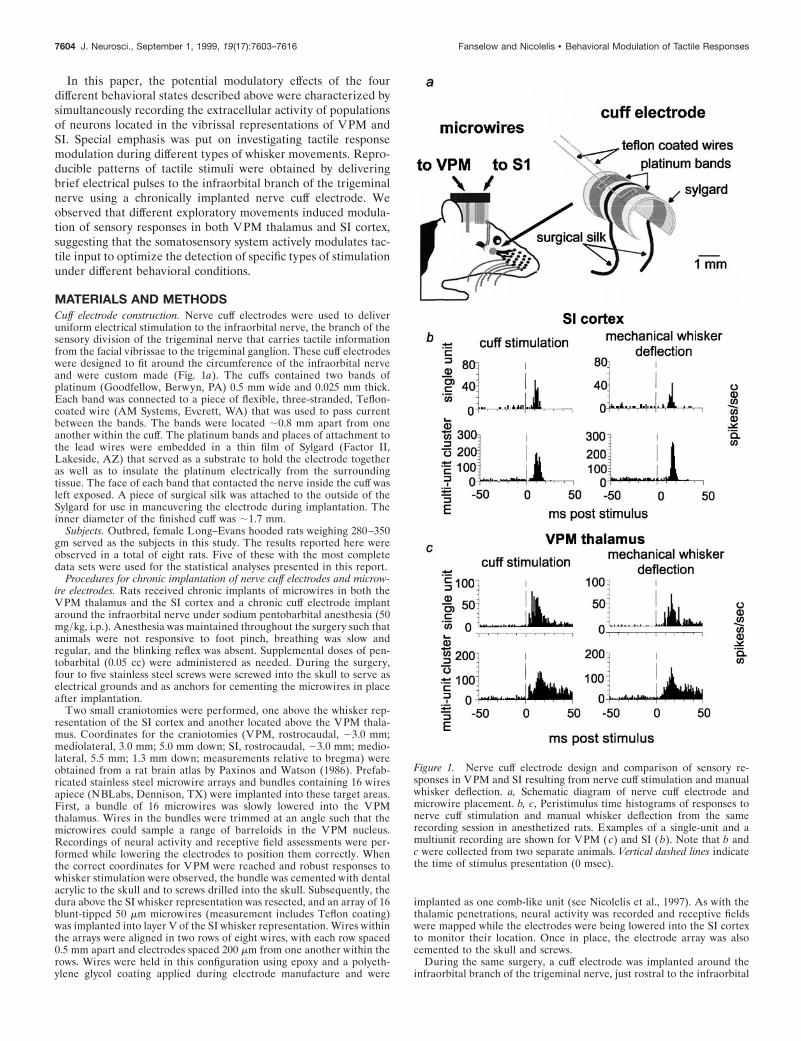
MATERIALS AND METHODSCuff electrode construction. Nerve cuff electrodes were used to deliveruniform electrical stimulation to the infraorbital nerve, the branch of thesensory division of the trigeminal nerve that carries tactile informationfrom the facial vibrissae to the trigeminal ganglion. These cuff electrodeswere designed to fit around the circumference of the infraorbital nerveand were custom made (Fig. 1a). The cuffs contained two bands ofplatinum (Goodfellow, Berwyn, PA) 0.5 mm wide and 0.025 mm thick.Each band was connected to a piece of flexible, three-stranded, Teflon-coated wire (AM Systems, Everett, WA) that was used to pass currentbetween the bands. The bands were located ;0.8 mm apart from oneanother within the cuff. The platinum bands and places of attachment tothe lead wires were embedded in a thin film of Sylgard (Factor II,Lakeside, AZ) that served as a substrate to hold the electrode togetheras well as to insulate the platinum electrically from the surroundingtissue. The face of each band that contacted the nerve inside the cuff wasleft exposed. A piece of surgical silk was attached to the outside of theSylgard for use in maneuvering the electrode during implantation. Theinner diameter of the finished cuff was ;1.7 mm.
Subjects. Outbred, female Long–Evans hooded rats weighing 280–350gm served as the subjects in this study. The results reported here wereobserved in a total of eight rats. Five of these with the most completedata sets were used for the statistical analyses presented in this report.
Procedures for chronic implantation of nerve cuff electrodes and microw-ire electrodes. Rats received chronic implants of microwires in both theVPM thalamus and the SI cortex and a chronic cuff electrode implantaround the infraorbital nerve under sodium pentobarbital anesthesia (50mg/kg, i.p.). Anesthesia was maintained throughout the surgery such thatanimals were not responsive to foot pinch, breathing was slow andregular, and the blinking reflex was absent. Supplemental doses of pen-tobarbital (0.05 cc) were administered as needed. During the surgery,four to five stainless steel screws were screwed into the skull to serve aselectrical grounds and as anchors for cementing the microwires in placeafter implantation.
Two small craniotomies were performed, one above the whisker rep-resentation of the SI cortex and another located above the VPM thala-mus. Coordinates for the craniotomies (VPM, rostrocaudal, 23.0 mm;mediolateral, 3.0 mm; 5.0 mm down; SI, rostrocaudal, 23.0 mm; medio-lateral, 5.5 mm; 1.3 mm down; measurements relative to bregma) wereobtained from a rat brain atlas by Paxinos and Watson (1986). Prefab-ricated stainless steel microwire arrays and bundles containing 16 wiresapiece (NBLabs, Dennison, TX) were implanted into these target areas.First, a bundle of 16 microwires was slowly lowered into the VPMthalamus. Wires in the bundles were trimmed at an angle such that themicrowires could sample a range of barreloids in the VPM nucleus.Recordings of neural activity and receptive field assessments were per-formed while lowering the electrodes to position them correctly. Whenthe correct coordinates for VPM were reached and robust responses towhisker stimulation were observed, the bundle was cemented with dentalacrylic to the skull and to screws drilled into the skull. Subsequently, thedura above the SI whisker representation was resected, and an array of 16blunt-tipped 50 mm microwires (measurement includes Teflon coating)was implanted into layer V of the SI whisker representation. Wires withinthe arrays were aligned in two rows of eight wires, with each row spaced0.5 mm apart and electrodes spaced 200 mm from one another within therows. Wires were held in this configuration using epoxy and a polyeth-ylene glycol coating applied during electrode manufacture and were
implanted as one comb-like unit (see Nicolelis et al., 1997). As with thethalamic penetrations, neural activity was recorded and receptive fieldswere mapped while the electrodes were being lowered into the SI cortexto monitor their location. Once in place, the electrode array was alsocemented to the skull and screws.
During the same surgery, a cuff electrode was implanted around theinfraorbital branch of the trigeminal nerve, just rostral to the infraorbital
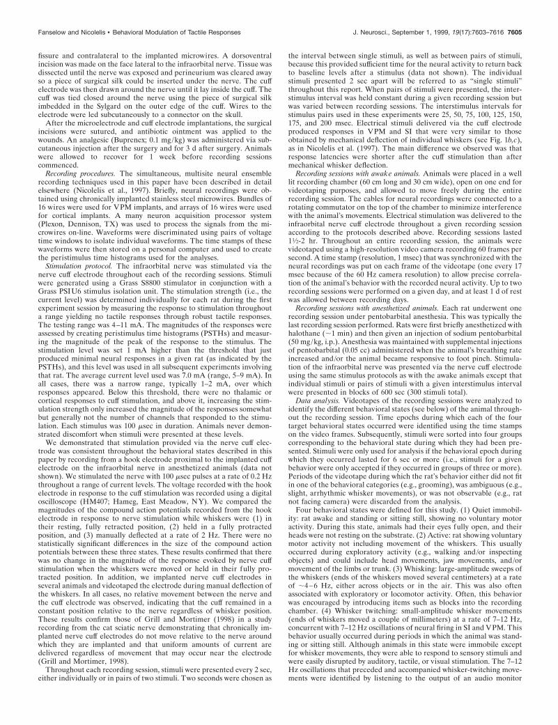
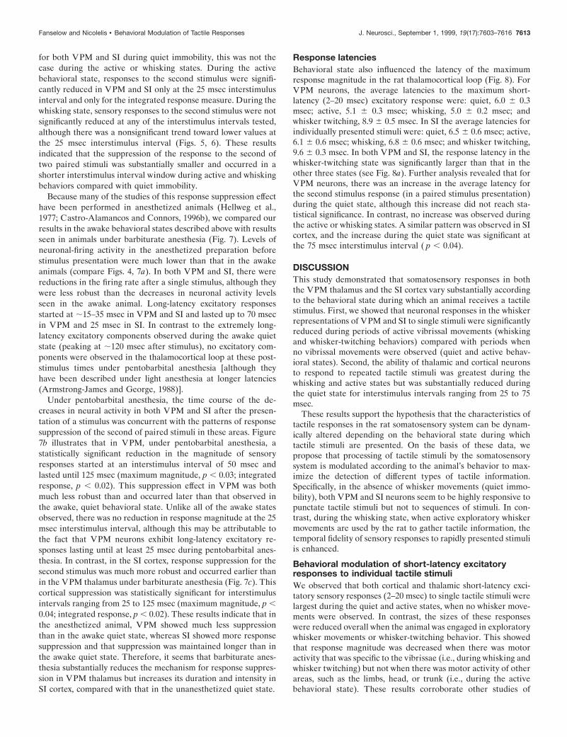
Figure 1. Nerve cuff electrode design and comparison of sensory re-sponses in VPM and SI resulting from nerve cuff stimulation and manualwhisker deflection. a, Schematic diagram of nerve cuff electrode andmicrowire placement. b, c, Peristimulus time histograms of responses tonerve cuff stimulation and manual whisker deflection from the samerecording session in anesthetized rats. Examples of a single-unit and amultiunit recording are shown for VPM ( c) and SI ( b). Note that b andc were collected from two separate animals. Vertical dashed lines indicatethe time of stimulus presentation (0 msec).
7604 J. Neurosci., September 1, 1999, 19(17):7603–7616 Fanselow and Nicolelis • Behavioral Modulation of Tactile Responses
fissure and contralateral to the implanted microwires. A dorsoventralincision was made on the face lateral to the infraorbital nerve. Tissue wasdissected until the nerve was exposed and perineurium was cleared awayso a piece of surgical silk could be inserted under the nerve. The cuffelectrode was then drawn around the nerve until it lay inside the cuff. Thecuff was tied closed around the nerve using the piece of surgical silkimbedded in the Sylgard on the outer edge of the cuff. Wires to theelectrode were led subcutaneously to a connector on the skull.
After the microelectrode and cuff electrode implantations, the surgicalincisions were sutured, and antibiotic ointment was applied to thewounds. An analgesic (Buprenex; 0.1 mg/kg) was administered via sub-cutaneous injection after the surgery and for 3 d after surgery. Animalswere allowed to recover for 1 week before recording sessionscommenced.
Recording procedures. The simultaneous, multisite neural ensemblerecording techniques used in this paper have been described in detailelsewhere (Nicolelis et al., 1997). Briefly, neural recordings were ob-tained using chronically implanted stainless steel microwires. Bundles of16 wires were used for VPM implants, and arrays of 16 wires were usedfor cortical implants. A many neuron acquisition processor system(Plexon, Dennison, TX) was used to process the signals from the mi-crowires on-line. Waveforms were discriminated using pairs of voltagetime windows to isolate individual waveforms. The time stamps of thesewaveforms were then stored on a personal computer and used to createthe peristimulus time histograms used for the analyses.
Stimulation protocol. The infraorbital nerve was stimulated via thenerve cuff electrode throughout each of the recording sessions. Stimuliwere generated using a Grass S8800 stimulator in conjunction with aGrass PSIU6 stimulus isolation unit. The stimulation strength (i.e., thecurrent level) was determined individually for each rat during the firstexperiment session by measuring the response to stimulation throughouta range yielding no tactile responses through robust tactile responses.The testing range was 4–11 mA. The magnitudes of the responses wereassessed by creating peristimulus time histograms (PSTHs) and measur-ing the magnitude of the peak of the response to the stimulus. Thestimulation level was set 1 mA higher than the threshold that justproduced minimal neural responses in a given rat (as indicated by thePSTHs), and this level was used in all subsequent experiments involvingthat rat. The average current level used was 7.0 mA (range, 5–9 mA). Inall cases, there was a narrow range, typically 1–2 mA, over whichresponses appeared. Below this threshold, there were no thalamic orcortical responses to cuff stimulation, and above it, increasing the stim-ulation strength only increased the magnitude of the responses somewhatbut generally not the number of channels that responded to the stimu-lation. Each stimulus was 100 msec in duration. Animals never demon-strated discomfort when stimuli were presented at these levels.
We demonstrated that stimulation provided via the nerve cuff elec-trode was consistent throughout the behavioral states described in thispaper by recording from a hook electrode proximal to the implanted cuffelectrode on the infraorbital nerve in anesthetized animals (data notshown). We stimulated the nerve with 100 msec pulses at a rate of 0.2 Hzthroughout a range of current levels. The voltage recorded with the hookelectrode in response to the cuff stimulation was recorded using a digitaloscilloscope (HM407; Hameg, East Meadow, NY). We compared themagnitudes of the compound action potentials recorded from the hookelectrode in response to nerve stimulation while whiskers were (1) intheir resting, fully retracted position, (2) held in a fully protractedposition, and (3) manually deflected at a rate of 2 Hz. There were nostatistically significant differences in the size of the compound actionpotentials between these three states. These results confirmed that therewas no change in the magnitude of the response evoked by nerve cuffstimulation when the whiskers were moved or held in their fully pro-tracted position. In addition, we implanted nerve cuff electrodes inseveral animals and videotaped the electrode during manual deflection ofthe whiskers. In all cases, no relative movement between the nerve andthe cuff electrode was observed, indicating that the cuff remained in aconstant position relative to the nerve regardless of whisker position.These results confirm those of Grill and Mortimer (1998) in a studyrecording from the cat sciatic nerve demonstrating that chronically im-planted nerve cuff electrodes do not move relative to the nerve aroundwhich they are implanted and that uniform amounts of current aredelivered regardless of movement that may occur near the electrode(Grill and Mortimer, 1998).
Throughout each recording session, stimuli were presented every 2 sec,either individually or in pairs of two stimuli. Two seconds were chosen as
the interval between single stimuli, as well as between pairs of stimuli,because this provided sufficient time for the neural activity to return backto baseline levels after a stimulus (data not shown). The individualstimuli presented 2 sec apart will be referred to as “single stimuli”throughout this report. When pairs of stimuli were presented, the inter-stimulus interval was held constant during a given recording session butwas varied between recording sessions. The interstimulus intervals forstimulus pairs used in these experiments were 25, 50, 75, 100, 125, 150,175, and 200 msec. Electrical stimuli delivered via the cuff electrodeproduced responses in VPM and SI that were very similar to thoseobtained by mechanical deflection of individual whiskers (see Fig. 1b,c),as in Nicolelis et al. (1997). The main difference we observed was thatresponse latencies were shorter after the cuff stimulation than aftermechanical whisker deflection.
Recording sessions with awake animals. Animals were placed in a welllit recording chamber (60 cm long and 30 cm wide), open on one end forvideotaping purposes, and allowed to move freely during the entirerecording session. The cables for neural recordings were connected to arotating commutator on the top of the chamber to minimize interferencewith the animal’s movements. Electrical stimulation was delivered to theinfraorbital nerve cuff electrode throughout a given recording sessionaccording to the protocols described above. Recording sessions lasted11⁄2-2 hr. Throughout an entire recording session, the animals werevideotaped using a high-resolution video camera recording 60 frames persecond. A time stamp (resolution, 1 msec) that was synchronized with theneural recordings was put on each frame of the videotape (one every 17msec because of the 60 Hz camera resolution) to allow precise correla-tion of the animal’s behavior with the recorded neural activity. Up to tworecording sessions were performed on a given day, and at least 1 d of restwas allowed between recording days.
Recording sessions with anesthetized animals. Each rat underwent onerecording session under pentobarbital anesthesia. This was typically thelast recording session performed. Rats were first briefly anesthetized withhalothane (;1 min) and then given an injection of sodium pentobarbital(50 mg/kg, i.p.). Anesthesia was maintained with supplemental injectionsof pentobarbital (0.05 cc) administered when the animal’s breathing rateincreased and/or the animal became responsive to foot pinch. Stimula-tion of the infraorbital nerve was presented via the nerve cuff electrodeusing the same stimulus protocols as with the awake animals except thatindividual stimuli or pairs of stimuli with a given interstimulus intervalwere presented in blocks of 600 sec (300 stimuli total).
Data analysis. Videotapes of the recording sessions were analyzed toidentify the different behavioral states (see below) of the animal through-out the recording session. Time epochs during which each of the fourtarget behavioral states occurred were identified using the time stampson the video frames. Subsequently, stimuli were sorted into four groupscorresponding to the behavioral state during which they had been pre-sented. Stimuli were only used for analysis if the behavioral epoch duringwhich they occurred lasted for 6 sec or more (i.e., stimuli for a givenbehavior were only accepted if they occurred in groups of three or more).Periods of the videotape during which the rat’s behavior either did not fitin one of the behavioral categories (e.g., grooming), was ambiguous (e.g.,slight, arrhythmic whisker movements), or was not observable (e.g., ratnot facing camera) were discarded from the analysis.
Four behavioral states were defined for this study. (1) Quiet immobil-ity: rat awake and standing or sitting still, showing no voluntary motoractivity. During this state, animals had their eyes fully open, and theirheads were not resting on the substrate. (2) Active: rat showing voluntarymotor activity not including movement of the whiskers. This usuallyoccurred during exploratory activity (e.g., walking and/or inspectingobjects) and could include head movements, jaw movements, and/ormovement of the limbs or trunk. (3) Whisking: large-amplitude sweeps ofthe whiskers (ends of the whiskers moved several centimeters) at a rateof ;4–6 Hz, either across objects or in the air. This was also oftenassociated with exploratory or locomotor activity. Often, this behaviorwas encouraged by introducing items such as blocks into the recordingchamber. (4) Whisker twitching: small-amplitude whisker movements(ends of whiskers moved a couple of millimeters) at a rate of 7–12 Hz,concurrent with 7–12 Hz oscillations of neural firing in SI and VPM. Thisbehavior usually occurred during periods in which the animal was stand-ing or sitting still. Although animals in this state were immobile exceptfor whisker movements, they were able to respond to sensory stimuli andwere easily disrupted by auditory, tactile, or visual stimulation. The 7–12Hz oscillations that preceded and accompanied whisker-twitching move-ments were identified by listening to the output of an audio monitor
Fanselow and Nicolelis • Behavioral Modulation of Tactile Responses J. Neurosci., September 1, 1999, 19(17):7603–7616 7605
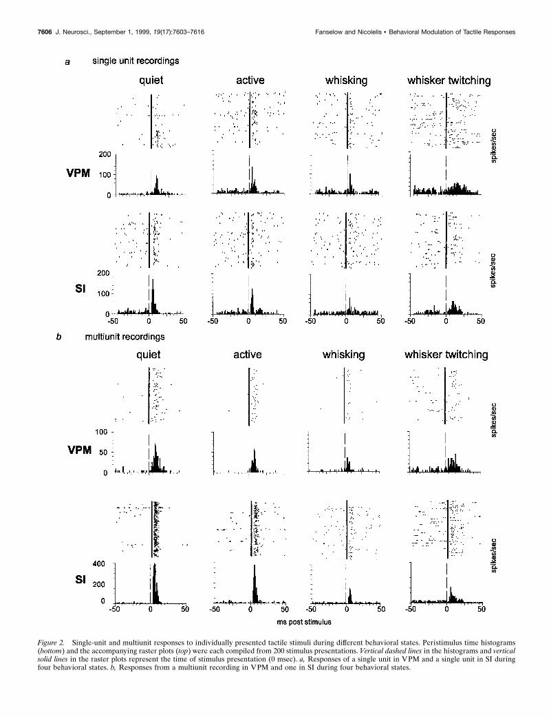
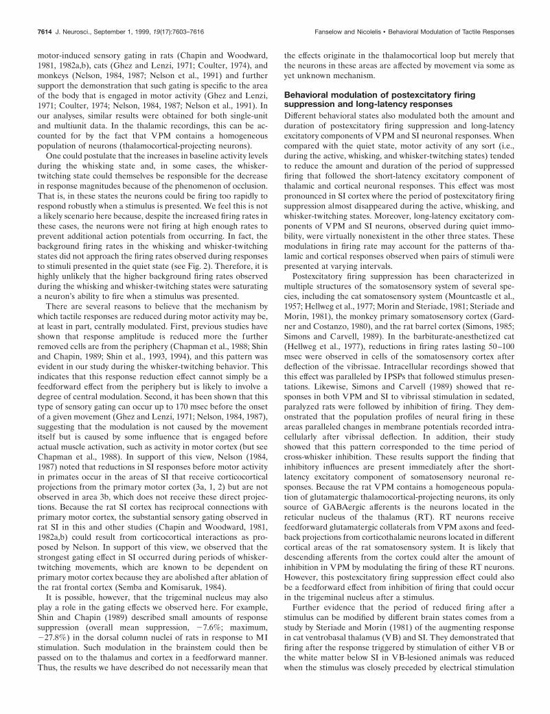
Figure 2. Single-unit and multiunit responses to individually presented tactile stimuli during different behavioral states. Peristimulus time histograms(bottom) and the accompanying raster plots (top) were each compiled from 200 stimulus presentations. Vertical dashed lines in the histograms and verticalsolid lines in the raster plots represent the time of stimulus presentation (0 msec). a, Responses of a single unit in VPM and a single unit in SI duringfour behavioral states. b, Responses from a multiunit recording in VPM and one in SI during four behavioral states.
7606 J. Neurosci., September 1, 1999, 19(17):7603–7616 Fanselow and Nicolelis • Behavioral Modulation of Tactile Responses
signal depicting ongoing multiunit neural activity that was recorded onthe videotape. The relationship between these oscillations and thewhisker-twitching movements have been described previously (Sembaand Komisaruk, 1984; Nicolelis et al., 1995).
Quantification of neural signals. Raster plots and PSTHs were gener-ated for responses on each channel to stimulation of the nerve cuffelectrode. These histograms were created using 200 stimuli for eachcondition and had a bin size of 1 msec. In all analyses, the beginning ofthe time period used was set at 2 msec to eliminate the electrical artifactthat could be observed in the 0–1 msec time bin on some channelsresulting from cuff stimulation. The peaks in the PSTHs correspondingto short-latency excitatory responses to the stimuli were assessed in twoways. First, the maximum height of the peak was measured (referred toas “maximum magnitude”). Second, the total amount of firing during aperiod from 2 to 20 msec after the stimulus was calculated. This secondmeasure was used to assess the overall amount of response a stimulusyielded, because some responses differed not only in magnitude but alsoin width and intensity across the experimental conditions. This secondmeasure will be referred to hereafter as the “integrated response.” Forboth of these measures, the background firing rate was calculated foreach channel (average firing rate during the 100 msec period precedingthe stimulus) and subtracted from the response. Both of these measureswere normalized to the quiet state activity levels. For pairs of stimuli,responses were normalized to the first stimulus of pairs presented duringthe quiet behavioral state. Responses were compared using ANOVAs,and Tukey’s honestly significant difference (HSD) tests were used whenpost hoc tests were needed. A response was considered significant if thep value was ,0.05. Only one recording session was conducted for eachstimulation protocol in each rat, ensuring that no neurons that couldpotentially have been recorded from in multiple sessions were statisticallypooled.
Overall levels of neural activity before and after short-latency excita-tory responses to stimuli were assessed by averaging histograms across allchannels used in the analyses for a given recording area (VPM or SI).The levels of neural activity before a stimulus were calculated by aver-aging the number of spikes per second during the 100 msec periodpreceding stimulus presentation. Spiking activity was also calculated atpoints every 25 msec after the stimulus.
Histology. After the entire battery of experiments was completed(usually 3–4 weeks after electrode implantation), rats received an over-dose of sodium pentobarbital and were then perfused transcardially with0.9% saline, followed by 5% formalin. The brains were sliced into 80 mmcoronal sections and stained using cresyl violet. In these sections, thelocation of the tips of the electrodes could be determined, and theirlocations in the whisker representations of VPM and SI were verified.
Parts of this paper have been published previously (Fanselow et al.,1997).
RESULTSTo evaluate the effects of different behavioral states (quiet, active,whisking, and whisker twitching) on the somatosensory responsesof VPM and SI neurons, both single-unit and multiunit recordingswere made during the experiments described here. On-line andoff-line waveform measurements, as well as off-line analysis ofinterspike interval histograms, were used to identify single unitsin our records. A recording was identified as a single unit if it metall three of the following criteria: (1) the presence of reproduciblewaveforms that could be isolated by our on-line recording proce-dure (Nicolelis et al., 1997), (2) interspike interval histogramsshowing that ,5% of the interspike intervals were shorter than1.2 msec, and 3) the negative-going component of the waveformfor each action potential being at least 75 mV in amplitude(average background noise levels were 15–20 mV, so the signal-to-noise ratios were 4–5). According to these criteria, we re-corded a total of 62 single units in the VPM thalamus and 61 inthe SI cortex in five awake, freely behaving rats. The other signalsobtained from these animals during the same experimental ses-sions were designated as multiunit recordings. A total of 165multiunit recordings were made in the VPM thalamus, and 157were made in the SI cortex. Because the rat VPM thalamus
contains only a single cell type (thalamocortical-projecting neu-rons), our multiunit data likely derived from a rather homogenouspopulation of neurons in this structure. For some of the statisticalanalyses in this paper, we decided to group single-unit and mul-tiunit recordings within each area. This decision was validated bythe fact that our single-unit results were similar to the findingsobtained with multiunit data (see Figs. 2, 3).
In each animal, after ;4 weeks of recording, a final recordingsession was performed under pentobarbital anesthesia. Becausethese recordings were performed several weeks after microwireimplantation, the neuronal yield was typically lower than inearlier recording sessions. Therefore during the experiments withanesthetized animals, a total of 25 single units were recorded inthe VPM thalamus, and 11 were recorded in the SI cortex; thetotal numbers of multiunit recordings in VPM and SI were 56 and47, respectively.
We compared cortical and thalamic somatosensory responsesproduced by electrical stimulation of the nerve cuff electrode withthose triggered by mechanical deflection of individual facial whis-kers. This was done by stimulating the infraorbital nerve via thecuff electrode at 1 Hz in anesthetized animals and comparing theresponses of SI (Fig. 1b) and VPM (Fig. 1c) neurons with thoseobtained via 1 Hz mechanical displacement of individual whis-kers. Whiskers were mechanically displaced in discrete steps (100msec duration) using a motor-driven manipulator that was placednext to each whisker individually and activated by a Grass S8800stimulator. This analysis revealed that the parameters for electri-cal stimulation of the infraorbital nerve used in this study (seeMaterials and Methods) could produce sensory responses verysimilar to those obtained by mechanical whisker deflection (forexamples, see Fig. 1b,c). Cortical and thalamic sensory responsesto cuff stimulation were of similar shape and amplitude to thoseobtained by manual whisker deflection. Electrical nerve stimula-tion did, however, induce faster sensory responses in both VPMthalamus and SI cortex. In addition, these control experimentsrevealed that the nerve cuff could be used for long periods of timeafter implantation and that reproducible stimuli could be deliv-ered chronically. Our results are consistent with data obtainedusing a similar cuff electrode design to stimulate the optic nerve(Weliky and Katz, 1997).
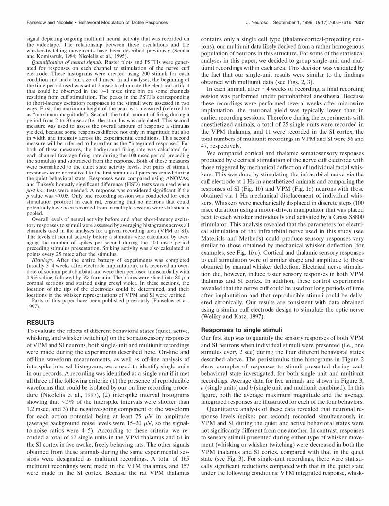
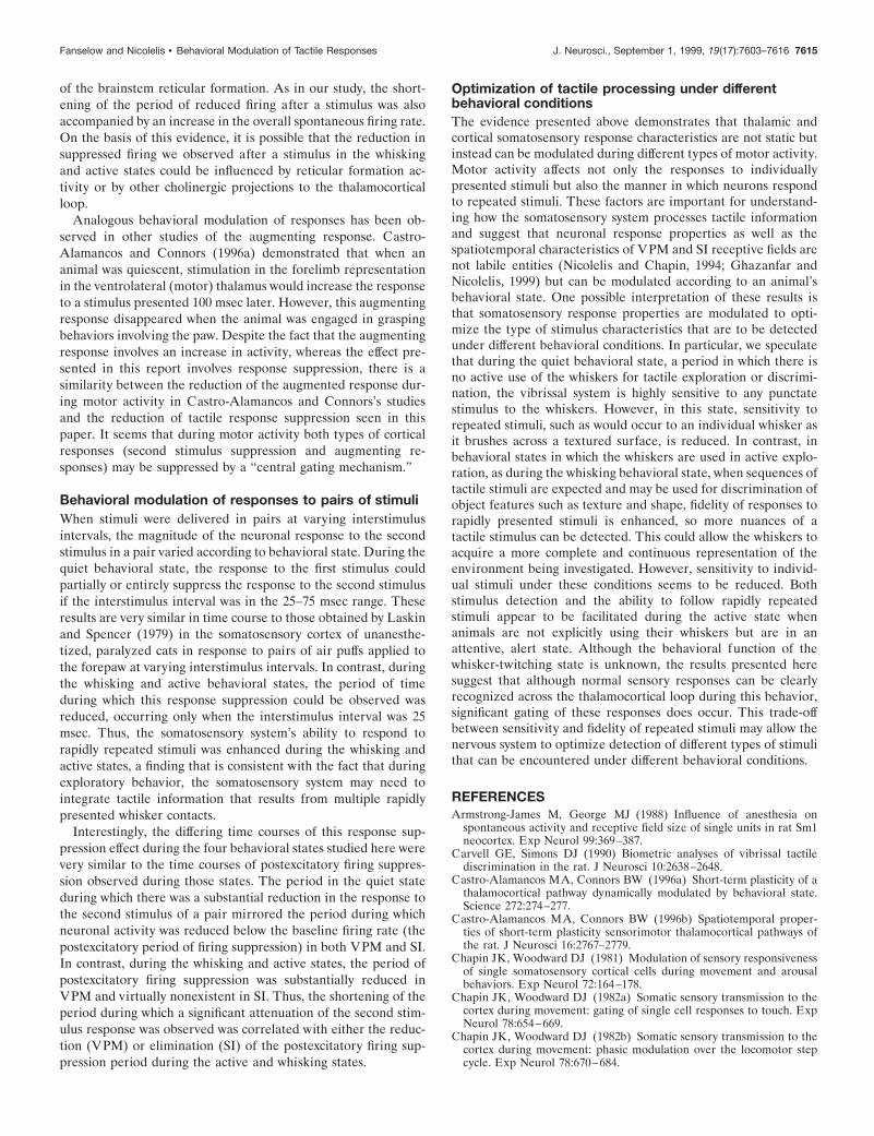
Responses to single stimuliOur first step was to quantify the sensory responses of both VPMand SI neurons when individual stimuli were presented (i.e., onestimulus every 2 sec) during the four different behavioral statesdescribed above. The peristimulus time histograms in Figure 2show examples of responses to stimuli presented during eachbehavioral state investigated, for both single-unit and multiunitrecordings. Average data for five animals are shown in Figure 3,a (single units) and b (single unit and multiunit combined). In thisfigure, both the average maximum magnitude and the averageintegrated responses are illustrated for each of the four behaviors.
Quantitative analysis of these data revealed that neuronal re-sponse levels (spikes per second) recorded simultaneously inVPM and SI during the quiet and active behavioral states werenot significantly different from one another. In contrast, responsesto sensory stimuli presented during either type of whisker move-ment (whisking or whisker twitching) were decreased in both theVPM thalamus and SI cortex, compared with that in the quietstate (see Fig. 3). For single-unit recordings, there were statisti-cally significant reductions compared with that in the quiet stateunder the following conditions: VPM integrated response, whisk-
Fanselow and Nicolelis • Behavioral Modulation of Tactile Responses J. Neurosci., September 1, 1999, 19(17):7603–7616 7607
ing (280.3 6 13.1%; p , 0.001); SI maximum magnitude, whisk-ing (230.0 6 10.7%; p , 0.05) and whisker twitching (245.1 611.7%; p , 0.02); and SI integrated response, whisking (241.29 618.3%; p , 0.001) and whisker twitching (255.6 6 8.3%; p ,0.001). For the pooled single-unit and multiunit data, there werestatistically significant reductions observed in the following cases:VPM maximum magnitude, whisker twitching (239.7 6 9.7%;p , 0.02); VPM integrated response, whisking (272.5 6 5.4%;p , 0.01); SI maximum magnitude, whisker twitching (253.22 68.4%; p , 0.02); and SI integrated response, whisking (251.2 6
5.8%; p , 0.002) and whisker twitching (248.5 6 8.4%; p ,0.005).
Magnitude of neuronal activity before and afterstimulus presentationThe demonstration that the magnitude of sensory neuronal re-sponses varied according to the animal’s behavior led us toexamine whether further variations in firing rate could be ob-served before and after the stimulus and initial excitatory re-sponse. This analysis revealed that the background levels of firing
Figure 3. Average responses to single stimuli during different behavioral states. In each graph, responses were normalized to responses during the quietstate. a, Average responses of single units. b, Average responses of single-unit and multiunit recordings combined. Top graphs in a and b represent themaximum magnitude (max mag) of the responses. Bottom graphs in a and b represent the integrated response (intg) values (see Materials and Methodsfor a description of these measures). Asterisks indicate values significantly different from the quiet state in each graph. Error bars represent 6 SEM.ANOVA results: a, VPM max mag, F 5 1.99, p 5 0.187; VPM intg, F 5 8.77, p 5 0.0012; SI max mag, F 5 5.74, p 5 0.0022; SI intg, F 5 10.99, p 50.000023. b, VPM max mag, F 5 9.82, p 5 0.0099; VPM intg, F 5 10.22, p 5 0.0090; SI max mag, F 5 9.82, p 5 0.0099; SI intg, F 5 23.94, p 5 0.00097.Please see text for p values from post hoc tests (Tukey’s HSD). A, Active; Q, quiet; W, whisking; WT, whisker twitching.
7608 J. Neurosci., September 1, 1999, 19(17):7603–7616 Fanselow and Nicolelis • Behavioral Modulation of Tactile Responses
before the presentation of a stimulus differed from one behavioralstate to the next. The average prestimulus firing rate values inspikes per second (averaged over the 100 msec before the stimu-lus) for single units in VPM were: quiet, 1.3 6 0.2; active, 2.7 60.7; whisking, 4.2 6 0.9; and whisker twitching, 5.8 6 0.5. In SI,the single-unit values were: quiet, 2.3 6 0.4; active, 2.7 6 0.5;whisking, 3.6 6 0.7; and whisker twitching, 2.0 6 0.3. In VPMthalamus, the prestimulus single-unit firing levels during the ac-tive, whisking, and whisker-twitching states were significantlyhigher than that during the quiet state ( p , 0.01). In SI, theprestimulus single-unit firing rate during whisking was signifi-cantly higher than that during the quiet and whisker-twitchingstates ( p , 0.02). We also calculated average prestimulus firingrates using both the single-unit and multiunit recordings com-bined (see Fig. 4 before stimulus presentation at 0 msec) andfound similar results overall, although the values for the combinedanalysis were larger in magnitude, as would be expected whenincluding the multiunit recordings in which more neurons wererecorded per channel. The combined values in VPM in spikes persecond were: quiet, 7.5 6 3.7; active, 9.8 6 4.9; whisking, 21.3 69.0; and whisker twitching, 5.8 6 3.4. In SI, the average firing ratevalues were: quiet, 5.5 6 1.8; active, 4.3 6 1.2; whisking, 8.1 6 1.8;
and whisker twitching, 3.0 6 1.3. In VPM, the multiunit firinglevel during the whisking state was significantly higher than thatin the quiet state ( p , 0.01). In SI, the prestimulus multiunitfiring rate during whisking was significantly higher than thatduring the active state ( p , 0.03).
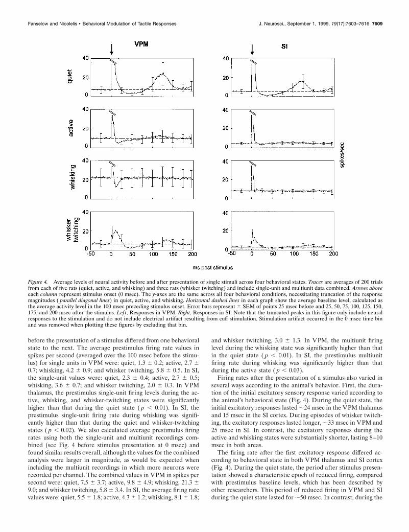
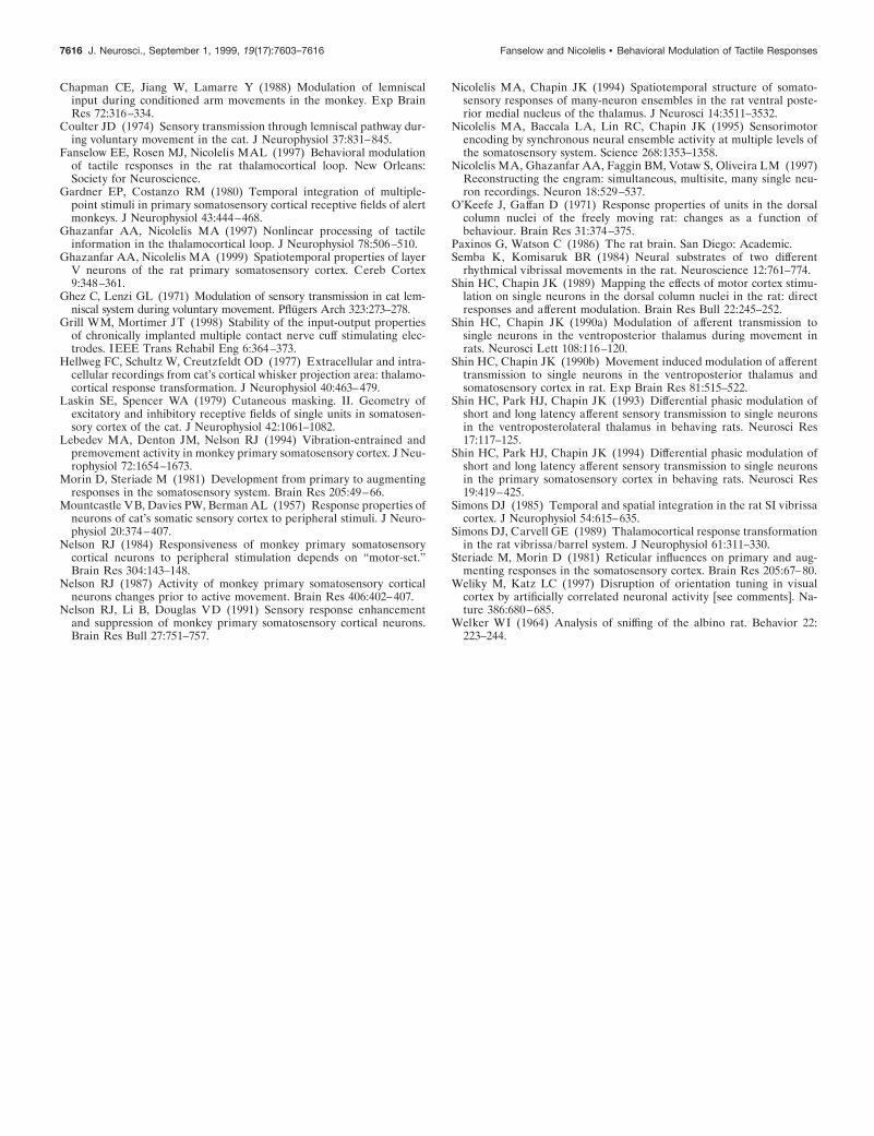
Firing rates after the presentation of a stimulus also varied inseveral ways according to the animal’s behavior. First, the dura-tion of the initial excitatory sensory response varied according tothe animal’s behavioral state (Fig. 4). During the quiet state, theinitial excitatory responses lasted ;24 msec in the VPM thalamusand 15 msec in the SI cortex. During episodes of whisker twitch-ing, the excitatory responses lasted longer, ;33 msec in VPM and25 msec in SI. In contrast, the excitatory responses during theactive and whisking states were substantially shorter, lasting 8–10msec in both areas.
The firing rate after the first excitatory response differed ac-cording to behavioral state in both VPM thalamus and SI cortex(Fig. 4). During the quiet state, the period after stimulus presen-tation showed a characteristic epoch of reduced firing, comparedwith prestimulus baseline levels, which has been described byother researchers. This period of reduced firing in VPM and SIduring the quiet state lasted for ;50 msec. In contrast, during the
Figure 4. Average levels of neural activity before and after presentation of single stimuli across four behavioral states. Traces are averages of 200 trialsfrom each of five rats (quiet, active, and whisking) and three rats (whisker twitching) and include single-unit and multiunit data combined. Arrows aboveeach column represent stimulus onset (0 msec). The y-axes are the same across all four behavioral conditions, necessitating truncation of the responsemagnitudes ( parallel diagonal lines) in quiet, active, and whisking. Horizontal dashed lines in each graph show the average baseline level, calculated asthe average activity level in the 100 msec preceding stimulus onset. Error bars represent 6 SEM of points 25 msec before and 25, 50, 75, 100, 125, 150,175, and 200 msec after the stimulus. Left, Responses in VPM. Right, Responses in SI. Note that the truncated peaks in this figure only include neuralresponses to the stimulation and do not include electrical artifact resulting from cuff stimulation. Stimulation artifact occurred in the 0 msec time binand was removed when plotting these figures by excluding that bin.
Fanselow and Nicolelis • Behavioral Modulation of Tactile Responses J. Neurosci., September 1, 1999, 19(17):7603–7616 7609
active and whisking states, this postexcitatory period of reducedfiring was substantially shorter and, in most cases, yielded lessspike suppression. During the active behavioral state, there was a35 msec period of reduced activity in the VPM thalamus, whereasno such period was detected in the SI cortex. Likewise, duringwhisking, there was a 20 msec period of reduced firing in theVPM thalamus but none in the SI cortex. During episodes ofwhisker twitching, the period of suppressed firing (;50 msec induration) in VPM was much more evident than in the SI cortex.This postexcitatory reduction of firing ended at approximatelythe same poststimulus time in both the quiet and the whisker-
twitching states. However, the beginning of this period was de-layed 10 msec in the latter state.
Finally, during quiet immobility, a pronounced long-latencyexcitatory response component was observed in both VPM andSI. These long-latency responses started at ;82 msec after thestimulus presentation in both VPM and SI, peaked at ;120 msecafter the stimulus in both structures, and lasted until 175 msecafter the stimulus in the VPM thalamus and 150 msec after thestimulus in the SI cortex. Minimal traces of such long-latencyresponses were observed during the active and whisker-twitchingstates in VPM. However, no long-latency components were ob-
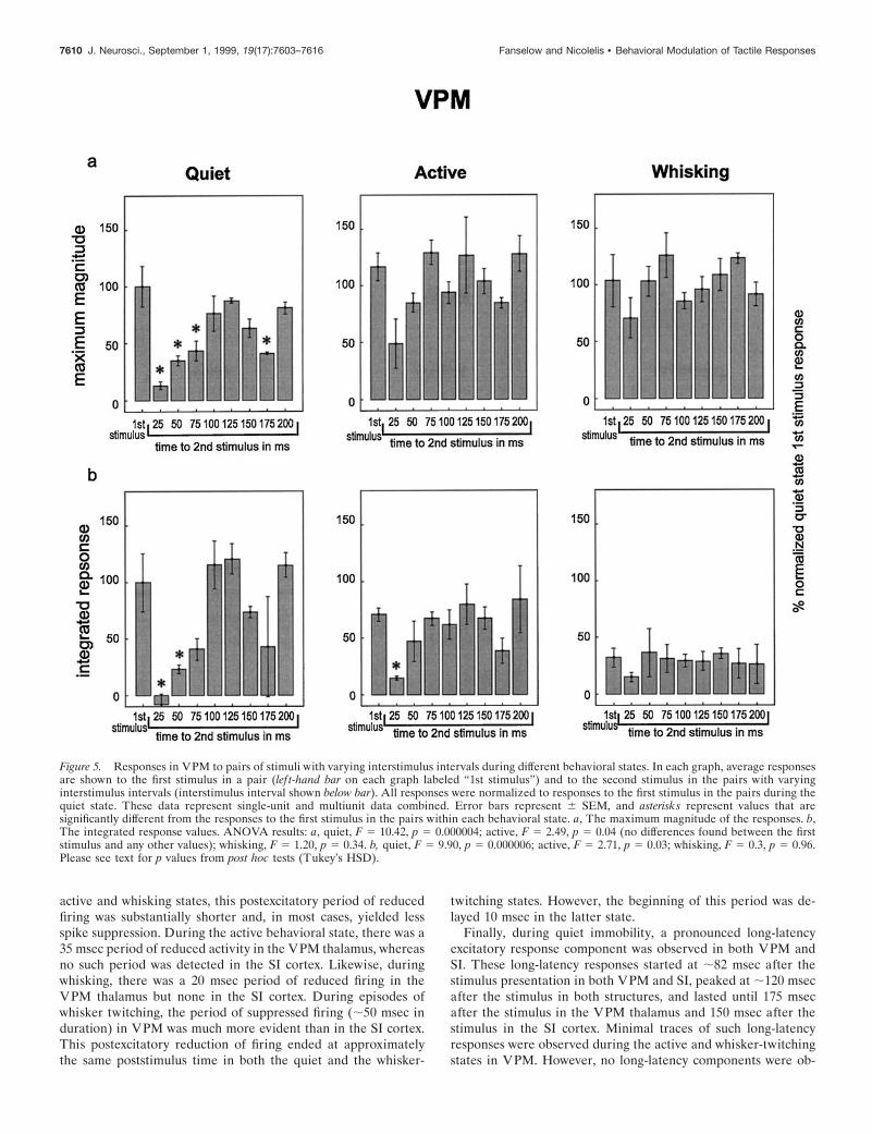
Figure 5. Responses in VPM to pairs of stimuli with varying interstimulus intervals during different behavioral states. In each graph, average responsesare shown to the first stimulus in a pair (lef t-hand bar on each graph labeled “1st stimulus”) and to the second stimulus in the pairs with varyinginterstimulus intervals (interstimulus interval shown below bar). All responses were normalized to responses to the first stimulus in the pairs during thequiet state. These data represent single-unit and multiunit data combined. Error bars represent 6 SEM, and asterisks represent values that aresignificantly different from the responses to the first stimulus in the pairs within each behavioral state. a, The maximum magnitude of the responses. b,The integrated response values. ANOVA results: a, quiet, F 5 10.42, p 5 0.000004; active, F 5 2.49, p 5 0.04 (no differences found between the firststimulus and any other values); whisking, F 5 1.20, p 5 0.34. b, quiet, F 5 9.90, p 5 0.000006; active, F 5 2.71, p 5 0.03; whisking, F 5 0.3, p 5 0.96.Please see text for p values from post hoc tests (Tukey’s HSD).
7610 J. Neurosci., September 1, 1999, 19(17):7603–7616 Fanselow and Nicolelis • Behavioral Modulation of Tactile Responses
served in cortical recordings in any state other than the quietstate.
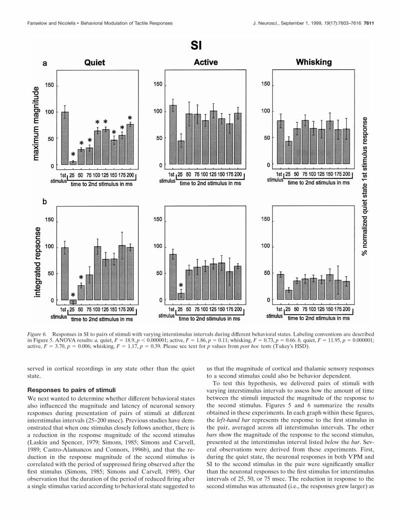
Responses to pairs of stimuliWe next wanted to determine whether different behavioral statesalso influenced the magnitude and latency of neuronal sensoryresponses during presentation of pairs of stimuli at differentinterstimulus intervals (25–200 msec). Previous studies have dem-onstrated that when one stimulus closely follows another, there isa reduction in the response magnitude of the second stimulus(Laskin and Spencer, 1979; Simons, 1985; Simons and Carvell,1989; Castro-Alamancos and Connors, 1996b), and that the re-duction in the response magnitude of the second stimulus iscorrelated with the period of suppressed firing observed after thefirst stimulus (Simons, 1985; Simons and Carvell, 1989). Ourobservation that the duration of the period of reduced firing aftera single stimulus varied according to behavioral state suggested to
us that the magnitude of cortical and thalamic sensory responsesto a second stimulus could also be behavior dependent.
To test this hypothesis, we delivered pairs of stimuli withvarying interstimulus intervals to assess how the amount of timebetween the stimuli impacted the magnitude of the response tothe second stimulus. Figures 5 and 6 summarize the resultsobtained in these experiments. In each graph within these figures,the lef t-hand bar represents the response to the first stimulus inthe pair, averaged across all interstimulus intervals. The otherbars show the magnitude of the response to the second stimulus,presented at the interstimulus interval listed below the bar. Sev-eral observations were derived from these experiments. First,during the quiet state, the neuronal responses in both VPM andSI to the second stimulus in the pair were significantly smallerthan the neuronal responses to the first stimulus for interstimulusintervals of 25, 50, or 75 msec. The reduction in response to thesecond stimulus was attenuated (i.e., the responses grew larger) as
Figure 6. Responses in SI to pairs of stimuli with varying interstimulus intervals during different behavioral states. Labeling conventions are describedin Figure 5. ANOVA results: a, quiet, F 5 18.9, p , 0.000001; active, F 5 1.86, p 5 0.11; whisking, F 5 0.73, p 5 0.66. b, quiet, F 5 11.95, p 5 0.000001;active, F 5 3.70, p 5 0.006; whisking, F 5 1.17, p 5 0.39. Please see text for p values from post hoc tests (Tukey’s HSD).
Fanselow and Nicolelis • Behavioral Modulation of Tactile Responses J. Neurosci., September 1, 1999, 19(17):7603–7616 7611
the interstimulus interval increased until the interstimulus inter-val reached 100 msec, at which point the response to the secondstimulus in the pair was not statistically different from the first inVPM but remained slightly (and statistically significantly) belowthe first stimulus values in SI throughout the 200 msec interstimu-lus intervals tested in this study. This effect mirrored the timingand duration of the period of postexcitatory firing suppressionobserved in VPM and SI (see Fig. 4). Interestingly, the responsesuppression effect returned at 150–175 msec interstimulus inter-vals during the quiet state (see maximum magnitude plots in Figs.5, 6), a period during which neuronal firing in the VPM thalamusand SI cortex tended to return to baseline levels after the presen-tation of individual stimuli (see Fig. 4).
The magnitude and time course of response suppression to thesecond stimulus in a pair varied according to behavioral state.Although this suppression effect was significant and long-lasting
Figure 7. Sensory responses during pentobarbital anesthesia. a, Averagelevels of neural activity in VPM and SI before and after stimulus presen-tation. Labeling conventions for a are described in Figure 4. b, c, Re-sponse magnitude and integrated response to pairs of stimuli at varyinginterstimulus intervals. Labeling for b and c is described in Figure 5.These data represent single-unit and multiunit data combined. ANOVAresults: b, max mag, F 5 6.45, p 5 0.003; intg, F 5 7.12, p 5 0.002; c, maxmag, F 5 4.44, p 5 0.014; intg, F 5 6.02, p 5 0.004. Please see text for pvalues from post hoc tests (Tukey’s HSD).
Figure 8. Response latencies in VPM and SI across behavioral states. a,Latencies for single stimuli. b, c, Latencies for pairs of stimuli. Labelingconventions are described in Figure 5. These latency values were obtainedfrom single-unit and multiunit data combined. ANOVA results: a, VPM,F 5 35.86, p 5 0.0003; SI, F 5 18.33, p 5 0.002; b, quiet, F 5 1.55, p 50.19; active, F 5 0.86, p 5 0.57; whisking, F 5 5.79, p 5 0.011 (no valuesfound significantly different from the first stimulus value); c, quiet, F 53.62, p 5 0.013; active, F 5 0.18, p 5 0.99; whisking, F 5 0.93, p 5 0.51.Please see text for p values from post hoc tests (Tukey’s HSD).
7612 J. Neurosci., September 1, 1999, 19(17):7603–7616 Fanselow and Nicolelis • Behavioral Modulation of Tactile Responses
for both VPM and SI during quiet immobility, this was not thecase during the active or whisking states. During the activebehavioral state, responses to the second stimulus were signifi-cantly reduced in VPM and SI only at the 25 msec interstimulusinterval and only for the integrated response measure. During thewhisking state, sensory responses to the second stimulus were notsignificantly reduced at any of the interstimulus intervals tested,although there was a nonsignificant trend toward lower values atthe 25 msec interstimulus interval (Figs. 5, 6). These resultsindicated that the suppression of the response to the second oftwo paired stimuli was substantially smaller and occurred in ashorter interstimulus interval window during active and whiskingbehaviors compared with quiet immobility.
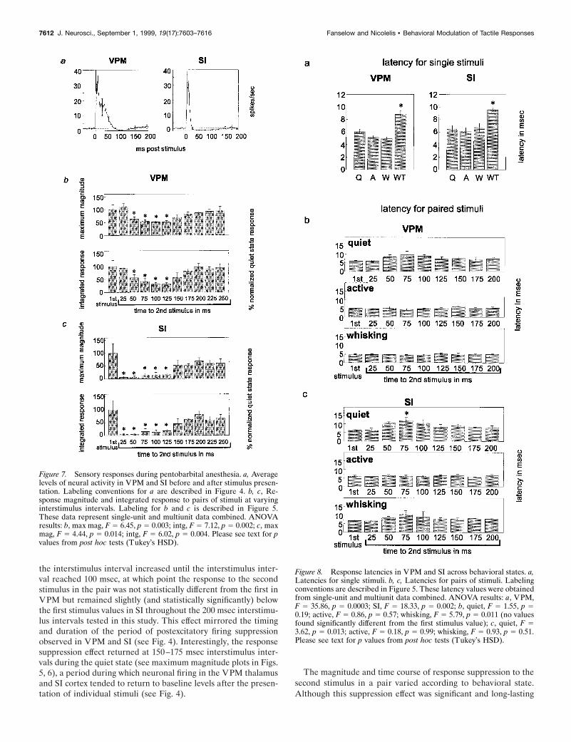
Because many of the studies of this response suppression effecthave been performed in anesthetized animals (Hellweg et al.,1977; Castro-Alamancos and Connors, 1996b), we compared ourresults in the awake behavioral states described above with resultsseen in animals under barbiturate anesthesia (Fig. 7). Levels ofneuronal-firing activity in the anesthetized preparation beforestimulus presentation were much lower than that in the awakeanimals (compare Figs. 4, 7a). In both VPM and SI, there werereductions in the firing rate after a single stimulus, although theywere less robust than the decreases in neuronal activity levelsseen in the awake animal. Long-latency excitatory responsesstarted at ;15–35 msec in VPM and SI and lasted up to 70 msecin VPM and 25 msec in SI. In contrast to the extremely long-latency excitatory components observed during the awake quietstate (peaking at ;120 msec after stimulus), no excitatory com-ponents were observed in the thalamocortical loop at these post-stimulus times under pentobarbital anesthesia [although theyhave been described under light anesthesia at longer latencies(Armstrong-James and George, 1988)].
Under pentobarbital anesthesia, the time course of the de-creases in neural activity in both VPM and SI after the presen-tation of a stimulus was concurrent with the patterns of responsesuppression of the second of paired stimuli in these areas. Figure7b illustrates that in VPM, under pentobarbital anesthesia, astatistically significant reduction in the magnitude of sensoryresponses started at an interstimulus interval of 50 msec andlasted until 125 msec (maximum magnitude, p , 0.03; integratedresponse, p , 0.02). This suppression effect in VPM was bothmuch less robust than and occurred later than that observed inthe awake, quiet behavioral state. Unlike all of the awake statesobserved, there was no reduction in response magnitude at the 25msec interstimulus interval, although this may be attributable tothe fact that VPM neurons exhibit long-latency excitatory re-sponses lasting until at least 25 msec during pentobarbital anes-thesia. In contrast, in the SI cortex, response suppression for thesecond stimulus was much more robust and occurred earlier thanin the VPM thalamus under barbiturate anesthesia (Fig. 7c). Thiscortical suppression was statistically significant for interstimulusintervals ranging from 25 to 125 msec (maximum magnitude, p ,0.04; integrated response, p , 0.02). These results indicate that inthe anesthetized animal, VPM showed much less suppressionthan in the awake quiet state, whereas SI showed more responsesuppression and that suppression was maintained longer than inthe awake quiet state. Therefore, it seems that barbiturate anes-thesia substantially reduces the mechanism for response suppres-sion in VPM thalamus but increases its duration and intensity inSI cortex, compared with that in the unanesthetized quiet state.
Response latenciesBehavioral state also influenced the latency of the maximumresponse magnitude in the rat thalamocortical loop (Fig. 8). ForVPM neurons, the average latencies to the maximum short-latency (2–20 msec) excitatory response were: quiet, 6.0 6 0.3msec; active, 5.1 6 0.3 msec; whisking, 5.0 6 0.2 msec; andwhisker twitching, 8.9 6 0.5 msec. In SI the average latencies forindividually presented stimuli were: quiet, 6.5 6 0.6 msec; active,6.1 6 0.6 msec; whisking, 6.8 6 0.6 msec; and whisker twitching,9.6 6 0.3 msec. In both VPM and SI, the response latency in thewhisker-twitching state was significantly larger than that in theother three states (see Fig. 8a). Further analysis revealed that forVPM neurons, there was an increase in the average latency forthe second stimulus response (in a paired stimulus presentation)during the quiet state, although this increase did not reach sta-tistical significance. In contrast, no increase was observed duringthe active or whisking states. A similar pattern was observed in SIcortex, and the increase during the quiet state was significant atthe 75 msec interstimulus interval ( p , 0.04).
DISCUSSIONThis study demonstrated that somatosensory responses in boththe VPM thalamus and the SI cortex vary substantially accordingto the behavioral state during which an animal receives a tactilestimulus. First, we showed that neuronal responses in the whiskerrepresentations of VPM and SI to single stimuli were significantlyreduced during periods of active vibrissal movements (whiskingand whisker-twitching behaviors) compared with periods whenno vibrissal movements were observed (quiet and active behav-ioral states). Second, the ability of thalamic and cortical neuronsto respond to repeated tactile stimuli was greatest during thewhisking and active states but was substantially reduced duringthe quiet state for interstimulus intervals ranging from 25 to 75msec.
These results support the hypothesis that the characteristics oftactile responses in the rat somatosensory system can be dynam-ically altered depending on the behavioral state during whichtactile stimuli are presented. On the basis of these data, wepropose that processing of tactile stimuli by the somatosensorysystem is modulated according to the animal’s behavior to max-imize the detection of different types of tactile information.Specifically, in the absence of whisker movements (quiet immo-bility), both VPM and SI neurons seem to be highly responsive topunctate tactile stimuli but not to sequences of stimuli. In con-trast, during the whisking state, when active exploratory whiskermovements are used by the rat to gather tactile information, thetemporal fidelity of sensory responses to rapidly presented stimuliis enhanced.
Behavioral modulation of short-latency excitatoryresponses to individual tactile stimuliWe observed that both cortical and thalamic short-latency exci-tatory sensory responses (2–20 msec) to single tactile stimuli werelargest during the quiet and active states, when no whisker move-ments were observed. In contrast, the sizes of these responseswere reduced overall when the animal was engaged in exploratorywhisker movements or whisker-twitching behavior. This showedthat response magnitude was decreased when there was motoractivity that was specific to the vibrissae (i.e., during whisking andwhisker twitching) but not when there was motor activity of otherareas, such as the limbs, head, or trunk (i.e., during the activebehavioral state). These results corroborate other studies of
Fanselow and Nicolelis • Behavioral Modulation of Tactile Responses J. Neurosci., September 1, 1999, 19(17):7603–7616 7613
motor-induced sensory gating in rats (Chapin and Woodward,1981, 1982a,b), cats (Ghez and Lenzi, 1971; Coulter, 1974), andmonkeys (Nelson, 1984, 1987; Nelson et al., 1991) and furthersupport the demonstration that such gating is specific to the areaof the body that is engaged in motor activity (Ghez and Lenzi,1971; Coulter, 1974; Nelson, 1984, 1987; Nelson et al., 1991). Inour analyses, similar results were obtained for both single-unitand multiunit data. In the thalamic recordings, this can be ac-counted for by the fact that VPM contains a homogeneouspopulation of neurons (thalamocortical-projecting neurons).
One could postulate that the increases in baseline activity levelsduring the whisking state and, in some cases, the whisker-twitching state could themselves be responsible for the decreasein response magnitudes because of the phenomenon of occlusion.That is, in these states the neurons could be firing too rapidly torespond robustly when a stimulus is presented. We feel this is nota likely scenario here because, despite the increased firing rates inthese cases, the neurons were not firing at high enough rates toprevent additional action potentials from occurring. In fact, thebackground firing rates in the whisking and whisker-twitchingstates did not approach the firing rates observed during responsesto stimuli presented in the quiet state (see Fig. 2). Therefore, it ishighly unlikely that the higher background firing rates observedduring the whisking and whisker-twitching states were saturatinga neuron’s ability to fire when a stimulus was presented.
There are several reasons to believe that the mechanism bywhich tactile responses are reduced during motor activity may be,at least in part, centrally modulated. First, previous studies haveshown that response amplitude is reduced more the furtherremoved cells are from the periphery (Chapman et al., 1988; Shinand Chapin, 1989; Shin et al., 1993, 1994), and this pattern wasevident in our study during the whisker-twitching behavior. Thisindicates that this response reduction effect cannot simply be afeedforward effect from the periphery but is likely to involve adegree of central modulation. Second, it has been shown that thistype of sensory gating can occur up to 170 msec before the onsetof a given movement (Ghez and Lenzi, 1971; Nelson, 1984, 1987),suggesting that the modulation is not caused by the movementitself but is caused by some influence that is engaged beforeactual muscle activation, such as activity in motor cortex (but seeChapman et al., 1988). In support of this view, Nelson (1984,1987) noted that reductions in SI responses before motor activityin primates occur in the areas of SI that receive corticocorticalprojections from the primary motor cortex (3a, 1, 2) but are notobserved in area 3b, which does not receive these direct projec-tions. Because the rat SI cortex has reciprocal connections withprimary motor cortex, the substantial sensory gating observed inrat SI in this and other studies (Chapin and Woodward, 1981,1982a,b) could result from corticocortical interactions as pro-posed by Nelson. In support of this view, we observed that thestrongest gating effect in SI occurred during periods of whisker-twitching movements, which are known to be dependent onprimary motor cortex because they are abolished after ablation ofthe rat frontal cortex (Semba and Komisaruk, 1984).
It is possible, however, that the trigeminal nucleus may alsoplay a role in the gating effects we observed here. For example,Shin and Chapin (1989) described small amounts of responsesuppression (overall mean suppression, 27.6%; maximum,227.8%) in the dorsal column nuclei of rats in response to MIstimulation. Such modulation in the brainstem could then bepassed on to the thalamus and cortex in a feedforward manner.Thus, the results we have described do not necessarily mean that
the effects originate in the thalamocortical loop but merely thatthe neurons in these areas are affected by movement via some asyet unknown mechanism.
Behavioral modulation of postexcitatory firingsuppression and long-latency responsesDifferent behavioral states also modulated both the amount andduration of postexcitatory firing suppression and long-latencyexcitatory components of VPM and SI neuronal responses. Whencompared with the quiet state, motor activity of any sort (i.e.,during the active, whisking, and whisker-twitching states) tendedto reduce the amount and duration of the period of suppressedfiring that followed the short-latency excitatory component ofthalamic and cortical neuronal responses. This effect was mostpronounced in SI cortex where the period of postexcitatory firingsuppression almost disappeared during the active, whisking, andwhisker-twitching states. Moreover, long-latency excitatory com-ponents of VPM and SI neurons, observed during quiet immo-bility, were virtually nonexistent in the other three states. Thesemodulations in firing rate may account for the patterns of tha-lamic and cortical responses observed when pairs of stimuli werepresented at varying intervals.
Postexcitatory firing suppression has been characterized inmultiple structures of the somatosensory system of several spe-cies, including the cat somatosensory system (Mountcastle et al.,1957; Hellweg et al., 1977; Morin and Steriade, 1981; Steriade andMorin, 1981), the monkey primary somatosensory cortex (Gard-ner and Costanzo, 1980), and the rat barrel cortex (Simons, 1985;Simons and Carvell, 1989). In the barbiturate-anesthetized cat(Hellweg et al., 1977), reductions in firing rates lasting 50–100msec were observed in cells of the somatosensory cortex afterdeflection of the vibrissae. Intracellular recordings showed thatthis effect was paralleled by IPSPs that followed stimulus presen-tations. Likewise, Simons and Carvell (1989) showed that re-sponses in both VPM and SI to vibrissal stimulation in sedated,paralyzed rats were followed by inhibition of firing. They dem-onstrated that the population profiles of neural firing in theseareas paralleled changes in membrane potentials recorded intra-cellularly after vibrissal deflection. In addition, their studyshowed that this pattern corresponded to the time period ofcross-whisker inhibition. These results support the finding thatinhibitory influences are present immediately after the short-latency excitatory component of somatosensory neuronal re-sponses. Because the rat VPM contains a homogeneous popula-tion of glutamatergic thalamocortical-projecting neurons, its onlysource of GABAergic afferents is the neurons located in thereticular nucleus of the thalamus (RT). RT neurons receivefeedforward glutamatergic collaterals from VPM axons and feed-back projections from corticothalamic neurons located in differentcortical areas of the rat somatosensory system. It is likely thatdescending afferents from the cortex could alter the amount ofinhibition in VPM by modulating the firing of these RT neurons.However, this postexcitatory firing suppression effect could alsobe a feedforward effect from inhibition of firing that could occurin the trigeminal nucleus after a stimulus.
Further evidence that the period of reduced firing after astimulus can be modified by different brain states comes from astudy by Steriade and Morin (1981) of the augmenting responsein cat ventrobasal thalamus (VB) and SI. They demonstrated thatfiring after the response triggered by stimulation of either VB orthe white matter below SI in VB-lesioned animals was reducedwhen the stimulus was closely preceded by electrical stimulation
7614 J. Neurosci., September 1, 1999, 19(17):7603–7616 Fanselow and Nicolelis • Behavioral Modulation of Tactile Responses
of the brainstem reticular formation. As in our study, the short-ening of the period of reduced firing after a stimulus was alsoaccompanied by an increase in the overall spontaneous firing rate.On the basis of this evidence, it is possible that the reduction insuppressed firing we observed after a stimulus in the whiskingand active states could be influenced by reticular formation ac-tivity or by other cholinergic projections to the thalamocorticalloop.
Analogous behavioral modulation of responses has been ob-served in other studies of the augmenting response. Castro-Alamancos and Connors (1996a) demonstrated that when ananimal was quiescent, stimulation in the forelimb representationin the ventrolateral (motor) thalamus would increase the responseto a stimulus presented 100 msec later. However, this augmentingresponse disappeared when the animal was engaged in graspingbehaviors involving the paw. Despite the fact that the augmentingresponse involves an increase in activity, whereas the effect pre-sented in this report involves response suppression, there is asimilarity between the reduction of the augmented response dur-ing motor activity in Castro-Alamancos and Connors’s studiesand the reduction of tactile response suppression seen in thispaper. It seems that during motor activity both types of corticalresponses (second stimulus suppression and augmenting re-sponses) may be suppressed by a “central gating mechanism.”
Behavioral modulation of responses to pairs of stimuliWhen stimuli were delivered in pairs at varying interstimulusintervals, the magnitude of the neuronal response to the secondstimulus in a pair varied according to behavioral state. During thequiet behavioral state, the response to the first stimulus couldpartially or entirely suppress the response to the second stimulusif the interstimulus interval was in the 25–75 msec range. Theseresults are very similar in time course to those obtained by Laskinand Spencer (1979) in the somatosensory cortex of unanesthe-tized, paralyzed cats in response to pairs of air puffs applied tothe forepaw at varying interstimulus intervals. In contrast, duringthe whisking and active behavioral states, the period of timeduring which this response suppression could be observed wasreduced, occurring only when the interstimulus interval was 25msec. Thus, the somatosensory system’s ability to respond torapidly repeated stimuli was enhanced during the whisking andactive states, a finding that is consistent with the fact that duringexploratory behavior, the somatosensory system may need tointegrate tactile information that results from multiple rapidlypresented whisker contacts.
Interestingly, the differing time courses of this response sup-pression effect during the four behavioral states studied here werevery similar to the time courses of postexcitatory firing suppres-sion observed during those states. The period in the quiet stateduring which there was a substantial reduction in the response tothe second stimulus of a pair mirrored the period during whichneuronal activity was reduced below the baseline firing rate (thepostexcitatory period of firing suppression) in both VPM and SI.In contrast, during the whisking and active states, the period ofpostexcitatory firing suppression was substantially reduced inVPM and virtually nonexistent in SI. Thus, the shortening of theperiod during which a significant attenuation of the second stim-ulus response was observed was correlated with either the reduc-tion (VPM) or elimination (SI) of the postexcitatory firing sup-pression period during the active and whisking states.
Optimization of tactile processing under differentbehavioral conditionsThe evidence presented above demonstrates that thalamic andcortical somatosensory response characteristics are not static butinstead can be modulated during different types of motor activity.Motor activity affects not only the responses to individuallypresented stimuli but also the manner in which neurons respondto repeated stimuli. These factors are important for understand-ing how the somatosensory system processes tactile informationand suggest that neuronal response properties as well as thespatiotemporal characteristics of VPM and SI receptive fields arenot labile entities (Nicolelis and Chapin, 1994; Ghazanfar andNicolelis, 1999) but can be modulated according to an animal’sbehavioral state. One possible interpretation of these results isthat somatosensory response properties are modulated to opti-mize the type of stimulus characteristics that are to be detectedunder different behavioral conditions. In particular, we speculatethat during the quiet behavioral state, a period in which there isno active use of the whiskers for tactile exploration or discrimi-nation, the vibrissal system is highly sensitive to any punctatestimulus to the whiskers. However, in this state, sensitivity torepeated stimuli, such as would occur to an individual whisker asit brushes across a textured surface, is reduced. In contrast, inbehavioral states in which the whiskers are used in active explo-ration, as during the whisking behavioral state, when sequences oftactile stimuli are expected and may be used for discrimination ofobject features such as texture and shape, fidelity of responses torapidly presented stimuli is enhanced, so more nuances of atactile stimulus can be detected. This could allow the whiskers toacquire a more complete and continuous representation of theenvironment being investigated. However, sensitivity to individ-ual stimuli under these conditions seems to be reduced. Bothstimulus detection and the ability to follow rapidly repeatedstimuli appear to be facilitated during the active state whenanimals are not explicitly using their whiskers but are in anattentive, alert state. Although the behavioral function of thewhisker-twitching state is unknown, the results presented heresuggest that although normal sensory responses can be clearlyrecognized across the thalamocortical loop during this behavior,significant gating of these responses does occur. This trade-offbetween sensitivity and fidelity of repeated stimuli may allow thenervous system to optimize detection of different types of stimulithat can be encountered under different behavioral conditions.
REFERENCESArmstrong-James M, George MJ (1988) Influence of anesthesia on
spontaneous activity and receptive field size of single units in rat Sm1neocortex. Exp Neurol 99:369–387.
Carvell GE, Simons DJ (1990) Biometric analyses of vibrissal tactilediscrimination in the rat. J Neurosci 10:2638–2648.
Castro-Alamancos MA, Connors BW (1996a) Short-term plasticity of athalamocortical pathway dynamically modulated by behavioral state.Science 272:274–277.
Castro-Alamancos MA, Connors BW (1996b) Spatiotemporal proper-ties of short-term plasticity sensorimotor thalamocortical pathways ofthe rat. J Neurosci 16:2767–2779.
Chapin JK, Woodward DJ (1981) Modulation of sensory responsivenessof single somatosensory cortical cells during movement and arousalbehaviors. Exp Neurol 72:164–178.
Chapin JK, Woodward DJ (1982a) Somatic sensory transmission to thecortex during movement: gating of single cell responses to touch. ExpNeurol 78:654–669.
Chapin JK, Woodward DJ (1982b) Somatic sensory transmission to thecortex during movement: phasic modulation over the locomotor stepcycle. Exp Neurol 78:670–684.
Fanselow and Nicolelis • Behavioral Modulation of Tactile Responses J. Neurosci., September 1, 1999, 19(17):7603–7616 7615
Chapman CE, Jiang W, Lamarre Y (1988) Modulation of lemniscalinput during conditioned arm movements in the monkey. Exp BrainRes 72:316–334.
Coulter JD (1974) Sensory transmission through lemniscal pathway dur-ing voluntary movement in the cat. J Neurophysiol 37:831–845.
Fanselow EE, Rosen MJ, Nicolelis MAL (1997) Behavioral modulationof tactile responses in the rat thalamocortical loop. New Orleans:Society for Neuroscience.
Gardner EP, Costanzo RM (1980) Temporal integration of multiple-point stimuli in primary somatosensory cortical receptive fields of alertmonkeys. J Neurophysiol 43:444–468.
Ghazanfar AA, Nicolelis MA (1997) Nonlinear processing of tactileinformation in the thalamocortical loop. J Neurophysiol 78:506–510.
Ghazanfar AA, Nicolelis MA (1999) Spatiotemporal properties of layerV neurons of the rat primary somatosensory cortex. Cereb Cortex9:348–361.
Ghez C, Lenzi GL (1971) Modulation of sensory transmission in cat lem-niscal system during voluntary movement. Pflugers Arch 323:273–278.
Grill WM, Mortimer JT (1998) Stability of the input-output propertiesof chronically implanted multiple contact nerve cuff stimulating elec-trodes. IEEE Trans Rehabil Eng 6:364–373.
Hellweg FC, Schultz W, Creutzfeldt OD (1977) Extracellular and intra-cellular recordings from cat’s cortical whisker projection area: thalamo-cortical response transformation. J Neurophysiol 40:463–479.
Laskin SE, Spencer WA (1979) Cutaneous masking. II. Geometry ofexcitatory and inhibitory receptive fields of single units in somatosen-sory cortex of the cat. J Neurophysiol 42:1061–1082.
Lebedev MA, Denton JM, Nelson RJ (1994) Vibration-entrained andpremovement activity in monkey primary somatosensory cortex. J Neu-rophysiol 72:1654–1673.
Morin D, Steriade M (1981) Development from primary to augmentingresponses in the somatosensory system. Brain Res 205:49–66.
Mountcastle VB, Davies PW, Berman AL (1957) Response properties ofneurons of cat’s somatic sensory cortex to peripheral stimuli. J Neuro-physiol 20:374–407.
Nelson RJ (1984) Responsiveness of monkey primary somatosensorycortical neurons to peripheral stimulation depends on “motor-set.”Brain Res 304:143–148.
Nelson RJ (1987) Activity of monkey primary somatosensory corticalneurons changes prior to active movement. Brain Res 406:402–407.
Nelson RJ, Li B, Douglas VD (1991) Sensory response enhancementand suppression of monkey primary somatosensory cortical neurons.Brain Res Bull 27:751–757.
Nicolelis MA, Chapin JK (1994) Spatiotemporal structure of somato-sensory responses of many-neuron ensembles in the rat ventral poste-rior medial nucleus of the thalamus. J Neurosci 14:3511–3532.
Nicolelis MA, Baccala LA, Lin RC, Chapin JK (1995) Sensorimotorencoding by synchronous neural ensemble activity at multiple levels ofthe somatosensory system. Science 268:1353–1358.
Nicolelis MA, Ghazanfar AA, Faggin BM, Votaw S, Oliveira LM (1997)Reconstructing the engram: simultaneous, multisite, many single neu-ron recordings. Neuron 18:529–537.
O’Keefe J, Gaffan D (1971) Response properties of units in the dorsalcolumn nuclei of the freely moving rat: changes as a function ofbehaviour. Brain Res 31:374–375.
Paxinos G, Watson C (1986) The rat brain. San Diego: Academic.Semba K, Komisaruk BR (1984) Neural substrates of two different
rhythmical vibrissal movements in the rat. Neuroscience 12:761–774.Shin HC, Chapin JK (1989) Mapping the effects of motor cortex stimu-
lation on single neurons in the dorsal column nuclei in the rat: directresponses and afferent modulation. Brain Res Bull 22:245–252.
Shin HC, Chapin JK (1990a) Modulation of afferent transmission tosingle neurons in the ventroposterior thalamus during movement inrats. Neurosci Lett 108:116–120.
Shin HC, Chapin JK (1990b) Movement induced modulation of afferenttransmission to single neurons in the ventroposterior thalamus andsomatosensory cortex in rat. Exp Brain Res 81:515–522.
Shin HC, Park HJ, Chapin JK (1993) Differential phasic modulation ofshort and long latency afferent sensory transmission to single neuronsin the ventroposterolateral thalamus in behaving rats. Neurosci Res17:117–125.
Shin HC, Park HJ, Chapin JK (1994) Differential phasic modulation ofshort and long latency afferent sensory transmission to single neuronsin the primary somatosensory cortex in behaving rats. Neurosci Res19:419–425.
Simons DJ (1985) Temporal and spatial integration in the rat SI vibrissacortex. J Neurophysiol 54:615–635.
Simons DJ, Carvell GE (1989) Thalamocortical response transformationin the rat vibrissa/barrel system. J Neurophysiol 61:311–330.
Steriade M, Morin D (1981) Reticular influences on primary and aug-menting responses in the somatosensory cortex. Brain Res 205:67–80.
Weliky M, Katz LC (1997) Disruption of orientation tuning in visualcortex by artificially correlated neuronal activity [see comments]. Na-ture 386:680–685.
Welker WI (1964) Analysis of sniffing of the albino rat. Behavior 22:223–244.
7616 J. Neurosci., September 1, 1999, 19(17):7603–7616 Fanselow and Nicolelis • Behavioral Modulation of Tactile Responses