Embed Size (px)
Citation preview
JPET #124719
1
Modulation Of Airway Responses To Influenza A/PR/8/34 By Delta-9-
Tetrahydrocannabinol In C57BL/6 Mice
John P. Buchweitz, Peer W. F. Karmaus, Jack R. Harkema , Kurt J. Williams, Norbert E.
Kaminski
Michigan State University, Department of Pharmacology and Toxicology (J.P.B., N.E.K); the
Center for Integrative Toxicology (J.P.B., N.E.K., P.W.F.K., J.R.H); and Department of
Pathobiology and Diagnostic Investigation (J.R.H., K.J.W.)
JPET Fast Forward. Published on August 28, 2007 as DOI:10.1124/jpet.107.124719
Copyright 2007 by the American Society for Pharmacology and Experimental Therapeutics.

JPET #124719
2
Running Title: 9-THC modifies host-resistance to influenza
Corresponding Author: Norbert E Kaminski, 315 National Food Safety and Toxicology
Center, Michigan State University, East Lansing, MI 48824-1317.
Tel: (517) 353-3786, Fax: (517) 432-3218, E-mail address: [email protected].
Number of Text pages: 21
Number of Tables: 3
Number of Figures: 6
Number of References: 34
Number of Words in Abstract: 211
Number of Words in Introduction: 486
Number of Words in Discussion: 1417
Recommended section assignment: Inflammation, Immunopharmacology, and Asthma
Abbreviations: AB, alcian blue; ANOVA, analysis of variance; BALF, bronchoalveolar lavage
fluid; CAS-3, Caspase-3; CTL, cytotoxic T-lymphocytes; dpi, days post-infection; FCM, flow
cytometer; H&E, hematoxylin and eosin; H1, hemagglutinin 1; MCM, mucous cell metaplasia;
pfu, plaque forming units; PR8, Influenza A/PR/8/34; 9-THC, 9-tetrahydrocannabinol; TH2, T
helper type 2 cell.
JPET #124719
3
Abstract
Delta-9-tetrahydrocannabinol ( 9-THC) is widely established as a modulator of host-immune
responses. Accordingly, the objective of the present study was to examine the effects of 9-THC
on the immune response within the lungs and associated changes in the morphology of the
bronchiolar epithelium after one challenge with a non-lethal dose of the influenza virus A/PR/8
(PR8). C57BL/6 mice were treated by oral gavage with 9-THC and/or vehicle (corn oil) for 5
consecutive days. On day three, mice were instilled intranasally with 50 plaque-forming units of
PR8 and/or vehicle (saline) four hours prior to 9-THC exposure. Mice were subsequently killed
7 and 10 days post-infection (dpi). Viral hemagglutinin 1 (H1) mRNA levels in the lungs were
increased in a dose-dependent manner with 9-THC treatment. Enumeration of inflammatory
cell types in bronchoalveolar lavage fluid showed an attenuation of macrophages and CD4+ and
CD8+ T cells in 9-THC treated mice when compared with control. Likewise, the magnitude of
inflammation and virus-induced mucous cell metaplasia (MCM), as assessed by histopathology,
was reduced in 9-THC treated mice by 10 dpi. Collectively, these results suggest that 9-THC
treatment increased viral load, as assessed by H1 mRNA levels, through a decrease in
recruitment of macrophages and lymphocytes, particularly CD4+ and CD8+ T cells, to the lung.
JPET #124719
4
Introduction
Influenza viruses are common human respiratory pathogens throughout the world.
Aerosol droplets released from infected individuals spread influenza virus person-to-person. In
immunocompetent human hosts, the virus infects the airway epithelium lining the respiratory
tract (Van Reeth, 2000) eliciting an immune response in which both innate and acquired
immunity play a critical role in viral clearance. Aside from the lytic activity of the virus alone,
infected epithelial cells are also destroyed by the actions of CD8+ cytotoxic T cells (CTL)
(Topham et al., 1997). In addition, there is growing support for the role of CD4+ T cells as
contributing immune effectors in the protection against influenza (Brown et al., 2004; Brown et
al., 2006; Swain et al., 2006). CD4+ T cells carry out this role through the promotion of long-
lasting CD8+ T memory cells, mediating the clearance of virus in an IFN-!-dependent
mechanism, by direct cytolytic effects on infected cells via Fas-FasL interactions, or by a
combination of these functions (Brown et al., 2006). The aforementioned findings illustrate the
complex, as well as redundant, mechanisms employed by the immune system to defend the host
against infectious pathogens.
Delta-9-tetrahydrocannabinol ( 9-THC) is the primary psychoactive component in
marijuana and is one of more than sixty structurally-related cannabinoids identified in the plant,
Cannabis sativa (Dewey, 1986). Exposure to 9-THC occurs either through inhalation as a part
of a complex mixture of chemicals including cannabinoids and pyrolysis products of smoked
marijuana (e.g., recreational use) or via oral consumption as a synthetically-derived therapeutic
for the treatment of such symptoms as AIDS-induced wasting or chemotherapy-induced emesis
(e.g., dronabinol). For the latter immunocompromised groups, cancer patients and those suffering
from AIDS, there is concern over their use of potentially immunosuppressive cannabinoids.
Cannabinoids, including 9-THC, are widely established as immunomodulators, affecting innate,
JPET #124719
5
humoral, and cell-mediated immune responses in a variety of animal and cell-based models
[reviewed by Berdyshev, 2000]. Accordingly, decreased host-resistance to opportunistic
pathogens (Morahan et al., 1979; Cabral et al., 1986; Specter et al., 1991; Klein et al., 2000) has
rendered 9-THC exposure as a potential determinant of susceptibility.
The objective of the current study was to evaluate the effect of 9-THC exposure on the
primary immune response to influenza infection (Buchweitz et al., 2007) and affected airways. In
particular, we sought to characterize the effects of 9-THC on the levels of viral H1 expressed in
the lung and on immune cell recruitment and soluble chemokines/cytokines in the BALF.
Morphologic changes in the surface epithelium lining the intrapulmonary conducting airway that
occur as a consequence of viral infection and the associated immune response were also
assessed. The results of this study suggest that 9-THC increased viral H1 load, as measured by
mRNA levels, through diminished recruitment of CD4+ and CD8+ T lymphocytes. Additionally,
there were significant decreases in airway epithelial cell apoptosis and in mucous cell metaplasia
associated with 9-THC treatment and the diminished immune response.
JPET #124719
6
Methods
Animals. Pathogen and respiratory disease free female C57BL/6 mice, 8-10 weeks old, (Charles
River, Portage, MI) were randomly assigned to and housed in plastic cages containing sawdust
bedding (five mice per cage). Mice were quarantined for 1 week upon arrival and then used in
accordance with guidelines set forth by the Institutional Animal Care and Use Committee at
Michigan State University. Food (Purina Certified Laboratory Chow) and water were provided
ad libitum and mice were not used for experimentation until their body weight was 17–20 g.
Animal holding rooms were maintained at 21–24°C and 40–60% relative humidity with a 12-h
light/dark cycle.
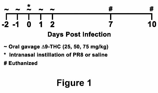
Experimental design. Mice, five per treatment group, were administered 9-THC (25, 50, or 75
mg/kg) and/or vehicle (corn oil) by oral gavage for five consecutive days (Figure 1). On the third
day of treatment, mice were instilled intranasally with influenza virus four hours prior to 9-THC
treatment. Mice were killed at 7 and 10 dpi. Day 7 post-infection was previously established as
the transition day between neutrophils and lymphocytes infiltrating the airways in response to
PR8 infection and the peak day for the detection of immune mediators released by leukocytes in
the airways. Day 10 post-infection was previously established as the day that marked the
transition from epithelial cell death (7 dpi) to epithelial regeneration and the onset of mucous cell
metaplasia (Buchweitz et al., 2007). Accordingly, two separate experiments were conducted for
the measurements of mRNA and BALF at 7 dpi and a third experiment was conducted for
routine histopathology and immunohistochemistry at both 7 and 10 dpi. Experimental values
obtained for the 9-THC control group administered 75 mg/kg 9-THC and intranasally instilled
with saline were removed from graphs and tables so as to not draw unwarranted comparisons
between this control and PR8 co-treatment groups receiving different dosage levels of 9-THC.
JPET #124719
7
Also, as would be anticipated, values for the 75 mg/kg 9-THC control were not different than
the corn oil control.
"9-tetrahydrocannabinol. 9-THC was provided by the National Institute on Drug Abuse
(Bethesda, MD) as a resin, which was solubilized in corn oil.
Influenza A/PR/8/34 instillation. Influenza A/PR/8/34 (PR8) was generously supplied by the
laboratory of Dr. Alan Harmsen (Montana State University, MT). Mice were anesthetized with
4% isoflurane in oxygen, and 50 µl of PR8 in pyrogen-free saline was instilled as 25 µl per nare
at a total dose of 50 plaque forming units (pfu).
Necropsy, lavage collection, and tissue preparation. Mice were anesthetized by an
intraperitoneal injection of 0.1 ml of 12% pentobarbital solution, a midline laparotomy was
performed, and mice were exsanguinated by cutting the abdominal aorta. Immediately after
death, the trachea was exposed and then cannulated, the heart and lung were excised en bloc. In
the first experiment, lung lobes were immersed in TRI reagent and stored at –80oC until RNA
isolation. In the second experiment, 1 ml of sterile saline was instilled through the tracheal
cannula and withdrawn to recover bronchoalveolar lavage fluid (BALF). A second saline lavage
was performed and combined with the first. In the third experiment, the left and right lung lobes
were inflated under constant pressure (30 cm H2O) with 10% neutral buffered formalin (Sigma
Chemical Co., St. Louis, MO) for 1 h. The tracheal airway was ligated and the inflated lobes
were stored in a large volume of the same fixative for at least 24 h until further processing for
histopathology.
RNA isolation. Total RNA was isolated from the lung lobes by using the TRI-reagent method
(Sigma Chemical, St Louis, MO). The evaluation of the relative expression levels of H1 and
JPET #124719
8
MUC5AC mRNA were determined using the TaqMan real-time multiplex RT-PCR with custom
designed TaqMan primers and probe to the target gene and the manufacturer’s pre-developed
primers and probe to 18S (Applied Biosystems, Foster City, CA). Measurements of caspase-3
(CAS-3) were determined similarly by using the manufacturer's pre-developed primers and probe
for CAS-3. The primers and probe to both the target gene and endogenous reference gene were
specifically designed to exclude detection of genomic DNA. In brief, aliquots of isolated tissue
RNA (1 µg total RNA) were converted to cDNA using random primers (Invitrogen, Carlsbad,
CA). The resultant cDNA (2µl) was added to a reaction mixture that consisted of the target gene
primers and probe, endogenous reference primers and probe (18S ribosomal RNA), and Taqman
universal master mix. Following PCR, amplification plots (change in dye fluorescence versus
cycle number) were examined, and a dye fluorescence threshold within the exponential phase of
the reaction was set separately for the target gene and the endogenous reference (18S). The cycle
number at which each amplified product crosses the set threshold represents the CT value. The
amount of target gene normalized to its endogenous reference was calculated by subtracting the
endogenous reference CT from the target gene CT ("CT). Relative mRNA expression was
calculated by subtracting the mean "CT of the control samples from the "CT of the treated
samples (""CT). The amount of target mRNA, normalized to the endogenous reference and
relative to the calibrator (i.e., RNA from control) is calculated by using the formula 2–""CT.
Total protein. Total protein was quantified in BALF using the bicinchoninic acid (BCA) method
as provided by the manufacturer (Pierce, Rockford, IL). In brief, BALF samples were
centrifuged at 600 rpm for 5 min to pellet cellular debris. Supernatants were removed and stored
at -20°C until testing was performed. 25 µl of BALF supernatant or bovine serum albumin
protein standard (25-2000 µg/ml) was incubated in a 96-well microplate with 200 µl of a 50:1
JPET #124719
9
mixture of reagents A (sodium carbonate, sodium bicarbonate, bicinchoninic acid and sodium
tartrate in 0.1M sodium hydroxide) and B (4% cupric sulfate) for 30 minutes at 37°C. Samples
were read on a Biotek microplate reader at 562 nm.
Bronchoalveolar lavage cellularity. The total number of leukocytes in BALF was counted with
a hemacytometer. In brief, 10 µl of BALF supernatant was added to a hemacytometer and the
number of leukocytes in the four etched corner quadrants were counted and multiplied by 2500
to yield the number of leukocytes per ml of sample. The percent of total leukocytes consisting of
eosinophils, lymphocytes, macrophages, and neutrophils were determined from counts of 200
cells in a cytospin sample stained with Diff-Quick (Dade Behring, Newark, DE). The percentage
of eosinophils, lymphocytes, macrophages, and neutrophils were multiplied by the total number
of leukocytes determined from the hemacytometer to yield the respective number of each cell
type per ml of sample.
Secreted inflammatory cytokines. BALF samples were centrifuged at 600 rpm for 5 min to
pellet cellular debris. Supernatants were removed and stored at -20°C until testing was
performed. The cytokines IL-6, IL-10, MCP-1, IFN-!, TNF-#, and IL-12p70 were detected
simultaneously by using the cytometric bead array (CBA) mouse inflammation kit (BD
Pharmingen, San Diego, CA). In brief, 50 µl of BALF from each sample was incubated
individually with a mixture of capture beads and 50 µl of PE detection reagent consisting of PE-
conjugated anti-mouse IL-6, IL-10, MCP-1, IFN-!, TNF-#, and IL-12p70. The samples were
incubated at room temperature for 2 h in the dark. After incubation, samples were washed once
and resuspended in 300 µl of wash buffer before acquisition on a BD FACSCalibur flow
cytometer. Data were analyzed using CBA software (BD Pharmingen, San Diego, CA). Standard
JPET #124719
10
curves were generated for each cytokine using the mixed cytokine standard provided with the kit.
The concentration of each cytokine was determined by interpolation from the corresponding
standard curve. The range of detection was 20 – 5000 pg/ml for each cytokine measured.
Analysis of BALF associated T cells by flow cytometry. After enumerating the retrieved cells
in BALF, the cells were pelleted by centrifugation and reconstituted in 150 µl flow cytometer
(FCM) buffer (PBS supplemented with 2% (w/v) bovine serum albumin and 0.09% (w/v) sodium
azide) with purified anti-mouse CD16/CD32 (Fc! III/II Receptor) antibody (BD Biosciences,
San Jose, CA) to block for 30 min. The samples were then split into two groups of equal volume,
washed and reconstituted in FCM buffer. One group received antibodies for CD3 (APC anti-
mouse CD3 ), CD4 [PE anti-mouse CD4(L3T4)] and CD8 [FITC anti-mouse CD8a(Ly-2)],
while the other group received the cognate isotype control antibodies. Samples were allowed to
incubate 1 h at 4oC, washed twice and then fixed for 10 min with Cytofix. Samples were then
washed again and reconstituted to a volume of 300 µl for analysis on the BD FACSCalibur flow
cytometer. The total number of events taken per each sample was 10,000.
Microdisssection and routine histopathology. The intrapulmonary airways of the fixed left
lung lobe from each rodent were microdissected according to a modified version of the technique
of Plopper et al., which is fully described in a previous publication (Harkema and Hotchkiss,
1992). Beginning at the lobar bronchus, airways are split down the long axis of the main axial
airway through the twelfth airway generation. Two transverse tissue blocks were excised at the
level of the fifth (proximal) and eleventh (distal) airway generation. Transverse tissue blocks
were also excised from the middle of the four right lung lobes perpendicular to the largest airway
branch entering each lobe. Tissue blocks from the left and right lung lobes were embedded in
JPET #124719
11
paraffin, sectioned at a thickness of 5 µm, and then stained with hematoxylin and eosin (H&E)
for light microscopic examination. Other paraffin sections were stained with Alcian Blue (pH
2.5)/Hematoxylin (AB/H) to identify acidic intraepithelial mucosubstances.
Immunocytochemistry. Hydrated paraffin sections (5 µm thick) from formalin-fixed lung
tissues were treated with 0.05% proteinase K for 2 min and washed with 1 N HCI for 1 h.
Sections were then treated with 3% H202 (in methanol) to block endogenous peroxide and then
incubated with a polyclonal antibody to CAS-3 (Abcam, Cambridge, MA) at 20 µg/ml for 1 h.
Immunoreactive CAS-3 was visualized with the Vectastain Elite ABC kit (Vectastain
Laboratories Inc., Burlingame, CA) using 3`,3`-diaminobenzidine (DAB) tetrahydrochloride
(Sigma Chemical Co., St. Louis, MO) as a chromagen.
Histopathology scores for inflammation. A histopathologic score was established based upon
the numbers and distribution of inflammatory cells within the tissues, as well as non-
inflammatory changes such as evidence of bronchiolar epithelial injury and repair. The scores
assigned were: 0 = no inflammation, 1 = mild, inflammatory cell infiltrate of the
perivascular/peribronchiolar compartment, 2 = moderate, inflammatory cell infiltrate of the
perivascular/peribronchiolar space with modest extension into the alveolar parenchyma, and 3 =
severe, inflammatory cell infiltrate of the perivascular/peribronchiolar space with a greater
magnitude of inflammatory foci found in the alveolar parenchyma. A certified pathologist scored
each lung section independently without prior knowledge of the treatment groups. A mean score
with standard error of the mean was calculated for each treatment group.
CAS-3 and alcian blue numeric cell densities. Slides of lung sections either stained
immunohistochemically for CAS-3 or stained for alcian blue (acidic mucosubstances) were
JPET #124719
12
examined. Numeric cell densities were determined for epithelial cells immunohistochemically
reactive to CAS-3 via light microscopy by counting the number of nuclear profiles of these
immunoreactive epithelial cells lining the bronchiolar epithelium at generation 5 and dividing by
the length of the underlying basal lamina. Numeric cell densities for CAS-3 were expressed as
the number of immunoreactive cells per mm basal lamina. In a similar manner, numeric cell
densities were determined for epithelial cells staining with alcian blue (acidic mucosubstances).
The numeric cell density of epithelial cells staining for alcian blue (acidic mucosubstances) was
expressed as the number of alcian blue reactive epithelial cells per mm basal lamina.
Statistical analysis. Data are expressed as mean ± standard error of the mean (SEM). Outliers
were identified and removed from sample sets using Grubb's test. The difference between saline
controls and mice treated with PR8 in the absence of 9-THC was evaluated by using a Student's
t-test. The differences between groups treated with PR8 in the presence or absence of 9-THC
were determined by one-way analysis of variance (ANOVA) and multiple comparisons by the
Student Newman Keuls post hoc test. For data that was not normally distributed a Kruskall
Wallis ANOVA on Ranks with Dunn’s post hoc test was utilized. Statistics were performed with
SigmaStat software version 2.03 from Jandel Scientific (San Rafael, CA). The criterion for
significance was taken to be p <0.05.
JPET #124719
13
Results
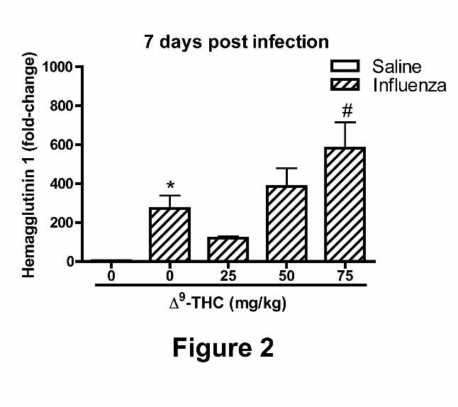
Expression of H1 in mouse lungs challenged with PR8. Hemagglutinin 1 is an influenza virus
surface protein that is a specific antigen target for B cell-derived immunoglobulin. H1 mRNA
levels in the lungs of mice challenged with influenza in the absence of 9-THC were
significantly elevated at 7 dpi as compared with the saline control (Figure 2). The H1 mRNA
levels in the lungs of mice treated with PR8 and 9-THC at 25 mg/kg was mildly attenuated as
compared with mice challenged with PR8 alone. Viral H1 mRNA levels increased with
increasing doses of 9-THC and were significantly elevated in mice administered 75 mg/kg 9-
THC as compared with PR8 challenged mice in the absence of 9-THC.
Measurements of injury, immune cell recruitment, and soluble chemical mediators in
bronchoalveolar lavage fluid (BALF). As a measure of alveolar/capillary membrane integrity,
total protein in BALF was assayed. There were marked increases in BALF-associated total
protein in the influenza group at 7 dpi as compared with saline control, but there were no
differences observed in the protein concentration recovered in the BALF from mice treated with
any dose of 9-THC (data not shown).
Consistent with an immune response to viral challenge, the total number of leukocytes in
BALF was significantly elevated in mice receiving only influenza at 7 dpi (Table 1). There were
apparent trends indicating dose-dependent effects of 9-THC on total cell counts. In spite of this
finding, there were marked decreases observed in the number of lymphocytes retrieved in BALF
from mice treated with 9-THC at all dose levels. In addition, treatment of mice with 9-THC at
doses of 25 mg/kg and 50 mg/kg led to decreases in the number of macrophages retrieved in
BALF. Flow cytometry was performed to evaluate differences in T lymphocyte subsets in
JPET #124719
14
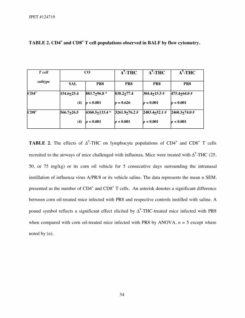
BALF. The absolute numbers of CD4+ and CD8+ T cells were determined (Table 2). 9-THC
treatment significantly decreased the number of CD8+ T cells at all dose levels with respect to
influenza alone; a similar effect on the number of CD4+ T cells was found in mice administered
50 and 75 mg/kg 9-THC when compared with the influenza group in the absence of 9-THC.
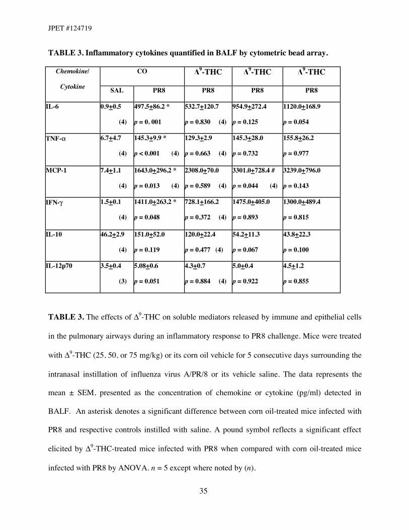
Secreted inflammatory chemokines and cytokines were measured as an indicator of
immune cell function in response to PR8 challenge. Mice challenged with PR8 had significantly
increased BALF concentrations of TNF-#, IFN-!, IL-6, MCP-1, and IL-10 at 7 dpi as compared
with the saline control (Table 3). In mice treated with 9-THC, an increase in MCP-1 at a dose of
50 mg/kg was observed. 9-THC treatment had no effect on BALF associated concentrations of
TNF-#, IFN-!, IL-6, IL-10 or IL-12p70 at any dose when compared with mice treated with PR8
alone.
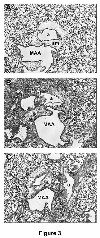
Descriptive pulmonary histopathology. Exposure of mice to the corn oil vehicle or 9-THC
alone did not result in significant histologic changes within the control mice (Figure 3A).
Infection of mice with influenza induced a significant cellular and inflammatory reaction 7 dpi in
all lung regions examined. The inflammatory infiltrate was centered upon the bronchiolo-
alveolar duct junction, and extended out into the surrounding alveolar parenchyma. The
inflammatory cells were a mix of predominantly lymphocytes and neutrophils, with fewer
macrophages and plasma cells. The lymphocytic and neutrophilic population often filled the
alveoli, and there was moderate alveolar interstitial infiltration with similar inflammatory cells.
The lymphocytes often formed well-organized perivascular and peribronchiolar aggregates.
Acute epithelial necrosis was present in small numbers of bronchioles, and the remaining
epithelial cells were moderately attenuated. At 10 dpi with influenza the inflammation was more
severe, and often obscured the alveolar parenchyma (Figure 3B). The inflammatory cells 10 dpi
JPET #124719
15
were primarily lymphocytes, with smaller numbers of neutrophils. The bronchiolar epithelium
10 dpi was moderately hyperplastic and hypertrophied, and there were scattered foci of alveolar
bronchiolarization (extension of bronchiolar epithelium into the adjacent alveolar spaces).
Treatment of influenza challenged mice with 9-THC resulted in no observed decreases in
inflammation 7 dpi at each 9-THC dose. At 10 dpi, treatment with 9-THC at all dose levels
resulted in a mild to moderate decrease in histologically apparent inflammation within the lungs
(Figure 3C). The inflammation within the mice was not uniformly distributed throughout all
lung regions, as found in the control PR8-infected mice. The decreases in inflammation included
both decreases in inflammatory cell numbers, as well as extent of distribution within the tissue.
The bronchiolar epithelial changes noted above were still present 10 dpi in influenza infected
mice treated with 9-THC.
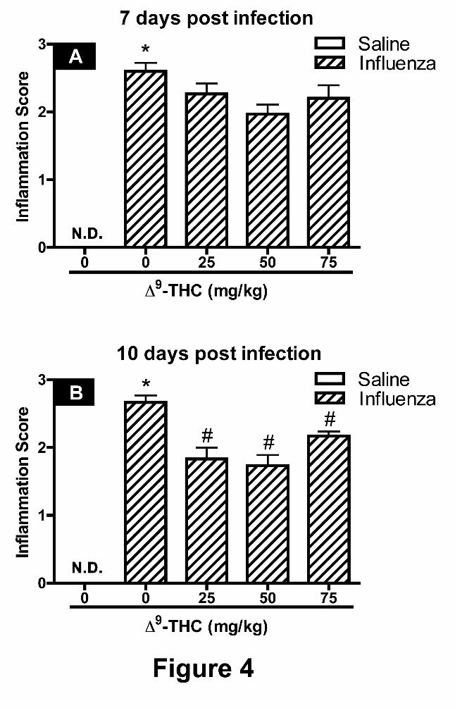
Inflammation scores from histopathology. The magnitude and severity of inflammation
observed in histological sections of lung isolated from the right and left lobes were
independently scored, as described under Methods, and compared between treatment groups at 7
dpi (Figures 4A) and 10 dpi (Figure 4B). There was no observed inflammation in the lungs of
mice intranasally instilled with saline. In contrast, a marked increase in the inflammation score
was noted for lungs in the influenza alone treatment group, representing a moderate to severe
inflammatory response. The inflammation scores for sections from mice challenged with PR8
and administered 9-THC were significantly attenuated at 10 dpi, representing mild to moderate
levels of inflammation.
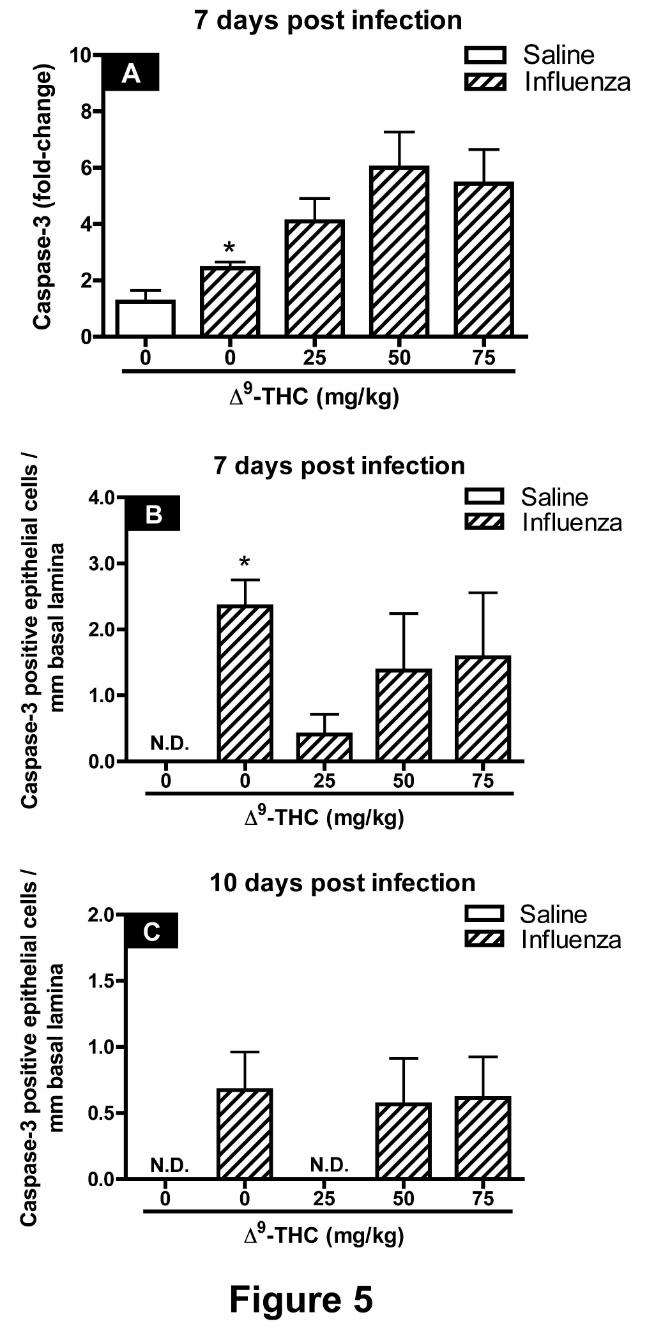
Caspase-3 mRNA expression levels and G5 main axial airway numeric cell densities. There
were markedly higher CAS-3 mRNA levels at 7 dpi in total lung homogenates from mice treated
JPET #124719
16
with influenza alone as compared with saline control suggesting an increase in apoptotic cell
death in response to PR8 infection (Figure 5A). There was also an apparent trend toward
increasing levels of CAS-3 mRNA with increasing doses of 9-THC; however, these increases
were not statistically significant. In addition, immunohistochemical staining for CAS-3 yielded
marked increases in the number of bronchiolar epithelial cells immunoreactive to CAS-3 with
influenza treatment alone at 7 dpi (Figures 5B). There was a decrease (p = 0.056) in CAS-3
immunoreactive epithelial cells at 7 dpi with 25 mg/kg 9-THC treatment (Figure 5B). There
were no significant differences observed in numeric cell densities for CAS-3 with any of the
treatments at 10 dpi (Figure 5C).
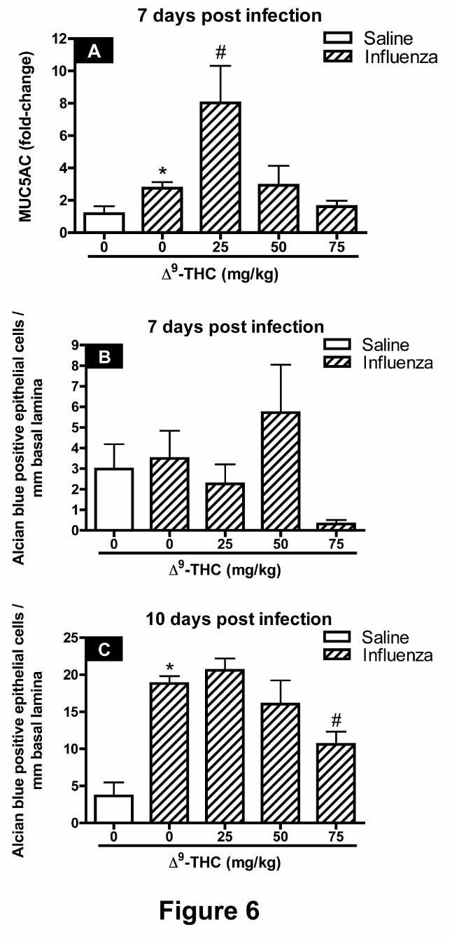
MUC5AC mRNA expression levels and G5 main axial airway numeric cell densities. The
levels of MUC5AC mRNA were increased in mice challenged with influenza alone (Figure 6A).
In mice challenged with PR8 and treated with 25 mg/kg 9-THC a four-fold increase in
MUC5AC mRNA levels was observed when compared with the influenza alone treatment group.
There were also marked increases in the number of epithelial cells staining for alcian blue (acidic
mucosubstances) with influenza treatment alone by 10 dpi but not at 7 dpi (Figures 7B and C).
9-THC treatment did not affect the number of alcian blue-stained epithelial cells observed along
the main axial airway at 7 dpi (Figure 6B), but did attenuate the number of alcian blue-positive
cells observed at 10 dpi in mice receiving 75 mg/kg 9-THC (Figure 6C) as compared with mice
challenged with PR8 alone.
JPET #124719
17
Discussion
The objective of the present investigation was to characterize the effects of 9-THC on
the intrapulmonary immune response and associated changes in the morphology of the
bronchiolar epithelium following challenge with a nonlethal dose of PR8. In the current study
we demonstrated that oral administration of 9-THC decreased immune competence as
evidenced by an increase in influenza viral load, as assessed by H1 mRNA levels, and decreased
lymphocytic and monocytic recruitment into the lungs. Oral administration was chosen since it
is a relevant route of 9-THC exposure, as in its synthetic therapeutic form, also known as
Marinol. More importantly, 9-THC is highly lipophilic requiring a nonaqueous diluent for
delivery, which in itself would have the potential for inducing irritation and damage to the
airways. In contrast, oral administration of 9-THC in corn oil, as employed in this study, is well
tolerated, albeit, poorly absorbed from the gastrointestinal tract resulting in modest blood
concentrations of 9-THC and its metabolites. Preliminary studies revealed that oral
administration of 75 mg/kg 9-THC for 5 consecutive days led to serum concentrations of 66.2
ng/ml of the parent compound (unpublished observation). These levels correlate well with a
previous report by Azorlosa and coworkers in 1992 (Azorlosa et al., 1992) in which human
plasma levels ranged from 57 to 268 ng/ml. In the current study, PR8 was administered by
intranasal instillation in saline on Day 3, with 9-THC co-administration surrounding the day of
infection. The rationale for this dosing paradigm was to investigate the effects of 9-THC
treatment on the immune response to PR8 during the early stages of infection.
9-THC-treated mice exhibited higher levels of lung-associated H1 viral mRNA than
corn oil-treated mice infected with PR8. In the current study, viral H1 mRNA was measured by
real-time PCR, which allowed rapid, sensitive and quantitative analysis of numerous tissue
samples while yielding results similar to those typically obtained with more conventional
JPET #124719
18
methods (Spackman et al., 2002; Jaspers et al., 2005). The increased H1 mRNA levels induced
by 9-THC treatment were dose-dependent albeit without an effect on mortality, suggesting that
9-THC administration impaired immune effectors involved in PR8 clearance. By 10 dpi H1
mRNA levels approached the level of detection (data not shown) in all groups, suggesting that
viral clearance had occurred. The timeline of viral clearance is consistent with uncomplicated
infections in humans (Hayden et al., 1998).
The effects of 9-THC on immune cell function were assessed by the measurement of
edema, immune cell recruitment, and cytokine production in response to PR8 challenge.
Epithelial cell death in response to PR8 infection begins as early as 3 dpi and continues through
7 dpi when the epithelium has begun to regenerate (Buchweitz et al., 2007). 9-THC had no
effect on BALF total protein, suggesting that 9-THC did not significantly affect the kinetics of
inflammatory mediators released by immune cells accruing in alveolar tissue that increase
vascular permeability and leakage at the alveolar/capillary interface. Likewise, there was no
difference in the total number of leukocytes retrieved in BALF between mice challenged with
PR8 in the presence or absence of 9-THC treatment. In spite of trends toward decreased total
leukocytes in BALF with 9-THC treatment, differential cell counts indicated that 9-THC
treatment decreased, in a dose-related manner, the number of macrophages and lymphocytes in
the airways. Dose-dependent modulation of immune cell recruitment to the lungs by 9-THC
during inflammation has been previously reported (Berdyshev et al., 1998). Lymphocytes
infiltrating the airways were comprised of CD4+ and CD8+ T cells. One of the most significant
findings in this study was that 9-THC administration markedly attenuated both CD4+ and CD8+
T cell counts in BALF. Given the marked lymphocytic and monocytic infiltration of the airway
submucosa, these data were considered in conjunction with histopathology, which suggests that
9-THC modulated the immune response to PR8 through an influence on leukocyte migration.
JPET #124719
19
These findings are consistent with cannabinoid effects on lymphocyte and monocyte chemotaxis
reported by others (Joseph et al., 2004; Sacerdote et al., 2005; Sacerdote et al., 2000; Stefano et
al., 1998).
As previously reported (Buchweitz et al., 2007), the immune response to PR8 challenge
included a marked increase in IL-6, TNF-#, IFN-!, MCP-1 and IL-10 by 7 dpi in BALF.
Collectively, no clear profile of activity emerged concerning the effects of 9-THC on PR8
cytokine or chemokine levels in BALF. This is not altogether surprising as each cytokine and
chemokine is regulated in its own unique manner with its own distinct kinetics. In addition,
molecular mechanisms by which certain cytokines and chemokines are modulated by
cannabinoids are only partially understood. In spite of this, several inflammatory mediators
evaluated in this study exhibited dose-dependent modulation in response to 9-THC treatment.
Specifically, mice treated with 9-THC at 50 and 75 mg/kg led to increased levels of MCP-1 and
IL-6 in BALF, whereas IL-10 concentrations were decreased at these doses. The modulation of
these cytokines by cannabinoid treatment is consistent with previous findings in other
experimental models (Molina-Holgado et al., 1998; Sacerdote et al., 2005).
Histopathology revealed that 9-THC, at all dose levels, attenuated the magnitude of
inflammation at 10 dpi. The inflammatory response in the lungs of mice challenged with
influenza alone extended beyond the perivascular/peribronchiolar submucosal compartment and
into the alveolar parenchyma. With 9-THC treatment, the inflammatory response was centered
on the submucosal compartment. The histopathology supports our finding of decreased numbers
of macrophages and lymphocytes observed in BALF from 9-THC treated mice. Since clearance
of influenza virus is critically dependent on both CD4+ and CD8+ T cells, a decrease in their
numbers is consistent with the increase in H1 mRNA observed with 9-THC treatment.
JPET #124719
20
Examination of epithelial cell changes in airways showed that PR8 treatment alone
induced CAS-3, a well-known marker for committed activation of apoptosis (Fischer et al.,
2003), as evidenced by mRNA levels and by tissue staining. Since apoptosis can be initiated
either directly by the virus in infected host cells (Takizawa et al., 1999; Wurzer et al., 2003), or
by effector cells, specifically cytotoxic T cells and/or NK cells, it is presently unclear which
mechanism(s) are predominately responsible for the observed increase in CAS-3. Interestingly, a
dose-related decrease in CAS-3 tissue staining, but not mRNA levels was observed with 9-THC
treatment suggesting that 9-THC may in part interfere with PR8-induced translation of CAS-3
mRNA or at the level of protein synthesis.
Following bronchiolar epithelial shedding at 3 and 7 dpi in response to PR8 infection and
concomitant with the cell-mediated immune response, the regenerative airway epithelium
undergoes metaplastic changes resulting in increased numbers of mucus goblet cells at 10 dpi
(Buchweitz et al., 2007). MCM is an adaptive response of the epithelium brought about by
soluble mediators of inflammation (Jamil et al., 1997; Dabbagh et al., 1999; Shim et al., 2001;
Justice et al., 2002; Kawano et al., 2002; Reader et al., 2003). An early indicator of increased
mucin production, and possibly MCM, is the expression of MUC5AC mRNA that encodes for
the goblet cell-derived mucin MUC5AC. Corn oil-treated mice challenged with PR8 exhibited an
increase in both the levels of MUC5AC mRNA and alcian blue staining, as shown previously
(Buchweitz et al., 2007). Interestingly, there was a correlative trend in the dose-dependent effects
of 9-THC treatment on MUC5AC mRNA at 7 dpi and alcian blue staining at 10 dpi, suggesting
that the effects of 9-THC on MUC5AC occur at the level of gene transcription. It is unclear
whether 9-THC treatment can directly interfere with the up-regulation of MUC5AC gene
transcription or whether the effect is mediated indirectly through the suppression of the
inflammatory response that induces MUC5AC transcription.
JPET #124719
21
In conclusion, 9-THC administration modulated the host-immune response to PR8 as
evidenced by an increased viral load and a decreased magnitude of macrophage and lymphocyte
(CD4+ and CD8+) recruitment. These findings were supported by histopathology and are
consistent with similar studies which demonstrated increased susceptibility to the respiratory
pathogen, Legionella pneumophila, after 9-THC administration (Klein et al., 2000). As in the
present study with PR8, Klein and coworkers similarly showed that 9-THC affected T cell and
macrophage function as suggested by altered regulation of T cell-derived cytokines and a
suppression of T cell activation to Legionella by attenuation of the expression of co-stimulatory
and polarizing molecules on the antigen presenting dendritic cells (Klein et al., 2000; Lu et al.,
2006). Further studies will need to be conducted to determine the effects of 9-THC on the later
stages of the immune response to PR8, as well as to ascertain the role of cannabinoid receptors,
CB1 and CB2, on the 9-THC-mediated effects observed.
JPET #124719
22
Acknowledgments: We would like to extend our thanks to Dr. James Klaunig at Indiana
University-Purdue University Indianapolis (IUPUI) for providing analytical support in the
measurement of 9-THC and its metabolites. We also thank Robert Crawford and Lori Bramble
for providing technical assistance with flow cytometry, and Kim Hambleton for her clerical
support in the submission of this manuscript.
JPET #124719
23
References:
Azorlosa JL, Heishman SJ, Stitzer ML and Mahaffey JM (1992) Marijuana smoking: effect of
varying delta 9-tetrahydrocannabinol content and number of puffs. J Pharmacol Exp Ther
261:114-122.
Berdyshev E, Boichot E, Corbel M, Germain N and Lagente V (1998) Effects of cannabinoid
receptor ligands on LPS-induced pulmonary inflammation in mice. Life Sci 63:PL125-
129.
Berdyshev EV (2000) Cannabinoid receptors and the regulation of immune response. Chem Phys
Lipids 108:169-190.
Brown DM, Dilzer AM, Meents DL and Swain SL (2006) CD4 T cell-mediated protection from
lethal influenza: perforin and antibody-mediated mechanisms give a one-two punch. J
Immunol 177:2888-2898.
Brown DM, Roman E and Swain SL (2004) CD4 T cell responses to influenza infection. Semin
Immunol 16:171-177.
Buchweitz JP, Harkema JR and Kaminski NE (2007) Time-dependent airway epithelial and
inflammatory cell responses induced by influenza virus A/PR/8/34 in C57BL/6 mice. Tox
Path 35:424-435
JPET #124719
24
Cabral GA, Lockmuller JC and Mishkin EM (1986) Delta 9-tetrahydrocannabinol decreases
alpha/beta interferon response to herpes simplex virus type 2 in the B6C3F1 mouse. Proc
Soc Exp Biol Med 181:305-311.
Dabbagh K, Takeyama K, Lee HM, Ueki IF, Lausier JA and Nadel JA (1999) IL-4 induces
mucin gene expression and goblet cell metaplasia in vitro and in vivo. J Immunol
162:6233-6237.
Dewey WL (1986) Cannabinoid pharmacology. Pharmacol Rev 38:151-178.
Fischer U, Janicke RU and Schulze-Osthoff K (2003) Many cuts to ruin: a comprehensive update
of caspase substrates. Cell Death Differ 10:76-100.
Harkema JR and Hotchkiss JA (1992) In vivo effects of endotoxin on intraepithelial
mucosubstances in rat pulmonary airways. Quantitative histochemistry. Am J Pathol
141:307-317.
Hayden FG, Fritz R, Lobo MC, Alvord W, Strober W and Straus SE (1998) Local and systemic
cytokine responses during experimental human influenza A virus infection. Relation to
symptom formation and host defense. J Clin Invest 101:643-649.
Jamil S, Breuer R and Christensen TG (1997) Abnormal mucous cell phenotype induced by
neutrophil elastase in hamster bronchi. Exp Lung Res 23:285-295.
JPET #124719
25
Jaspers I, Ciencewicki JM, Zhang W, Brighton LE, Carson JL, Beck MA and Madden MC
(2005) Diesel exhaust enhances influenza virus infections in respiratory epithelial cells.
Toxicol Sci 85:990-1002.
Joseph J, Niggemann B, Zaenker KS and Entschladen F (2004) Anandamide is an endogenous
inhibitor for the migration of tumor cells and T lymphocytes. Cancer Immunol
Immunother 53:723-728.
Justice JP, Crosby J, Borchers MT, Tomkinson A, Lee JJ and Lee NA (2002) CD4(+) T cell-
dependent airway mucus production occurs in response to IL-5 expression in lung. Am J
Physiol Lung Cell Mol Physiol 282:L1066-1074.
Kawano H, Haruta A, Tsuboi Y, Kim Y, Schachern PA, Paparella MM and Lin J (2002)
Induction of mucous cell metaplasia by tumor necrosis factor alpha in rat middle ear: the
pathological basis for mucin hyperproduction in mucoid otitis media. Ann Otol Rhinol
Laryngol 111:415-422.
Klein TW, Newton CA, Nakachi N and Friedman H (2000) Delta 9-tetrahydrocannabinol
treatment suppresses immunity and early IFN-gamma, IL-12, and IL-12 receptor beta 2
responses to Legionella pneumophila infection. J Immunol 164:6461-6466.
Lu T, Newton C, Perkins I, Friedman H and Klein TW (2006) Cannabinoid treatment suppresses
the T-helper cell-polarizing function of mouse dendritic cells stimulated with Legionella
pneumophila infection. J Pharmacol Exp Ther 319:269-276.
JPET #124719
26
Molina-Holgado F, Molina-Holgado E and Guaza C (1998) The endogenous cannabinoid
anandamide potentiates interleukin-6 production by astrocytes infected with Theiler's
murine encephalomyelitis virus by a receptor-mediated pathway. FEBS Lett 433:139-142.
Morahan PS, Klykken PC, Smith SH, Harris LS and Munson AE (1979) Effects of cannabinoids
on host resistance to Listeria monocytogenes and herpes simplex virus. Infect Immun
23:670-674.
Plopper CG, Mariassy AT and Lollini LO (1983) Structure as revealed by airway dissection. A
comparison of mammalian lungs. Am Rev Respir Dis 128:S4-7.
Reader JR, Hyde DM, Schelegle ES, Aldrich MC, Stoddard AM, McLane MP, Levitt RC and
Tepper JS (2003) Interleukin-9 induces mucous cell metaplasia independent of
inflammation. Am J Respir Cell Mol Biol 28:664-672.
Sacerdote P, Martucci C, Vaccani A, Bariselli F, Panerai AE, Colombo A, Parolaro D and Massi
P (2005) The nonpsychoactive component of marijuana cannabidiol modulates
chemotaxis and IL-10 and IL-12 production of murine macrophages both in vivo and in
vitro. J Neuroimmunol 159:97-105.
Sacerdote P, Massi P, Panerai AE and Parolaro D (2000) In vivo and in vitro treatment with the
synthetic cannabinoid CP55, 940 decreases the in vitro migration of macrophages in the
rat: involvement of both CB1 and CB2 receptors. J Neuroimmunol 109:155-163.
JPET #124719
27
Shim JJ, Dabbagh K, Ueki IF, Dao-Pick T, Burgel PR, Takeyama K, Tam DC and Nadel JA
(2001) IL-13 induces mucin production by stimulating epidermal growth factor receptors
and by activating neutrophils. Am J Physiol Lung Cell Mol Physiol 280:L134-140.
Spackman E, Senne DA, Myers TJ, Bulaga LL, Garber LP, Perdue ML, Lohman K, Daum LT
and Suarez DL (2002) Development of a real-time reverse transcriptase PCR assay for
type A influenza virus and the avian H5 and H7 hemagglutinin subtypes. J Clin
Microbiol 40:3256-3260.
Specter S, Lancz G, Westrich G and Friedman H (1991) Delta-9-tetrahydrocannabinol augments
murine retroviral induced immunosuppression and infection. Int J Immunopharmacol
13:411-417.
Stefano GB, Salzet M, Rialas CM, Mattocks D, Fimiani C and Bilfinger TV (1998) Macrophage
behavior associated with acute and chronic exposure to HIV GP120, morphine and
anandamide: endothelial implications. Int J Cardiol 64 Suppl 1:S3-13.
Swain SL, Agrewala JN, Brown DM, Jelley-Gibbs DM, Golech S, Huston G, Jones SC,
Kamperschroer C, Lee WH, McKinstry KK, Roman E, Strutt T and Weng NP (2006)
CD4+ T-cell memory: generation and multi-faceted roles for CD4+ T cells in protective
immunity to influenza. Immunol Rev 211:8-22.
JPET #124719
28
Takizawa T, Tatematsu C, Ohashi K and Nakanishi Y (1999) Recruitment of apoptotic cysteine
proteases (caspases) in influenza virus-induced cell death. Microbiol Immunol 43:245-
252.
Topham DJ, Tripp RA and Doherty PC (1997) CD8+ T cells clear influenza virus by perforin or
Fas-dependent processes. J Immunol 159:5197-5200.
Van Reeth K (2000) Cytokines in the pathogenesis of influenza. Vet Microbiol 74:109-116.
Wurzer WJ, Planz O, Ehrhardt C, Giner M, Silberzahn T, Pleschka S and Ludwig S (2003)
Caspase 3 activation is essential for efficient influenza virus propagation. Embo J
22:2717-2728.
JPET #124719
29
Footnotes:
1. Support: This work was supported by the MSU Foundation strategic partnership grant,
NIH grant DA07908, and the NIEHS training grant T32 ES07255.
JPET #124719
30
Legends for Figures
Figure 1. Experimental time course. Time course of 9-THC (~) administration in relationship
to influenza virus (*) challenge and times of sacrifice (#).
Figure 2. Pulmonary hemagluttinin 1 mRNA levels. Effects of "9-THC on hemagglutinin 1
mRNA levels after influenza challenge in whole lung homogenates at 7 dpi. Data represents the
mean ± SEM, presented as the fold-change in gene expression of hemagglutinin 1 normalized to
mice instilled with saline. An asterisk denotes a significant difference between corn oil-treated
mice infected with PR8 and respective controls instilled with saline. A pound symbol reflects a
significant effect elicited by "9-THC-treated mice infected with PR8 when compared with corn
oil-treated mice infected with PR8 by ANOVA.
Figure 3. Histopathology of inflammatory response to influenza challenge at generation 5.
Effects of "9-THC on the inflammatory response observed at generation 5 of the main axial
airway on day 10 post infection. Light photomicrographs were taken for lung sections at
generation 5 of the main axial airway (MAA) to include the bronchiolar artery (a) and alveolar
parenchyma (p). A = lung section from a mouse receiving corn oil gavage surrounding a saline
instillation. There was no evidence of inflammation in the perivascular/peribronchiolar
submucosal compartment (sm) or alveolar parenchyma. B = lung section from a mouse receiving
corn oil gavage surrounding an influenza instillation. There was severe inflammation of the
perivascular/peribronchiolar submucosal compartment with marked extension into the alveolar
JPET #124719
31
parenchyma. C = lung section from a mouse receiving "9-THC (75 mg/kg) gavage surrounding
an influenza instillation. There was severe inflammation of the perivascular/peribronchiolar
submucosal compartment with modest extension into the alveolar parenchyma. Bar = 100
microns.
Figure 4. Combined histopathology-based inflammation scores for the right and left lung
lobes. Effects of "9-THC on the inflammatory response gathered within the subepithelial
interstitium and alveolar parenchyma following influenza challenge. Inflammation scores were
recorded from lung sections taken on 7 dpi (A) and 10 dpi (B). Scores were tabulated and
averaged for sections of the 4 right lobes and 2 sections taken from generations 5 and 11 of the
left lung lobe. Scores: 0 = no inflammation, 1 = mild, inflammatory cell infiltrate of the
perivascular/peribronchiolar compartment, 2 = moderate, inflammatory cell infiltrate of the
perivascular/peribronchiolar space with modest extension into the alveolar parenchyma, and 3 =
severe, inflammatory cell infiltrate of the perivascular/peribronchiolar space with a greater
magnitude of inflammatory foci found in the alveolar parenchyma. Data represents the mean ±
SEM, presented as an inflammation score. An N.D. denotes that inflammation was not
detectable. An asterisk denotes a significant difference between corn oil-treated mice infected
with PR8 and respective controls instilled with saline. A pound symbol reflects a significant
effect elicited by "9-THC-treated mice infected with PR8 when compared with corn oil-treated
mice infected with PR8 by ANOVA.
Figure 5. Quantification of pulmonary apoptotic cell death by caspase-3 mRNA expression
and numeric cell densities of caspase-3 immunopositive cells. The effects of "9-THC on
JPET #124719
32
apoptotic cell death in response to influenza challenge were measured by the expression of CAS-
3 mRNA (A) and immunohistochemical staining of CAS-3 in the epithelium lining the main
axial airway at generation 5 of the left lung lobe at 7 dpi (B) and 10 dpi (C). In panel A data
represents the mean ± SEM, presented as the fold-change in gene expression of Caspase-3
normalized to mice instilled with saline. In panels B and C data represents the number of
Caspase-3 positive epithelial cells per mm of basal lamina. An N.D. denotes that inflammation
was not detectable. An asterisk denotes a significant difference between corn oil-treated mice
infected with PR8 and respective controls instilled with saline.
Figure 6. Quantification of bronchiolar epithelial mucous cell metaplasia by MUC5AC
mRNA levels and alcian blue numeric cell densities. The effects of "9-THC on the
development of mucous cell metaplasia in the epithelium lining the main axial airway following
influenza challenge and subsequent inflammatory cell responses. Expression levels of MUC5AC
mRNA (A) and generation 5 main axial airway numeric cell densities of alcian blue were
determined at 7 dpi (B) and 10 dpi (C). In panel A data represents the mean ± SEM, presented as
the fold-change in gene expression of MUC5AC normalized to mice instilled with saline. In
panels B and C data represents the number of alcian blue positive epithelial cells per mm of basal
lamina. An asterisk denotes a significant difference between corn oil-treated mice infected with
PR8 and respective controls instilled with saline. A pound symbol reflects a significant effect
elicited by "9-THC-treated mice infected with PR8 when compared with corn oil-treated mice
infected with PR8 by ANOVA.
JPET #124719
33
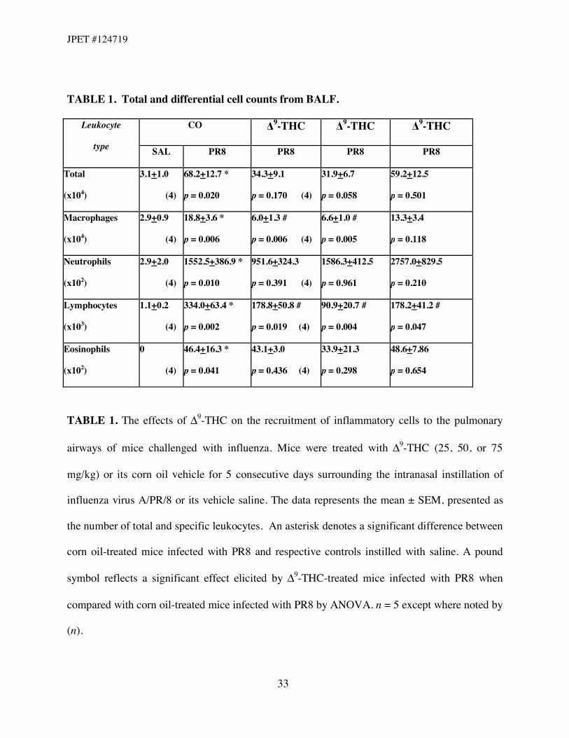
TABLE 1. Total and differential cell counts from BALF.
CO 9-THC 9-THC 9-THC Leukocyte
type SAL PR8 PR8 PR8 PR8
Total
(x104)
3.1+1.0
(4)
68.2+12.7 *
p = 0.020
34.3+9.1
p = 0.170 (4)
31.9+6.7
p = 0.058
59.2+12.5
p = 0.501
Macrophages
(x104)
2.9+0.9
(4)
18.8+3.6 *
p = 0.006
6.0+1.3 #
p = 0.006 (4)
6.6+1.0 #
p = 0.005
13.3+3.4
p = 0.118
Neutrophils
(x102)
2.9+2.0
(4)
1552.5+386.9 *
p = 0.010
951.6+324.3
p = 0.391 (4)
1586.3+412.5
p = 0.961
2757.0+829.5
p = 0.210
Lymphocytes
(x103)
1.1+0.2
(4)
334.0+63.4 *
p = 0.002
178.8+50.8 #
p = 0.019 (4)
90.9+20.7 #
p = 0.004
178.2+41.2 #
p = 0.047
Eosinophils
(x102)
0
(4)
46.4+16.3 *
p = 0.041
43.1+3.0
p = 0.436 (4)
33.9+21.3
p = 0.298
48.6+7.86
p = 0.654
TABLE 1. The effects of "9-THC on the recruitment of inflammatory cells to the pulmonary
airways of mice challenged with influenza. Mice were treated with "9-THC (25, 50, or 75
mg/kg) or its corn oil vehicle for 5 consecutive days surrounding the intranasal instillation of
influenza virus A/PR/8 or its vehicle saline. The data represents the mean ± SEM, presented as
the number of total and specific leukocytes. An asterisk denotes a significant difference between
corn oil-treated mice infected with PR8 and respective controls instilled with saline. A pound
symbol reflects a significant effect elicited by "9-THC-treated mice infected with PR8 when
compared with corn oil-treated mice infected with PR8 by ANOVA. n = 5 except where noted by
(n).
JPET #124719
34
TABLE 2. CD4+ and CD8+ T cell populations observed in BALF by flow cytometry.
CO 9-THC 9-THC 9-THC T cell
subtype SAL PR8 PR8 PR8 PR8
CD4+
154.6+25.4
(4)
883.7+96.8 *
p < 0.001
838.2+77.4
p = 0.626
304.4+15.5 #
p < 0.001
475.4+64.0 #
p < 0.001
CD8+
566.7+26.5
(4)
4360.5+133.4 *
p < 0.001
3261.9+76.2 #
p < 0.001
2483.4+52.1 #
p < 0.001
2460.3+74.0 #
p < 0.001
TABLE 2. The effects of "9-THC on lymphocyte populations of CD4+ and CD8+ T cells
recruited to the airways of mice challenged with influenza. Mice were treated with "9-THC (25,
50, or 75 mg/kg) or its corn oil vehicle for 5 consecutive days surrounding the intranasal
instillation of influenza virus A/PR/8 or its vehicle saline. The data represents the mean ± SEM,
presented as the number of CD4+ and CD8+ T cells. An asterisk denotes a significant difference
between corn oil-treated mice infected with PR8 and respective controls instilled with saline. A
pound symbol reflects a significant effect elicited by "9-THC-treated mice infected with PR8
when compared with corn oil-treated mice infected with PR8 by ANOVA. n = 5 except where
noted by (n).
JPET #124719
35
TABLE 3. Inflammatory cytokines quantified in BALF by cytometric bead array.
CO 9-THC 9-THC 9-THC Chemokine/
Cytokine SAL PR8 PR8 PR8 PR8
IL-6
0.9+0.5
(4)
497.5+86.2 *
p = 0. 001
532.7+120.7
p = 0.830 (4)
954.9+272.4
p = 0.125
1120.0+168.9
p = 0.054
TNF-#
6.7+4.7
(4)
145.3+9.9 *
p < 0.001 (4)
129.3+2.9
p = 0.663 (4)
145.3+28.0
p = 0.732
155.8+26.2
p = 0.977
MCP-1
7.4+1.1
(4)
1643.0+296.2 *
p = 0.013 (4)
2308.0+70.0
p = 0.589 (4)
3301.0+728.4 #
p = 0.044 (4)
3239.0+796.0
p = 0.143
IFN-!
1.5+0.1
(4)
1411.0+263.2 *
p = 0.048
728.1+166.2
p = 0.372 (4)
1475.0+405.0
p = 0.893
1300.0+489.4
p = 0.815
IL-10
46.2+2.9
(4)
151.0+52.0
p = 0.119
120.0+22.4
p = 0.477 (4)
54.2+11.3
p = 0.067
43.8+22.3
p = 0.100
IL-12p70
3.5+0.4
(3)
5.08+0.6
p = 0.051
4.3+0.7
p = 0.884 (4)
5.0+0.4
p = 0.922
4.5+1.2
p = 0.855
TABLE 3. The effects of "9-THC on soluble mediators released by immune and epithelial cells
in the pulmonary airways during an inflammatory response to PR8 challenge. Mice were treated
with "9-THC (25, 50, or 75 mg/kg) or its corn oil vehicle for 5 consecutive days surrounding the
intranasal instillation of influenza virus A/PR/8 or its vehicle saline. The data represents the
mean ± SEM, presented as the concentration of chemokine or cytokine (pg/ml) detected in
BALF. An asterisk denotes a significant difference between corn oil-treated mice infected with
PR8 and respective controls instilled with saline. A pound symbol reflects a significant effect
elicited by "9-THC-treated mice infected with PR8 when compared with corn oil-treated mice
infected with PR8 by ANOVA. n = 5 except where noted by (n).