Embed Size (px)
Citation preview
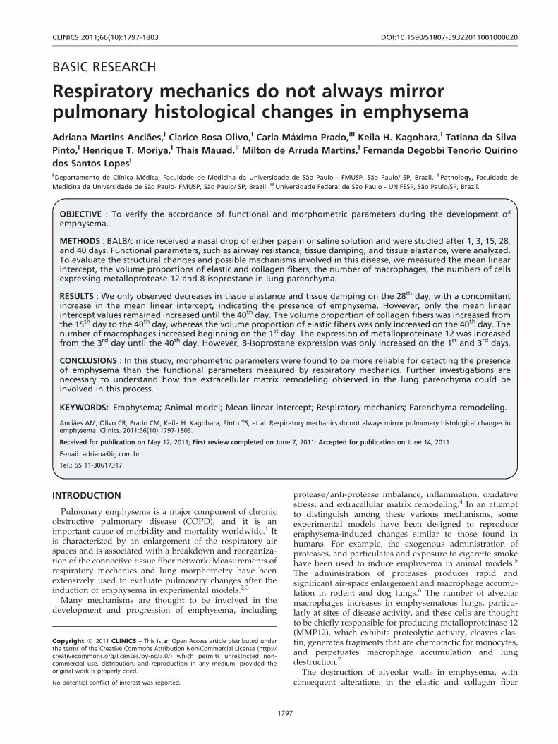
BASIC RESEARCH
Respiratory mechanics do not always mirrorpulmonary histological changes in emphysemaAdriana Martins Anciaes,I Clarice Rosa Olivo,I Carla Maximo Prado,III Keila H. Kagohara,I Tatiana da Silva
Pinto,I Henrique T. Moriya,I Thais Mauad,II Mılton de Arruda Martins,I Fernanda Degobbi Tenorio Quirino
dos Santos LopesI
I Departamento de Clınica Medica, Faculdade de Medicina da Universidade de Sao Paulo - FMUSP, Sao Paulo/ SP, Brazil. II Pathology, Faculdade de
Medicina da Universidade de Sao Paulo- FMUSP, Sao Paulo/ SP, Brazil. III Universidade Federal de Sao Paulo - UNIFESP, Sao Paulo/SP, Brazil.
OBJECTIVE : To verify the accordance of functional and morphometric parameters during the development ofemphysema.
METHODS : BALB/c mice received a nasal drop of either papain or saline solution and were studied after 1, 3, 15, 28,and 40 days. Functional parameters, such as airway resistance, tissue damping, and tissue elastance, were analyzed.To evaluate the structural changes and possible mechanisms involved in this disease, we measured the mean linearintercept, the volume proportions of elastic and collagen fibers, the number of macrophages, the numbers of cellsexpressing metalloprotease 12 and 8-isoprostane in lung parenchyma.
RESULTS : We only observed decreases in tissue elastance and tissue damping on the 28th day, with a concomitantincrease in the mean linear intercept, indicating the presence of emphysema. However, only the mean linearintercept values remained increased until the 40th day. The volume proportion of collagen fibers was increased fromthe 15th day to the 40th day, whereas the volume proportion of elastic fibers was only increased on the 40th day. Thenumber of macrophages increased beginning on the 1st day. The expression of metalloproteinase 12 was increasedfrom the 3rd day until the 40th day. However, 8-isoprostane expression was only increased on the 1st and 3rd days.
CONCLUSIONS : In this study, morphometric parameters were found to be more reliable for detecting the presenceof emphysema than the functional parameters measured by respiratory mechanics. Further investigations arenecessary to understand how the extracellular matrix remodeling observed in the lung parenchyma could beinvolved in this process.
KEYWORDS: Emphysema; Animal model; Mean linear intercept; Respiratory mechanics; Parenchyma remodeling.
Anciaes AM, Olivo CR, Prado CM, Keila H. Kagohara, Pinto TS, et al. Respiratory mechanics do not always mirror pulmonary histological changes inemphysema. Clinics. 2011;66(10):1797-1803.
Received for publication on May 12, 2011; First review completed on June 7, 2011; Accepted for publication on June 14, 2011
E-mail: [email protected]
Tel.: 55 11-30617317
INTRODUCTION
Pulmonary emphysema is a major component of chronicobstructive pulmonary disease (COPD), and it is animportant cause of morbidity and mortality worldwide.1 Itis characterized by an enlargement of the respiratory airspaces and is associated with a breakdown and reorganiza-tion of the connective tissue fiber network. Measurements ofrespiratory mechanics and lung morphometry have beenextensively used to evaluate pulmonary changes after theinduction of emphysema in experimental models.2,3
Many mechanisms are thought to be involved in thedevelopment and progression of emphysema, including
protease/anti-protease imbalance, inflammation, oxidativestress, and extracellular matrix remodeling.4 In an attemptto distinguish among these various mechanisms, someexperimental models have been designed to reproduceemphysema-induced changes similar to those found inhumans. For example, the exogenous administration ofproteases, and particulates and exposure to cigarette smokehave been used to induce emphysema in animal models.5
The administration of proteases produces rapid andsignificant air-space enlargement and macrophage accumu-lation in rodent and dog lungs.6 The number of alveolarmacrophages increases in emphysematous lungs, particu-larly at sites of disease activity, and these cells are thoughtto be chiefly responsible for producing metalloproteinase 12(MMP12), which exhibits proteolytic activity, cleaves elas-tin, generates fragments that are chemotactic for monocytes,and perpetuates macrophage accumulation and lungdestruction.7
The destruction of alveolar walls in emphysema, withconsequent alterations in the elastic and collagen fiber
Copyright � 2011 CLINICS – This is an Open Access article distributed underthe terms of the Creative Commons Attribution Non-Commercial License (http://creativecommons.org/licenses/by-nc/3.0/) which permits unrestricted non-commercial use, distribution, and reproduction in any medium, provided theoriginal work is properly cited.
No potential conflict of interest was reported.
CLINICS 2011;66(10):1797-1803 DOI:10.1590/S1807-59322011001000020
1797

networks, alters lung viscoelastic properties, which areevaluated by functional parameters, such as lung elastancemeasurements. Many studies use the constant-phase modelto describe the input impedance of the respiratory systemfrom data obtained by the forced oscillation technique (FOT)and to calculate tissue elastance (Htis), tissue damping(Gtis), and airway resistance (Raw). Raw includes contribu-tions from both the chest wall tissues and the pulmonaryairways; Gtis and Htis characterize the viscoelastic proper-ties of the respiratory tissues.8-10
In addition, lung morphometry has been extensively usedto verify pulmonary changes in animal models of emphy-sema, where it remains one of the most reliable techniquesfor evaluating alveolar destruction.11 An increase in themean linear intercept (Lm) correlates with alveolar enlarge-ment and parenchymal wall destruction in mice and ratswith lung emphysema.12 Moreover, many studies havefound an increase in the total number of collagen and elasticfibers, which indicates extracellular matrix remodeling inthis disease.3,13
However, the results obtained by these differentmethodologies are not always consistent because struc-tural changes follow different courses than functionalchanges in emphysema.14 In a previous study, we foundan increase in Lm and in the amount of elastic andcollagen fibers in the parenchyma two months afterpapain administration in mice,15 but no significantdifferences were detected in the animals’ respiratorymechanics. The distribution of emphysema throughoutthe lung is associated with the altered composition ofelastic and collagen fibers and might be important forexplaining these results.
To verify how respiratory mechanics and morphometryparameters change during the development of emphysemain mice, we examined whether changes in oscillatorymechanics were in accordance with changes in parenchymalstructures at different time intervals after protease admin-istration in mice.
MATERIALS AND METHODS
This study was approved by the Institutional ReviewBoard. Six- to eight-week-old male BALB/c mice were used.All the animals received care in compliance with the‘‘Principles of Laboratory Animal Care’’ published by theNational Institutes of Health.
Induction of emphysemaSeventy BALB/c mice (23-25 g) received a nasal admin-
istration of 50 mL of a 10-mg/mL papain solution (20 mg/kg, 6,000 UI/mg; Valdequimica, Sao Paulo, Brazil). Thisdose of papain has previously been shown to inducepulmonary emphysema in mice.12,15 The control micereceived 50 mL of 0.9% NaCl (saline), the vehicle used forthe papain.
Experimental groupsAfter the administration of papain (group P) or the same
amount of saline solution (group S), mice from each groupwere randomly assigned to five subgroups that corre-sponded to the days on which they were euthanized afternasal administration (P 1, 3, 15, 28, and 40 as well as S 1, 3,15, 28, and 40).
Assessment of respiratory mechanicsThe animals were deeply anesthetized by an intraper-
itoneal injection of thiopental (70 mg/kg), tracheostomizedand then connected to a ventilator for small animals(Flexivent, Scireq, Montreal) with a tidal volume of10 mL/kg (120 breaths/min) and a positive end expiratorypressure (PEEP) of 5 cm H2O. The experimental data fromthe forced oscillation technique were obtained only after theanimals had been paralyzed with pancuronium bromide(0.2 mg/kg). On the basis of a previously describedmodel,16 respiratory mechanics were characterized by thefollowing parameters: Raw, Gtis, and Htis.
Sample preparationAt the end of the respiratory mechanics evaluation, the
abdominal wall was opened, and the animals wereexsanguinated via the abdominal aorta. The thoracic cavitywas then opened, and the lungs were removed. Both lungswere fixed using 10% buffered formalin infused through thetrachea at a constant pressure of 20 cm H20 for 24 hours andwere then embedded in paraffin. Lung tissue sections(5 mm) were stained with H&E, Sirius red, or resorcin-fuchsin for lung structure analysis, collagen fiber evaluationor elastic fiber evaluation, respectively.
ImmunohistochemistryThe sections were deparaffinized and hydrated. After
blocking endogenous peroxidase activity, antigen retrievalwas performed with either high-temperature citrate buffer(pH = 6.0) or trypsin. The following primary antibodies wereused: goat polyclonal anti-mouse MMP12 (151,000, SantaCruz Biotechnology, CA, USA), anti-mouse macrophagemarker Mac-2 (1510,000, clone M3/38, Cedarlane, ON,Canada), and polyclonal goat anti-8-epi-PGF2a (151,200,Oxford Biomedical Research, Oxford, England). TheVectastain ABC Kit (Vector Laboratories, Burlingame, CA,USA) provided the secondary antibody, and 303-diamino-benzidine (DAB, Sigma, St. Louis, MO, USA) was used asthe chromogen. The sections were counterstained withHarris hematoxylin. For the negative control, the primaryantibody was omitted from the procedure, and BSA wasused instead.
MorphometryFor conventional morphometry, an eyepiece with a
coherent system of 50 lines, 100 points, and a known areaattached to the microscope ocular were used. The meanlinear intercept (Lm), an indicator of the mean alveolardiameter,17 was assessed in 20 non-overlapping fields oflung parenchyma per animal at 200x magnification. Thevolume proportion of collagen or elastic fibers in thealveolar tissue was determined by dividing the number ofpoints contacting the collagen or elastic fibers by the totalnumber of points contacting the alveolar septa.18 All of themeasurements were performed on 10 non-overlappingfields in each animal at 400x magnification.
The numbers of macrophages and MMP12-expressingcells in the alveolar parenchyma were also assessed bypoint-counting. Using the eyepiece (62,500 mm2 at 400xmagnification), the number of points in each field contactingalveolar tissue was counted. The alveolar tissue area in eachfield was calculated as the number of points contactingalveolar tissue as a proportion of the total grid area. The
Respiratory mechanicsAnciaes AM et al.
CLINICS 2011;66(10):1797-1803
1798

number of positive cells within the alveolar tissue area wascounted, and the results were expressed in cells/mm.2,19 Theexpression of 8-isoprostane was assessed using a digitalanalysis system and specific software (Image Pro Plus v. 4.5for Windows, Media Cybernetics, USA). Sections werestained with an 8-isoprostane antibody and captured usinga microscope (E200, Nikon, Japan) connected to a camera(Infinity 2-1 Monochrome CCD camera, Lumenera,Canada), and the images were fed into a computer. Theisoprostane-stained area (%) was expressed as the amountof isoprostane in a specific frame relative to the total area oftissue within that frame.
Statistical analysisAll of the data are expressed as means ¡ SD. Statistical
analyses were performed using SigmaStat software (SPSSInc., Chicago, IL). Student’s paired t-test was used at eachtime point (1, 3, 15, 28, and 40 days) to compare the papainand saline groups. A p-value of less than 0.05 wasconsidered to be significant.
RESULTS
Mechanics AssessmentThe mean (¡SD) values of Raw, Gtis, and Htis measured
at various times after papain administration are presentedin Figures 1A, 1B, and 1C, respectively. When comparing
the papain group (P) with the saline group (S) at each timepoint, an increase in tissue damping was observed on the 1st
day (p = 0.009), and decreases in tissue elastance and tissuedamping were only observed on the 28th day after papainadministration (p = 0.0012 and p = 0.03, respectively). Whenanalyzing the airway resistance values, no differences wereobserved at any time.
Lung MorphometryFigure 2A shows the mean Lm values (¡SD) for each
experimental group. Papain administration resulted insubstantial alveolar wall destruction, resulting in anenlargement of the distal air spaces. An increase in Lmwas observed on the 28th day and remained until the 40th
day (p#0.001 and p = 0.008, respectively).The volume proportions of collagen and elastic fibers in
the alveolar tissue are shown in Figures 2B and 2C,respectively. Papain administration resulted in a significantincrease in the proportion of collagen fibers in the alveolarwalls on the 15th (p = 0.003), 28th (p = 0.03), and 40th days(p = 0.012). On the 40th day, the mice that received papainalso exhibited an increased proportion of elastic fibers intheir alveolar tissue as compared to the mice that receivedsaline (p = 0.009) (Figure 2C).
Both the total number of macrophages (Figure 3A) andthe number of matrix metalloproteinase 12 (MMP12)-positive cells (Figure 3B) were increased in the mice that
Figure 1 - The Htis, Gtis, and Raw for the papain and saline groups at each time point are represented in Figures 1A, 1B, and 1C. (A) TheHtis results are separated into the 1st, 3rd, 15th, 28th, and 40th days. When comparing the groups that received intranasal saline to thepapain groups, a significant difference was found on the 28th day (* p = 0.012). (B) Increases in the Gtis values were observed on the 1st
(* p = 0.09) and 28th days (* p = 0.03) as compared with the groups that received the intranasal vehicle (saline). (C) No significantdifferences were found in the Raw values between the experimental groups over time. The values are expressed as means ¡ SD.
CLINICS 2011;66(10):1797-1803 Respiratory mechanicsAnciaes AM et al.
1799

received papain. Figure 3A shows the mean (¡SD)macrophage numbers for the parenchymas of the differentgroups. The number of macrophages was increased on the1st (p = 0.07), 3rd, 15th, 28th, and 40th days (p#0.001), whilethe number of MMP12-positive cells was increased from the3rd day (p = 0.034) until the 40th day (p#0.001).
Figure 3C shows the mean values (¡SD) of 8-isoprostaneexpression in the parenchyma. Increases in 8-isoprostaneexpression were only observed in the lung tissue of the micethat received papain on the 1st and 3rd days afteradministration (p = 0.04).
DISCUSSION
The current study shows that the parameters evaluated byrespiratory mechanics do not always mirror the changesdetected by morphometric analysis at various time intervalsafter papain administration in mice. A significant increase inthe mean linear intercept (Lm) associated with decreases intissue elastance and tissue damping was observed only onthe 28th day after papain administration. Additionally, at thesame time point, a morphometric evaluation revealed thatthe number of macrophages, the number of MMP12-expressing cells and the number of collagen fibers in thealveolar parenchyma were increased.
However, on the 40th day, we did not observe differencesin the respiratory mechanics parameters; however, all ofthe morphometric parameters, including the mean linear
intercept values, remained increased, indicating the pre-sence of lung emphysema.
When analyzing the volume proportions of elastic andcollagen fibers in the parenchyma, an increase in collagenfiber deposition was observed starting on the 15th day,whereas an increase in the number of elastic fibers was onlyobserved on the 40th day. Taken together, these resultssuggest that extracellular matrix remodeling, particularlythat of collagen and elastic fibers, most likely interferes withrespiratory mechanics. An increase in elastic force inducedby the deposition of collagen and elastic fibers may have theopposite effect on the elastic properties of pulmonary tissue,such as a decrease in alveolar surface area. This effect couldexplain the lack of a significant difference in tissue elastancewhen comparing the papain and saline groups at that time.
Although respiratory mechanics measurements have beenconsidered an important strategy for analyzing lungchanges in animal models of emphysema, some disparitiesexist between functional measurements and morphometricanalysis. In an earlier study, Foronjy et al.20 found nocorrelation between the emphysema measured by lungmorphometry and that measured by pulmonary compliancein A/J mice exposed to cigarette smoke. They observed thatthis murine smoke-induced model produced histologicalemphysema with no changes in pulmonary compliance. Inanother study, Guerassimov et al.21 analyzed various strainsof mice with differential susceptibilities to the developmentof smoking-induced emphysema and observed that changes
Figure 2 - (A) Mean linear intercept values measured for the experimental groups. Increases in Lm were observed on the 28th
(* p#0.001) and 40th days (* p = 0.008). The values are expressed as means ¡ SD. (B) Increases in the volume proportion of collagenfibers in the parenchyma were observed on the 15th (* p = 0.003), 28th (* p = 0.003) and 40th days (* p = 0.012). The values are expressedas means ¡ SD. (C) The volume proportion of elastic fibers in the parenchyma of the experimental groups was only increased on the40th day (* p = 0.009). The values are expressed as means ¡ SD.
Respiratory mechanicsAnciaes AM et al.
CLINICS 2011;66(10):1797-1803
1800

in Lm were not always mirrored by changes in lungmechanics after 6 months of smoke exposure. They believedthat to alter the mechanical characteristics of a lung, athreshold change in airspace enlargement was necessary.The strains with the greatest change in compliance(elastance) also showed the greatest increase in Lm.
Aside from alveolar destruction, studies have suggestedthat both the organization and the amount of elastic andcollagen fibers determine altered lung function in emphy-sema. There is evidence for the breakdown and resynthesisof matrix components during the development of experi-mental emphysema.
22,23,24 The manner in which theresynthesis of these matrix components interferes with lungfunction requires further investigation. Many morphometricand biomechanical studies in humans and animal modelshave suggested that the presence of collagen in emphyse-matous lungs is abnormal. In the present study, an increasein collagen deposition was observed on the 15th day,whereas an increase in the number of elastic fibers wasonly observed on the 40th day.
Four weeks after elastase administration, Kononov et al.25
observed significant remodeling in rat lungs, and thisremodeling led to thickened elastin and collagen fibers.Furthermore, Kononov et al. observed that, during stretch-ing, the newly deposited elastin and collagen fibers under-went substantially greater distortion than normal tissues.The threshold for the mechanical failure of collagen, whichprovides mechanical stability to the normal lung, is reduced
during stretching, which suggests that the mechanical forcesproduced during breathing are capable of causing thefailure of the remodeled extracellular matrix and contribut-ing to the progression of emphysema.
The majority of experimental studies of emphysema withprotease administration have only analyzed functional andmorphometric data after the 21st day of disease induction,and these studies have only found alterations in the tissueelastance and mean linear intercept.3,4,25 Thus far, to ourknowledge, no studies have evaluated these parameters atvarious time points after the induction of emphysema in ananimal model to examine how the elastance, mean linearintercept, and remodeling of extracellular matrix fiberschange during the development of emphysema.
Animal studies have been important in determining howvarious mechanisms are involved in the pathophysiology ofemphysema.6,26,27 Macrophages are the predominantdefense cells, both in normal individuals and COPDpatients. MMPs are mainly produced by these cells,27 andan increase in MMP activation in the lungs forms the basisof the protease/anti-protease imbalance hypothesis, whichis the prevailing mechanism used to explain the pathogen-esis of emphysema. Thus, we decided to verify the numberof macrophages and metalloproteinase 12 (MMP12) expres-sion in the lung parenchyma.
Corroborating the results of Shapiro et al.,6 our study showsthat a single administration of papain induces an increase inthe number of macrophages in the lung parenchyma,
Figure 3 - (A) The density of macrophages (cells immunostained for MAC-2) was increased on the 1st (* p = 0.007), 3rd, 15th, 28th, and 40th
days (p#0.005 for these groups). (B) MMP12-immunopositive cells in the lung alveolar tissue of the experimental groups, increaseswere observed on the 3rd (* p = 0.034), 15th, 28th, and 40th days (p#0.005 for these groups). The values are expressed as means ¡ SD. (C)Expression of 8-isoprostane measured in the different groups revealed increases on the 1st and 3rd days (* p #0.05 for both groups).
CLINICS 2011;66(10):1797-1803 Respiratory mechanicsAnciaes AM et al.
1801
beginning on the 1st day and remaining until the 40th day.MMP12 expression is increased on the 3rd day, and thisincrease is maintained until the 40th day. Such results supportthe idea that, in this experimental model, the development ofemphysema depends on a protease/anti-protease imbalance,starting at the beginning of disease progression.
Oxidative stress is another important mechanism thatexplains the development of emphysema, and there isevidence for increased lung tissue and systemic oxidativestress in COPD patients.28 Thus, 8-isoprostane expressionwas also evaluated. Isoprostanes are prostaglandin-likecompounds formed by the peroxidation of arachidonic acidand are considered to be accurate oxidative stress markersin vivo, both in humans and experimental animals.29 In aprevious study, we found a worsening of emphysemaconcomitant with an increase in 8-isoprostane expression inmice exposed to air pollution, which contrasted withemphysema in mice that were maintained in a chamberwithout air pollution.15 In this study, increases in 8-isoprostane expression were observed only on the 1st and3rd days, suggesting the presence of an oxidant/antioxidantbalance starting on the 3rd day. However, this hypothesisneeds to be investigated further.
Our study has some limitations. Although cigarettesmoking could have been used as a model to induceemphysema (because it more closely mimics the human
disease), the development of emphysema in such animalmodels is lengthy and may only lead to a mild case of thedisease.30 Therefore, increases in the numbers of collagenand elastic fibers in these models requires many months ofexposure to cigarette smoke. Thus, a protease model wasused to induce emphysema. The short time required fordisease development can be used to determine whichmeasurements of respiratory mechanics and lung morpho-metry accurately mirror changes in the lungs and todetermine which of these measurements best representsthe lung changes in this experimental model.
In conclusion, in this protease-induced model of emphy-sema by the administration of papain solution, morphometricparameters were found to be more reliable for detecting thepresence of emphysema as compared to functional para-meters measured by respiratory mechanics. The deposition ofcollagen and elastic fibers may interfere with the mechanicalproperties of the respiratory system and could explain theimpaired accuracy of the functional measurements.
ACKNOWLEDGEMENTS
The authors would like to thank Angela B. dos Santos and Maria Cristina
Medeiros for the assistance with immunohistochemical staining.
This study was supported by the Conselho Nacional de
Desenvolvimento Cientıfico e Tecnologico (CNPq) and the Laboratorios
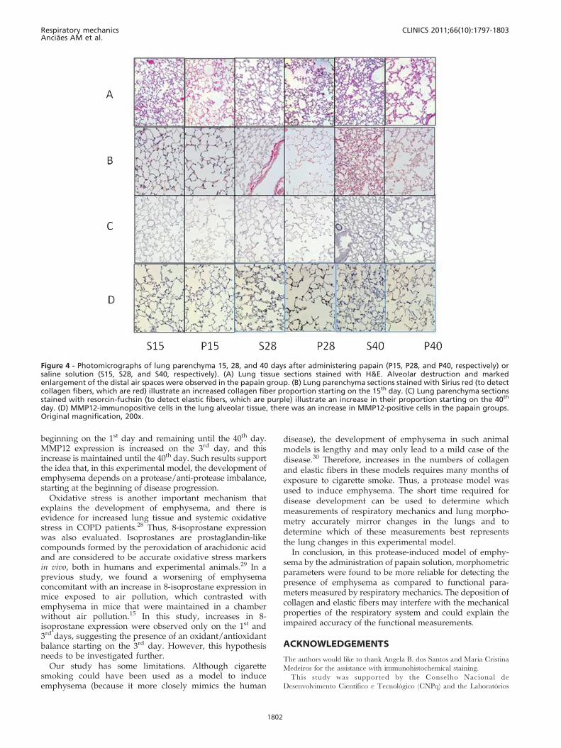
Figure 4 - Photomicrographs of lung parenchyma 15, 28, and 40 days after administering papain (P15, P28, and P40, respectively) orsaline solution (S15, S28, and S40, respectively). (A) Lung tissue sections stained with H&E. Alveolar destruction and markedenlargement of the distal air spaces were observed in the papain group. (B) Lung parenchyma sections stained with Sirius red (to detectcollagen fibers, which are red) illustrate an increased collagen fiber proportion starting on the 15th day. (C) Lung parenchyma sectionsstained with resorcin-fuchsin (to detect elastic fibers, which are purple) illustrate an increase in their proportion starting on the 40th
day. (D) MMP12-immunopositive cells in the lung alveolar tissue, there was an increase in MMP12-positive cells in the papain groups.Original magnification, 200x.
Respiratory mechanicsAnciaes AM et al.
CLINICS 2011;66(10):1797-1803
1802
de Investigacao Medica do Hospital das Clınicas da Faculdade de
Medicina da Universidade de Sao Paulo (LIM/HC).
REFERENCES
1. Global Initiative for Chronic Lung Disease, 2010. Global strategy for thediagnosis, management, and prevention of chronic obstructive pulmon-ary disease. Available: /http://www.goldcopd.org/S.
2. Takubo Y, Guerassimov A, Ghezzo H, Triantafillopoulos A, Bates JHT,Hoidal JR, et al. a1-Antitrypsin determines the pattern of emphysemaand function in tobacco smoke–exposed mice: parallels with humandisease. Am J Respir Crit Care Med. 2002;166:1596–603.
3. Ito S, Ingenito EP, Brewer KK, Black LD, Parameswaran H, Lutchen KR,et al. Mechanics, nonlinearity, and failure strength of lung tissue in amouse model of emphysema: possible role of collagen remodeling. J ApplPhysiol. 2005;98:503-11, doi: 10.1152/japplphysiol.00590.2004.
4. Suki B, Lutchen KR, Ingenito EP. On the progressive nature ofemphysema roles of proteases, inflammation, and mechanical forces.Am J Respir Crit Care Med. 2003;168:516–21.
5. March TH, Green FHY, Fletcher FH, Kinula KJ. Animal models ofemphysema and their relevance to studies of particle-induced disease.Inhalation Toxicology. 2000;12:155-87, doi: 10.1080/089583700750019558.
6. Gross P, Pfitzer EA, Tolker E, Babyak MA, Kaschak M. Experimentalemphysema. Its production with papain in normal and silicotic rats.Arch Environ Health. 1965;11:50–8.
7. Shapiro SD. Animal models for COPD. Chest. 2000;117:223S–227S.8, doi:10.1378/chest.117.5_suppl_1.223S.
8. Bates JHT, Abe T, Romero PV, Sato J. Measurement alveolar pressure inclose chest dogs during flow interruption. J Appl Physiol. 1989;67:488-92.
9. Gomes RFM, Shen X, Ramchandani R, Tepper RS, Bates JHT.Comparative respiratory system mechanics in rodents. J Appl Physiol.2000;89:908-916.
10. Faria AC, Costa AA, Lopes AJ, Jansen JM, Melo PL. Forced oscillationtechnique in the detection of smoking-induced respiratory alterations:diagnostic accuracy and comparison with spirometry. Clinics.2010;64:443-50.
11. Pastor LM, Sanchez-Gascon F, Girona JC, Bernal-Manas CM, Morales E,Beltran-Frutos E, et al. Morphogenesis of rat experimental pulmonaryemphysema induced by intratracheally administered papain: changes inelastic fibres. Histol. Histopathol. 2006;21:1309-19.
12. Flo C, Lopes FDTQS, Kasahara DI, Silva ACD, Jesus RCC, Rivero DHRF,et al. Effects of exercise training on papain-induced pulmonaryemphysema in Wistar rats. J Appl Physiol. 2006; 100:2815, doi: 10.1152/japplphysiol.00024.2005.
13. Shifren A, Mecham RP. The stumbling block in lung repair of emphysema:elastic fiber assembly. Proc Am Thorac Soc Vol. 2006;3428-433.
14. Bates JH, Davis GS, Majumdar A, Butnor KJ, Suki B. Linkingparenchymal disease progression to changes in lung mechanicalfunction by percolation. Am J Crit Care Med. 2007;176:617-23, doi: 10.1164/rccm.200611-1739OC.
15. Lopes FDTQS, Pinto TS, Costa FMA, Moriya HT, Biselli PJC, Ferraz LFS,et al. Exposure to ambient levels of particles emitted by traffic worsens
emphysema in mice. Envir Res. 2009;544-51, doi: 10.1016/j.envres.2009.03.002.
16. Hantos Z, Daroczy B, Suki B, Nagy S, Fredberg JJ. Input impedance andperipheral inhomogeneity of dog lungs. J Appl Physiol. 1992;72:168-78,doi: 10.1063/1.352153.
17. Margraf LR, Tomashefski JF, Bruce MC, Dahms BB. Morphometricanalysis of the lung in bronchopulmonary dysplasia. Am Rev Respir Dis.1991;143:391-400.
18. Lancas T, Kasahara DI, Prado CM, Tiberio IF, Martins MA, DolhnikoffM. Comparison of early and late responses to antigen of sensitizedguinea pig parenchymal lung strips. J Appl Physiol. 2006;100:1610-6, doi:10.1152/japplphysiol.00828.2005.
19. Simoes SM, Santos MA, Oliveira MS, Fontes ES, Fernezliant S, GarippotAL, et al. Inflammatory cell mapping of the respiratory tract in fatalasthma. Clin Exp Allergy. 2005;35:602-11.
20. Foronjy RF, Mercer BA, Maxfield MW, Powell CA, D’Armiento J, OkadaY. Structural emphysema does not correlate with lung compliance:lessons from the mouse smoke model. Experimental Lung Research.2005;31:547-62, doi: 10.1080/019021490951522.
21. Guerassimov A, Hoshino Y, Takubo Y, Turcotte A, Yamamoto M,Ghezzo H, et al. The development of emphysema in cigarette smoke-exposed mice is strain dependent. Am J Respir Crit Care Med.2004;170:974–80, doi: 10.1164/rccm.200309-1270OC.
22. Wright JL, Churg A. Smoke-induced emphysema in guinea pigs isassociated with morphometric evidence of collagen breakdown andrepair. Am J Physiol. 1995;268:17–20.
23. Finlay GA, O’Driscoll LR, Russell KJ, D’Arcy EM, Masterson JB, Fitz-Gerald MX, et al. Matrix metalloproteinase expression and production byalveolar macrophages in emphysema. Am J Respir Crit Care Med.1997;156:240–7.
24. Vlahovic G, Russell ML, Mercer RR, Crapo JD. Cellular and connectivetissue changes in alveolar septal walls in emphysema. Am J Respir CritCare Med. 1999;160:2086–92.
25. Kononov S, Brewer K, Sakai H, Cavalcante FSA, Sabayanagam CR,Ingenito EP, et al. Roles of mechanical forces and collagen failure in thedevelopment of elastase-induced emphysema. Am J Respir Crit CareMed. 2001;164:920–6.
26. Mahadeva R, Shapiro SD. Review Series: Chronic obstructive pulmonarydisease 3: Experimental animal models of pulmonary emphysema.Thorax. 2002;57:908-14, doi: 10.1136/thorax.57.10.908.
27. Shapiro SD. The macrophage in chronic obstructive: pulmonary disease.Am J Respir Crit Care Med. 1999;160:S29–S32.
28. Drost EM, Skwarsk KM, Sauleda J, Soler N, Roca J, Augusti A, et al.Oxidative stress and airway inflammation in severe exacerbations ofCOPD. Thorax. 2005;60:293-300, doi: 10.1136/thx.2004.027946.
29. Morrow JD, Roberts LJ. Their role as an index of oxidant stress status inhuman pulmonary disease. Am J Respir Crit Care Med. 2002;166:S25–S30, doi: 10.1164/rccm.2206011.
30. Churg A, Wright J. Animal models of cigarette smoke-induced chronicobstructive lung disease. Contrib Microbiol. 2007;14:113–25, doi: 10.1159/000107058.
CLINICS 2011;66(10):1797-1803 Respiratory mechanicsAnciaes AM et al.
1803