Embed Size (px)
Citation preview
Bacterial Leaf Scorch of Oak
Forest Health MonitoringA. B. Gould and J. H. Lashomb
April, 2008
Bacterial Leaf Scorch (BLS) Working Group Dr. Ann Gould, Plant Pathology Dr. James Lashomb, Entomology Dr. George Hamilton, Entomology Dr. Mark Vodak, Natural Resources Dr. Jason Grabosky, Natural Resources Jason Zhang, Entomology Halina Staniszewska, Plant Pathology
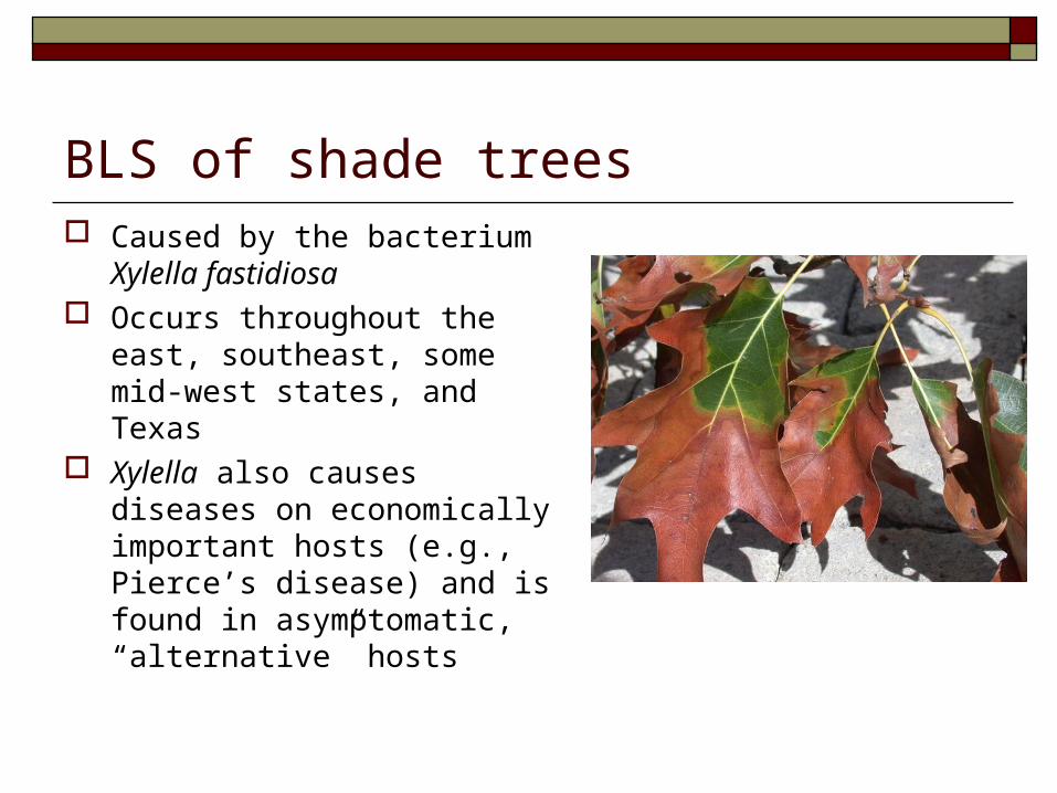
BLS of shade trees Caused by the bacterium
Xylella fastidiosa Occurs throughout the
east, southeast, some mid-west states, and Texas
Xylella also causes diseases on economically important hosts (e.g., Pierce’s disease) and is found in asymptomatic, “alternative” hosts
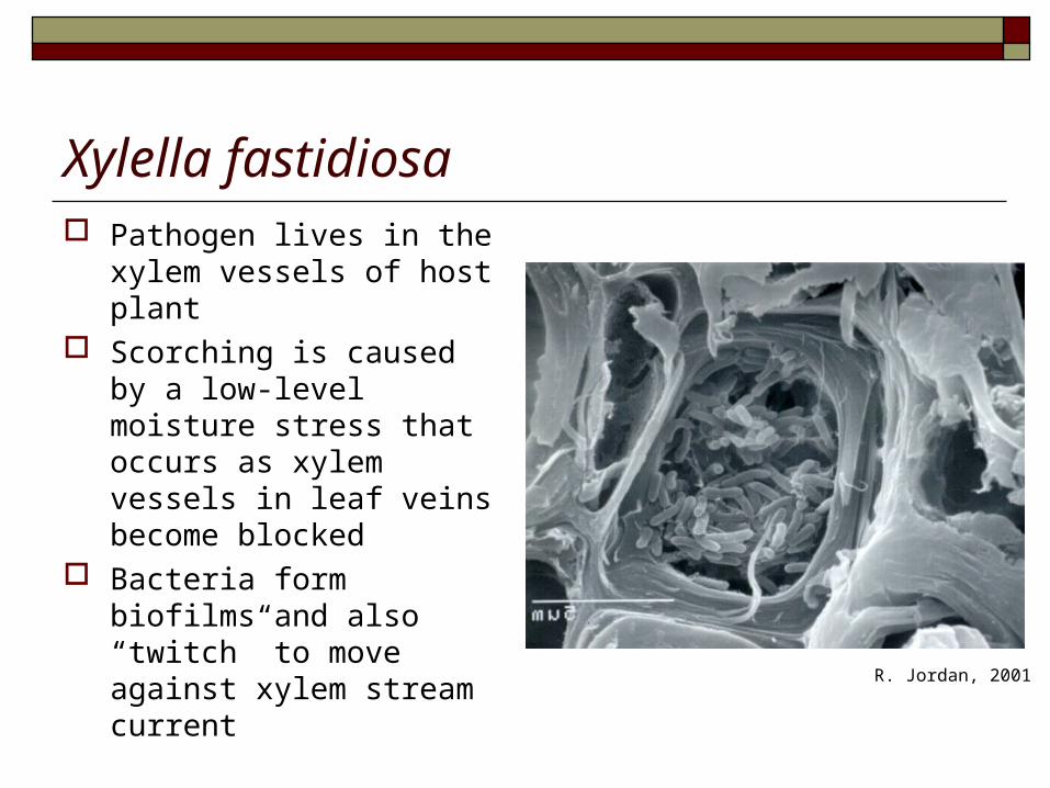
Xylella fastidiosa Pathogen lives in the
xylem vessels of host plant
Scorching is caused by a low-level moisture stress that occurs as xylem vessels in leaf veins become blocked
Bacteria form biofilms and also “twitch” to move against xylem stream current
R. Jordan, 2001
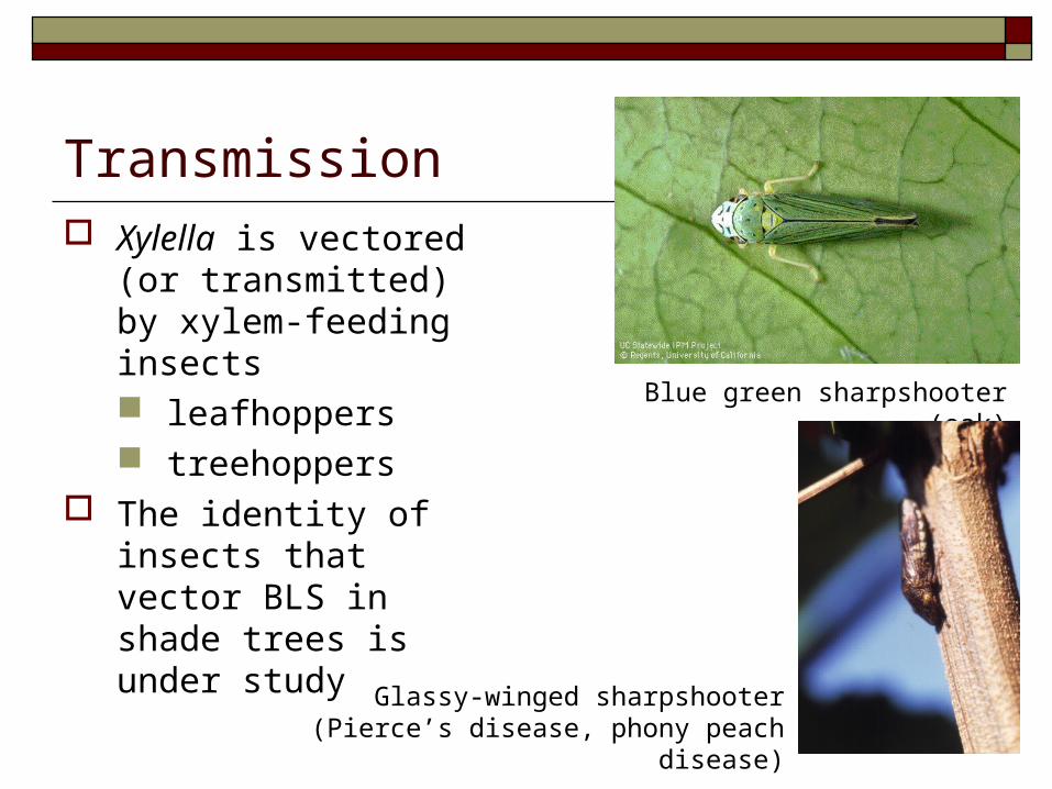
Transmission Xylella is vectored (or
transmitted) by xylem-feeding insects leafhoppers treehoppers
The identity of insects that vector BLS in shade trees is under study
Blue green sharpshooter (oak)
Glassy-winged sharpshooter (Pierce’s disease, phony peach disease)
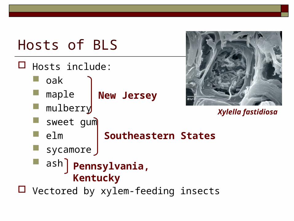
Hosts of BLS Hosts include:
oak maple mulberry sweet gum elm sycamore ash
Vectored by xylem-feeding insects
New Jersey
Southeastern States
Pennsylvania, Kentucky
Xylella fastidiosa
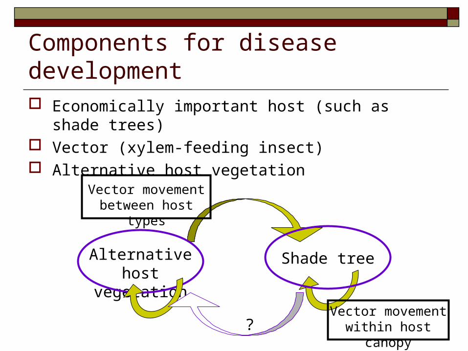
Components for disease development Economically important host (such as shade
trees) Vector (xylem-feeding insect) Alternative host vegetation
Alternative host
vegetation
Shade tree
Vector movement between host types
Vector movement within host canopy?
Bacterial Leaf Scorch of Shade Trees
Xylella fastidiosa
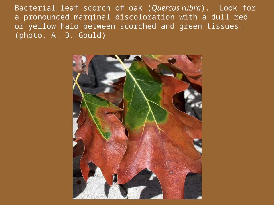
Bacterial leaf scorch of oak (Quercus rubra). Look for a pronounced marginal discoloration with a dull red or yellow halo between scorched and green tissues. (photo, A. B. Gould)
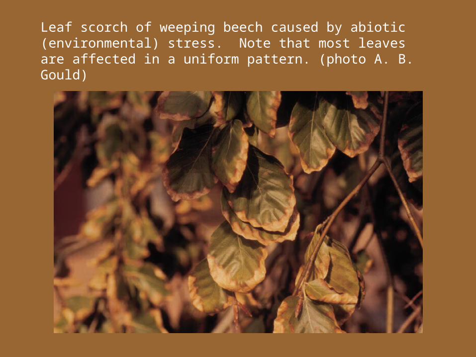
Leaf scorch of weeping beech caused by abiotic (environmental) stress. Note that most leaves are affected in a uniform pattern. (photo A. B. Gould)
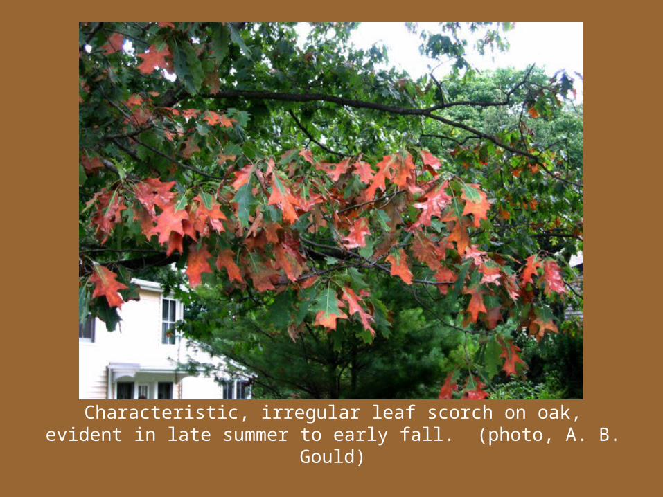
Characteristic, irregular leaf scorch on oak, evident in late summer to early fall. (photo, A. B. Gould)
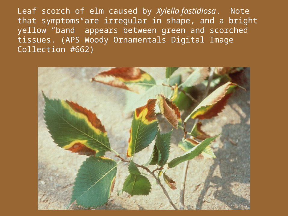
Leaf scorch of elm caused by Xylella fastidiosa. Note that symptoms are irregular in shape, and a bright yellow “band” appears between green and scorched tissues. (APS Woody Ornamentals Digital Image Collection #662)
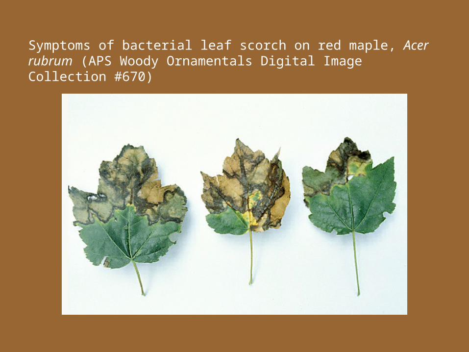
Symptoms of bacterial leaf scorch on red maple, Acer rubrum (APS Woody Ornamentals Digital Image Collection #670)
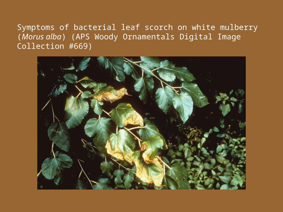
Symptoms of bacterial leaf scorch on white mulberry (Morus alba) (APS Woody Ornamentals Digital Image Collection #669)
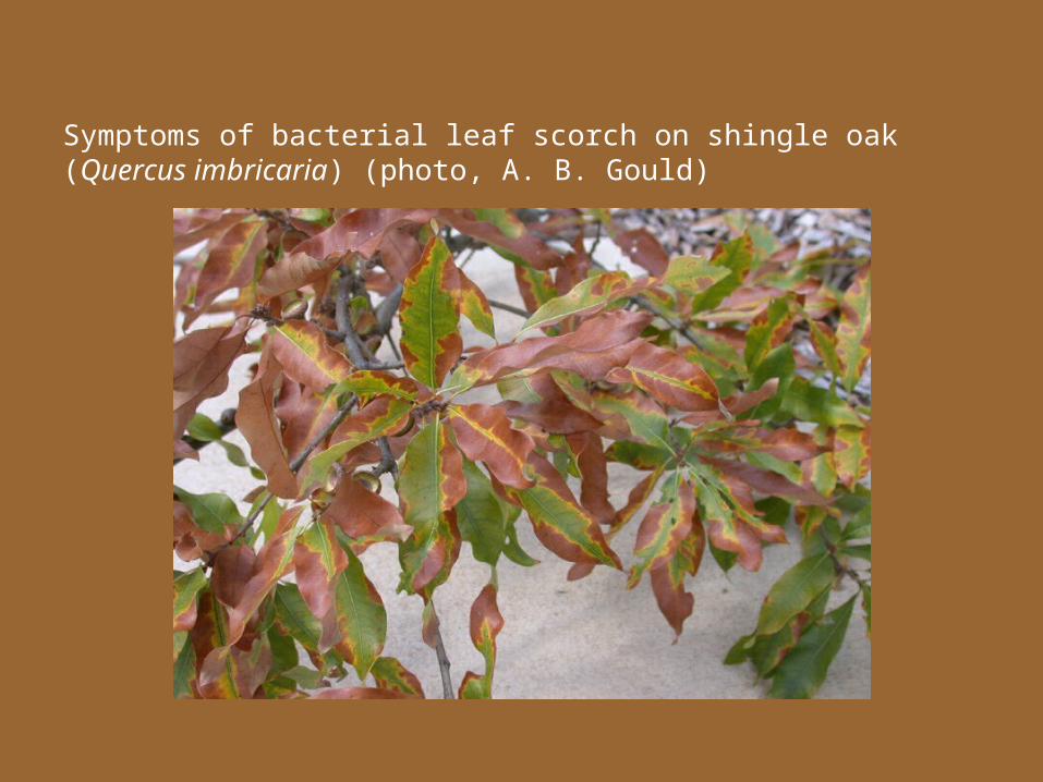
Symptoms of bacterial leaf scorch on shingle oak (Quercus imbricaria) (photo, A. B. Gould)
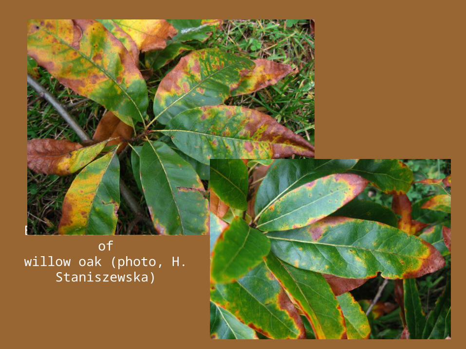
Bacterial leaf scorch ofwillow oak (photo, H.
Staniszewska)
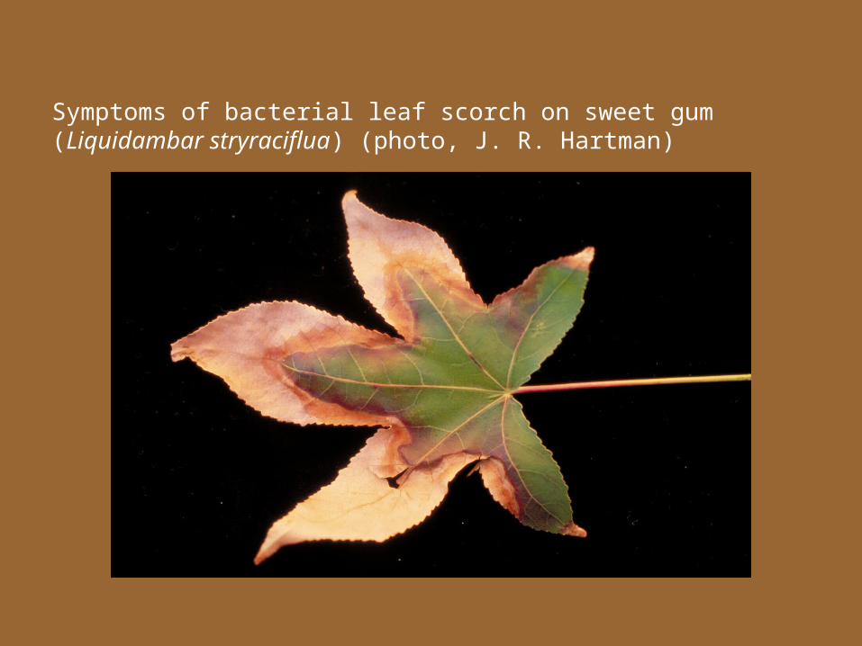
Symptoms of bacterial leaf scorch on sweet gum (Liquidambar stryraciflua) (photo, J. R. Hartman)
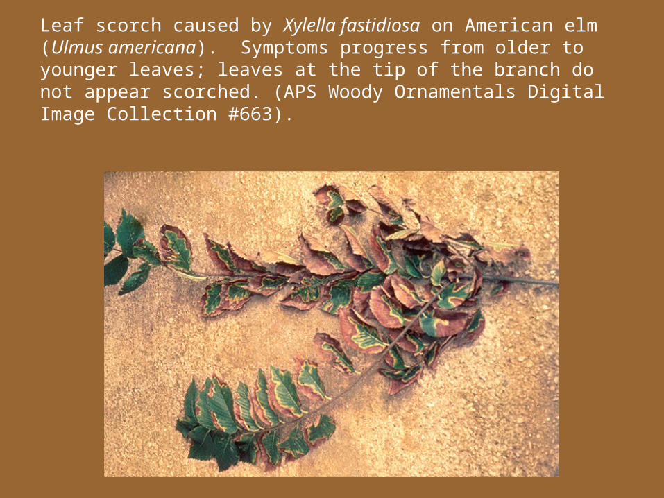
Leaf scorch caused by Xylella fastidiosa on American elm (Ulmus americana). Symptoms progress from older to younger leaves; leaves at the tip of the branch do not appear scorched. (APS Woody Ornamentals Digital Image Collection #663).
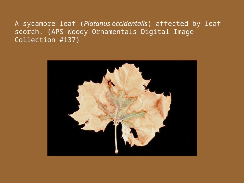
A sycamore leaf (Platanus occidentalis) affected by leaf scorch. (APS Woody Ornamentals Digital Image Collection #137)
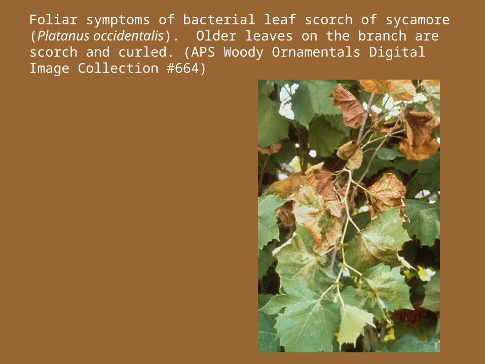
Foliar symptoms of bacterial leaf scorch of sycamore (Platanus occidentalis). Older leaves on the branch are scorch and curled. (APS Woody Ornamentals Digital Image Collection #664)
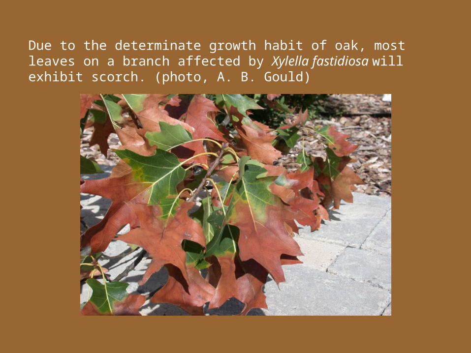
Due to the determinate growth habit of oak, most leaves on a branch affected by Xylella fastidiosa will exhibit scorch. (photo, A. B. Gould)
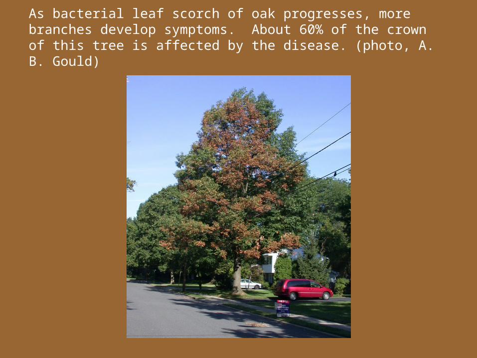
As bacterial leaf scorch of oak progresses, more branches develop symptoms. About 60% of the crown of this tree is affected by the disease. (photo, A. B. Gould)
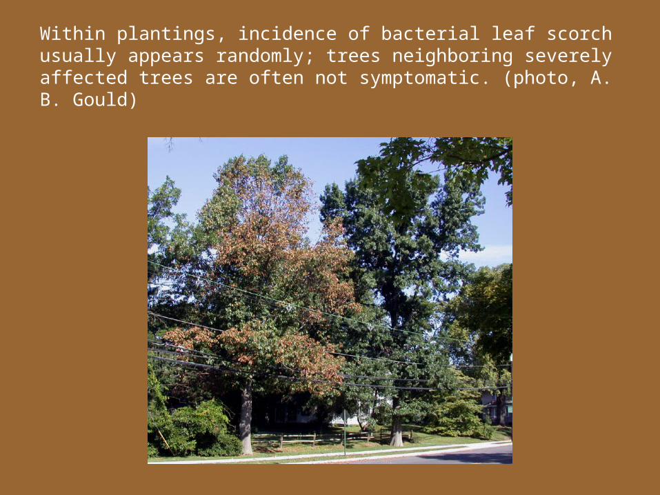
Within plantings, incidence of bacterial leaf scorch usually appears randomly; trees neighboring severely affected trees are often not symptomatic. (photo, A. B. Gould)
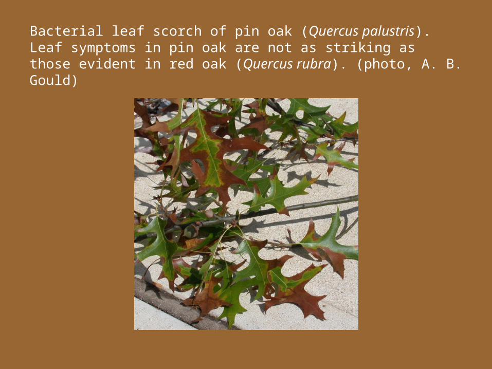
Bacterial leaf scorch of pin oak (Quercus palustris). Leaf symptoms in pin oak are not as striking as those evident in red oak (Quercus rubra). (photo, A. B. Gould)
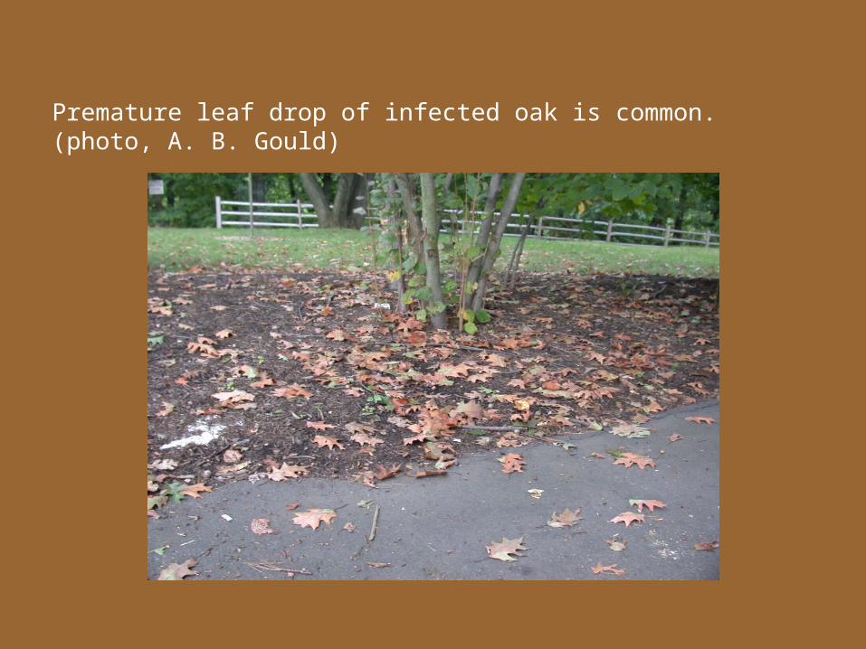
Premature leaf drop of infected oak is common. (photo, A. B. Gould)
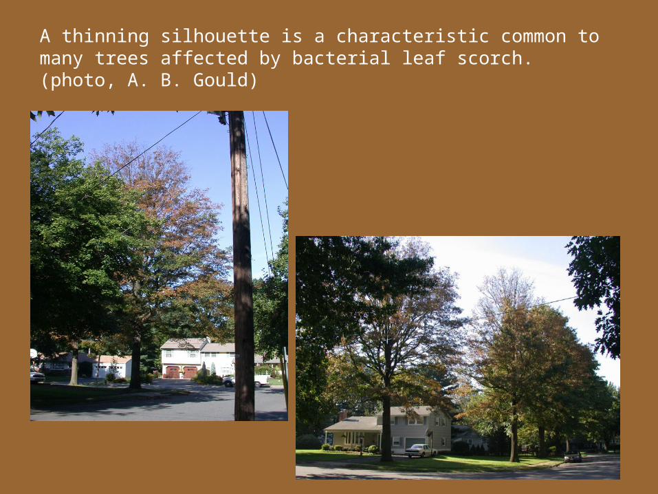
A thinning silhouette is a characteristic common to many trees affected by bacterial leaf scorch. (photo, A. B. Gould)
Bacterial Leaf Scorch of Oak
Xylella fastidiosa subsp. multiplex
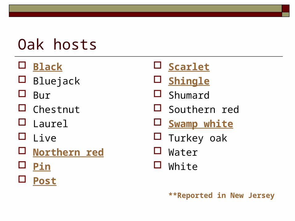
Oak hosts Black Bluejack Bur Chestnut Laurel Live Northern red Pin Post
Scarlet Shingle Shumard Southern red Swamp white Turkey oak Water White
**Reported in New Jersey
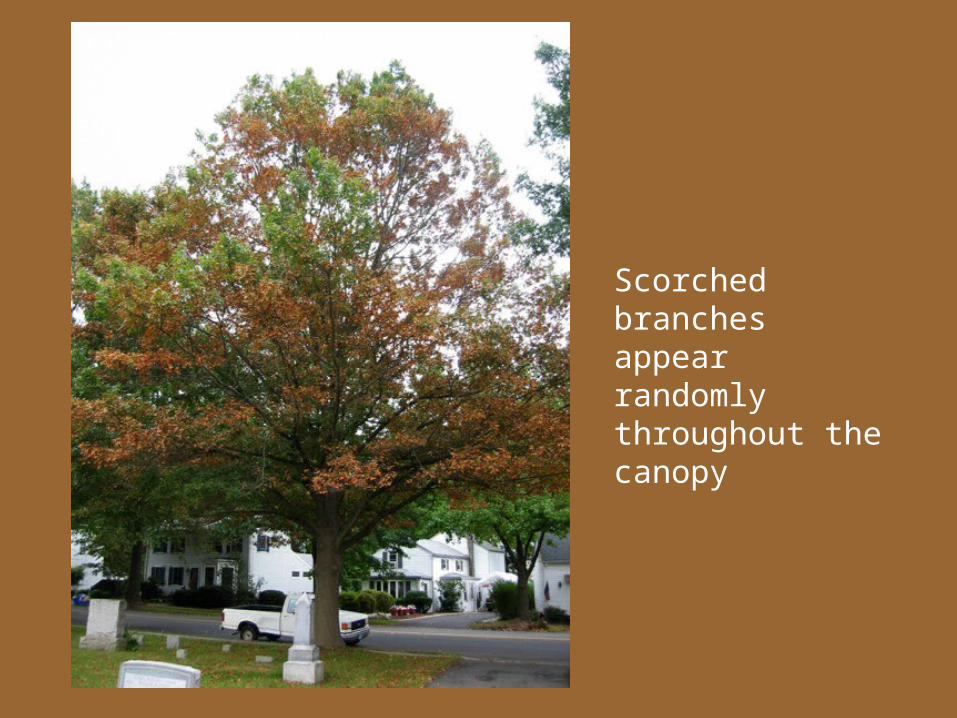
Scorched branches appear randomly throughout the canopy
Extent of BLS of oak in New Jersey Ground Surveys Detection Techniques Vector Relationships
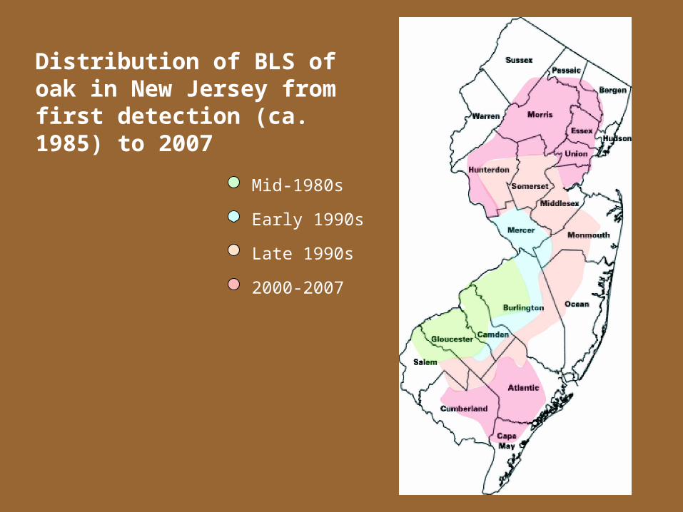
Mid-1980s
Early 1990s
Late 1990s
2000-2007
Distribution of BLS of oak in New Jersey from first detection (ca. 1985) to 2007
Disease incidence: 5-year survey Ground survey of street-side oak trees in three
communities with established disease incidence Evaluated:
disease severity (% of canopy with scorch, branch dieback, or transparency estimated)
disease incidence (number of trees in a population with symptoms)
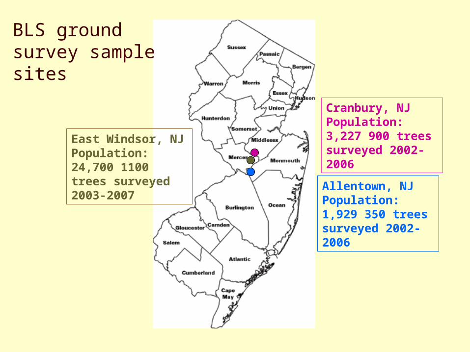
Cranbury, NJ Population: 3,227 900 trees surveyed 2002-2006
Allentown, NJ Population: 1,929 350 trees surveyed 2002-2006
East Windsor, NJ Population: 24,700 1100 trees surveyed 2003-2007
BLS ground survey sample sites
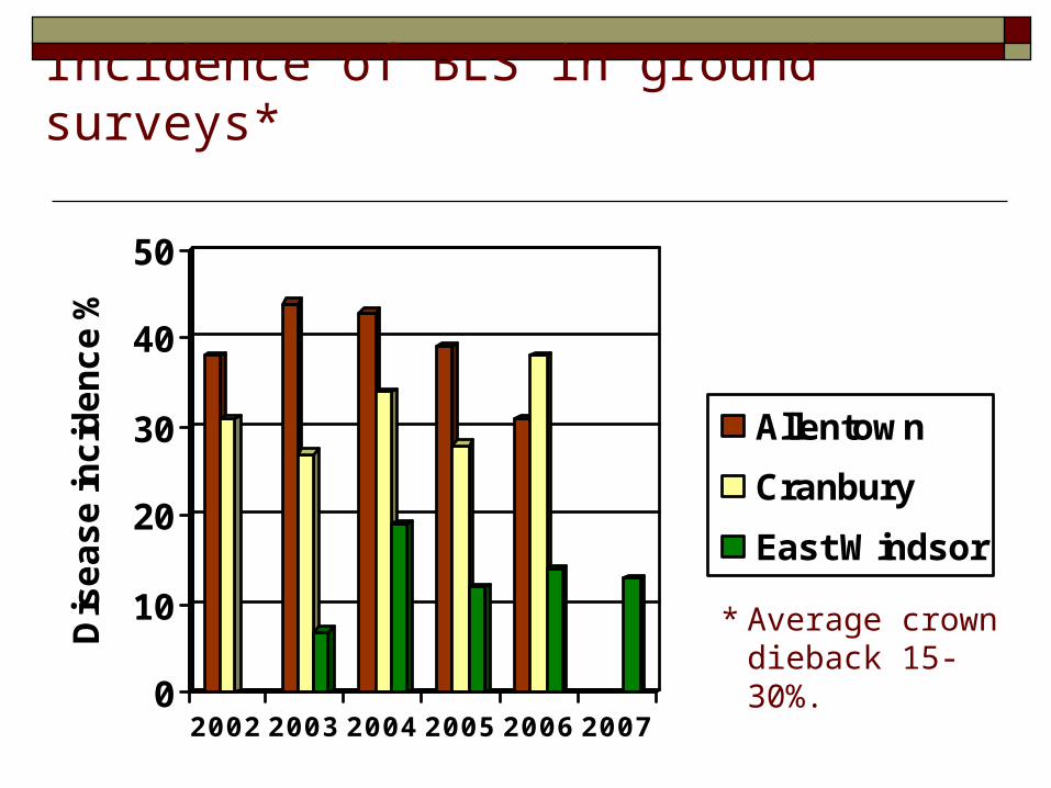
Incidence of BLS in ground surveys*
0
10
20
30
40
50
Dis
ea
se
inc
ide
nc
e %
2002 2003 2004 2005 2006 2007
Allentown
Cranbury
East Windsor
* Average crown dieback 15-30%.
What’s next? Disease incidence in these communities is high What’s next?
We need to look for the “leading edge” of the epidemic
Determine whether trees may be infected by the pathogen, but remain asymptomatic
Detection techniques used for Xylella Microscopic examination of xylem fluid Culturing on special agar medium ELISA (enzyme-linked immunosorbent assay) PCR (polymerase chain reaction) real-time PCR (QRT-PCR)
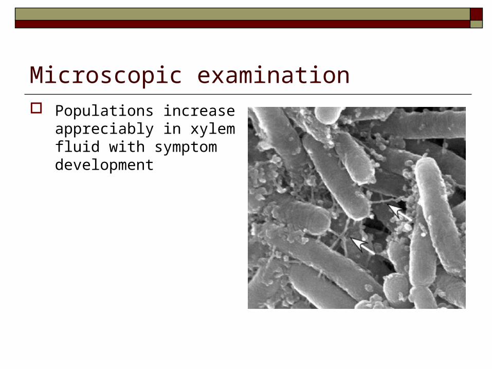
Microscopic examination Populations increase
appreciably in xylem fluid with symptom development
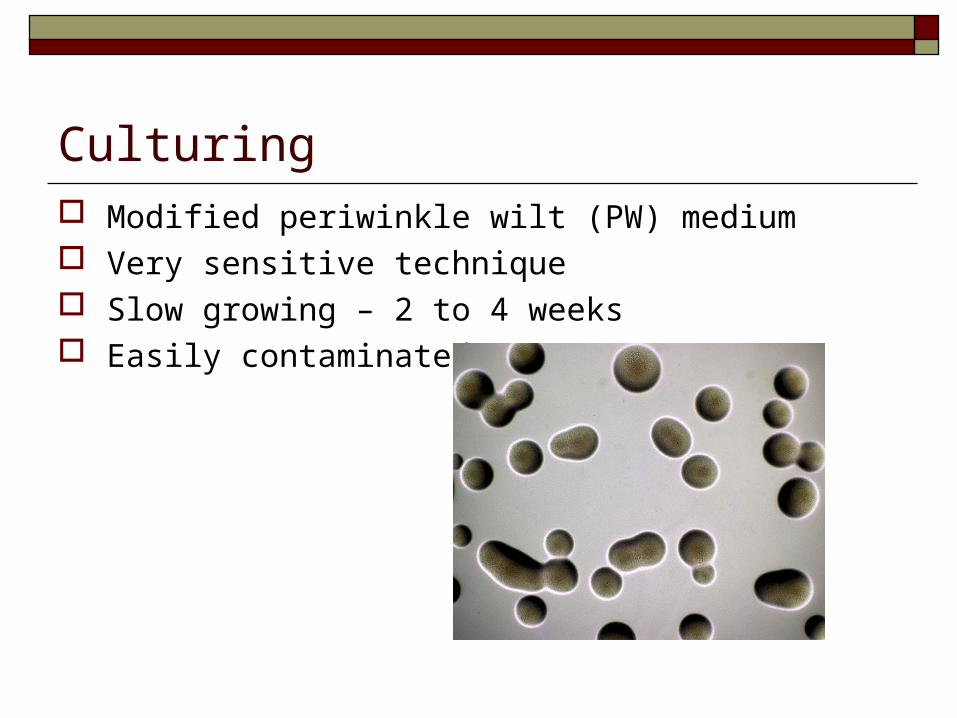
Culturing Modified periwinkle wilt (PW) medium Very sensitive technique Slow growing – 2 to 4 weeks Easily contaminated
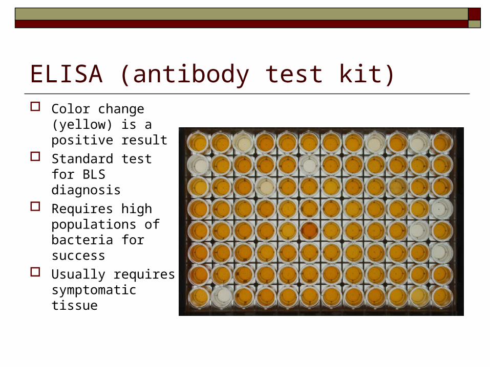
ELISA (antibody test kit) Color change
(yellow) is a positive result
Standard test for BLS diagnosis
Requires high populations of bacteria for success
Usually requires symptomatic tissue
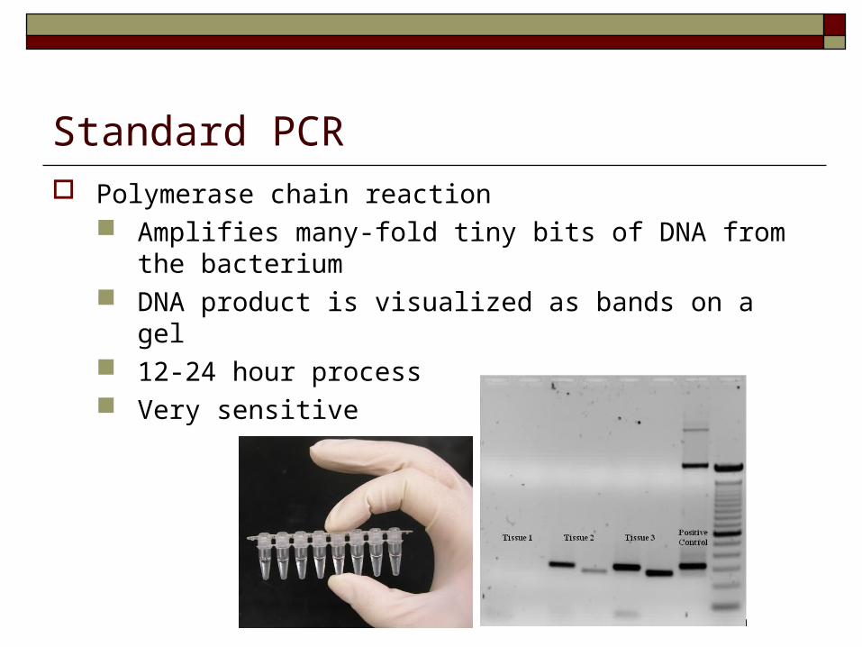
Standard PCR Polymerase chain reaction
Amplifies many-fold tiny bits of DNA from the bacterium
DNA product is visualized as bands on a gel 12-24 hour process Very sensitive
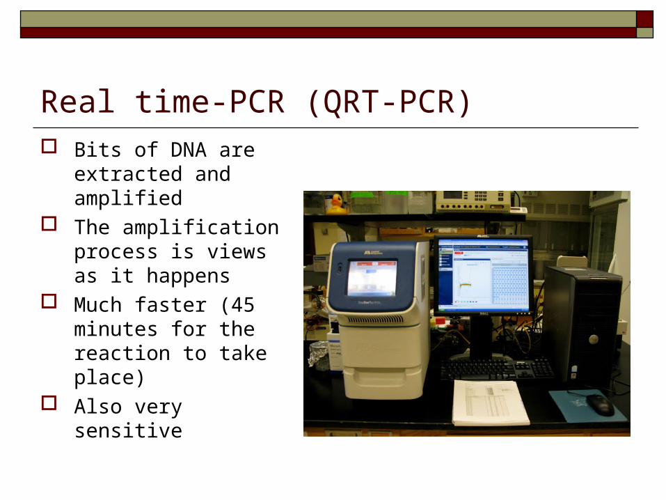
Real time-PCR (QRT-PCR) Bits of DNA are
extracted and amplified
The amplification process is views as it happens
Much faster (45 minutes for the reaction to take place)
Also very sensitive
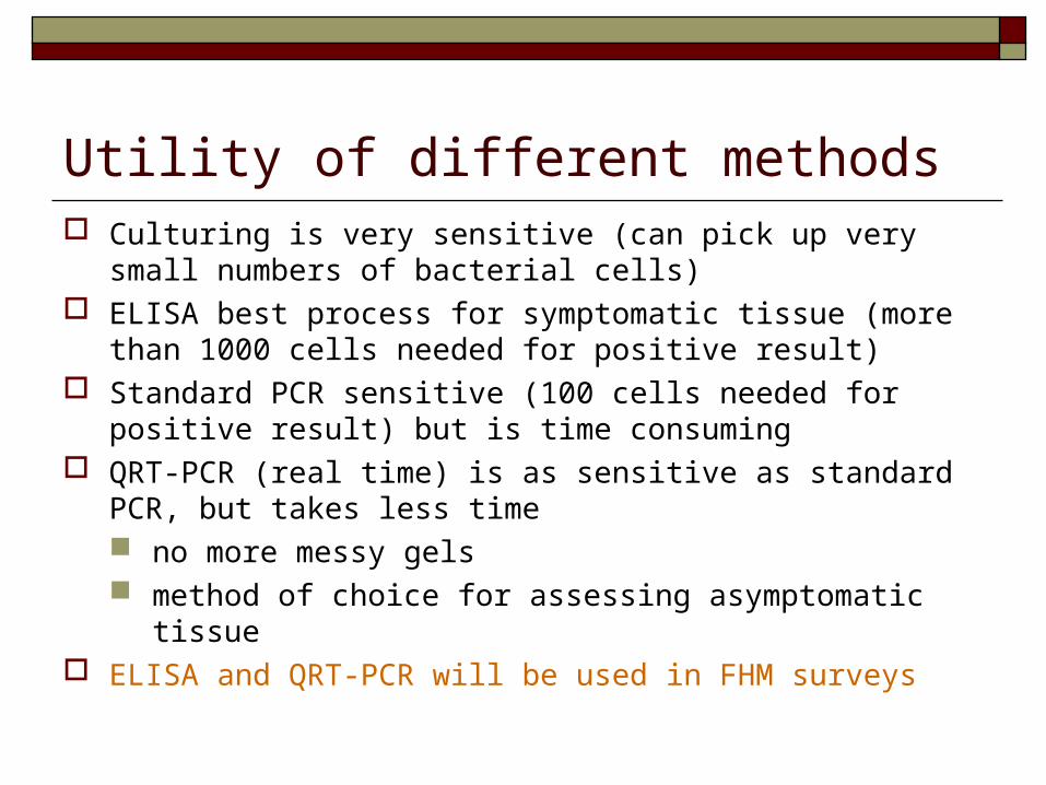
Utility of different methods Culturing is very sensitive (can pick up very small
numbers of bacterial cells) ELISA best process for symptomatic tissue (more than
1000 cells needed for positive result) Standard PCR sensitive (100 cells needed for positive
result) but is time consuming QRT-PCR (real time) is as sensitive as standard PCR,
but takes less time no more messy gels method of choice for assessing asymptomatic tissue
ELISA and QRT-PCR will be used in FHM surveys
ThanksSupport for these research projects was received from: Penn-Del Chapter ISA ISA Tree Fund Horticultural Research Institute U.S. Forest Service State and Hatch Act Funds Duke Farms Mercer Community College Townships of Allentown, Cranbury, and East Windsor
Bodies (2007 season): R. Gorazniak, L. Beirn, R. Orr, M. Cantarella, R. Obal