Embed Size (px)
Citation preview
HORTSCIENCE 44(2):413–417. 2009.
Bacterial Leaf Scorch, a New BlueberryDisease Caused by Xylella fastidiosaChung-Jan Chang1 and Ruth DonaldsonDepartment of Plant Pathology, University of Georgia, Griffin Campus, 1109Experiment Street, Griffin, GA 30223
Phil BrannenDepartment of Plant Pathology, University of Georgia, 3307 Miller PlantScience Building, Athens, GA 30602
Gerard KrewerDepartment of Horticulture, P.O. Box 748, University of Georgia,Horticulture Building, Tifton, GA 31793
Robert BolandUniversity of Georgia, Brantley County Cooperative Extension Service, P.O.Box 275, Nahunta, GA 31553
Additional index words. rabbiteye blueberry, southern highbush blueberry, Pierce’s disease
Abstract. Since 2004, growers and scientists have observed a disorder described as‘‘yellow twig’’ or ‘‘yellow stem’’ affecting a major selection of southern highbushblueberry, FL 86-19, in the south Georgia blueberry production region. The initialsymptom observed was leaf marginal chlorosis and subsequent necrosis, which eventu-ally progressed throughout the whole leaf resulting in early leaf fall. Thin, yellow twigs oryellow stems became evident on some cultivars. The described symptoms on blueberrywere similar to those exhibited on grapes with Pierce’s disease and on plum with leafscald disease. This prompted the enzyme-linked immunosorbent assay (ELISA) tests andisolations of Xylella fastidiosa, which is the causal agent of the previously mentionedgrape and plum diseases. Two leaf and two root tissue samples were collected from adiseased FL 86-19 plant for isolation and ELISA testing on 2 Mar. 2006. ELISA resultsindicated all four tissues tested positive for the bacterial pathogen, X. fastidiosa, whereasonly the two root tissues provided positive isolations. One leaf and one root tissue samplewere later collected from each of five additional diseased plants for isolation and ELISAtesting. Both isolation and ELISA testing methods obtained positive results. Cultureswere multiplied to inoculate seedlings of three cultivars: ‘Southern Belle’ (eight plants),‘Premier’ (six), and ‘Powderblue’ (six) on 23 May 2006 and one selection, FL 86-19(eight), on 31 May 2006. Two FL 86-19 plants started to show symptoms of marginalnecrosis 54 days postinoculation, whereas one plant each of ‘Southern Belle’ and‘Powderblue’ started to show symptoms of marginal necrosis 63 days postinoculationand ‘Premier’ stayed symptomless. All eight culture-inoculated FL 86-19 plants (100%)showed symptoms 72 days postinoculation, but no symptoms were observed on thecontrol plants. One hundred twenty-six days postinoculation, two ‘Powderblue’ and four‘Southern Belle’ plants showed mild symptoms, whereas all ‘Premier’ plants wereasymptomatic. Positive reisolations of the bacteria from the inoculated symptomaticplants, not from asymptomatic plants, fulfilled Koch’s postulates, which confirmed X.fastidiosa was the causal bacterium of the new blueberry disorder, the bacterial leafscorch of blueberry.
Relative to total sales, blueberries are thenumber one fruit commodity in the state ofGeorgia, surpassing even peaches. Produc-tion is concentrated in the southern coastalflatwoods. Rabbiteye blueberry (Vacciniumvirgatum Aiton), a native species, has longbeen the predominant blueberry species cul-tivated in Georgia. More recently, however,growers have increased the production of thesouthern highbush cultivars (V. corymbosum
interspecific hybrids) as a result of a veryfavorable market window (Scherm andKrewer, 2003). Several new diseases suchas necrotic ringspot caused by tobacco ring-spot virus and blueberry ringspot virus (Har-ald Scherm, personal communication) haveappeared with the increased production ofsouthern highbush cultivars. Recently,growers and scientists observed a new disor-der affecting the southern highbush selectionFL 86-19 in the Georgia blueberry produc-tion region. An initial symptom was marginalleaf scorch (burn) of the older leaves, similarto that observed with extreme drought orfertilizer salt burn. New developing shootswere usually abnormally thin with a reduced
number of flower buds. Leaf drop eventuallyoccurred with young twigs or stems of thesouthern highbush selection FL 86-19 devel-oping a yellow, ‘‘skeleton-like’’ appearance.At this stage, the root system still appearedhealthy, except for the possible loss of finenew roots. Whole plants or individual canesshowed symptoms. The plant eventually diedafter leaf drop, typically during the secondyear of observation.
The previously mentioned symptoms wereinconsistent with any previously reportedblueberry disease. It was noticed, however,that the symptoms were similar to diseasescaused by X. fastidiosa in other plants in southGeorgia such as plum leaf scald (Raju et al.,1982; Wells et al., 1987) or pecan bacterialleaf scorch (Sanderlin and Heyderick-Alger,2000). This observation prompted the initia-tion of testing to determine whether X. fastid-iosa could cause these symptoms, andsubsequent death, of blueberry plants.
Several reports in the past few years haverevealed that X. fastidiosa is associated withdiseases in a growing number of plant hosts(Almeida et al., 2005; Baumgartner andWarren, 2005; Hernandez-Martinez et al.,2007; Montero-Astua et al., 2008; Myerset al., 2007; Randall et al., 2007). No reports,however, specifically describe the aforemen-tioned blueberry disease. Using a multiprimerpolymerase chain reaction (PCR) system, ran-dom amplified polymorphic DNA–PCR, andsequence analysis of the 16S-23S rDNAintergenic spacer region, Hernandez-Martinezet al. (2007) reported the phylogenetic rela-tionships of X. fastidiosa strains isolated fromlandscape ornamentals in southern California.They found that strains isolated from daylily,jacaranda, and magnolia clustered with mem-bers of X. fastidiosa subsp. sandyi and causedoleander leaf scorch but did not cause Pierce’sdisease on grapes. Strains isolated from Span-ish broom and cherry and one strain isolatedfrom western redbud clustered with X. fastid-iosa subsp. fastidiosa members, whereasstrains isolated from purple-leafed plum,olive, peach, plum, sweet gum, maidenhairtree, crape myrtle, and another western redbudstrain clustered with members of X. fastidiosasubsp. multiplex. All the strains isolated frommulberry and one isolated from heavenlybamboo formed a separate cluster that hasyet to be defined as a subspecies. In a separateexperiment using nested PCR, Myers et al.(2007) identified the four most abundantsharpshooter leafhoppers in the eastern Pied-mont region and the northeastern CoastalPlain in North Carolina: Oncometopia orbona,Graphocephala versuta, Parahlepsius irrora-tus, and Agolliota constricta. Among them, O.orbona, G. versuta, and P. irroratus testedpositive for the presence of X. fastidiosa.Additionally, Myers et al. (2007) observedfor the first time in the southeast that O.orbona and G. versuta were able to transmitX. fastidiosa to grapevines.
Almeida et al. (2005) showed that Homa-lodisca coagulata was able to inoculate X.fastidiosa into dormant grapes, although fieldacquisition experiments did not result in
Received for publication 9 Oct. 2008. Accepted forpublication 18 Dec. 2008.1To whom reprint requests should be addressed;e-mail [email protected].
HORTSCIENCE VOL. 44(2) APRIL 2009 413
PLANT PATHOLOGY

transmission. Montero-Astua et al. (2008)reported the finding of X. fastidiosa in avocadoplants in Costa Rica based on the positiveresults of enzyme-linked immunosorbentassay (ELISA) and PCR, although attemptsto cultivate the bacterium in periwinkle wilt(PW) medium (Davis et al., 1981) and buff-ered charcoal yeast extract medium (Feeleyet al., 1979; Raju et al., 1982) were negative.
Furthermore, in their study of the persis-tence of X. fastidiosa in riparian hosts nearnorth California vineyards, Baumgartner andWarren (2005) explained the higher Pierce’sdisease incidence in Napa vineyards relativeto plants in Mendocino. This was based ontheir finding of higher X. fastidiosa popula-tions in California grapes, Himalayan black-berries, and periwinkles in Napa and muchless in the same three mentioned plants inMendocino. Randall et al. (2007) concludedthat X. fastidiosa is present in New Mexicoand that the common landscape ornamentalchitalpa is a host for X. fastidiosa based onpositive results obtained from ELISA, PCR,and cultivation. At the time, direct pathoge-nicity testing was still in progress.
Based on ELISA and PCR techniques, it isclear that the association of X. fastidiosa withnew hosts is increasing at a fast pace;however, most of the studies are awaitingfulfillment of Koch’s postulates before therelationship between pathogen and host canbe asserted. The research presented in thisarticle provides clear evidence that X. fastid-iosa is the causal bacterium for a newblueberry disease—herein named the bacte-
rial leaf scorch of blueberry—by demonstrat-ing the fulfillment of Koch’s postulates.
Materials and Methods
Diseased blueberry tissues. A single dis-eased blueberry bush of the selection FL 89-16 was excavated from a blueberry farm insouth Georgia on 2 Feb. 2006. The bush wassubsequently stored under cold room condi-tions (5 �C)—in a plastic trash bag to preventmoisture loss—until attempted detection ofX. fastidiosa using direct isolation andELISA tests (Agdia, Inc., Elkhart, IN). Fromthis initial plant, two samples of young stemsand two root samples were collected andprocessed for isolation and ELISA assayson 2 Mar. 2006. The diseased blueberry bushwas then moved to a greenhouse and plantedin a 30.5-cm diameter pot. This originaldiseased plant was used to monitor thesurvival of the bacterium and symptomdevelopment on new growth after beingstored for 48 d at 5 �C. Five additionaldiseased plants and five healthy appearingplants were then collected from south Geor-gia. On 12 Mar. 2006, five symptomaticstems and five roots were collected fromeach diseased plant, and five asymptomaticstems were collected from the five healthy-appearing plants. These samples were storedat 5 �C until a second round of attemptedisolations and ELISA tests.
Bacterial isolation and enzyme-linkedimmunosorbent assay. Each sample (1 g rootor stem tissue) was surface-sterilized with a
0.8% sodium hypochlorite solution for 3 minfollowed by three rinses with sterilized water(5 min/rinse). The sterilized tissues weremacerated in 3 mL of PW broth (Daviset al., 1981) with a sterilized razor blade.The resulting sap suspension was streakedonto PW, CS20 (Chang and Walker, 1988),and XF-26 (Chang and Donaldson, 1993)plates for the direct isolation of X. fastidiosa.Streaked plates were sealed in a plastic bagand incubated at 28 �C for at least 4 weeksand observed on a weekly basis for colonydevelopment using a dissecting scope. Theremaining sap suspension was mixed with anequal volume of antigen buffer (Agdia, Inc.)in a sterile 5 mL polystyrene tube (12 · 75mm). This mixture was either immediatelyused for ELISA testing or frozen for later use.A commercial kit (DAS ELISA for Xylellafastidiosa; Agdia, Inc.) was used for ELISAtesting according to the manufacturer’s direc-tion. The ELISA reader used for this workwas a Labsystems Multiskan RC Version 6.0(Helsinki, Finland) purchased from Fisher(Pittsburg, PA). The wavelength selectedfor reading the plates was 620 nm, suffi-ciently close to the 650 nm recommended bythe Agdia ELISA Kit. Data values for eachwell were calculated from the raw data bycorrection relative to a buffer-only well set aszero. Calculated data values are presented inTable 1 and Table 2; those values greater than0.100 are considered positive (Agdia, Inc.).
Pathogenicity tests of the cultured X.fastidiosa. Bacterial cell suspensions wereprepared by scraping colonies from five 7- to
Table 1. The reisolation and enzyme-linked immunosorbent assay (ELISA) results on cultivars Premier, Powderblue, and Southern Belle.
Cultivar
Date 1: 23 May/28 Junez
Reisolation
Date 2: 23 May/2 Aug.
Reisolation
Date 3: 23 May/7 Sept.
Reisolation
Date 4: 23 May/12 Oct.
ReisolationELISA ELISA ELISA ELISA
Premier PW1y 0/2x –0.011w 0/2 0/2 0.065 0/2 0/2 –0.029 0/2 0/2 0.050 0/2PW2 –0.012 0.020 –0.011 0.045S1 0/6 –0.019 0/6 0/6 –0.013 0/6 0/6 0.022 0/6 0/6 0.052 0/6S2 –0.012 0.062 0.034 0.077S3 –0.013 –0.012 0.001 0.073R1 –0.006 –0.004 –0.008 0.042R2 –0.011 –0.011 –0.011 0.048R3 –0.013 –0.019 0.038 0.051
Powder-blue
PW1 0/2 –0.004 0/2 0/2 –0.004 0/2 0/2 –0.027 0/2 0/2 0.042 0/2PW2 –0.012 0.010 0.029 –0.001S1 0/6 –0.009 0/6 1/6 0.032 1/6 0/6 0.020 0/6 1/6 0.153 + 0/6S2 –0.010 0.154 +v + –0.020 0.058S3 –0.017 0.024 –0.005 0.022R1 –0.019 –0.013 0.042 0.056R2 –0.015 –0.023 0.035 0.036R3 –0.012 –0.002 0.011 0.058
SouthernBelle
PW1 0/2 –0.019 0/2 0/2 –0.005 0/2 0/2 –0.011 0/2 0/2 0.011 0/2PW2 –0.020 –0.010 –0.011 –0.005S1 0/8 –0.026 0/8 1/8 –0.014 2/8 0/8 –0.027 0/8 4/8 0.124 + 1/8 +S2 –0.021 –0.010 –0.028 0.314 +S3 –0.015 –0.006 –0.035 –0.004S4 –0.006 –0.016 –0.030 –0.001R1 –0.012 –0.006 –0.029 0.113 +R2 –0.012 –0.013 + –0.036 0.223 +R3 –0.003 0.122 + + –0.032 0.012R4 –0.019 –0.004 –0.028 0.011
zInocualtion date/reisolation and ELISA testing date.yBlueberry plants received inoculation with periwinkle wilt broth (PW), strain of cell suspension originally isolated from diseased stem tissues (S), and that fromdiseased root tissues (R).xTotal number that gave positive ELISA or reisolation/total number of inoculated plants.wELISA raw data values (calculated), which were the values based on the values of buffer wells set as zero. The values greater than 0.100 were considered ELISApositives.v‘‘+’’ represented positive result either based on ELISA or reisolation of the tissues collected from that particular blueberry plant.
414 HORTSCIENCE VOL. 44(2) APRIL 2009
10–d-old PW agar plates into 5 mL PWbroth. The concentration of the cell suspen-sions was determined by a serial of 10-folddilutions to 10–9 in PW broth. Diluted cul-tures were incubated at 28 �C for at least4 weeks before the reading of the color-changing units (CCUs) was concluded. TheCCU was used for the measurement ofSpiroplasma citri cell populations in thepistil, sepal, corolla tube, stem, and petalsof infected periwinkles (Chang and Zheng,1999). The color of fresh PW broth mediumwith phenol red is yellowish at a pH of 6.7. X.fastidiosa blueberry strains change the pH ofthe medium from acidic to basic duringgrowth; this corresponds with a change inthe color of the medium from yellow to red orpink when growth reaches the exponential orstationary phase, respectively. Thus, CCUsare useful for a close determination of theconcentration of cell suspensions. After incu-bation at 28 �C for 4 weeks, the concentra-tions of cell suspensions for stem and rootisolates used for 23 May and 31 May inoc-ulations were 107 CCUs/mL and 108 CCUs/mL, respectively.
For each inoculation, a cell suspensiondrop was placed on stems of healthy blueberryplants near the soil line; this was followed bypricking the stems through the cell suspensiona few times with a syringe needle until thesuspension was absorbed into the xylem. Theinoculation sites were marked with stickytape. All plants were kept in the greenhouseand fertilized monthly with Peters 10-10-10.Symptom development was closely observedand recorded on a weekly basis.
Pathogenicity tests were carried out ontwo different dates, 23 May 2006 and 31 May2006. Eight ‘Premier’, eight ‘Powderblue’,and 10 ‘Southern Belle’ plants grown in 15.2-cm diameter pots were used for inoculationon 23 May, whereas 12 FL 86-19 were usedfor inoculation on 31 May. For the first threecultivars, two plants of each cultivar weremechanically inoculated with PW broth ascontrols, whereas the rest were mechanicallyinoculated with bacterial cell suspensions
(half originally isolated from stems and halforiginally isolated from roots). Of the 12 FL86-19 plants, four were mechanically inocu-lated with PW broth; four were mechanicallyinoculated with cell suspension of stemorigin, and four with that of root origin.
Reisolation of X. fastidiosa from inoculatedplants. The protocols for isolations and ELISAtests for X. fastidiosa from inoculated plantswere similar to those described previously,except the ratio of 1:3 (grams of tissue tomilliliters of PW broth medium) was appliedwhen sample weights were less than 1 g.Several isolations and ELISA tests wereperformed throughout the course of the study.Thirty-seven d postinoculation, leaves 1, 3,and 5 above the inoculation point were col-lected from each plant of the three cultivars(Premier, Powderblue, and Southern Belle).Leaves from the initial field-diseased blue-berry bush were also collected for isolationand ELISA tests. Forty-one d postinoculation,leaves 1, 3, and 5 above the inoculation pointof the FL 86-19 plants were collected forisolation and ELISA tests. Sixty-three d post-inoculation, leaves 2, 4, and 6 above theinoculation site were collected from FL 86-19 plants; 71 d postinoculation, leaves in thesame position were collected from the threeother cultivars for isolation and ELISA tests.Ninety-nine d postinoculation, leaves 7, 8, and9 above the inoculation site were collectedfrom FL 86-19 plants and at 107 d, leaves ofthe same position were collected from theother three cultivars for isolation and ELISA.At 113 d postinoculation, leaves from selec-tion FL 86-19 were collected again for iso-lation and ELISA. At 142 d postinoculation,leaves from cultivars Premier, Powderblue,and Southern Belle were once more collectedfor isolation and ELISA.
Results
Isolation and enzyme-linked immunosorbentassay results. All were positive by ELISA, butisolations were only successful from roots in thedirect isolation and ELISA test from stem and
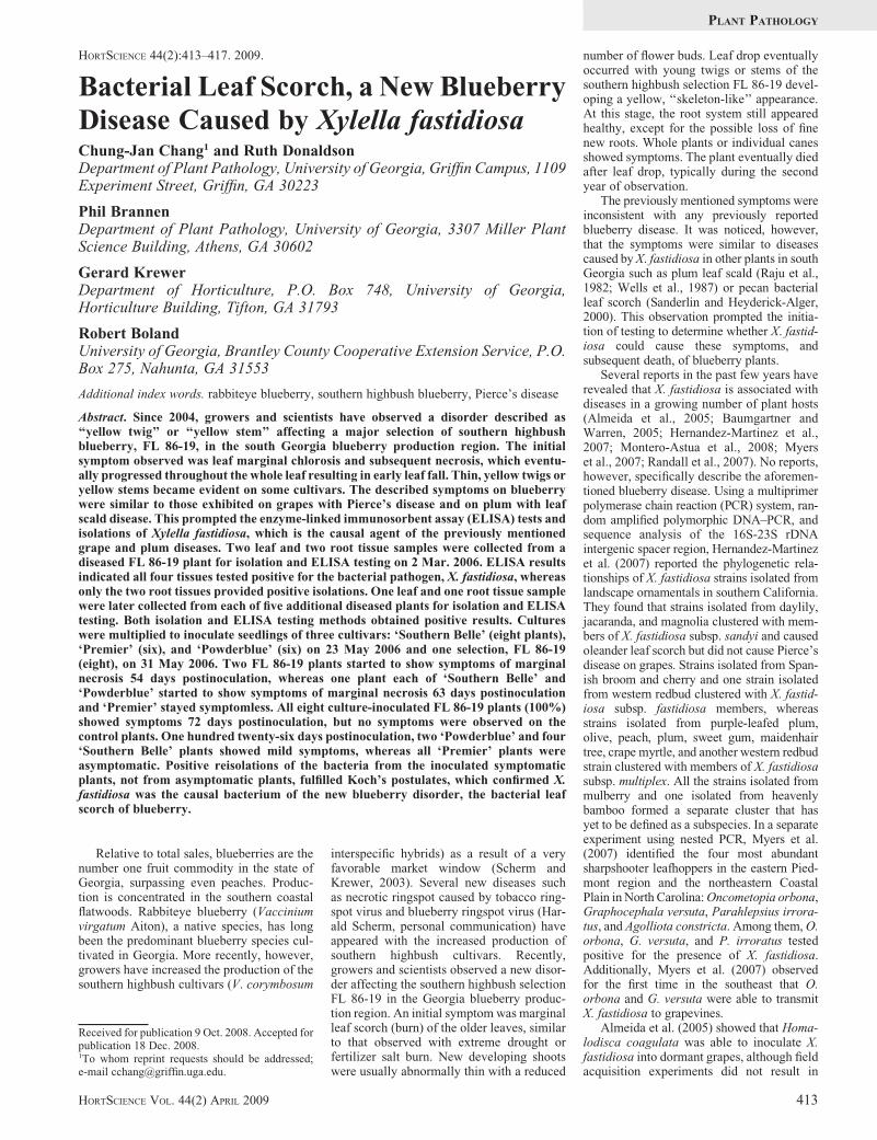
root tissues of the original diseased blueberryplant. Five stem and root samples from fiveadditional symptomatic plants also gave posi-tive results in direct isolations and ELISA tests.Five stem samples taken from the five asymp-tomatic plants gave negative results for bothisolation and ELISA. These results provide astrong association of X. fastidiosa to the leaf-scorched blueberry plants. Colony developmentof the blueberry strains was relatively slow,taking 10 to 14 d to become visible (Fig. 1A).The colonies were opalescent white and reached0.03 to 0.07 mm in diameter in 2 weeks.
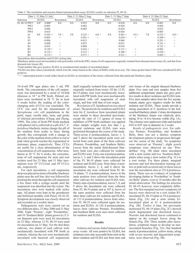
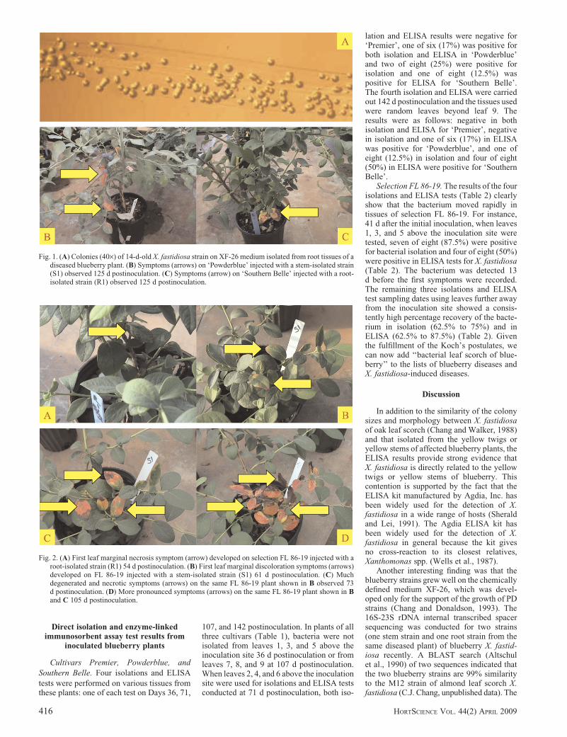
Results of pathogenicity tests. For culti-vars Premier, Powderblue, and SouthernBelle, there was not a distinct symptomdevelopment pattern or progression through-out the course of the study. No symptomswere observed on ‘Premier’; slight scorchsymptoms were observed on one ‘Pow-derblue’ (Fig. 1B) plant when inoculatedwith a stem isolate and two ‘Southern Belle’plants when using a stem isolate (Fig. 1C) ora root isolate. For these plants, marginalnecrosis and leaf discoloration (turning yel-low or pink/red) occurred only to those leavesnear the inoculation sites at 125 d postinocu-lation. There was no evidence of symptomsdeveloping further in ‘Powderblue’ or ‘South-ern Belle’ plants, even at 14 months after theinitial observation. The findings for selectionFL 86-19, however, were completely differ-ent. The first marginal necrosis symptoms (ofleaves) were observed on a root isolate R1-inoculated FL 86-19 plant at 54 d postinocu-lation (Fig. 2A) and a stem isolate S1-inoculated plant at 61 d postinoculation(Fig. 2B). Twelve d later, the previouslymentioned FL 86-19 plant (Fig. 2C) showedmuch degenerated and discolored leaves.Necrotic and discolored leaves continued toappear on the younger leaves along thebranch (Fig. 2D). Three months after inocu-lation, symptoms began to develop on non-inoculated branches (Fig. 3A). One hundredninety d postinoculation, yellow stems, alongwith severe necrotic and degenerated symp-toms, were observed (Fig. 3B).
Table 2. The reisolation and enzyme-linked immunosorbent assay (ELISA) results on selection FL 89-16.
Selection
Date 1: 31 May/11 Julyz
Reisolation
Date 2: 31 May/2 Aug.
Reisolation
Date 3: 31 May/7 Sept.
Reisolation
Date 4: 31 May/21 Sept.
ReisolationELISA ELISA ELISA ELISA
FL 86-19 PW1y 0/4x 0.000w 0/4 0/4 0.019 0/4 0/4 –0.030 0/4 0/4 –0.001 0/4PW2 –0.005 0.002 –0.028 0.002PW3 0.006 –0.002 –0.024 0.023PW4 0.014 –0.020 –0.005S1 4/8 0.303 +v 7/8 + 5/8 0.325 + 6/8 + 5/8 0.463 + 5/8 + 7/8 0.532 + 6/8 +S2 –0.003 + 0.581 + + 0.630 + + 0.300 + +S3 –0.004 –0.013 0.004 0.291 + +S4 0.008 + 0.023 + –0.031 0.761 + +R1 0.055 + 0.004 –0.018 0.721 + +R2 0.130 + + 0.262 + + 0.131 + + –0.003R3 0.174 + + 0.213 + + 0.308 + + 0.220 +R4 0.172 + + 0.571 + + 0.521 + + 0.603 + +
zInocualtion date/reisolation and ELISA testing date.yBlueberry plants received inoculation with periwinkle wilt broth (PW), strain of cell suspension originally isolated from diseased stem tissues (S), and that fromdiseased root tissues (R).xTotal number that gave positive ELISA or reisolation/total number of inoculated plants.wELISA raw data values (calculated), which were the values based on the values of buffer wells set as zero. The values greater than 0.100 were considered ELISApositives.v‘‘+’’ represented positive result either based on ELISA or reisolation of the tissues collected from that particular blueberry plant.
HORTSCIENCE VOL. 44(2) APRIL 2009 415
Direct isolation and enzyme-linkedimmunosorbent assay test results from
inoculated blueberry plants
Cultivars Premier, Powderblue, andSouthern Belle. Four isolations and ELISAtests were performed on various tissues fromthese plants: one of each test on Days 36, 71,
107, and 142 postinoculation. In plants of allthree cultivars (Table 1), bacteria were notisolated from leaves 1, 3, and 5 above theinoculation site 36 d postinoculation or fromleaves 7, 8, and 9 at 107 d postinoculation.When leaves 2, 4, and 6 above the inoculationsite were used for isolations and ELISA testsconducted at 71 d postinoculation, both iso-
lation and ELISA results were negative for‘Premier’, one of six (17%) was positive forboth isolation and ELISA in ‘Powderblue’and two of eight (25%) were positive forisolation and one of eight (12.5%) waspositive for ELISA for ‘Southern Belle’.The fourth isolation and ELISA were carriedout 142 d postinoculation and the tissues usedwere random leaves beyond leaf 9. Theresults were as follows: negative in bothisolation and ELISA for ‘Premier’, negativein isolation and one of six (17%) in ELISAwas positive for ‘Powderblue’, and one ofeight (12.5%) in isolation and four of eight(50%) in ELISA were positive for ‘SouthernBelle’.
Selection FL 86-19. The results of the fourisolations and ELISA tests (Table 2) clearlyshow that the bacterium moved rapidly intissues of selection FL 86-19. For instance,41 d after the initial inoculation, when leaves1, 3, and 5 above the inoculation site weretested, seven of eight (87.5%) were positivefor bacterial isolation and four of eight (50%)were positive in ELISA tests for X. fastidiosa(Table 2). The bacterium was detected 13d before the first symptoms were recorded.The remaining three isolations and ELISAtest sampling dates using leaves further awayfrom the inoculation site showed a consis-tently high percentage recovery of the bacte-rium in isolation (62.5% to 75%) and inELISA (62.5% to 87.5%) (Table 2). Giventhe fulfillment of the Koch’s postulates, wecan now add ‘‘bacterial leaf scorch of blue-berry’’ to the lists of blueberry diseases andX. fastidiosa-induced diseases.
Discussion
In addition to the similarity of the colonysizes and morphology between X. fastidiosaof oak leaf scorch (Chang and Walker, 1988)and that isolated from the yellow twigs oryellow stems of affected blueberry plants, theELISA results provide strong evidence thatX. fastidiosa is directly related to the yellowtwigs or yellow stems of blueberry. Thiscontention is supported by the fact that theELISA kit manufactured by Agdia, Inc. hasbeen widely used for the detection of X.fastidiosa in a wide range of hosts (Sheraldand Lei, 1991). The Agdia ELISA kit hasbeen widely used for the detection of X.fastidiosa in general because the kit givesno cross-reaction to its closest relatives,Xanthomonas spp. (Wells et al., 1987).
Another interesting finding was that theblueberry strains grew well on the chemicallydefined medium XF-26, which was devel-oped only for the support of the growth of PDstrains (Chang and Donaldson, 1993). The16S-23S rDNA internal transcribed spacersequencing was conducted for two strains(one stem strain and one root strain from thesame diseased plant) of blueberry X. fastid-iosa recently. A BLAST search (Altschulet al., 1990) of two sequences indicated thatthe two blueberry strains are 99% similarityto the M12 strain of almond leaf scorch X.fastidiosa (C.J. Chang, unpublished data). The
Fig. 1. (A) Colonies (40·) of 14-d-old X. fastidiosa strain on XF-26 medium isolated from root tissues of adiseased blueberry plant. (B) Symptoms (arrows) on ‘Powderblue’ injected with a stem-isolated strain(S1) observed 125 d postinoculation. (C) Symptoms (arrow) on ‘Southern Belle’ injected with a root-isolated strain (R1) observed 125 d postinoculation.
Fig. 2. (A) First leaf marginal necrosis symptom (arrow) developed on selection FL 86-19 injected with aroot-isolated strain (R1) 54 d postinoculation. (B) First leaf marginal discoloration symptoms (arrows)developed on FL 86-19 injected with a stem-isolated strain (S1) 61 d postinoculation. (C) Muchdegenerated and necrotic symptoms (arrows) on the same FL 86-19 plant shown in B observed 73d postinoculation. (D) More pronounced symptoms (arrows) on the same FL 86-19 plant shown in Band C 105 d postinoculation.
416 HORTSCIENCE VOL. 44(2) APRIL 2009
genetic relationship among the blueberrystrains, the PD strains, and the almond leafscorch strains warrants further investigation.
Symptoms of marginal necrosis began todevelop on the original diseased blueberrybush �5 months after it was moved from thecold room to the greenhouse and by 6 months,symptoms were observed on all new growth.Unfortunately, the original blueberry bushdied �24 Oct. 2006, less than 9 months afterexcavation. This original blueberry bush pro-vided valuable information on the survivabil-ity of the X. fastidiosa blueberry strain. Thebacterium was able to survive at 5 �C for 48d when the bush was kept in a plastic bagbefore being planted in a large pot and kept inthe greenhouse. On 10 July 2006, tissuesfrom this bush were collected for isolationand ELISA as described previously; theresults were positive for both methods. Theblueberry industry—particularly growers—in the southeastern United States will find thisinformation especially important, becausethe research suggests that the bacteria is ableto survive in aboveground tissues through thesouth Georgia winter because it is unlikelyfor the temperature to remain at 5 �C 24 h aday for a consecutive 48 d in the winter.Furthermore, the source of inoculum fortransmission would likely be available year-round. Additional research is necessary toconfirm this preliminary observation on sur-vivability, because no repeat of the same coldtreatment was conducted for this project.
By 3 months after initial inoculation, alleight X. fastidiosa-injected FL 86-19 plantsshowed symptoms, whereas all four PWmedium-only-injected plants remained asymp-tomatic. For the other three cultivars, only twoof six ‘Powderblue’ and four of eight ‘Southern
Belle’ showed mild symptoms, whereas zero ofsix ‘Premier’ plants were symptomatic even at4 months postinoculation. Both ELISA anddirect isolations confirmed the presence of X.fastidiosa in symptomatic plants. Yellow stemsor twigs were a strong symptomatic indicatorof X. fastidiosa infection. There seemed to be adifferent degree of susceptibility among thethree cultivars and one selection with selectionFL 86-19 clearly being the most susceptibleconsistent with what had been previouslyobserved in the field. More work, includingrepeating the comparison of the previouslymentioned three cultivars plus other cultivarsand selection FL 86-19, is necessary before acultivar is considered resistant or tolerant toblueberry X. fastidiosa. In 2007, blueberry X.fastidiosa was isolated from the followingthree diseased rabbiteye blueberry cultivars,Star, Palmetto, Ochlockonee, and one south-ern highbush Millennia (C.J. Chang, unpub-lished data). In the past 2 years, X. fastidiosahas become a major disease of southernhighbush blueberries in Georgia and Florida(Brannen et al., 2007). Given these findings, itis critical that the blueberry industry beginsregular screening for cultivars that are resis-tant or tolerant to X. fastidiosa.
Literature Cited
Almeida, R.P.P., C. Wistrom, B.L. Hill, J. Hashim,and A.H. Purcell. 2005. Vectors transmissionof Xylella fastidiosa to dormant grape. PlantDis. 89:419–424.
Altschul, S.F., W. Gish, W. Miller, E.W. Myers,and D.J. Lipman. 1990. Basic local alignmentsearch tool. J. Mol. Biol. 215:403–410.
Baumgartner, K. and J.G. Warren. 2005. Persis-tence of Xylella fastidiosa in riparian hosts nearnorthern California vineyards. Plant Dis. 89:1097–1102.
Brannen, P., G. Krewer, B. Boland, D. Horton, andC.J. Chang. 2007. Bacterial leaf scorch of blue-berry. 7 Nov. 2007. <http://www.smallfruits.org/blueberries/pestinformation/2007/blueberryxylella.pdf>.
Chang, C.J. and R.C. Donaldson. 1993. Xylellafastidiosa: Cultivation in chemically definedmedium. Phytopathology 83:192–194.
Chang, C.J. and T.J. Walker. 1988. Bacterial leafscorch of northern red oak: Isolation, cultiva-tion, and pathogenicity of xylem-limited bac-terium. Plant Dis. 72:730–733.
Chang, C.J. and B. Zheng. 1999. Isolation ofSpiroplasma citri from flowers and seeds col-lected from infected periwinkles. Plant Dis.83:60–61.
Davis, M.J., W.J. French, and N.W. Schaad. 1981.Axenic culture of the bacteria associated withphony disease of peach and plum leaf scald.Curr. Microbiol. 6:309–314.
Feeley, J.C., R.J. Gibson, G.W. Gorman, N.C.Langford, J.K. Rasheed, D.C. Mackel, andW.B. Baine. 1979. Charcoal-yeast extract agar:Primary isolation medium for Legionella pneu-mophila. J. Clin. Microbiol. 10:437–441.
Hernandez-Martinez, R., K.A. de la Cerda, H.S.Costa, D.A. Cooksey, and F.P. Wong. 2007.Phylogenetic relationships of Xylella fastidiosastrains isolated from landscape ornamentals insouthern California. Phytopathology 97:857–864.
Montero-Astua, M., G. Saborio-R., C. Chacon-Diaz, L. Garita, W. Villalobos, L. Moreira,J.S. Hartung, and C. Rivera. 2008. First reportof Xylella fastidiosa in avocado in Costa Rica.Plant Dis. 92:175.
Myers, A.L., T.B. Sutton, J.A. Abad, and G.G.Kennedy. 2007. Pierce’s disease of grapevines:Identification of the primary vectors in NorthCarolina. Phytopathology 97:1440–1450.
Raju, B.C., J.M. Wells, G. Nyland, R.H. Brlansky,and S.K. Lowe. 1982. Plum leaf scald: Iso-lation, culture, and pathogenicity of the causalagent. Phytopathology 72:1460–1466.
Randall, J.J., M. Radionenko, J.M. French, M.W.Olsen, N.P. Goldberg, and S.F. Hanson. 2007.Xylella fastidiosa detected in New Mexico inchitalpa, a common landscape ornamental plant.Plant Dis. 91:329.
Sanderlin, R.S. and K.I. Heyderick-Alger. 2000.Evidence that Xylella fastidiosa can cause leafscorch disease of pecan. Plant Dis. 84:1282–1286.
Scherm, H. and G. Krewer. 2003. Blueberry pro-duction in Georgia: Historical overview andrecent trends. Small Fruits Rev. 2:83–91.
Sherald, J.L. and J.D. Lei. 1991. Evaluation of arapid ELISA test kit for detection of Xylellafastidiosa in landscape trees. Plant Dis. 75:200–203.
Wells, J.M., B.C. Raju, H.-Y. Hung, W.G. Weis-burg, L. Mandelco-Paul, and D.J. Brenner.1987. Xylella fastidiosa gen. nov., sp. nov:Gram-negative xylem-limited, fastidious plantbacteria related to Xanthomonas spp. Intl. J.Syst. Bacteriol. 36:136–143.
Fig. 3. (A) Symptoms (arrow) on FL 86-19 with necrotic and discolored tissues on leaves appeared onbranches that were not injected 153 d postinoculation. Note the branch with a green tape (star) wasinjected with a stem strain. (B) Symptoms (arrow) of yellow stems and severe necrotic and degeneratedleaves were recorded on FL 86-19 plant injected with a stem strain 190 d postinoculation.
HORTSCIENCE VOL. 44(2) APRIL 2009 417


![Blueberry Hill - Kiama Blowhole Buskers€¦ · Blueberry Hill [C] /// /// [F] /// /// [G7] stop [Tacet] I found my [F]thrill ..... on Blueberry [C]Hill ..... On Blueberry [G] Hill](https://img.pdfslide.us/doc/110x75/5f334258c6a82b4839519c93/blueberry-hill-kiama-blowhole-buskers-blueberry-hill-c-f-g7.jpg)


![[BIRU] the Maze Runner (the Scorch Trials) BluRay](https://img.pdfslide.us/doc/110x75/5695cff91a28ab9b02905cc1/biru-the-maze-runner-the-scorch-trials-bluray.jpg)