Embed Size (px)
Citation preview
HEINI POHJANKOSKI
Juvenile Idiopathic Arthritis
ACADEMIC DISSERTATIONTo be presented, with the permission of
the board of the School of Medicine of the University of Tampere,for public discussion in the Päijät-Häme District Central Hospital,
Lecture room 1, Keskussairaalankatu 7, Lahti, on August 24th, 2012, at 12 o’clock.
UNIVERSITY OF TAMPERE
Studies on associated autoimmune diseases and drug therapy
Reviewed byDocent Oili Kaipiainen-SeppänenUniversity of Eastern FinlandFinlandDocent Anne Putto-LaurilaUniversity of TurkuFinland
DistributionBookshop TAJUP.O. Box 61733014 University of TampereFinland
Tel. +358 40 190 9800Fax +358 3 3551 7685 [email protected]/tajuhttp://granum.uta.fi
Cover design byMikko Reinikka
Acta Universitatis Tamperensis 1752ISBN 978-951-44-8874-0 (print)ISSN-L 1455-1616ISSN 1455-1616
Acta Electronica Universitatis Tamperensis 1225ISBN 978-951-44-8875-7 (pdf )ISSN 1456-954Xhttp://acta.uta.fi
Tampereen Yliopistopaino Oy – Juvenes PrintTampere 2012
ACADEMIC DISSERTATIONUniversity of Tampere, School of Medicine Päijät-Häme District Central Hospital, Department of paediatricsRheumatism Foundation Hospital, HeinolaFinland
Supervised byDocent Anneli SavolainenUniversity of TampereFinlandProfessor Matti KorppiUniversity of TampereFinland
Copyright ©2012 Tampere University Press and the author
3
To my family
and to my patients
4
CONTENTS 4
LIST OF ORIGINAL PAPERS 7
ABSTRACT 8
TIIVISTELMÄ 10
ABBREVIATIONS 12
LITERATURE REVIEW 13
1. JUVENILE IDIOPATHIC ARTHRITIS (JIA) 13
1.1. Classification and diagnostic criteria
1.2. Clinical features
1.2.1. Oligoarthritis, persistent
1.2.2. Oligoarthritis, extended
1.2.3. Polyarthritis seronegative
1.2.4. Polyarthritis seropositive
1.2.5. Systemic arthritis
1.2.6.Seronegative spondyloarthropathies
1.3. Epidemiology
1.4. Uveitis
1.5. Drug treatment
1.6. Outcome
1.6.1. Psychological outcome
2. OTHER AUTOIMMUNE DISEASES 27
2.1. Diabetes mellitus type 1 (DM1)
2.1.1. Definition, diagnostic criteria, clinical characteristics
2.1.2. Epidemiology
2.2. Celiac disease, Autoimmune thyroiditis, Multiple sclerosis
5
3. CONNECTION BETWEEN JUVENILE IDIOPATHIC ARTHRITIS 28
AND OTHER AUTOIMMUNE DISEASES
3.1. Autoimmune diseases in children with JIA
3.1.1. DM1 in children with JIA
3.1.2. Other autoimmune diseases in children with JIA
3.2. Autoimmune diseases in JIA children’s first-degree relatives
PURPOSE OF THE STUDY 32
PATIENTS AND METHODS 33
1. Autoimmune diseases in children with JIA
2. Autoimmune diseases in JIA children’s first-degree relatives
3. Simultaneous occurrence of JIA and DM1 in the same patient
4. Trends in the medical treatment practice of JIA in years 2000-2007
5. The effect of simultaneous DM1 on the drug treatment for JIA
6. Statistical methods
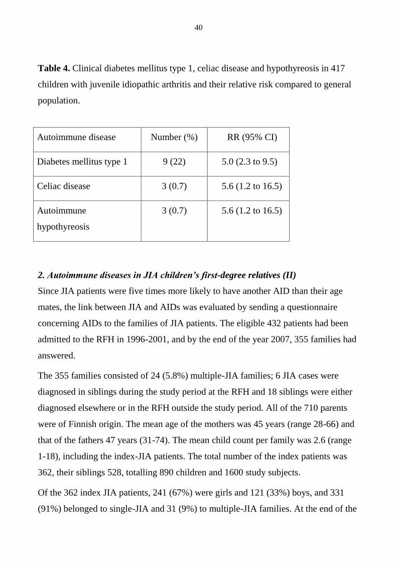
RESULTS 39
1. Autoimmune diseases in children with JIA
2. Autoimmune diseases in JIA children’s first-degree relatives
2.1. Prevalence of AIDs in the index patients’ families
3. Simultaneous occurrence of JIA and DM1 in the same patient
3.1. Occurrence
3.2. JIA or DM1 first
3.3. Age at onset
3.4. Laboratory findings
3.5. Erosions
3.6. Use of glucocorticoids
3.7. Use of biologic agents
6
3.8. Uveitis
3.9. Additional autoimmune diseases
3.10. Psychiatric diseases
4. Trends in the medical treatment practice of JIA in years 2000-2007
4.1. Drug treatment
4.2. Early treatment strategy
5. The effect of simultaneous DM1 on the drug treatment for JIA
DISCUSSION 52
1. Background research
1.1. Connection between different autoimmune diseases
1.2. Autoimmune diseases’ response to drug treatment
1.3 Epidemiological aspects
2. Autoimmune diseases in children with JIA
3. Autoimmune diseases in JIA children’s first-degree relatives
4. Simultaneous occurrence of JIA and DM1 in the same patient
5. Trends in the medical treatment practice of JIA in years 2000-2007
6. The effect of simultaneous DM1 on the drug treatment for JIA
METHODOLOGICAL ASPECTS 63
CONCLUSIONS AND CLINICAL RECOMMENDATIONS 65
REFERENCES 66
ACKNOWLEDGEMENTS 85
ORIGINAL PUBLICATIONS
7
LIST OF ORIGINAL PAPERS
This thesis is based on the following original papers referred to in the text by their
Roman numerals I-IV. In addition, the thesis contains unpublished data.
The original papers are printed with the permission of the copyright holders.
I. Pohjankoski H, Kautiainen H, Kotaniemi K, Korppi M, Savolainen A.
Autoimmune diseases in children with juvenile idiopathic arthritis. Scand J
Rheumatol 2010;39:435-6.
II. Pohjankoski H, Kautiainen H, Kotaniemi K, Korppi M, Savolainen A.
Diabetes, coeliac disease, multiple sclerosis and chronic arthritis in first-degree
relatives of patients with juvenile idiopathic arthritis. Acta Paediatr
2012;101:767-71
III. Pohjankoski H, Kautiainen H, Korppi M, Savolainen A. Simultaneous juvenile
idiopathic arthritis and diabetes mellitus type 1 - a Finnish nationwide study. J
Rheumatol 2012;39:377-81.
IV.Pohjankoski H, Latva K, Kautiainen H, Säilä H, Klaukka T, Virta L, Puolakka
K, Pohjolainen T, Savolainen A. First-year purchases of disease modifying
drugs of incident patients with chronic juvenile arthritis in Finland. Clin Exp
Rheumatol 2011;29:878-81.
8
ABSTRACT
Autoimmune diseases (AIDs) are chronic conditions that involve an immune attack
on one or more organ systems. Juvenile idiopathic arthritis (JIA) belongs to AIDs
with an unknown specific aetiology, with some evidence referring to genetic and
environmental factors. Over the recent years, several studies have revealed that the
prevalence of AIDs is higher among JIA patients than in controls.
The occurrence of other AIDs was studied in 417 children referred to the
Rheumatism Foundation Hospital (RFH) because of JIA during years 1992-2000.
Children with JIA had approximately over 5-fold occurrence of diabetes mellitus type
1 (DM1), celiac disease (CD) or hypothyreosis (HT) compared to the population data
available.
The occurrence of AIDs was studied by questionnaires addressed to the families of
the 362 JIA patients referred to the RFH in years 1996-2001. The questions
concerned the AID diagnoses in these families: chronic arthritis (CA), DM1, CD and
multiple sclerosis (MS). The families were also asked whether the patients or their
first-degree relatives were entitled to a special reimbursement for medicines for
diabetes or chronic arthritis by the Social Insurance Institution (SII), and what their
exact diagnosis was. The diagnoses ultimately made by a physician and recorded as
per the ICD-10 classification were then gathered. Almost a quarter, 21.4%, of the
families had a member with another AID. The occurrence of JIA and DM1 among the
JIA patients’ relatives was clearly higher than in the previously published studies.
The characteristics of JIA were studied in 82 patients with both JIA and DM1
identified from the SII registers in years 1976-2005. The simultaneous occurrence of
JIA and DM1 had increased over 5-fold as the first ten-year period (1976-1985) was
compared with the last ten-year period (1996-2005). A quarter of these patients
(22%) had a third AI disease. Seropositivity was recorded more often than expected
and the proportion of uveitis was low. Almost a fifth of children in this group (16%)
had serious psychiatric problems.
9
The data on prescribed medication for JIA, reimbursed by SII for the first time in
years 2000-2007, were collected from the SII registers. The number of identified
patients younger than 16 years was 1970. The use of methotrexate (mtx) increased
significantly whereas the use of prednisolone decreased during the study period.
The influence of simultaneous DM1 to the drug treatment for JIA was studied in 1970
JIA patients identified in the SII registers from years 2000-2007. The diagnosis of
DM1 was already made in 23 children. Mtx was introduced to 83% of these children
during the first 3 disease months, compared to 59% in all Finnish JIA patients.
10
TIIVISTELMÄ
Autoimmuunitaudit ovat kroonisia sairauksia, joissa elimistön oma poikkeavasti
toimiva puolustusmekanismi kohdistuu yhteen tai useampaan elimeen tai kudokseen.
Lastenreuma on autoimmuunitauti, jonka syy on edelleen avoin. Tutkimukset
viittaavat sekä perimän että ympäristön vaikutukseen taudin synnyssä. Geneettiset
että epidemiologiset tutkimukset eri puolilta maailmaa ovat vahvistaneet, että
normaaliväestöön verrattuna autoimmuunitauteja on enemmän sekä
lastenreumapotilailla että heidän sukulaisillaan.
Tutkimme kaikkien vuosina 1992-2000 Reumasäätiön Sairaalaan tutkimuksiin ja
hoitoon ensi kertaa lähetettyjen lastenreumapotilaiden (417) sairauskertomukset
Totesimme, että potilailla oli keskimäärin yli 5-kertainen todennäköisyys saada joko
nuoruusiän diabetes, keliakia tai hypothyreoosi.
Lisäksi selvitimme lastenreumapotilaiden ensimmäisen asteen sukulaisten (isä, äiti ja
täyssisarus) autoimmuunitaudit. Vuosina 1996-2001 Reumasäätiön sairaalaan
lähetettyjen 432 potilaan perheille postitettiin kyselylomake, jossa tiedustelimme
perheen autoimmuunitauteja: krooninen reumatauti, keliakia, MS-tauti ja nuoruusiän
diabetes. Kroonisesta reumataudista ja nuoruusiän diabeteksesta halusimme tietää,
milloin erityiskorvattavat lääkkeet oli myönnetty, ja muista taudeista halusimme
varmistua, että diagnoosin oli tehnyt lääkäri. Saimme vastauksen 355 perheeltä, joissa
oli yhteensä 362 potilasta. Noin viidesosassa (21,4 %) perheistä oli lastenreuman
lisäksi joku muu autoimmuunitauti. Lastenreuman ja nuoruusiän diabeteksen
esiintyvyys oli selvästi suurempi kuin aikaisemmissa väestötutkimuksissa.
Väitöskirjatyössä tarkastettiin valtakunnallisesti vuosilta 1976-2005 kaikkien
lastenreumaa ja samanaikaista nuoruusiän diabetesta sairastavien potilaiden
sairauskertomukset. Tutkimuksessa selvitettiin näiden tapausten ilmaantuvuus ja
lastenreuman taudinkuva. Ilmaantuvuus oli noussut yli viisinkertaiseksi kolmen
vuosikymmenen aikana. Tutkituista potilaista useampi kuin lastenreumapotilaista
yleensä sairasti taudin reumafaktoripositiivista muotoa ja heillä esiintyi vähemmän
11
kroonista reumaattista silmätulehdusta. Lähes neljäsosalla (22 %) oli joku kolmas
autoimmuunitauti. Heillä oli myös huomattavasti enemmän (16 %) vaikeita
psykiatrisia ongelmia muuhun väestöön verrattuna.
Kansaeläkelaitoksen (KELA) tiedostoista kerättiin tiedot erityiskorvattavista
lääkkeistä, jotka oli myönnetty kroonisen niveltulehduksen hoitoon alle 16-vuotiaille
lapsille vuosina 2000-2007 ja samanaikaisista reseptiostoista. Seurantajakson aikana
metotreksaatin käyttö lisääntyi merkittävästi samalla kun kortisonin käyttö väheni.
Selvitimme myös, miten samanaikainen nuoruusiän diabeteksen sairastaminen
vaikutti lastenreuman lääkitykseen. KELAn tiedostoista löytyi 23 potilasta, joilla oli
jo diagnosoitu DM1 ja joille myönnettiin korvattavuus kroonista artriittia hoitaviin
lääkkeisiin. Näistä 19:lle (83 %) aloitettiin metotreksaatti ensimmäisen kolmen
kuukauden aikana. Vastaava luku kaikilla lastenreumaa sairastavilla potilailla (1970)
valtakunnallisesti oli 59 %.
12
ABBREVIATIONS
ACR American College of Rheumatology
AID autoimmune disease
AIT autoimmune thyroid disease
ANA antinuclear antibodies
ARA American Rheumatoid Association
AS ankylosing spondylitis
CD celiac disease
CA chronic arthritis
CJA chronic juvenile arthritis
CI confidence interval
CRP C-reactive protein
DM1 diabetes mellitus type 1
DMARD disease modifying antirheumatic drugs
ERA enthesitis-related arthritis
ESR erythrocyte sedimentation rate
EULAR European League Against Rheumatism
HLA human leukocyte antigen
HT hypothyreosis,
HRQL health related quality of life
IgM immunoglobulin M
IL interleukin
ILAR International League of Associations for Rheumatology
JAIT juvenile autoimmune thyreoiditis
JCA juvenile chronic arthritis
JIA juvenile idiopathic arthritis
JRA juvenile rheumatoid arthritis
mtx methotrexate
MS multiple sclerosis
NSAID non-steroidal anti-inflammatory drug
PRR prevalence rate ratio
PsA psoriatic arthritis
RA rheumatoid arthritis
RFH Rheumatism Foundation Hospital
RF IgM rheumatoid factor
RR relative risk
SI sacroiliac
SII Social Insurance Institution
SD standard deviation
Th1 T-helper 1 cell
13
LITERATURE REVIEW
I. JUVENILE IDIOPATHIC ARTHRITIS
Juvenile idiopathic arthritis (JIA), previously also called juvenile chronic arthritis
(JCA) and juvenile rheumatoid arthritis (JRA) belongs to autoimmune diseases (AID)
and is the most common cause of chronic arthritis (CA) in children. JIA is not a
single disease, but merely an umbrella term for various clinical entities of CA in
children and adolescents, forming a complex group of diseases where other reasons
for arthritis are excluded. According to the current concept, system-onset JIA is
rather an autoinflammatory than autoimmune disease.
JIA has various phenotypes, which refer to the involvement of multiple susceptibility
genes. Genetic studies have identified both general autoimmune genes and JIA-
specific genes, explaining the variability of different phenotypes (Phelan et al 2006,
Angeles-Han and Prahalad 2010).
The balance of immune system, including both innate and acquired immunity, is
disturbed in JIA patients. There is evidence that inflammatory cells are trafficking to
the synovium causing local inflammation and further progressive joint damage.
Inflammation includes T-cell and macrophage infiltrations, Th1-oriented cytokine
predominance in inflamed tissues, evidence of T-cell clonality and by inference,
antigen presentation (Phelan et al 2006).
1.1. Classification and diagnostic criteria
JIA is defined as a CA in a child aged less than 16 years and lasting more than six
weeks in the absence of any known cause. There are no specific diagnostic tests for
JIA.
During the past decades, rheumatologists have struggled to identify and classify the
disease in order to understand the disease more clearly (Haines 2007). The oldest
classification made by the American Rheumatoid Association (ARA) in 1973 used
14
the term juvenile rheumatoid arthritis (JRA). In 1977 the European League Against
Rheumatism (EULAR) recommended the term juvenile chronic arthritis (JCA). The
latest classification from 1995 was proposed by the task force of the Pediatric
Standing Committee of the International League of Associations of Rheumatology
(ILAR). In this proposition, further revised in 1997, 2001 and 2004, earlier chronic
juvenile arthritis, is called JIA, juvenile idiopathic arthritis (Petty et al 1998 and
2004). The purpose was to change the classification better suited for clinical work
and to help the research on the etiology, pathogenesis, epidemiology, outcome and
treatment of the disease (Ravelli and Martini 2007).
The ILAR criteria of JIA demand on over 6-week duration of the disease and the
patient must be younger than 16 years at the disease onset. The onset-type is the type
of JIA defined 6 months after the onset of the disease, and course-type is the type
which the disease develops later.
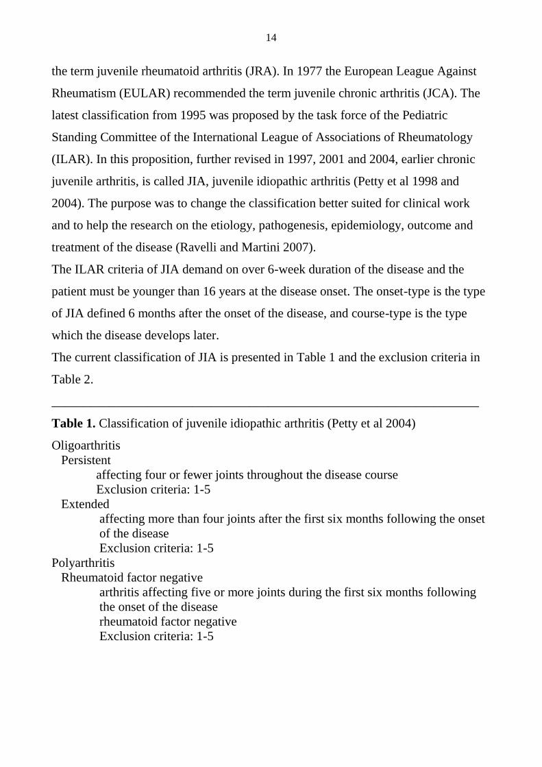
The current classification of JIA is presented in Table 1 and the exclusion criteria in
Table 2.
___________________________________________________________________
Table 1. Classification of juvenile idiopathic arthritis (Petty et al 2004)
Oligoarthritis
Persistent
affecting four or fewer joints throughout the disease course
Exclusion criteria: 1-5
Extended
affecting more than four joints after the first six months following the onset
of the disease
Exclusion criteria: 1-5
Polyarthritis
Rheumatoid factor negative
arthritis affecting five or more joints during the first six months following
the onset of the disease
rheumatoid factor negative
Exclusion criteria: 1-5
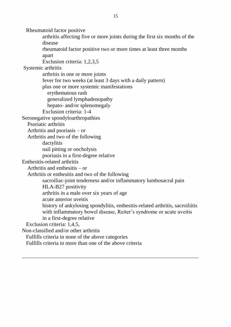
15
Rheumatoid factor positive
arthritis affecting five or more joints during the first six months of the
disease
rheumatoid factor positive two or more times at least three months
apart
Exclusion criteria: 1,2,3,5
Systemic arthritis
arthritis in one or more joints
fever for two weeks (at least 3 days with a daily pattern)
plus one or more systemic manifestations
erythematous rash
generalized lymphadenopathy
hepato- and/or splenomegaly
Exclusion criteria: 1-4
Seronegative spondyloarthropathies
Psoriatic arthritis
Arthritis and psoriasis – or
Arthritis and two of the following
dactylitis
nail pitting or oncholysis
psoriasis in a first-degree relative
Enthesitis-related arthritis
Arthritis and enthesitis – or
Arthritis or enthesitis and two of the following
sacroiliac-joint tenderness and/or inflammatory lumbosacral pain
HLA-B27 positivity
arthritis in a male over six years of age
acute anterior uveitis
history of ankylosing spondylitis, enthesitis-related arthritis, sacroiliitis
with inflammatory bowel disease, Reiter’s syndrome or acute uveitis
in a first-degree relative
Exclusion criteria: 1,4,5,
Non-classified and/or other arthritis
Fulfills criteria in none of the above categories
Fulfills criteria in more than one of the above criteria
_________________________________________________________________
16
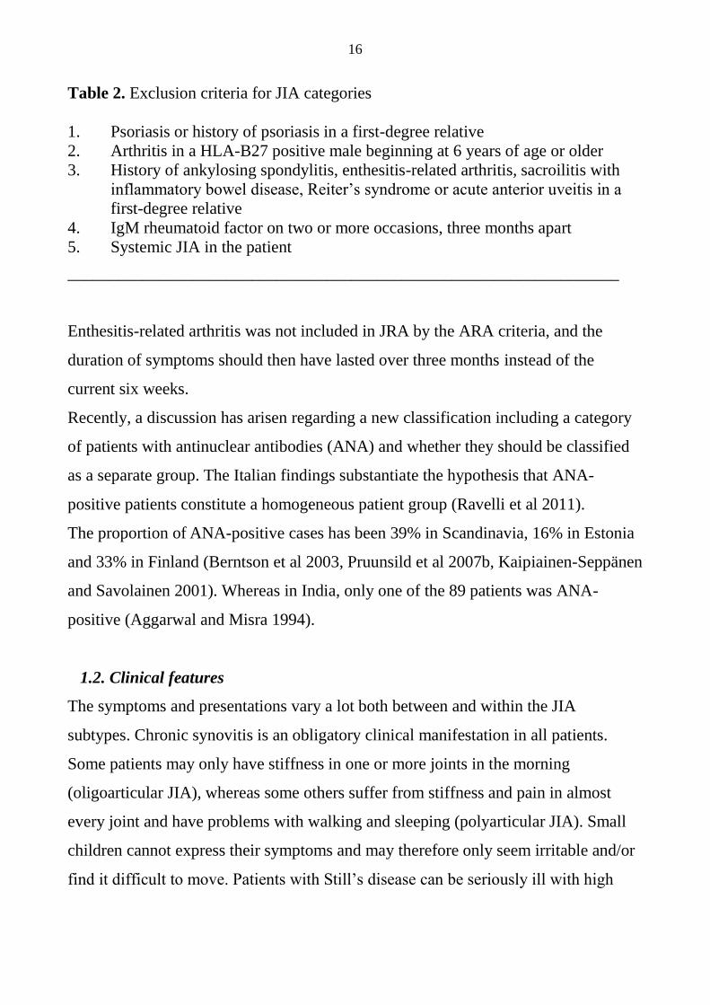
Table 2. Exclusion criteria for JIA categories
1. Psoriasis or history of psoriasis in a first-degree relative
2. Arthritis in a HLA-B27 positive male beginning at 6 years of age or older
3. History of ankylosing spondylitis, enthesitis-related arthritis, sacroilitis with
inflammatory bowel disease, Reiter’s syndrome or acute anterior uveitis in a
first-degree relative
4. IgM rheumatoid factor on two or more occasions, three months apart
5. Systemic JIA in the patient
__________________________________________________________________
Enthesitis-related arthritis was not included in JRA by the ARA criteria, and the
duration of symptoms should then have lasted over three months instead of the
current six weeks.
Recently, a discussion has arisen regarding a new classification including a category
of patients with antinuclear antibodies (ANA) and whether they should be classified
as a separate group. The Italian findings substantiate the hypothesis that ANA-
positive patients constitute a homogeneous patient group (Ravelli et al 2011).
The proportion of ANA-positive cases has been 39% in Scandinavia, 16% in Estonia
and 33% in Finland (Berntson et al 2003, Pruunsild et al 2007b, Kaipiainen-Seppänen
and Savolainen 2001). Whereas in India, only one of the 89 patients was ANA-
positive (Aggarwal and Misra 1994).
1.2. Clinical features
The symptoms and presentations vary a lot both between and within the JIA
subtypes. Chronic synovitis is an obligatory clinical manifestation in all patients.
Some patients may only have stiffness in one or more joints in the morning
(oligoarticular JIA), whereas some others suffer from stiffness and pain in almost
every joint and have problems with walking and sleeping (polyarticular JIA). Small
children cannot express their symptoms and may therefore only seem irritable and/or
find it difficult to move. Patients with Still’s disease can be seriously ill with high
17
fever and inner organ manifestations like pericarditis or myocarditis and sometimes
need intensive care (Ravelli and Martini 2007).
1.2.1. Oligoarthritis, persistent
In the oligoarticular-type of JIA patients suffer from an inflammation in four or fewer
joints during the first six months of the disease as well as during the whole disease
course. This subtype is more frequent in girls than boys, and the onset is most
common in young children (peak age 2-4 years). Patients are often ANA-positive
(70-80%) and have chronic uveitis (Kotaniemi et al 2003, Petty et al 2003). The
involvement of joints is generally asymmetric and mostly affects knees and ankles.
1.2.2 .Oligoarthritis, extended
In this subtype, patients first have oligoarthritis, but they develop a polyarticular
course.
Persistent and extended oligoarthritis types form a clinically homogenous group,
except that the outcome is more severe in the extended oligoarthritis (Ravelli and
Martini 2007).
1.2.3. Polyarthritis seronegative
This subtype is defined as an arthritis affecting five or more joints during the first six
months of the disease in the absence of IgM rheumatoid factor (RF). Some patients in
this group resemble the adult-onset, seronegative rheumatoid arthritis (RA) with
symmetric synovitis of large and small joints. Some patients have dry synovitis which
causes flexion contractures (Ravelli and Martini 2007).
1.2.4. Polyarthritis seropositive
Patients with RF resemble adult-onset RA. Adolescent girls are dominant in this
group, and their arthritis affects small and large joints symmetrically with an
increased risk for erosive changes.
18
1.2.5. Systemic arthritis
This subgroup, also called Still’s disease, presents with daily, spiking fever for at
least two weeks and arthritis. Patients may have hepato- and/or splenomegaly,
lymphadenopathy, serositis and a typical rash, appearing simultaneously with the
fever spikes. Laboratory findings consist of elevated acute-phase reactants, such as
trombosytosis, leukosytosis and increased ferritin level. Systemic arthritis is equally
common in boys and girls.
1.2.6. Seronegative spondyolarthropathies
Psoriatic arthritis: Arthritis and psoriasis rash can appear separately many years
apart.
Enthesitis-related arthritis: Enthesitis most commonly affects the achilles tendon,
plantar fascia or tarsal area. Synovitis often affects joints of lower extremities. Some
patients have inflammation in SI and spinal joints, thus producing clinically
ankylosing spondylitis (AS). Most patients are HLA-B27 positive boys.
Finally, because of the wide heterogeneity in the symptom presentation of the disease
it is difficult to reach a 100% valid classification.
1.3. Epidemiology
JIA is the most common cause of CA in children. The published incidence figures of
JIA vary depending on geographical factors, ethnicity of the target population and
patient selection, and also on the difference in community-based and hospital-based
studies. The highest prevalence was reported in an Australian community-based
study, which included children with previously diagnosed JIA and with JIA
diagnosed during the study (Manners and Diepeveen 1996). Differences in
epidemiology have been found between various ethnic groups. In a Canadian study,
children of European descent had an increased risk of developing JIA (Saurenmann et
al 2007). Studies on environmental influences show seasonal trends suggesting highly
plausible, possible etiological factors, such as infections (Oen et al 1995). The
19
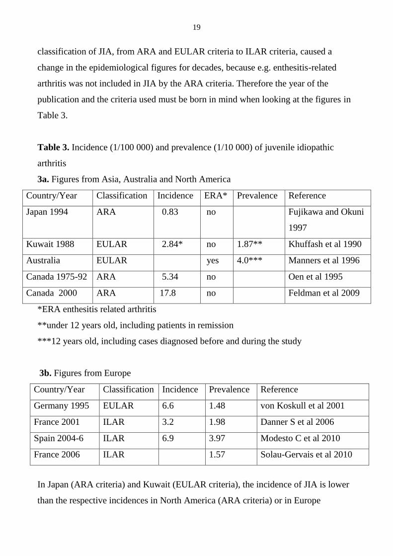
classification of JIA, from ARA and EULAR criteria to ILAR criteria, caused a
change in the epidemiological figures for decades, because e.g. enthesitis-related
arthritis was not included in JIA by the ARA criteria. Therefore the year of the
publication and the criteria used must be born in mind when looking at the figures in
Table 3.
Table 3. Incidence (1/100 000) and prevalence (1/10 000) of juvenile idiopathic
arthritis
3a. Figures from Asia, Australia and North America
Country/Year Classification Incidence ERA* Prevalence Reference
Japan 1994 ARA 0.83 no Fujikawa and Okuni
1997
Kuwait 1988 EULAR 2.84* no 1.87** Khuffash et al 1990
Australia EULAR yes 4.0*** Manners et al 1996
Canada 1975-92 ARA 5.34 no Oen et al 1995
Canada 2000 ARA 17.8 no Feldman et al 2009
*ERA enthesitis related arthritis
**under 12 years old, including patients in remission
***12 years old, including cases diagnosed before and during the study
3b. Figures from Europe
Country/Year Classification Incidence Prevalence Reference
Germany 1995 EULAR 6.6 1.48 von Koskull et al 2001
France 2001 ILAR 3.2 1.98 Danner S et al 2006
Spain 2004-6 ILAR 6.9 3.97 Modesto C et al 2010
France 2006 ILAR 1.57 Solau-Gervais et al 2010
In Japan (ARA criteria) and Kuwait (EULAR criteria), the incidence of JIA is lower
than the respective incidences in North America (ARA criteria) or in Europe
20
(EULAR criteria or ILAR criteria). The prevalence figures are not comparable due to
the different age limits and research strategies.
Berntson et al (2003) compared the incidence rates of JIA by the ILAR criteria and of
JCA by the EULAR criteria in the Nordic countries. The conclusion was that the rates
by the ILAR criteria were slightly higher, obviously due to two reasons: a shorter
duration of symptoms required for the diagnosis and the inclusion of children with
enthesitis-related arthritis.
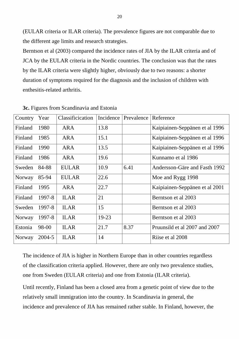
3c. Figures from Scandinavia and Estonia
Country Year Classificication Incidence Prevalence Reference
Finland 1980 ARA 13.8 Kaipiainen-Seppänen et al 1996
Finland 1985 ARA 15.1 Kaipiainen-Seppänen et al 1996
Finland 1990 ARA 13.5 Kaipiainen-Seppänen et al 1996
Finland 1986 ARA 19.6 Kunnamo et al 1986
Sweden 84-88 EULAR 10.9 6.41 Andersson-Gäre and Fasth 1992
Norway 85-94 EULAR 22.6 Moe and Rygg 1998
Finland 1995 ARA 22.7 Kaipiainen-Seppänen et al 2001
Finland 1997-8 ILAR 21 Berntson et al 2003
Sweden 1997-8 ILAR 15 Berntson et al 2003
Norway 1997-8 ILAR 19-23 Berntson et al 2003
Estonia 98-00 ILAR 21.7 8.37 Pruunsild et al 2007 and 2007
Norway 2004-5 ILAR 14 Riise et al 2008
The incidence of JIA is higher in Northern Europe than in other countries regardless
of the classification criteria applied. However, there are only two prevalence studies,
one from Sweden (EULAR criteria) and one from Estonia (ILAR criteria).
Until recently, Finland has been a closed area from a genetic point of view due to the
relatively small immigration into the country. In Scandinavia in general, the
incidence and prevalence of JIA has remained rather stable. In Finland, however, the
21
incidence of another AID, DM1, is the highest in the world and still on the increase
(Harjutsalo et al 2008).
1.4. Uveitis
Approximately 20% of JIA patients will develop chronic uveitis during a longer
follow-up period. Uveitis is particularly common in ANA-positive girls with an
early-onset (before 4 years of age) oligoarthritis during the first years of the disease
(Kotaniemi et al 2001). Patients with JIA need regular ophthalmologist’s
consultations because chronic uveitis is usually asymptomatic.
The prevalence and incidence of uveitis also seem to vary in different countries.
Among 89 Indian JIA patients, the median duration of JIA was 3.5 years, and only
one had uveitis and only one was ANA-positive (Aggarwal and Misra 1996). In
Sweden, 15% developed uveitis during a 4-year follow-up (Andersson- Gäre and
Fasth 1992). Overall, 8.6% of JIA patients developed uveitis during the first six
months of the disease in the Scandinavian countries (Berntsson et al 2003). In
Finland, the presence of uveitis was 5% among patients at the time of the JIA
diagnosis, when 33% of the patients were ANA-positive (Kaipiainen-Seppänen and
Savolainen 2001). In previous Finnish studies the proportion of JIA patients with
uveitis has been 21%, 16% and 24%, respectively (Kunnamo et al 1986, Kotaniemi et
al 1999 and 2001).
1.5. Drug treatment
The treatment of JIA has been somewhat experimental and has undergone significant
changes in the past. In the 1970s in Finland, intramuscular gold, glucocorticoids,
chloroquine preparations and non-steroidal anti-inflammatory drugs (NSAID)
dominated the therapy (Immonen et al 2007). In the early 1980s, azathioprine
(Savolainen et al 1997, Kvien et al 1986, Dale 1972 and Kölle et al 1972) and in the
late 1980s methotrexate (mtx) (Giannini et al 1992) were introduced into the
treatment of JIA. In the early 1990s, the superiority of mtx became widely recognized
22
(Giannini et al 1992, Haapasaari 2006). Thereafter, mtx gained position as the
second-line drug in JIA medication and the gold standard in therapeutic trials
replacing gold proper (Ruperto et al 2004, Ortiz-Alvarez et al 2004).
Among other disease modifying drugs (DMARDs) used for JIA, both sulphasalazine,
which was initially used to treat juvenile spondyloarthropathies (van Rossum et al
1998 and 2007), and an oral gold preparation auranofin are safe but their clinical
effect is poor or only modest (Giannini et al 1990). D-penicillamine and cyclosporin
A have some effect on JIA (van Kerckhove et al 1988, Prieur et al 1985, Gerloni et al
2001, Ruperto et al 2006). Leflunomide has been nearly as efficient and equally well
tolerated as mtx (Silverman et al 2005). Chlorambucil, a potent, established and
historical drug, has only been used in the most severe JIA cases (Savolainen 1999).
Mtx and cyclosporin A are used for severe uveitis (Kotaniemi et al 2003, Petty et al
2003).
In the late 1990s and early 2000s biologic agents, gradually became more widely
used for JIA patients in Finland (Haapasaari 2006). Anti TNFalfa therapy: etanercept,
adalimumab and infliximab, is commonly used in combination with mtx in cases
resistant to mtx alone (Horneff et al 2009, Beukelman 2011). Etanercept has a history
of numeral studies demonstrating its efficacy and safety in JIA (Giannini et al 2009).
Adalimumab has been effective in polyarticular JIA with some effect on uveitis, too
(Lovell et al 2008, Kotaniemi et al 2011).
In the most recent study on a very early drug therapy for JIA from Finland,
infliximab and mtx combined were superior to a combination of three DMARDs and
strikingly superior to mtx alone (Tynjälä et al 2011).
Abatacept blocks T-cell activation. It is recommended for JIA patients who are
resistant to mtx (or other DMARDs) and TNF alpha inhibitors (Beukelman et al
2011).
Rituximab, a CD-20-cell blocker, can be introduced to JIA patients with a high
disease activity after receiving mtx, TNFalpha and abatacept therapy.
23
Anakinra, IL-1-receptor antagonist, and tocilizumab, an antibody against IL-6-
receptor, are especially indicated in systemic JIA (Ilowhite et al 2003, Beukelman
2011).
However, side effects, even rare, should be considered (Scheinfeld 2005). They are
most commonly connected with abnormal laboratory measurements (blood cell
counts, liver enzymes and/or serum creatinine) or infections. Therefore all patients
need regular laboratory monitoring and pediatric rheumatologist screening
(Beukelman et al 2011). The possible long-term side-effects of biologic drugs need
consideration. In Sweden, biologics-naive patients with JIA, who had been followed
up for 40 years, did not have an increased risk of cancer. In stratified analyses,
patients with JIA diagnosed 20-40 years ago did not have an elevated risk, whereas
patients with JIA diagnosed within the past 20 years had an elevated risk of
lymphoproliferative cancers (Simard et al 2010). In a study from the USA, children
with JIA had an increased rate of incident malignancy compared to children without
JIA. The treatment for JIA, including TNF inhibitors, did not appear to be
significantly associated with the development of malignancy, but as for TNF
inhibitors, the follow-up was only two years (Beukelman et al 2012). Until now
respective data have not been available regarding the long-term users of biologic
drugs, and any possible connection between malignancies and biologic medication
needs both register monitoring and controlled studies (Ruperto and Martini 2011).
Biologic medication may also have other rare side-effects, such as lupus or
demyelinating disorders, but any evidence of this is only preliminary (Kahn 2011).
The data gained from adult patients with RA show that an early aggressive
intervention even with conventional medication or by combining the drugs (Korpela
et al 2004, Möttönen et al 1999) helps to achieve clinical remission.
Regarding JIA, recent studies recommend an early introduction of mtx to achieve
maximal effect on rheumatic inflammation (Albers et al 2009b, Cespedes-Cruz et al
2008). The time from symptom onset to diagnosis and treatment was a significant
predictor for inactive disease at 6 months in 356 prospectively studied patients
24
(Oen et al 2009). Inactive disease at 1 year from the disease onset was associated
with the use of mtx and/or oral corticosteroids within the first 6 months in
polyarticular JIA patients (Ringold et al 2009). Remission, even in the short term,
may prevent later joint damage and consequently also functional impairment (Albers
et al 2009). Patients responding favorably to mtx at 6 months from the disease onset
had an improved outcome (Bartoli et al 2008). The improvement was seen in the
physical domain of health-related quality of life (HRQL) and responses in the ACR
pediatric 70 definition (Cespedes-Cruz et al 2008, Giannini et al 1997).
1.6. Outcome
The outcome of JIA has varied depending on the study period. Laaksonen studied
544 JIA patients treated at the RFH in 1951-1961. The disease was active in 42% of
the patients depending on the duration of the disease: in 41%, 46% and 41%, if the
duration was 3-7, 8-15 or 16 or more years, respectively. Only 30.1% of the patients
had preserved complete functional capacity: 51%, 34.5% and 9.6%, if the duration
was 3-7, 8-15, 16 or more years, respectively (Laaksonen 1966).
Ylijoki studied 174 JIA patients at 15 years after the onset of the disease in 1965-
1974. The functional capacity was normal in two-thirds of the patients, and severe or
moderate lack of functional capacity was present in only 10%. Thus, the outcome was
significantly better than in the earlier study of Laaksonen (Ylijoki 1998).
Arkela-Kautiainen et al studied 123 JIA patients, born in 1976-1980 diagnosed and
treated at the RFH, at the age of 21-26. Spousal relationship, educational level and
employment status were similar to those of the controls. Their HRQL results were
similar to those in the controls with only one exception, the physical functions scale.
As expected, patients with an active disease had poorer HRQL when weighted
against the physical component. The extended oligoarthritis group had the lowest
physical and mental scores in HRQL among all JIA subgroups
25
(Arkela-Kautiainen et al 2005). The authors concluded that JIA patients’ outcome has
improved with better diagnostic methods and improved treatment.
An early introduction of mtx improves the long-term outcome in JIA patients (Albers
et al 2009b, Cespedes-Cruz et al 2008), but mtx alone is not sufficient for all patients.
Therefore, it is very important to recognize the prognostic factors or indicators of this
poor outcome. The known risk factors for the poor outcome are the disease duration,
the presence of RF and HLAB-27 positivity (Savolainen et al 1998). In a population-
based, long-term Nordic cohort study the remission rate was highest in the
oligoarticular persistent and systemic JIA subgroups (Nordal et al 2011). The
outcome of juvenile psoriatic arthritis seems to be associated with an unfavorable
outcome compared to oligo- or polyarthritis (Flato et al 2009).
1.6.1. Psychological outcome
In general, children and adolescents with a chronic illness face a greater risk to
develop internalizing problems (e.g. anxiety, depression, social withdrawal), but there
are individual differences in their responses to life stress caused by the illness
(LeBovidge et al 2003).
In 1974, a study from the USA revealed that children with CA had more
psychological difficulties than their healthy peers (McArney et al 1974). In 1988, 363
Australian JIA patients filled out a questionnaire aimed at assessing disease activity,
psychosocial functioning and adjustment with the disease. Psychological function and
disease activity scores associated with adjustment scores in both elementary and high
school groups, whereas social scores associated with adjustment only in the high
school group. The associations between the measures of psychological functioning,
social relationships, severity of the disease and adjustment to the disease were minor
in young adults. The authors emphasized the importance of using a developmental
model to understand the adjustment of patients to JIA (Ungerer et al 1988).
In Finland, the rate of suicides among young people between 15 and 24 years of age
26
has increased in the 1980s, being about 22/100 000. The incidence among patients
with JIA was two-fold which is alarming (Savolainen and Isomäki 1993).
In 82 adults with JIA from the UK with an average of 21 year disease duration and 30
years of age, mental summation scores were lower than in controls. The result was
similar in all subtypes of JIA and independent of the degree of functional disability.
Furthermore, despite an excellent educational attainment, there was a high rate of
unemployment among the patients (Foster et al 2003).
In view of the recent studies from western countries, JIA patients seem to have quite
a normal psychosocial outcome. This is shown in a study from the UK regarding 60
polyarthritis type of JIA patients aged 7-18 (Ding et al 2008). A similar result was
found in a Finnish study of 123 JIA patients aged 21-26 (Arkela-Kautiainen et al
2005). Another Finnish study showed that 142 JIA patients aged 8-15 with their
disease lasting for at least one year seemed to cope quite well despite the disease. The
level of pain was not alarmingly high, self-efficacy was good and no severe
depression or anxiety was detected (Vuorimaa 2010).
In Norway, 55 young adults with JIA, in mean18.3 years after the onset of the
disease, did not differ in their psychosocial health compared to the general
Norwegian population. The level of education was even higher, and there was no
difference in their employment status (Ostile et al 2010).
In every aspect, the outcome of JIA has shown radical improvement over the past
decades.
27
2. OTHER AUTOIMMUNE DISEASES
2.1. Diabetes mellitus type 1
2.1.1. Definition, diagnostic criteria, clinical characteristics
DM1 is perceived as a chronic immune-mediated disease with a subclinical
asymptomatic, prodromal period of highly variable duration. The pathological
process causes selective loss of insulin-producing beta-cells in the pancreatic islets in
genetically susceptible subjects (Knip and Siljander 2008). DM1 is characterised by
chronic hyperglycemia resulting from defective insulin secretion. Diagnostic criteria
are based on blood glucose measurements, and when required also on an oral glucose
tolerance test. Serological markers of an autoimmune pathological process, including
islet cell antibodies (ICA), or insulin antibodies (IAA), are present in 85-90% of the
patients when fasting hyperglycemia is detected.
DM1 in children usually presents itself with characteristic symptoms such as
polyuria, polydipsia, fatigue and weight loss in association with glucosuria and
ketonuria (Craig et al 2009).
2.1.2. Epidemiology,
The incidence of DM1 varies greatly between different countries and ethnic groups.
Based on several studies, DM1 is more common in Finland than in any other country.
In 1996, the incidence was 45/100 000 children aged 14 years or younger
(Tuomilehto et al 1999). In fact, the incidence is the world’s highest in Finland and
still growing. During 1980-2005, the average age-standardized incidence was
42.9/100 000, increasing from 31.3/100 000 in 1980 to 64.2/100 000 in 2005
(Harjutsalo et al 2008, Myers et al 2008). In the most recent study, the prevalence of
pediatric DM1 in the Helsinki region was 3.7/1000 (Oilinki et al 2012).
28
2.2. Celiac disease, juvenile autoimmune thyroiditis and multiple
sclerosis
Celiac disease (CD) is an AID triggered by wheat, barley and rye proteins in
genetically susceptible individuals and is known to have classic symptoms caused by
malabsorption and failure to thrive. CD may remain non-symptomatic for years. In an
extensive population-based study, the prevalence of CD was 1% in Finnish
schoolchildren (Mäki et al 2003).
Juvenile autoimmune thyroiditis (JAIT), also called chronic lymphocytic thyroiditis
or Hashimoto’s disease, is the most common cause of thyroid enlargement in children
and adolescents. No population-based data are available on the occurrence of
autoimmune thyroiditis in Finland. In the USA, the incidence has varied from 5 to
6.5/1000 children (Rallison et al 1975). In Finland and Israel, JAIT was leading to
hypothyroidism (HT) in about 70% of the cases (Mäenpää et al 1985, de Vries et al
2009).
Multiple sclerosis (MS) is an AID with a complex aetiology with involvement of both
genetic and environmental factors. MS is very rare in children; less than 10% of all
cases occur under 18 years of age (Chabas et al 2008). The estimated MS prevalence
in Finland is 1.0/1000, in the over 10-year-old population, with pronounced regional
variations (Sumelahti et al 2001). In the UK, the prevalence was 1.46/1000 in 2005 in
the total population (Hirst et al 2008).
2. CONNECTION BETWEEN JUVENILE IDIOPATHIC ARTHRITIS AND
OTHER AUTOIMMUNE DISEASES
AIDs are chronic conditions, where the immunological environmental and genetic
factors are involved (Becker et al 1998). In the USA, the overall prevalence of
autoimmunity is estimated at 5% (Jacobson et al 1997). JIA has been described as a
complex genetic trait where multiple genes interact and result in a specific phenotype
(Angeles-Han and Prahalad 2010). Therefore, genetic studies of JIA, including those
29
regarding the link to other AIDs, are difficult. Many presenting phenotypes resulting
from different and complex pathogenetic mechanisms are likely to involve different
susceptibility genes (Phelan et al 2006).
The autoimmune regulatory (AIRE) gene mutations result in a defective AIRE
protein. This protein is essential for self-tolerance. AIRE gene mutations are
associated with multiple autoimmune diseases and e.g. systemic type of JIA
(Podkrajsek et al 2008). Autoimmune polyendocrine syndromes (APS) are rare
autosomal recessive disorders characterized by autoimmune multiorgan attack
(Weiler et al 2012).
3.1. Autoimmune diseases in children with JIA
The identification of genes behind JIA and other AIDs has improved over the recent
years (Albers et al 2009a, Hinks et al 2009, Hinks et al 2010). There is increasing
evidence that AIDs, such as JIA and DM1, share the same susceptibility genes. For
example, PTPN22 SNP has been strongly associated with both JIA and DM1 (Hinks
et al 2005). In addition, IL2RA/CD25, which regulates interleukin-2 receptor
expression, bears a connection with JIA and DM1 inheritance and represents a
susceptibility locus for both diseases (Hinks et al 2009). Albers et al recently
confirmed that autoimmunity locus 4q27 previously associated with DM1 was also
associated with the polyarticular JIA (Albers et al 2009a).
3.1.1. DM 1 in children with JIA
The incidence of JIA in Finnish children is quite similar to that in other Nordic
populations (Berntsson et al 2003). Instead, the incidence of DM1 in Finland is the
world’s highest and still on the increase (Harjutsalo et al 2008).
The few publications on patients having both JIA and DM1 are mostly case reports
(Castleman and McNeely 1968, Fisher et al 1980, Aggarwal and Meena 2003, Nagy
et al 2010).
The first pediatric patient with arthritis and diabetes whom we found in the literature
30
was a 7-year-old girl reported in 1968 (Castleman and McNeely 1968). Since then,
ten patients with JIA and DM1 have been reported, and nine of them also had
thyroiditis (Fisher et al 1980, Rudolf et al 1981, Aggarwal and Meena 2003, Nagy et
al 2010).
In the USA, seven patients with JIA were found among 200 diabetic children. Six of
them had polyarthritis with RF and/or ANAs and evidence of thyroid problems
(Rudolf et al 1981).
When 66 Italian JIA patients were screened for pre-diabetic autoantibodies, only 3%
showed any positivity (Alpigiani et al 2002). On the other hand, children with JIA in
the USA had a 6-fold prevalence of DM1 compared to the population prevalence
(Prahalad et al 2004).
3.1.2 Other autoimmune diseases in children with JIA
Studies in adults have shown that AIDs cluster in the same patients (Viljamaa et al
2005). There is preliminary evidence that a similar clustering may also take place in
children. In Finland, CD was discovered in 3 out of 150 JIA patients (Mäki et al
1988). In line, the prevalence of HT in Italian JIA patients was 4-fold (Stagi et al
2005) and the prevalence of CD 8-11-fold compared with the control goup (Lepore et
al 1996, Stagi et al 2005). As many as 44 % of the Bulgarian (Mihailova et al 1999),
14% of the Italian (Stagi et al 2005), 7-11% of the Israeli (Harel et al 2006), and 5%
of the Turkish (Unsal et al 2008) JIA patients had laboratory findings suggestive of
JAIT, although nearly all patients were euthyroid.
3.2. Autoimmune diseases in JIA children’s first-degree relatives
AIDs seem to cluster in children with JIA (Lepore et al 1996, Prahalad et al 2002,
Stagi et al 2005, Alpigiani et al 2008). Reports suggesting that AIDs, such as CA,
may also cluster in JIA children’s families have been published since the 1950s
(Wittinghill et al 1958, Ansell et al 1962, Andersson-Gäre and Fasth 1995, Säilä et al
31
2003). JIA itself has a tendency to cluster in families (Säilä et al 2003, Prahalad et al
2004 and 2010). The approximate effect of familial factors attributing to the risk for
JIA was recently calculated to be 13% in a study from the USA (Prahalad et al 2010).
In Finland, the sibling occurrence risk in JIA families is approximately 15-20-fold
(Säilä 2006).
The occurrence of AIDs in JIA families has been rarely compared with the population
data. The prevalence of spondyloarthropathy in parents of 70 children with JIA was
16-fold in the Netherlands (Hertzberger-ten and Dijkmans 1993). In the USA, the
population prevalence of AIDs is about 5%, and the figure was 16.1% among JIA
patients’ first-degree relatives (Prahalad et al 2002). In Taiwan, there was a family
member with AID in 4.5% of the 110 JIA patients’ families. Of individual family
members, 6.4% had an AID (Huang et al 2004).
There are two previous controlled studies from the USA regarding the clustering of
AIDs in families with JIA children (Prahalad et al 2002 and 2004). In an interview
study of 110 JIA families, the prevalence of AIDs was increased as compared to 45
control families. Among individual AIDs, the difference was statistically significant
only for thyroiditis (Prahalad et al 2002).
32
PURPOSES OF THE PRESENT STUDY
The main purposes of the thesis were to evaluate the clustering of AIDs in children
with JIA and in their family members, as well as the recent trends in medical
treatment of JIA.
The specific aims were
1) to assess the prevalence of AIDs, including DM1, CD and HT in JIA patients (I)
2) to evaluate the occurrence of AIDs, including DM1, CD, CA and MS in families
of children with JIA (II)
3) to establish the prevalence and to describe the clinical characteristics of patients
with simultaneous JIA and DM1 (III)
4) to establish the trends in medical treatment of JIA in years 2000-2007, with a
special focus on early treatment during the first 3 and 12 months after disease
onset, respectively (IV)
5) to estimate what effect DM1 has on the drug treatment for JIA (unpublished data)
33
PATIENTS AND METHODS
The Rheumatism Foundation Hospital (RFH) in Heinola, Finland, was a tertiary
centre for patients with chronic inflammatory articular diseases. Approximately 60-
70% of JIA patients were referred to the RFH from the early 1950s onwards. Two
original articles of the present thesis are based on the medical and family histories
and clinical characteristics of JIA patients who for the first time were admitted to the
RFH (I, II).
The Social Insurance Institution (SII) in Finland maintains registers on individuals
who are granted a special reimbursement for medication for defined chronic diseases.
To establish this entitlement, a doctor’s certificate based on clinical examination is
required. In the case of JIA and AIDs, the diagnoses must have been made by
paediatricians (DM1) according to the ICD-10 classification, and the treatment plan
must follow good clinical practice. The certificates are checked by an expert
physician at the SII, before any special reimbursement is granted. In addition, the SII
keeps a register of the prescription drugs which are entirely or partly free of charge.
Access to the SII registers to collect information from the patient files requires an
authorization from the Ministry of Social Affairs and Health. Two original articles
(III, IV) and the presented unpublished data are based on the data collected from the
SII registers.
1. Autoimmune diseases in children with JIA (I)
All children aged under 16 years admitted to the RFH during 1992-2000 and
diagnosed with JIA were recruited into the study. All diagnoses were revised from
the patient files according to the ILAR criteria (Petty et al 2004). The study subjects
were identified from the ophthalmologist’s register, because all patients admitted to
the RFH for the first time underwent an ophthalmologist’s evaluation. Patients
admitted solely for eye problems were excluded. Clinical data were collected,
including the age at JIA diagnosis, course type of JIA and the presence of uveitis,
34
from the patient registers by using a structured form. The number of patients was
417, consisting of 283 girls and 134 boys.
There were 5 sibling pairs, including one set of twins and one family with 3 affected
daughters. The follow-up of 191 patients lasted until their 16th birthday. The rest, 226
patients, were followed-up until 31st January 2006.
The population data were collected from the Official Statistics of Finland. Children
with DM1, CD and HT were identified from the SII registers. The average numbers
of children of 16 years or younger at the end of the years 2003, 2004, 2005 and 2006
were calculated from both population and SII registers, and the figures were then
compared with the respective figures of the 417 study patients.
2. Autoimmune diseases in JIA children’s first-degree relatives (II)
The families of all consecutive JIA patients admitted first time to the RFH in 1996-
2001 were identified by using the hospital registers. All diagnoses were revised by
HP from the patient files in March 2007 according to the ILAR criteria (Petty et al
2004).
In winter 2003-2004, a questionnaire was posted to 432 patients. In case they did not
reply, a new one was sent out within a year. No response was received from 46
patients, 15 letters were returned marked as “address unknown”, and nine patients
were excluded because of incomplete information (e.g. adoption, paternal
information unavailable). Complete data were thus obtained from 362 (84%) patients.
The questions concerned CA, CD, DM1 and MS diagnoses of the index patients’
first-degree relatives (mothers, fathers, full siblings). The diagnoses (recorded
according the ICD-10 classification) had to be made by a physician.
3. Simultaneous occurrence of JIA and DM1 in a same patient (III)
Starting from 1965, the SII has provided reimbursement for prescription drugs for
DM and from 1966 also for chronic rheumatic diseases including JIA. Injectable
35
drugs, e.g. intra-articular glucocorticoids are not reimbursed for and therefore not
registered since they are administered free in inpatient and outpatient clinics.
Biologic agents are administered in outpatient or inpatient clinics, or require specific
certificates when administered at home; data on these drugs were not collected.
The patients were identified from the SII registers on the basis of reimbursement for
medication for both CA and DM1 granted for the first time between 1 January 1976
and 31 December 2005. The register files of the SII provide basic data like the
patients’ birth date, gender and residential area and the date of the reimbursement
decision. In Finland, according to an established practice, certain chronic diseases in
children like DM1 and JIA are treated in the secondary-level central hospitals.
During the 30-year surveillance period, a total of 240 patients were reimbursed for
both CA and DM. The 112 patients who were reimbursed for arthritis at 21 years or
older were excluded. The age limit of 20 for JIA was chosen because of the
possibility that the application for reimbursement had been filed with delay, which for
DM is unlikely. The files of the remaining 128 patients were further checked to
ascertain the diagnoses and the patients’ exact age at the onset of the diseases. Twenty
had adult onset RA, three had DM secondary to glucocorticoids and five had DM
type 2. Thirteen patients had later been re-diagnosed as having another disease than
JIA. Four potential patients were excluded because their files could not be located
and one patient was not included in the study period and was thus excluded. The
remaining 82 patients who met the study criteria were classified according to the
ILAR criteria. Data on laboratory markers, radiological changes, uveitis, and the use
of intra-articular and systemic glucocorticoids, were collected by charting the
patients’ hospital records.
The study patients were divided into two groups depending on which disease, JIA or
DM1, had started first. The activity of JIA was assessed on the basis of inflammatory
parameters, erythrocyte sedimentation rate (ESR) and C-reactive protein (CRP), at
the onset of the disease, on the presence of erosions in native radiology and on the
use of systemic glucocorticoids. The need of systemic glucocorticoids or the need of
36
six or more intra-articular injections in any one year was set as criteria for an active
disease.
4. Trends in the medical treatment practice of JIA in years 2000-2007 (IV)
Patients with certain chronic and severe diseases, such as idiopathic inflammatory
rheumatic diseases, are entitled to a special (72% or 100%) reimbursement for
medication from the SII if their condition meets the defined criteria. In JIA, the
special reimbursement covers DMARDs and glucocorticoids but not NSAIDs. The
new biologic agents are granted separately with stringent criteria, and they are not
intended for the first-line use. The reimbursement decisions are gathered in a register
maintained by the SII, as are all reimbursable drugs purchased on doctor’s
prescription. From this database, the patients aged 16 years or younger, who were
first granted a special reimbursement for drugs for JIA in years 2000- 2007, were
identified.
Children with the following ICD-10 diagnoses were included: M08 (prolonged
childhood arthritis) either with a more specific subcategory (n=458) or without
(n=1316), L40.5 psoriatic arthritis (n=33), M45 spondyloarthropathies (n=13),
M46.1 sacroiliitis of no obvious other cause (n=6), M05 seropositive RA (n=24),
M06 seronegative or non-specified RA (n= 36), and M13.9 non-specified arthritis (n=
22). A total of 1970 children with CJA (ICD10 M08) were thus identified.
We collected the data on prescribed medication purchased by JIA patients in 2000-
2007. The introduction of DMARDs, whether single or combined, was the primary
interest of the study, with a special focus on mtx.
Intra-articular and intravenous glucocorticoids like all the intravenous medications
are not included in this study, because these are provided at inpatient and outpatient
clinics without any written prescription. With the exception of glucocorticoids,
intravenous medications were practically never given during the first year. The use of
oral glucocorticoids was not analyzed during 2006-2007 because of a temporary
change in the reimbursement system.
37
5. The effect of simultaneous DM1 on the drug treatment for JIA (unpublished
data)
As described above, patients with simultaneous JIA and DM1 were identified from
the SII registers from 1 January 2000 to 31
December 2007. Among the 1970 patients
who were granted a special reimbursement for drugs to treat CJA were patients who
were already reimbursed for the treatment of DM with insulin, were selected. Twenty
patients were reimbursed for drugs to treat CJA and DM1 and 3 to treat CJA, DM and
HT.
We collected the same data as in the study above on prescribed medication purchased
for JIA patients in 2000-2007 (IV). In addition, data were collected on prescribed
medication purchased by those 23 patients who already received reimbursement for
insulin to treat DM1 and for drugs to treat CJA.
The treatment of the 23 JIA patients with simultaneous DM1 was compared to the
treatment of all the 1970 JIA patients. Our primary interest was to check the possible
delay in the introduction of mtx, with a focus on treatment in the first 3 and 12
months of the disease.
6.Statistical methods
The results are expressed as mean figures with standard deviations (SD), medians
with interquartile ranges (IQR) or counts with percentages. Descriptive values were
expressed with 95% CI. Statistical comparison between groups was made by t-test,
Chi-Square test, Fisher exact test or permutation test (Monte Carlo p-value) where
appropriate. In the study, the occurrence of the common AI diseases DM1, CD and
HT as diagnosed by clinical criteria in children with JIA, their families and in the
general population was compared, the results were expressed as prevalence rate
ratios (PRR) with 95% CI. Gender- and age- matched samples of the general
population were obtained from data in the Official Statistics of Finland (I,II).
38
In the third study, the estimates of occurrence rate ratios (ORR) were calculated by
using Poisson regression models (III).
In the fourth and fifth study statistical significance for hypotheses of linearity was
evaluated by using Cochran-Armitage test (IV,V).
39
RESULTS
1. Autoimmune diseases in children with JIA (I)
The median age of the 417 patients admitted to the RFH in 1996-2000 and eligible to
the study was 7 years (range 12 months to 15 years). At the end of the study in 2006,
the median follow-up time from the JIA diagnosis was 7.5 years (range 5.5 - 9.5
years).
The course type of JIA was oligoarthritis in 22%, extended oligoarthritis in 36%, and
seronegative polyarthritis in 33%. All other course types each formed less than 5%.
Nine patients had DM1 [2.2% (95 CI 1.0 to 4.1)] (Table 4). DM1 preceded JIA in 7/9
(78%) cases. The PRR for DM1 between the JIA patients and the general population
was 5.0 (95% CI 2.3 to 9.5).
Three patients, all with seronegative polyarthritis, had CD [0.7% (0.1-2.1)] (Table 4).
All the 3 CD diagnoses were confirmed by a small bowel biopsy from children with
intestinal symptoms. CD was diagnosed before JIA in 1/3 cases. Gluten-free diet did
not improve their joint symptoms. The PRR for CD between the JIA patients and
general population was 5.6 (95% CI 1.2 to 16.5).
Three patients had HT [0.7% (0.1 to 2.1)], and they were all on thyroxin substitution
(Table 4). The JIA diagnosis was either extended oligoarthritis or seronegative
polyarthritis, and DM1 and CD were present in 2 cases. HT was diagnosed before
JIA in 2/3 (67%) cases. The PRR for HT between the JIA patients and the general
population was 5.6 (1.2 to 16.5).
In summary, 13/417 (3.1%) of the children with JIA had another AID. Only one
(7.7%) of them, a girl with seronegative polyarthritis and CD, had uveitis, compared
with 90 uveitis cases (22%) in the group of JIA patients without another AID
(p=0.18).
40
Table 4. Clinical diabetes mellitus type 1, celiac disease and hypothyreosis in 417
children with juvenile idiopathic arthritis and their relative risk compared to general
population.
Autoimmune disease Number (%) RR (95% CI)
Diabetes mellitus type 1 9 (22) 5.0 (2.3 to 9.5)
Celiac disease 3 (0.7) 5.6 (1.2 to 16.5)
Autoimmune
hypothyreosis
3 (0.7) 5.6 (1.2 to 16.5)
2. Autoimmune diseases in JIA children’s first-degree relatives (II)
Since JIA patients were five times more likely to have another AID than their age
mates, the link between JIA and AIDs was evaluated by sending a questionnaire
concerning AIDs to the families of JIA patients. The eligible 432 patients had been
admitted to the RFH in 1996-2001, and by the end of the year 2007, 355 families had
answered.
The 355 families consisted of 24 (5.8%) multiple-JIA families; 6 JIA cases were
diagnosed in siblings during the study period at the RFH and 18 siblings were either
diagnosed elsewhere or in the RFH outside the study period. All of the 710 parents
were of Finnish origin. The mean age of the mothers was 45 years (range 28-66) and
that of the fathers 47 years (31-74). The mean child count per family was 2.6 (range
1-18), including the index-JIA patients. The total number of the index patients was
362, their siblings 528, totalling 890 children and 1600 study subjects.
Of the 362 index JIA patients, 241 (67%) were girls and 121 (33%) boys, and 331
(91%) belonged to single-JIA and 31 (9%) to multiple-JIA families. At the end of the
41
study, the mean age of the girls was 15.5 years (range 7-27) and that of the boys 15.7
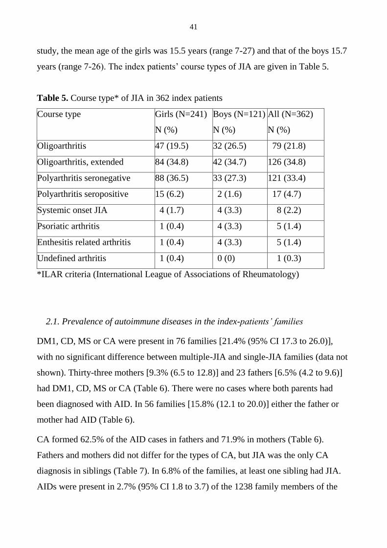
years (range 7-26). The index patients’ course types of JIA are given in Table 5.
Table 5. Course type* of JIA in 362 index patients
Course type Girls (N=241)
N (%)
Boys (N=121)
N (%)
All (N=362)
N (%)
Oligoarthritis 47 (19.5) 32 (26.5) 79 (21.8)
Oligoarthritis, extended 84 (34.8) 42 (34.7) 126 (34.8)
Polyarthritis seronegative 88 (36.5) 33 (27.3) 121 (33.4)
Polyarthritis seropositive 15 (6.2) 2 (1.6) 17 (4.7)
Systemic onset JIA 4 (1.7) 4 (3.3) 8 (2.2)
Psoriatic arthritis 1 (0.4) 4 (3.3) 5 (1.4)
Enthesitis related arthritis 1 (0.4) 4 (3.3) 5 (1.4)
Undefined arthritis 1 (0.4) 0 (0) 1 (0.3)
*ILAR criteria (International League of Associations of Rheumatology)
2.1. Prevalence of autoimmune diseases in the index-patients’ families
DM1, CD, MS or CA were present in 76 families [21.4% (95% CI 17.3 to 26.0)],
with no significant difference between multiple-JIA and single-JIA families (data not
shown). Thirty-three mothers [9.3% (6.5 to 12.8)] and 23 fathers [6.5% (4.2 to 9.6)]
had DM1, CD, MS or CA (Table 6). There were no cases where both parents had
been diagnosed with AID. In 56 families [15.8% (12.1 to 20.0)] either the father or
mother had AID (Table 6).
CA formed 62.5% of the AID cases in fathers and 71.9% in mothers (Table 6).
Fathers and mothers did not differ for the types of CA, but JIA was the only CA
diagnosis in siblings (Table 7). In 6.8% of the families, at least one sibling had JIA.
AIDs were present in 2.7% (95% CI 1.8 to 3.7) of the 1238 family members of the
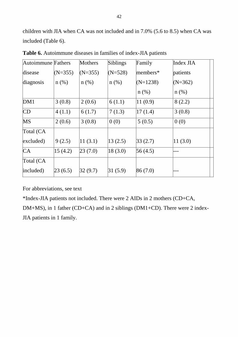
42
children with JIA when CA was not included and in 7.0% (5.6 to 8.5) when CA was
included (Table 6).
Table 6. Autoimmune diseases in families of index-JIA patients
Autoimmune
disease
diagnosis
Fathers
(N=355)
n (%)
Mothers
(N=355)
n (%)
Siblings
(N=528)
n (%)
Family
members*
(N=1238)
n (%)
Index JIA
patients
(N=362)
n (%)
DM1 3 (0.8) 2 (0.6) 6 (1.1) 11 (0.9) 8 (2.2)
CD 4 (1.1) 6 (1.7) 7 (1.3) 17 (1.4) 3 (0.8)
MS 2 (0.6) 3 (0.8) 0 (0) 5 (0.5) 0 (0)
Total (CA
excluded)
9 (2.5)
11 (3.1)
13 (2.5)
33 (2.7)
11 (3.0)
CA 15 (4.2) 23 (7.0) 18 (3.0) 56 (4.5) ---
Total (CA
included)
23 (6.5)
32 (9.7)
31 (5.9)
86 (7.0)
---
For abbreviations, see text
*Index-JIA patients not included. There were 2 AIDs in 2 mothers (CD+CA,
DM+MS), in 1 father (CD+CA) and in 2 siblings (DM1+CD). There were 2 index-
JIA patients in 1 family.
43
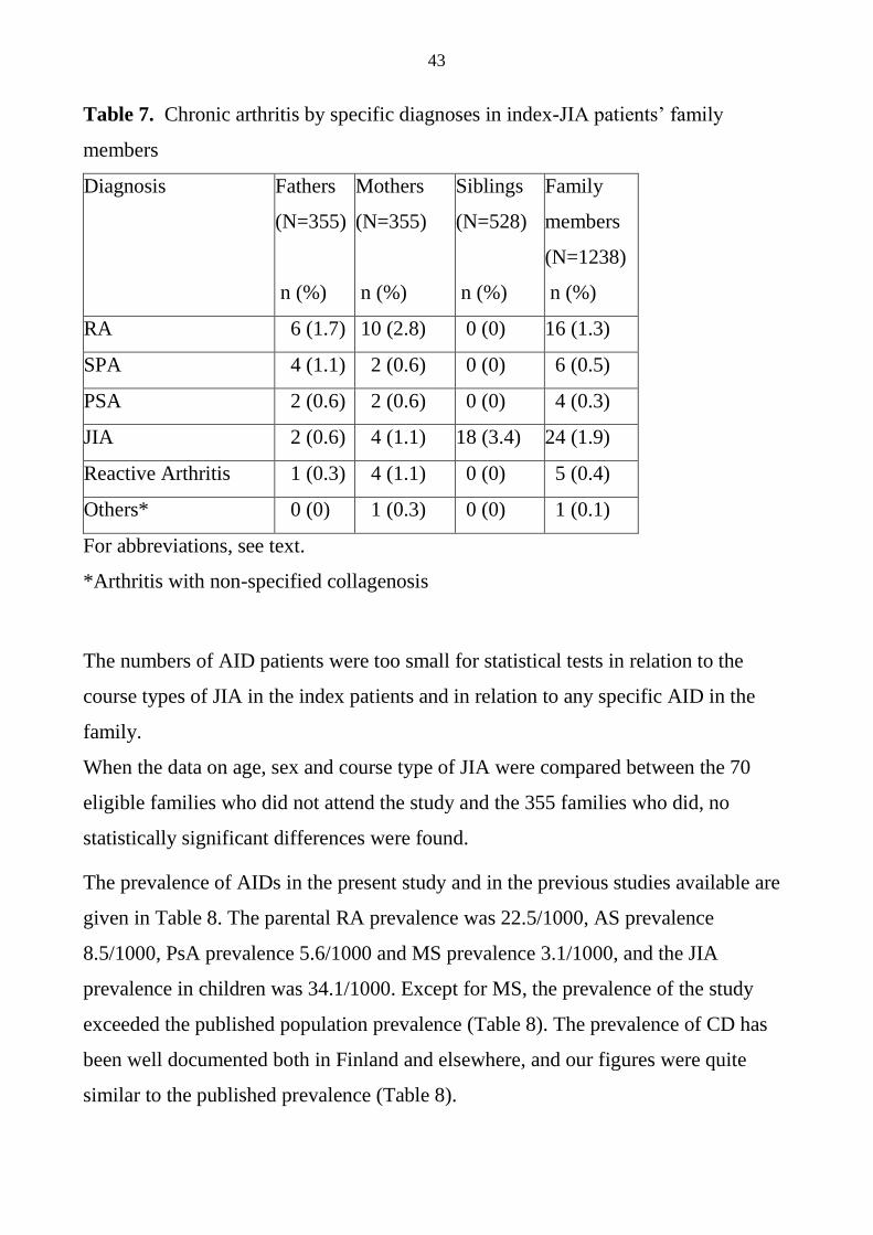
Table 7. Chronic arthritis by specific diagnoses in index-JIA patients’ family
members
Diagnosis Fathers
(N=355)
n (%)
Mothers
(N=355)
n (%)
Siblings
(N=528)
n (%)
Family
members
(N=1238)
n (%)
RA 6 (1.7) 10 (2.8) 0 (0) 16 (1.3)
SPA 4 (1.1) 2 (0.6) 0 (0) 6 (0.5)
PSA 2 (0.6) 2 (0.6) 0 (0) 4 (0.3)
JIA 2 (0.6) 4 (1.1) 18 (3.4) 24 (1.9)
Reactive Arthritis 1 (0.3) 4 (1.1) 0 (0) 5 (0.4)
Others* 0 (0) 1 (0.3) 0 (0) 1 (0.1)
For abbreviations, see text.
*Arthritis with non-specified collagenosis
The numbers of AID patients were too small for statistical tests in relation to the
course types of JIA in the index patients and in relation to any specific AID in the
family.
When the data on age, sex and course type of JIA were compared between the 70
eligible families who did not attend the study and the 355 families who did, no
statistically significant differences were found.
The prevalence of AIDs in the present study and in the previous studies available are
given in Table 8. The parental RA prevalence was 22.5/1000, AS prevalence
8.5/1000, PsA prevalence 5.6/1000 and MS prevalence 3.1/1000, and the JIA
prevalence in children was 34.1/1000. Except for MS, the prevalence of the study
exceeded the published population prevalence (Table 8). The prevalence of CD has
been well documented both in Finland and elsewhere, and our figures were quite
similar to the published prevalence (Table 8).
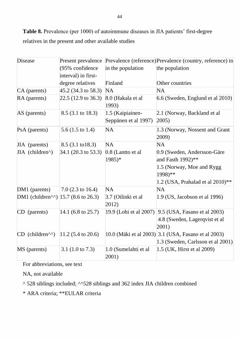
44
Table 8. Prevalence (per 1000) of autoimmune diseases in JIA patients’ first-degree
relatives in the present and other available studies
Disease
Present prevalence
(95% confidence
interval) in first-
degree relatives
Prevalence (reference)
in the population
Finland
Prevalence (country, reference) in
the population
Other countries
CA (parents) 45.2 (34.3 to 58.3) NA NA
RA (parents)
22.5 (12.9 to 36.3) 8.0 (Hakala et al
1993)
6.6 (Sweden, Englund et al 2010)
AS (parents)
8.5 (3.1 to 18.3)
1.5 (Kaipiainen-
Seppänen et al 1997)
2.1 (Norway, Backland et al
2005)
PsA (parents) 5.6 (1.5 to 1.4) NA 1.3 (Norway, Nossent and Grant
2009)
JIA (parents)
JIA (children^)
8.5 (3.1 to18.3)
34.1 (20.3 to 53.3)
NA
0.8 (Lantto et al
1985)*
NA
0.9 (Sweden, Andersson-Gäre
and Fasth 1992)**
1.5 (Norway, Moe and Rygg
1998)**
1.2 (USA, Prahalad et al 2010)**
DM1 (parents)
DM1 (children^^)
7.0 (2.3 to 16.4)
15.7 (8.6 to 26.3)
NA
3.7 (Oilinki et al
2012)
NA
1.9 (US, Jacobson et al 1996)
CD (parents)
CD (children^^)
14.1 (6.8 to 25.7)
11.2 (5.4 to 20.6)
19.9 (Lohi et al 2007)
10.0 (Mäki et al 2003)
9.5 (USA, Fasano et al 2003)
4.8 (Sweden, Lagerqvist et al
2001)
3.1 (USA, Fasano et al 2003)
1.3 (Sweden, Carlsson et al 2001)
MS (parents) 3.1 (1.0 to 7.3)
1.0 (Sumelahti et al
2001)
1.5 (UK, Hirst et al 2009)
For abbreviations, see text
NA, not available
^ 528 siblings included; ^^528 siblings and 362 index JIA children combined
* ARA criteria; **EULAR criteria

45
3. Simultaneous occurrence of JIA and DM1 in the same patient (III)
JIA patients and their first-degree relatives have a higher risk for AIDs than the
general population. This association was studied more accurately in terms of JIA and
DM1.
The SII statistics from 1976-2005 identified 82 patients with simultaneous JIA and
DM1, consisting of 55 (67%) girls and 27 (33%) boys. The subtypes of the patients,
classified according to the ILAR criteria, are presented in Table 9.
Table 9. Subtypes of JIA in 82 patients with both JIA and DM1.
JIA Onset type
N (%)
Course type
N (%)
Systemic onset arthritis 1 (1.2) 1 (1.2)
Oligoarthritis 45 (55) 33 (40)
Oligoarthritis, extended 11 (13)
Polyarthritis seropositive 12 (15) 12 (15)
Polyarthritis seronegative 22 (27) 22 (27)
Enthesitis related arthritis 2 (2) 2 (2)
Psoriatic arthritis 0 (0) 1 (1.2)
3.1. Occurrence
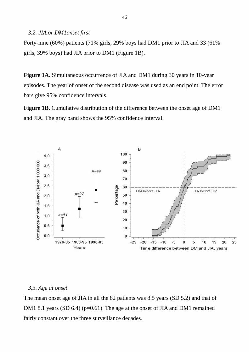
Simultaneous JIA and DM1 occurrence during the 30-year period is shown in Figure
1A. There was a statistically significant increase in this occurrence (age and sex
adjusted p for linearity <0.001): 4.49-fold [95% CI (2.32 to 8.69)] between the first
(1976-1985) and last (1996-2005) periods.
46
3.2. JIA or DM1onset first
Forty-nine (60%) patients (71% girls, 29% boys had DM1 prior to JIA and 33 (61%
girls, 39% boys) had JIA prior to DM1 (Figure 1B).
Figure 1A. Simultaneous occurrence of JIA and DM1 during 30 years in 10-year
episodes. The year of onset of the second disease was used as an end point. The error
bars give 95% confidence intervals.
Figure 1B. Cumulative distribution of the difference between the onset age of DM1
and JIA. The gray band shows the 95% confidence interval.
3.3. Age at onset
The mean onset age of JIA in all the 82 patients was 8.5 years (SD 5.2) and that of
DM1 8.1 years (SD 6.4) (p=0.61). The age at the onset of JIA and DM1 remained
fairly constant over the three surveillance decades.
47
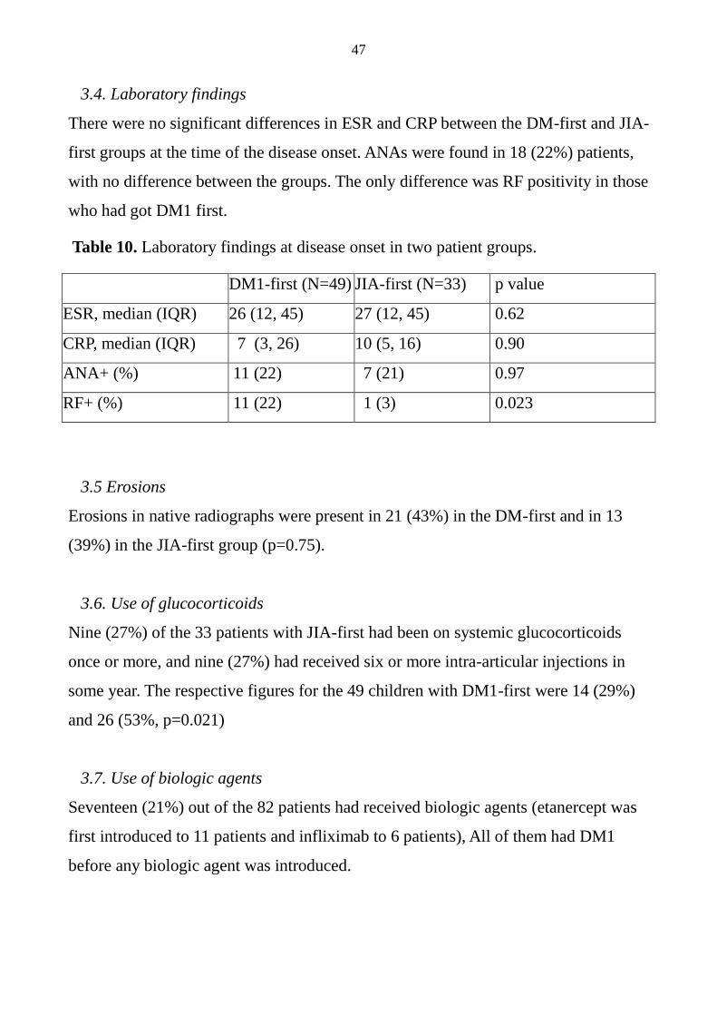
3.4. Laboratory findings
There were no significant differences in ESR and CRP between the DM-first and JIA-
first groups at the time of the disease onset. ANAs were found in 18 (22%) patients,
with no difference between the groups. The only difference was RF positivity in those
who had got DM1 first.
Table 10. Laboratory findings at disease onset in two patient groups.
DM1-first (N=49) JIA-first (N=33) p value
ESR, median (IQR) 26 (12, 45) 27 (12, 45) 0.62
CRP, median (IQR) 7 (3, 26) 10 (5, 16) 0.90
ANA+ (%) 11 (22) 7 (21) 0.97
RF+ (%) 11 (22) 1 (3) 0.023
3.5 Erosions
Erosions in native radiographs were present in 21 (43%) in the DM-first and in 13
(39%) in the JIA-first group (p=0.75).
3.6. Use of glucocorticoids
Nine (27%) of the 33 patients with JIA-first had been on systemic glucocorticoids
once or more, and nine (27%) had received six or more intra-articular injections in
some year. The respective figures for the 49 children with DM1-first were 14 (29%)
and 26 (53%, p=0.021)
3.7. Use of biologic agents
Seventeen (21%) out of the 82 patients had received biologic agents (etanercept was
first introduced to 11 patients and infliximab to 6 patients), All of them had DM1
before any biologic agent was introduced.
48
3.8. Uveitis
Six patients (7%) had chronic uveitis. Five of them had JIA before DM1, four had
oligoarthritis (one with extended arthritis) and one had seronegative polyarthritis. The
patient with seronegative polyarthritis had DM1 before JIA. Five patients with
chronic uveitis were ANA-negative. The seventh patient, a boy with oligoarthritis
(ANA -/HLA-B27+), had experienced several bouts of acute uveitis.
3.9 Additional autoimmune diseases
Eighteen (22%) out of 82 patients had a third AID, which in 12 cases was HT. Three
were RF-seropositive and all 12 were ANA-negative. Six had CD.
3.10. Psychiatric diseases
Thirteen (16%) patients, three male and 10 females, had psychiatric disorders
demanding regular or longstanding therapy or medication or even admittance to a
psychiatric hospital (five depressions necessitating drug therapy and psychiatric
therapy, one psychosis and drug abuse, one alcohol abuse, one had severe compliance
problems as a teenager, two anorexia and one bulimia with depression, one ADHD
and compliance problems).
4. Trends in the medical treatment practice of JIA in years 2000-2007 (IV)
The data were limited to the drug treatment during the early disease months, defined
in two ways: the first 3 months and first 12 months after the JIA diagnosis.
4.1. Drug treatment
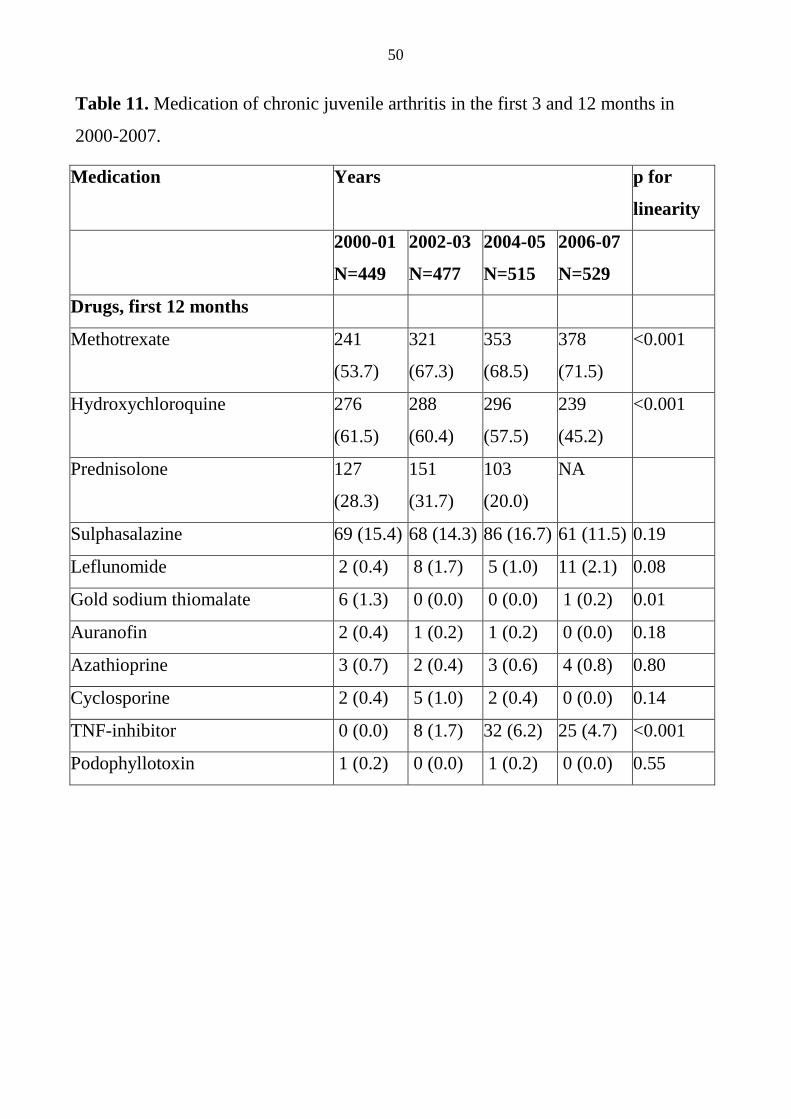
Mtx and hydroxychloroquine were the most commonly purchased first-year drugs
over the whole study period (Table 11). The use of mtx increased significantly (p for
linearity < 0.001) during the follow-up period, which meant 71.5 % of the patients in
2007. The use of hydroxychloroquine decreased (p < 0.001), and a declining trend
49
was also seen in the use of prednisolone (Table 11). The increased use of mtx was on
the same level as during the first 3 months in both single and combination therapies
(Figure 2).
4.2. Early treatment strategy
The most common early treatment (3-month) strategy was DMARD monotherapy.
The proportion of patients on a single DMARD remained constant, approx. 50%. The
number of patients receiving combination therapy increased between years 2000-
2003 (p < 0.001), but remained rather stable thereafter (Table 11).
Almost 20% of all patients received neither a DMARD nor a biologic agent during
the immediate 3 months after the disease onset, the proportion being rather stable
between 2000-2007.
Figure 2. Methotrexate used as single therapy and in combination with other
DMARDs during the first 3 disease months in patients with JIA in 2000-2007.
50
Table 11. Medication of chronic juvenile arthritis in the first 3 and 12 months in
2000-2007.
Medication Years p for
linearity
2000-01
N=449
2002-03
N=477
2004-05
N=515
2006-07
N=529
Drugs, first 12 months
Methotrexate 241
(53.7)
321
(67.3)
353
(68.5)
378
(71.5)
<0.001
Hydroxychloroquine 276
(61.5)
288
(60.4)
296
(57.5)
239
(45.2)
<0.001
Prednisolone 127
(28.3)
151
(31.7)
103
(20.0)
NA
Sulphasalazine 69 (15.4) 68 (14.3) 86 (16.7) 61 (11.5) 0.19
Leflunomide 2 (0.4) 8 (1.7) 5 (1.0) 11 (2.1) 0.08
Gold sodium thiomalate 6 (1.3) 0 (0.0) 0 (0.0) 1 (0.2) 0.01
Auranofin 2 (0.4) 1 (0.2) 1 (0.2) 0 (0.0) 0.18
Azathioprine 3 (0.7) 2 (0.4) 3 (0.6) 4 (0.8) 0.80
Cyclosporine 2 (0.4) 5 (1.0) 2 (0.4) 0 (0.0) 0.14
TNF-inhibitor 0 (0.0) 8 (1.7) 32 (6.2) 25 (4.7) <0.001
Podophyllotoxin 1 (0.2) 0 (0.0) 1 (0.2) 0 (0.0) 0.55
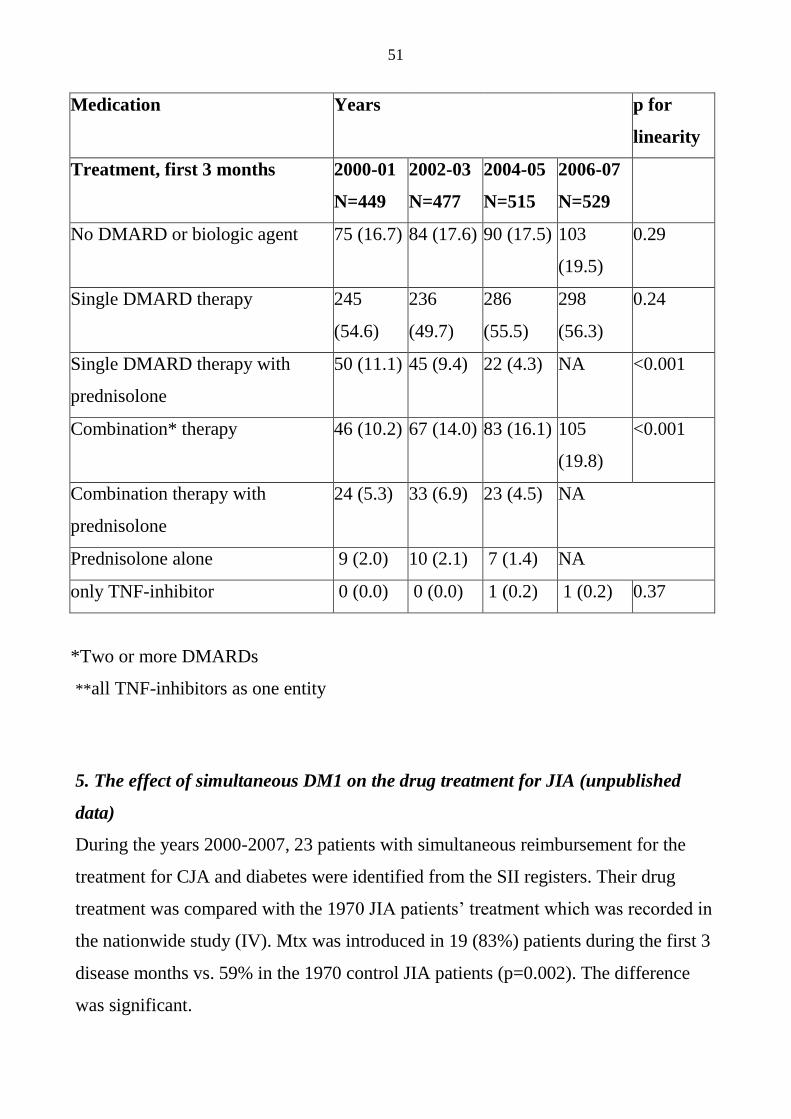
51
Medication Years p for
linearity
Treatment, first 3 months 2000-01
N=449
2002-03
N=477
2004-05
N=515
2006-07
N=529
No DMARD or biologic agent 75 (16.7) 84 (17.6) 90 (17.5) 103
(19.5)
0.29
Single DMARD therapy 245
(54.6)
236
(49.7)
286
(55.5)
298
(56.3)
0.24
Single DMARD therapy with
prednisolone
50 (11.1) 45 (9.4) 22 (4.3) NA <0.001
Combination* therapy 46 (10.2) 67 (14.0) 83 (16.1) 105
(19.8)
<0.001
Combination therapy with
prednisolone
24 (5.3) 33 (6.9) 23 (4.5) NA
Prednisolone alone 9 (2.0) 10 (2.1) 7 (1.4) NA
only TNF-inhibitor 0 (0.0) 0 (0.0) 1 (0.2) 1 (0.2) 0.37
*Two or more DMARDs
**all TNF-inhibitors as one entity
5. The effect of simultaneous DM1 on the drug treatment for JIA (unpublished
data)
During the years 2000-2007, 23 patients with simultaneous reimbursement for the
treatment for CJA and diabetes were identified from the SII registers. Their drug
treatment was compared with the 1970 JIA patients’ treatment which was recorded in
the nationwide study (IV). Mtx was introduced in 19 (83%) patients during the first 3
disease months vs. 59% in the 1970 control JIA patients (p=0.002). The difference
was significant.
52
DISCUSSION
1. Backround research
1.1. Connection between different autoimmune diseases
The epidemiology of autoimmune diseases is a topic of growing focus. AIDs are
considered having a complex etiology with a risk conferred by both genes and
environment (Ellis et al 2010).
A number of recent studies show evidence that AIDs, such as JIA, DM1, CD,
HT and MS, share some common genetic factors (Lettre et al 2008, Mysliwiec et al
2008, Albers et al 2009a, Hinks et al 2005 and 2010). Studies regarding the
pathogenesis of different AIDs e.g. the pathogenesis of CD and JIA show that they
may have several features in common. The lymphocyte cytotoxicity is abnormally
increased in the intestinal mucosa in patients with JIA. The same phenomenon is also
seen in patients with CD (Arvonen et al 2010). CD and HT also have a connection in
their pathogenesis. Serum transglutaminase-2 autoantibodies, which are very
common in patients with active CD, can react with transglutaminase-2 in thyroid
tissue and thereby contribute to the development of thyroid disease (Duntas 2009).
Environmental factors, an infection for example, may have a role in triggering
CA, such as JIA (Oen et al 1995, Ravelli and Martini 2007).
The occurrence of both JIA and DM1 have a seasonality trend (Levy et al 2008).
It is widely recognized that viral infections and low levels of vitamin D in children,
follow the same seasonal variation as the onset of DM1 and MS (Handel et al 2009).
Vitamin D has a role in regulating innate and adaptive immunity which may provide
a link between vitamin D and AIDs. Vitamin D regulates T and B cells, macrophages,
dendritic cells and ceratinocytes (Hewison 2010, Maruotti and Cantatore 2010). In
2001, a Finnish study showed that vitamin D supplementation may have a protective
effect against the development of DM1 (Hyppönen et al 2001). In a more recent
Finnish study, low levels of vitamin D were also linked to some incident cases of
53
DM1 (Oilinki et al 2011). It is worth noting, that according to another Finnish study
vitamin D supplements are not used as per to the dietary recommendations in a
substantial proportion of children (Räsänen et al 2006). Furthermore, vitamin D may
have a protective role in the pathogenesis of MS (Mirzaei F et al 2011).The studies
suggesting that low levels of vitamin D could be an etiological factor for JIA are few,
but the idea is fascinating (Ellis et al 2010).
1.2. Autoimmune diseases’ response to drug treatment
Similar responses to the same drugs in different AIDs have evidenced that there are
common mechanisms. In animal models on RA, DM1 and MS, certain potassium
channel blockers have eased the disease (Rangaraju et al 2009). Type 1 interferons
contribute to autoimmune pathogenesis, but they can also control inflammation and
as a therapeutic agent, have had favourable effects to RA, DM1 and MS. Some
positive effect, such as a trend toward preservation of pancreatic beta-cell function,
was observed in the recent-onset DM1 patients, who received oral recombinant INF-
alpha (Crow 2010). On the other hand, biologic agents, e.g. TNF blockers, may
contribute as a cause of triggering chronic rheumatoid diseases, systemic lupus
erythematosus, and another AID, MS (Kahn 2011).
1.3. Epidemiological aspects
In epidemiological studies the connection between DM1 and CD seems to be
obvious. The prevalence of CD was 7% in DM1 patients in Italy (Valetta et al 2007).
In another Italian study, the prevalence of CD was 5.7% among DM1 patients and
1.9% among the first-degree relatives of DM1 patients (Not et al 2001). All figures
differed significantly from the controls. In two recent studies, the consumption of
gluten resulted in an altered gut permeability and mucosal immunity, which may
predispose the patient to the development of DM1 (Sud et al 2010, Vaarala et al
2008). In patients with CD, a gluten-free diet may also reduce joint symptoms
54
(Alpigiani et al 2008), which was not, however, observed in the patients of the
present study.
A register-based study from the UK, made among an adult population regarding
AIDs in the same patient, found a connection between RA, AIT an DM1 at a higher
rate than expected, but was not found between RA and MS (Somers et al 2009)
In a Finnish study based on SII registers, 596 children and adolescents were
identified to have inflammatory bowel disease. The presence of CD, epilepsy, JIA
and DM1 in these patients was calculated. These children had more chronic diseases
than their controls. CD was associated with later development of ulcerative colitis.
Clear association was seen only with epilepsy and ulcerative colitis (Virta and Kolho
2012).
The present study showed that Finnish JIA patients and their families had AIDs more
often than the general population. This clustering offers indirect evidence that AIDs
may have similar mechanisms. Moreover, the simultaneous occurrence of JIA and
DM1 has increased significantly during the past 30 years.
2. Autoimmune diseases in children with JIA
The patients with JIA had more than 5-fold risk of getting another AID, such as
DM1, CD or HT a sequel of autoimmune thyroiditis. The results are in line with the
evidence obtained from adults (Viljamaa et al 2005). Compared with the population
data, the 1.1% prevalence of DM1 was 6-fold in 443 American children (Prahalad et
al 2004), and the 2.5% prevalence of CD was 8-fold in 119 Italian children with JIA
(Lepore et al 1996). Based on active screening, the 1.3% prevalence of subclinical
HT was 4-fold and the 6.7% prevalence of CD was 11-fold in 151 Italian children
with JIA, as compared with age- and sex-matched controls (Stagi et al 2005). In our
study, the 2.2% figure for DM1 was 2-fold compared with the American JIA patients,
probably due to the higher prevalence of DM1 in the Finnish population (Harjutsalo
55
et al 2008). The numbers of children with CD and HT were so small, i.e. less than 1%
of the JIA patients that to compare them with other results is not justified.
Three earlier studies give strong laboratory evidence for the connection between JIA
and AIT in children (Mihailova et al 1999, Stagi et al 2005, Harel et al 2006). Since
we did not use any systematic screening, only three clinically obvious cases were
diagnosed, and thus transient JAIT cases and probably also some mild HT cases were
missed.
An untreated autoimmune disease, such as CD, which often remains non-
symptomatic for years, may predispose the patient to another autoimmune disease
(Lepore et al 1996). In patients with CD, a gluten-free diet may also reduce joint
symptoms (Alpigiani et al 2008). In the present study, all three CD diagnoses were
based on histological findings in symptomatic patients, and thus represent an end-
stage of the autoimmune process. Therefore, a gluten-free diet reduced intestinal
symptoms, but did not reduce joint symptoms in patients with JIA. The connection
between JIA and CD should be evaluated according to studies which include
systematic laboratory screening for CD in both JIA patients and controls. There also
seems to be a connection between DM1 and CD. The prevalence of anti-tTG IgA
positivity among children and adolescents with DM1 was 8.6% (Kakleas et al 2010).
The present results provide preliminary evidence that the presence of multiple AIDs
may even protect a patient from uveitis, which commonly occurs jointly with
seronegative polyarthritis and extended oligoarthritis. This suggests that the
mechanisms leading to autoimmune disorders and uveitis are different. The
conclusion is based on an active over 5 year monitoring of the eye status of more than
400 JIA patients, mainly consisting of seronegative polyarthritis or extended
oligoarthritis cases.
56
3. Autoimmune diseases in JIA children’s first-degree relatives
Two conclusions can be drawn from the present retrospective study on the prevalence
of AIDs in 355 families with a JIA child. First, as many as 21.4% of the families had
at least one member with DM1, CD, MS or CA. This figure is high when compared
with the 4.5% prevalence in 110 JIA families in Taiwan (Huang et al 2004) and with
the 16.1% prevalence in 110 JIA patients families in the USA (Prahalad et al 2002).
The prevalence figures of AIDs were high, although we did not collect data on
thyroiditis, which has been a major AID in several previous studies (Prahalad et al
2002, Huang et al 2004, Zeft et al 2008).
Second, when compared with the population data available (Oilinki et al 2012,
Hakala et al 1993, Kaipiainen-Seppänen et al 1997, Sumelahti et al 2001), the
prevalence of DM1, RA, AS and MS was increased in JIA families. CD was as
prevalent as in the general population (Mäki et al 2003). However, there were no
significant differences in the occurrence or distribution of AIDs between the single-
JIA and multiple-JIA families, which disagreed with the previous US study (Prahalad
et al 2002).
CA formed over half of the AIDs in JIA families and occurred only in parents. The
parental 9.7% occurrence in single-JIA and 25% occurrence in multi-JIA families are
in line with the previously found 18% in Finnish multiple-JIA families (Säilä et al
2003). In the present study, RA was the most common specific diagnosis in parents,
especially mothers. The 23/1000 prevalence of RA was 3.5-fold (Hakala et al 1993)
and the 8/1000 prevalence of AS was 6.7-fold compared with the national population
data (Kaipiainen-Seppänen et al 1997). Surprisingly, the parents with AS had no sons
with juvenile AS, and 3 of the 4 index-JIA patients with a parent with AS were girls.
The prevalence of PsA in the Finnish adult population is not known; the present
6/1000 prevalence was 4.6-fold compared with the prevalence in Norway (Nossent et
al 2009).
57
The prevalence of JIA was found to be 0.8/1000 in northern Finland when the ARA
criteria was used in 1985 (Lantto and von Wendt 1985). In the present study, the
34.1/1000 prevalence of JIA in siblings was 42.6-fold, but the real difference must be
lower because ARA criteria excluded juvenile SPA. The prevalence in our study was
22.7- to 28.4-fold when compared with the figures by the EULAR criteria from the
USA, Sweden and Norway (Andersson-Gäre and Fasth 1995, Moe and Rygg 1998,
Andersson-Gäre 1999). The JIA prevalence in parents was 8.5/1000, but no
population data are available for JIA in adults. Since the prevalence of JIA in children
in the Nordic countries is about 1/1000 and the mean age of onset is 7-8 years, the
cumulative incidence of JIA in adults can be estimated at 2/1000. Thus, the parental
JIA prevalence in our study was approximately 4-fold.
The exact prevalence of DM1 in Finnish children in the whole country is not known,
although the incidence of pediatric DM1 is considered to be the world’s highest and
seems to increase all the time (Harjutsalo et al 2008, Myers and Zimmet 2008). In a
recent study, the prevalence of DM1 was 3.7/1000 in children in the Helsinki region
(Oilinki et al 2012). The estimate made over ten years ago in the USA was 2/1000
(Jacobsson et al 1997). The combined DM1 prevalence in first- and second-degree
relatives of JIA patients was only 2-fold in the US study (Prahalad et al 2002).
Compared with these figures, the prevalence of pediatric DM1 in the present study
was 3.3- to 5.7-fold when only siblings were included, and 4.2- to 8.3-fold, when
both siblings and index patients with JIA were included.
In some earlier studies, the prevalence of JIA was increased in the first-degree
relatives of CD patients (Neuhausen et al 2008). Contrary to expectations, the
prevalence of CD in the present study did not exceed the prevalence in Finnish adults
(14/1000 vs. 20/1000; Lohi et al 2007) or children (11/1000 vs. 10/1000; Mäki et al
2003). Unlike in the population studies, the present study subjects were not screened
and the diagnoses were made on clinical grounds only. In Sweden, the prevalence of
CD was 7/1000 among the 3004 non-screened children (Carlsson et al 2001). When
58
this figure was used as a reference, the prevalence of CD in JIA families was 1.9-fold
in the present study.
The estimated MS prevalence in Finland is 1.0/1000, with pronounced regional
variations (Sumelahti et al 2001). In the UK, the respective figure was 1.5 (Hirst et al
2009) and considered to be higher than in most other countries. In the USA, the
prevalence of MS was 4/1000 among first- and second-degree relatives of JIA
patients (Prahalad et al 2002). In the present study, the prevalence of MS in first-
degree relatives was 3.1/1000 when adults and children were combined, which is 3-
to 4-fold compared to the Finnish population data (Sumelahti et al 2001).
As expected, mothers had AIDs more often than fathers (96/1000 vs. 68/1000). This
is due to a higher prevalence of AIDs in females (Jacobsson et al 1997, Zeft et al
2008, de Vries et al 2009).
In conclusion, the most distinctive differences between the present and literature data
were seen in the presence of JIA in siblings and DM1 in JIA children.
4. Simultaneous occurrence of JIA and DM1 in the same patient
Four conclusions can be drawn from the study on simultaneous occurrence of JIA and
DM1. First, the number of patients increased between years 1975-2005. Second, the
prevalence of seropositivity was high. Third, the occurrence of uveitis was low. And
fourth, almost 25% had a third AID and 16% had psychiatric problems.
The increase of DM1 incidence in Finnish children is well-known (Harjutsalo et al
2008), and there may also be a slight increase in JIA incidence (Kunnamo et al 1986,
Kaipiainen-Seppänen and Savolainen 1996 and 2001, Berntsson et al 2003). Between
1965-1996, the average annual increase of DM1 incidence was calculated to be 3.4%
(Tuomilehto et al 1999). If there is an increase in JIA incidence, their number must be
small compared to DM1.
The distribution of JIA subtypes corresponds to that seen in JIA in general, with two
59
exceptions: more (15% vs. 2.6%) seropositive and fewer (1% vs. 4.6%) systemic
cases (Kaipiainen-Seppänen and Savolainen 1996 and 2001, Berntson et al 2003).
Some patients with systemic JIA might be missing here if they were treated with
NSAIDs and systemic glucocorticoids only.
Chronic uveitis was present in only 7% of patients with JIA and DM1. In line with
this, uveitis was present in only 8% of children with JIA and another AID. In earlier
Finnish studies, the proportion has been 21% (Kunnamo et al 1986) and 24%
(Kotaniemi et al 2001). The results suggest that the mechanisms of uveitis and AIDs
are different. Although 22% of the JIA patients were ANA-positive, the few patients
with chronic uveitis did not have ANAs.
A fifth of our study patients had a third AI disease, in line with some earlier studies
on clustering of AIDs in individuals with JIA and in their families (Andersson-Gäre
and Fasth 1995, Prahalad et al 2002 and 2004 and 2010). In contrast, no association
was found between the third AID and ANA- or RF-positivity, as previously observed
for simultaneous JIA, DM1 and HT (Rudolf et al 1981, Aggarwal and Meena 2003).
In the recent studies from western countries, JIA patients seem to have a fairly normal
psychosocial outcome, as shown in the UK study in 60 polyarthritis patients
aged 7-18 (Ding et al 2008) and in the Finnish study regarding 123 patients
aged 21-26 (Arkela-Kautiainen et al 2005). In the Netherlands, 4.7% of 233 DM1
patients aged 9-19, had received psychological care (de Witt and Snoek 2011).
In a large Finnish birth cohort, 12% of the boys and 4% of the girls born in 1981 had
used child mental health services by 2005 (Sourander et al 2008). In another study in
over 5000 subjects from Finland, 5.2% had been treated in psychiatric hospital
between 13-24 years of age (Gyllenberg et al 2010).
Compared with these figures, the 16% occurrence of serious psychiatric problems
was almost 3-fold in the JIA patients in the present study. Depression was the most
frequent diagnosis. The large number of patients with serious psychiatric problems
was an unexpected surprise, and emphasizes the importance of multiprofessional
60
care, including the psychosocial aspect.
There were only minor differences in glucocorticoid consumption between the groups
constructed by the presence of JIA and DM1. The 49 patients with DM1 preceding
JIA had been on systemic glucocorticoids at some stage of their arthritis just as
frequently as those 33 who had JIA before DM1, but they had been treated more
often (53% vs. 27%) with intra-articular corticosteroid injections. These observations
are against the common practice to avoid glucocorticoids, especially systemic
glucocorticoids, for diabetic patients. These observations raise the suspicion that JIA
is more severe when presenting with DM1. The aim of this more aggressive intra-
articular corticosteroid injection -treatment could also have been to cut down
inflammation to stabilize the glucose balance.
A case report from year 2000 describes the onset of DM1 only 5 months after the
introduction of the etanercept therapy to a patient with systemic JIA (Bloom 2000).
At least in theory, DM1 could be drug-induced. In our study, 17 JIA patients with
DM1 received biologic agents (11 etanercept, 6 infliximab), they had all got diabetes
prior to therapy with biologic agents.
This nationwide study used the SII registers which included patients who had
reimbursement for drugs for JIA and DM1. After 1994, when the reimbursement for
NSAIDs was withdrawn, a few mild oligoarthritis may have been missed, as well as
systemic JIA cases which were treated with NSAIDs and/or glucocorticoids only. The
treatment of JIA has become more aggressive over the last decades. The use of
effective, reimbursable drugs has increased, and more patients therefore, have been
included in the database. However, considering the current medication practices in
Finland, their number must be low. On the other hand, salicylates were the only
NSAIDs in the 70s and because they are cheap it may be that in some cases
reimbursement was not even applied for. This could explain the lower numbers in the
early years of our study.
61
5. Trends in the medical treatment practice of JIA in years 2000-2007
The present study covers the entire population of incident-JIA patients in the country
and documents the trends in the drug treatment modalities during eight consecutive
years. The most accurate method to examine the drug use is to collect data on the
actual intake. Since this approach is difficult, counting the purchases of the
medication rather than the number of prescriptions is the next-best estimate of the use
of medicines.
The use of mtx during both the first 3 and the first 12 months increased steadily
during the follow-up period. Mtx became the most common early-phase drug
whereas glucocorticoid consumption diminished. If the first DMARD was not
efficient enough, the tendency in the first study years was to add prednisolone into
the regimen, whereas later, the practice of adding another DMARD was preferred.
The treat to target with DMARDs is, besides gaining a remission, also administering
glucocorticoids shorter periods at lower doses. Ample evidence shows that the earlier
the treatment with DMARDs is started, the better the long-term outcome will be
(Albers et al 2009b). That is why an effective early treatment is emphasized. In line,
the present study revealed how an early use of mtx diminished the consumption of
glucocorticoids.
The impact of adult RA treatment, and also the impact of the FIN-RACo study can be
seen in the present study (Möttönen et al 1999). Both have emphasized an early
treatment with multiple drugs. Therefore, the use of e.g. sulphasalazine did not
decrease, since it was often part of combination therapy. Likewise,
hydroxychloroquine was used as combined with mtx (Haapasaari et al 2005).
Patients with no DMARDs or oral glucocorticoids during the first 3 months covered
almost 20% of all patients. We assume that many of these had mild oligoarthritis or
Still’s disease, which was treated with NSAIDs and intra-articular/intra-venous
glucocorticoids, but compliance problems cannot be excluded. The Finnish system to
treat is to actively inject the inflamed joints with glucocorticoids. The child is then
62
often symptom-free the next morning and the parents may think it is unnecessary to
start any other treatment e.g. mtx, and so they do not purchase the medicines.
There was a restricted supply of etanercept in Finland during the first study years
until summer 2003, which explains the limited use of TNF-inhibitors. Among the JIA
patients 4.6% have Still’s disease in this country (Kunnamo et al 1986, Kaipiainen-
Seppänen and Savolainen 2001). The treatment is often introduced with non-
reimbursable NSAIDs and glucocorticoids. In addition, the ICD-10 diagnoses in the
SII certificates are not necessarily in line with the ILAR classification.
In an earlier prospective Finnish incidence study on arthritis in 161 children, 28
(17.4%) had CA, and all but one (3.6%) of them met the ARA criteria for JRA
(Kunnamo et al 1986). By applying this figure, 70 or fever of the present 1970 JIA
patients might get another diagnosis if they were to be examined later.
To summarize, we identified several major changes in Finnish JIA patients’ drug
treatment in years 2000-2007. A lengthy follow-up is needed to see if the patients
have benefitted from an early introduction of DMARDs and the use of combination
therapy.
6. The effect of simultaneous DM1 on the drug treatment for JIA
When evaluating the drug treatment of JIA patients nationwide (IV), it was
interesting to compare the difference in drug treatment between JIA patients in
general and JIA patients with another AID (DM1).
In order to save on the use of glucocorticoids with patients who already had DM1,
systemic drug treatment must be used aggressively for a maximum effect. During the
first three disease months mtx was introduced to 83% of JIA patients who already
had DM1. The difference was significant (p=0.002) when we compared the drug
treatment of patients with both JIA and DM1 to the drug treatment of JIA patients in
the whole country. The earlier mtx is introduced the better a chance a patient has of
reaching remission, especially patients with two simultaneous but different AIDs.
63
METHODOLOGICAL ASPECTS
The main strength of the study on AIDs in children with JIA is the large number, over
400, of patients and a long follow-up of 5.5-9.5 years. Moreover, the SII registers
cover all children with certain chronic diseases in the country and allow population-
based comparisons of the occurrence of DM1, CD and HT in JIA patients. The main
limitation of the study was that the JIA patients were selected, i.e. admitted to a
tertiary centre due to moderate or severe JIA. In addition, subclinical CD or HT was
not actively screened. Since autoimmune diseases associated with JIA may develop
later, very long monitoring times until adulthood are needed to find all patients.
The study on AIDs in the JIA patients’ relatives was retrospective and the data were
collected by questionnaires only. On the other hand, patients in Finland are well
aware of their chronic diseases and exact diagnoses, since chronic diseases are
registered in the nationwide files of the SII for reimbursement of the drugs and other
treatment costs for certain diseases like DM1, MS and CA. The epidemiology of CD
has been under active study in Finland, and the prevalence figures are well known.
Since the data were collected from the files of the tertiary hospital and with the RFH
providing services in child rheumatology for the whole country, the number of our
study subjects was high: 362 index cases with JIA in families and 1238 family
members.
The study on patients with simultaneous JIA and DM1 was a nationwide,
epidemiological study, again based on the SII reimbursement registers. It is most
likely that the identified 82 patients represent all or a great majority of eligible
patients, and the information even allowed us to establish which disease had started
first.
Our study regarding the entire population of incident JIA patients documented the
general trends in the drug treatment during 8 consecutive years and covered as many
as 1970 patients.
64
Despite of the large number of study subjects and the modern statistics used, several
outcomes were so rare (e.g. 23 patients with JIA and DM1, years 2000-2007) that the
analyses were under-powered.
65
CONCLUSIONS AND CLINICAL RECOMMENDATIONS
1. AIDs tend to accumulate in children with JIA. AIDs also cluster in families
with JIA in a child. Therefore, clinicians who treat JIA patients should be
aware of the increased risk of other AIDs not only in their patients but also in
the patient’s family members.
2. The number of patients with simultaneous JIA and DM1 has increased in
Finland, in line with the increase of DM1. Patients with multiple AIDs need
multi-professional care and substantial support. Psychological support
especially, is necessary not only for the patients but also for the parents. In
addition, JIA may be more severe in children with DM1.
3. When we evaluated JIA patients’ drug treatment nationwide, several major
changes were identified between years 2000-2007. However, a long follow-up
until adulthood is needed to see how much the patients will benefit from an
early introduction of DMARDs and a combination therapy.
4. If a JIA patient already had DM1, he/she needs a more aggressive drug
treatment – partly in order to avoid corticosteroids.
The treatment of JIA patients has undergone major changes during the last
decades. It has become more challenging and the demands for the treatment
keep on growing. Patients’ problems are not just medical. Patients need ample
high-quality treatment and support to cope with the numerous of problems they
have to face.
66
REFERENCES
Aggarwal A and Misra R. Juvenile chronic arthritis in India: is it different from that
seen in Western countries? Rheumatol Int 1994;14:53-6
Aggarwal A and Misra RN. Juvenile rheumatoid arthritis in India – rarity of
antinuclear antibody and uveitis. Indian J Pediatr 1996;63:301-4
Aggarwal S and Meena PD. Simultaneous Occurrence of Type 1 Diabetes mellitus
and juvenile rheumatoid arthritis. Indian Pediatrics 2003;40:568-71
Albers HM, Kurreeman FA, Stoeken-Rijsbergen G, Brinkman DM, Kamphuis SS,
van Rossum MA, Girschick HJ, Wouters C, Saurenmann RK, Hoppenreijs E,
Slagboom P, Houwing-Duistermaat JJ, Verduijn W, Huizinga TWJ, ten Cate R, Toes
RE and Schilham MW. Association of the autoimmunity locus 4q27 with juvenile
idiopathic arthritis. Arthritis Rheum 2009a;60:901-4
Albers HM, Wessels JAM, van der Straaten RJHM, Brinkman DMC, Suijlekom-Smit
LWA, Kamphuis SSM, Girschick HJ, Wouters C, Schilham MW, le Cessie S,
Huizinga TWJ, ten Cate R and Guchelaar HJ. Time to treatment as an important
factor for the response to methotrexate in juvenile idiopathic arthritis. Arthritis
Rheum 2009b;61:46-51
Alpigiani MG, Cerboni M, Bertini I, d’Annunzio G, Haupt R, Iester A and Lorini L.
Endocrine autoimmunity in young patients with juvenile chronic arthritis. Clin Exp
Rheumatol 2002;20:565-8
Alpigiani M, Haupt R, Parodi S, Calcagno A, Poggi E and Lorini R. Coeliac disease
in 108 patients with juvenile idiopathic arthritis. Clin Exp Rheumatol 2008;26:162
Andersson-Gäre B. Juvenile arthritis - who gets it, where and when? A review of
current data on incidence and prevalence [review]. Clin Exp Rheumatol 1999;17:367-
74
Andersson-Gäre B and Fasth A. Epidemiology of juvenile chronic arthritis in
southwestern Sweden: a 5-year prospective population study. Pediatrics
1992;90:950-8.
67
Andersson-Gäre B and Fasth A. The Natural history of juvenile chronic arthritis: A
population based cohort study. Onset and Disease Process. J Rheumatol
1995;22:295-307
Angeles-Han S and Prahalad S. The genetics of juvenile idiopathic arthritis: What is
new in 2010? Curr Rheumatol Rep 2010;12:87-93
Ansell B, Bywaters E and Lawrence J. A family study of Still’s disease. Ann Rheum
Dis 1962;21:243-52
Arkela-Kautiainen M, Haapasaari J, Kautiainen H, Vilkkumaa I, Mälkiä E and
Leirisalo-Repo M. Favourable social functioning and health related quality of life of
patients with JIA in early adulthood. Ann Rheum Dis 2005;64:875-80
Arvonen M, Ikni L, Augustin M, Karttunen TJ and Vähäsalo P. Increase of duodenal
and ileal mucosal cytotoxic lymphocytes in juvenile idiopathic arthritis. Clin Exp
Rheumatol 2010;28:128-34
Backland G, Nossent HC and Grant JT. Incidence and prevalence of ankylosing
spondylitis in northern Norway. Arthritis Rheum 2005; 53:850-5
Bartoli M, Taro M, Magni-Manzoni S, Pistorio A, Traverso F, Viola S, Magnani A,
Gasparini C, Martini A and Ravelli A. The magnitude of early response to
methotrexate therapy predicts long-term outcome of patients with juvenile idiopathic
arthritis. Ann Rheum Dis 2008;67:370-4
Becker KG, Simon RM, Bailey-Wilson JE, Freidlin B, Biddison WE, McFarland HF
and Trent JM. Clustering of non-major histocompatibility complex susceptibility
candidate loci in human autoimmune diseases. Proc Natl Acad Sci USA
1998;95:9979-84
Berntson L, Andersson-Gäre B, Fasth A, Herlin T, Kristinsson J, Lahdenne P,
Marhaug G, Nielsen S, Pelkonen P, Rygg M and Nordic Study Group. Incidence of
juvenile idiopathic arthritis in the Nordic countries. A population based study with
special reference to the validity of the ILAR and EULAR criteria. J Rheumatol
2003;30:2275-82
68
Beukelman T, Patkar NM, Saag KG, Tolleson-Rinehart S, Cron RQ, DeWitt EM,
Ilowite NT, Kimura Y, Laxer RM, Lovell DJ, Martini A, Rabinovich CE and Ruperto
N. 2011 American college of rheumatology recommendations for the treatment of
juvenile idiopathic arthritis: Initiation and safety monitoring of therapeutic agents for
the treatment of arthritis and systemic features. Arthritis Care Res 2011;64:465-82
Beukelman T, Haynes K, Curtis JR, Xie F, Chen L. Bemrich-Stolz CJ, Delzell E,
Saag KG, Solomon DH and Lewis JD, on behalf of the Safety Assessment of
Biological Therapeutics Collaboration. Rates of malignancy associated with
juvenile idiopathic arthritis and its treatment. Arthritis Rheum 2012;
64:1263–1271
Bloom BJ. Development of diabetes mellitus during etanercept therapy in a child
with systemic onset juvenile rheumatoid arthritis. Arthritis Rheum 2000;43:2606-8
LeBovidge JS, Lavigne JV, Donenberg GR and Miller ML. Psychological adjustment
of children and adolescents with chronic arthritis: A Meta-analytic Review. J of
Pediatr Psychol 2003;28:29-39
Castleman B and McNeely BU. Case records of the Massachusetts General Hospital,
case 44. N Engl J Med 1968;279:987-96
Carlsson AK, Axelsson IE, Borulf SK, Bredberg AC and Ivarsson SA. Serological
screening for celiac disease in healthy 2.5-year-old children in Sweden. Pediatrics
2001; 107:42-5
Cespedes-Cruz A, Gutierres-Suarez R, Pistorio A, Ravelli A, Loy A, Murray KJ,
Gerloni V, Wulffraat N, Oliveira S, Walsh J, Penades IC, Alpigiani MG, Lahdenne P,
Saad-Magalhaes C, Cortis E, Lepore L, Kimura Y, Wouters C, Martini A and
Ruperto N. for the Pediatric Rheumatology International Trials Organization
(PRINTO). Methotrexate improves the health-related quality of life of children with
juvenile idiopathic arthritis. Ann Rheum Dis 2008;67:309-14
Chabas D, Strober J and Waubant E. Pediatric multiple sclerosis. Curr Neurol
Neuroscience Report 2008;8:434-41
69
Craig ME, Hattersley A and Donaghue KC. Definition, epidemiology and
classification of diabetes in children and adolescents. Pediatr Diabetes 2009:10(Suppl
12):3-12
Crow MK. Type 1 interferon in organ-targeted autoimmune and inflammatory
diseases. Arthritis Research Ther 2010;12(Suppl 1):s5
Dale I. The treatment of juvenile rheumatoid arthritis with azathioprine. Scand J
Rheumatol 1972:1;125-7
Ding T, Hall A, Jacobs K, and David J. Psychological functioning of children and
adolescents with juvenile idiopathic arthritis is related to physical disability but not to
disease status. Rheumatology 2008; 47:660-4
Danner S, Sordet C, Terzic J, Donato L, Velten M, Fischbach M and Sibilia J.
Epidemiology of juvenile idiopathic arthritis in Alsace, France. J Rheumatol
2006;33:1377-81
Duntas LH. Does celiac disease trigger autoimmune thyroiditis? Autoimmunity
2009;5:109-11
Ellis J, Munro J and Ponsonby A-L. Possible environmental determinants of juvenile
idiopathic arthritis. Rheumatology 2010;49:411-25
Englund M, Jöud A, Geborek B, Felson DT, Jacobsson LT and Petersson IF.
Prevalence and incidence of rheumatoid arthritis in southern Sweden 2008 and their
relation to prescribed biologics. Rheumatology 2010;49:1563-9
Fasano A, Berti I, Gerarduzzi T, Not T, Colletti RP, Drago S, Elitsur Y, Green PH,
Guandalini S, Hill ID, Pietzak M, Ventura A, Thorpe M, Kryszak D, Fornaroli F,
Wasserman SS, Murray JA and Horvath K. Prevalence of celiac disease in at-risk and
non-at-risk groups in the United States: a large multicenter study. Arch Int Med
2003;163:286-92
70
Feldman D, Bernatsky S and Houde M. The incidence of juvenile rheumatoid
arthritis in Quebec: a population data-based study. Pediatr Rheumatol Online J
2009;7:20
Fink CW. Proposal for the development of classification criteria for idiopathic
arthritides of childhood. J Rheumatol 1995;22:1566-9.
Fisher M, Nussbaum M, Abrams CAL and Shenker IR. Diabetes mellitus,
Hashimoto’s thyreoiditis and juvenile rheumatoid arthritis. Am J Dis Child 1980;134:
93-4
Flato B, Lien G, Smerdel-Ramoya A and Vinje O. Juvenile psoriatic arthritis: Long-
term outcome and differentiation from other subtypes of juvenile idiopathic arthritis.
J Rheumatol 2009;36:642-50
Foster HE, Marshall N, Myers A, Dunkley P and Griffiths ID. Outcome in adults with
juvenile idiopathic arthritis. Arthritis Rheum 2003;48:767-75
Fujikawa S and Okuni M. A nationwide surveillance study of rheumatic disease
among Japanese children. Acta Pediatr Jap 1997;39:242-4
Gerloni V, Cimaz R, Gattinara M, Arnoldi C, Pontikaki I and Fantini F. Efficacy and
safety profile of cyclosporin A in the treatment of juvenile chronic (idiopathic)
arthritis. Results of 10-year prospective study. Rheumatology 2001;40:907-13
Giannini EH, Brewer Jr EJ, Kuzimina N, Shaikov A and Wallin B for the USA
Pediatric Rheumatology Collaborative Study Group and the USSR Cooperative
Children’s Study Group. Auranofin in the treatment of juvenile rheumatoid arthritis.
Results of the USA-USSR double-blind, placebo-controlled trial. Arthritis Rheum
1990;33:466-76
Giannini EH, Brewer EJ, Kuzimina N, Shaikov A, Maximov A, Vorontsov I, Fink
CW, Newman AJ, Cassidy JT and Zemel LS, for the Pediatric Rheumatology
Collaborative Study Group and the Cooperative Children‘s Study Group.
Methotrexate in resistant juvenile rheumatoid arthritis. Results of the USA-USSR
double-blind, placebo-controlled trial. N Engl J Med 1992;326:1043-9

71
Giannini EH, Ruperto N, Ravelli A, Lovell DJ, Felson DT and Martini A.
Preliminary definition of improvement in juvenile arthritis. Arthritis Rheum
1997;40:1202-9
Giannini EH, Ilowite NT, Lovell DJ, Wallace CA, Rabinovich CE, Reiff A, Higgins
G, Gottlieb B and Singer NG, for the pediatric rheumatology collaborative study
group, Chon Y, Lin S-L, Baumgartner SW. Long-term safety and effectiveness of
etanercept in children with selected categories of juvenile idiopathic arthritis.
Arthritis Rheum 2009;60:2794-2804
Gyllenberg D, Sourander A, Niemelä S, Helenius H, Sillanmäki L, Piha J,
Kumpulainen K, Tamminen T, Moilanen I and Almqvist F. Childhood predictors of
later psychiatric hospital treatment: findings from the Finnish 1981 birth cohort study.
Eur Child Adolesc Psychiatry 2010;19:823-33
Haapasaari J. Disease modifying drug treatment in juvenile idiopathic arthritis.
(Thesis) Tampere: Acta Universitatis Tamperensis; 2006, p.28
Haapasaari J, Kautiainen H, Isomäki H and Hakala M. Hydroxychloroquine does not
decrease serum methotrexate concentrations in children with juvenile idiopathic
arthritis. Arthritis Rheum 2005;52:1621-2
Haines KA. Juvenile idiopathic arthritis: Therapies in the 21st century. Bull NYU
Hosp Jt Dis 2007;65:205-11
Hakala M, Pöllänen R and Nieminen P. The ARA 1987 revised criteria select patients
with clinical rheumatoid arthritis from a population based cohort of subjects with
chronic rheumatic diseases registered from drug reimbursement. J Rheumatol
1993;20:1674-8
Handel A, Handunnetthi L, Ebers G and Ramagopalan S. Type 1 diabetes mellitus
and multiple sclerosis: common etiological features. Nat Rev Endocrinol 2009;5:655-
64
Harel L, Prais D, Uziel Y, Mukamel M, Hashkes P, Harel G, Amir J, Monselise Y
and Press J. Increased prevalence of antithyroid antibodies and subclinical
72
hypothyroidism in children with juvenile idiopathic arthritis. J Rheumatol
2006;33:164-6
Harjutsalo V, Sjöberg L and Tuomilehto J. Time trends in the incidence of type 1
diabetes in Finnish children: a cohort study. Lancet 2008;371:1777-82
Hertzberger-ten Cate R and Dijkmans BAC. Increased prevalence of
spondylarthropathies in parents of children with pauciarticular juvenile chronic
arthritis, Type 1. Clin Rheumatol 1993;12:361-3
Hewison M. Vitamin D and the immune system: New perspectives on an old theme.
Endocrinol Metab Clin North Am 2010;39:365-79
Hinks A, Barton A, John S, Bruce I, Hawkins C, Griffiths CEM, Donn R, Thomson
W and Silman A, Worthington J. Association between the PTPN22 gene and
rheumatoid arthritis and juvenile idiopathic arthritis in a UK population. Arthritis
Rheum 2005;52:1694-9
Hinks A, Ke X, Barton A, Eyre S, Bowes J and Worthington J; the UK Rheumatoid
Arthritis Genetics Consortium; British Society of Pediatric and Adolescent
Rheumatology Study Group. Thompson SD, Langefeld CD, Glass DN, Thomson W.
Association of the IL2RA/CD25 gene with juvenile idiopathic arthritis. Arthritis
Rheum 2009;60:251-7
Hinks A, Martin P, Flynn E, Eyre S, Packham J and Barton A. Investigation of type 1
diabetes and celiac disease susceptibility loci for association with juvenile idiopathic
arthritis. Ann Rheum Dis 2010;69:2169-72
Hirst C, Ingram G, Pickergill T, Swingler S, Compston D and Robertson N.
Increasing prevalence and incidence of multiple sclerosis in South East Wales. J
Neurol Neurosurg Psychiatry 2009;80:386-91
Horneff G, De Bock F, Foeldvari I, Girschick HJ, Michels H, Moebius D and
Schmeling H. German and Austrian Pediatric Rheumatology Collaborative Study
Group. The safety and efficacy of combination of etanercept and methotrexate
compared to treatment with etanercept only. Ann Rheum Dis 2009;68:519-25
73
Huang CM, Yang YH and Chiang BL. Different familial association patterns of
autoimmune diseases between juvenile-onset systemic lupus erythematosus and
juvenile rheumatoid arthritis. J Microbiol Immunol Infect 2004;37:88-94
Hyppönen E, Läärä E, Reunanen A, Järvelin M-R and Virtanen SM. Intake of
vitamin D and a risk of type 1 diabetes: a birth-cohort study. Lancet 2001;358:1500-3
Ilowhite N, Porras O, Reiff A, Rudge S, Punaro M, Martin A, Allen R, Harville T,
Sun Y-N, Bevirt T, Aras G and Appleton B. Anakinra in the treatment of
polyarticular-course juvenile rheumatoid arthritis: safety and preliminary efficacy
results of a randomized multicenter study. Clin Rheumatol 2009;28:129-37
Immonen K, Savolainen HA and Hakala M. Why can we no longer find juvenile
idiopathic arthritis-associated amyloidosis in childhood or in adolescence in Finland?
(letter). Scand J Rheumatol 2007;36:402-9
Jacobson D, Gange S, Rose N and Graham N. Epidemiology and estimated
population burden of selected autoimmune diseases in the United States. Clin
Immunol Immunopathol 1997;84:223-43
Kahn P. Juvenile idiopathic arthritis. An update on pharmacotherapy. Bull NYU
Hosp Jt Dis.2011;69:264-76
Kaipiainen-Seppänen O, Aho K and Heliövaara M. Incidence and prevalence of
ankylosing spondylitis in Finland. J Rheumatol 1997;24:496-9
Kaipiainen-Seppänen O and Savolainen A. Incidence of chronic juvenile rheumatic
diseases in Finland during 1980-1990. Clin Exp Rheumatol 1996;14:441-4
Kaipiainen-Seppänen O and Savolainen A. Changes in the incidence of juvenile
rheumatoid arthritis in Finland. Rheumatol 2001;40:928-32
Kakleas K, Karayianni C, Critselis E, Papathanasiou A, Petrou V, Fotinou A and
Karavanaki K. The prevalence and risk factors for celiac disease among children and
adolescents with type 1 diabetes mellitus. Diabetes Res Clin Pract 2010;90:202-8
74
van Kerckhove C, Giannini EH and Lovell DJ. Temporal patterns of response to D-
penicillamine, hydroxychloroquine, and placebo in juvenile rheumatoid arthritis
patients. Arthritis Rheum 1988;10:1252-8
Khuffash FA, Majeed HA, Lubani MM, Najdi KN, Gunawardana SS and Bushnaq R.
Epidemiology of juvenile chronic arthritis and other connective tissue diseases
among children in Kuwait. Ann Trop Pediatr 1990;10:255-9
Knip M, andSiljander H. Autoimmune mechanisms in type 1 diabetes. Autoimmun
Rev 2008;7:550-7
Korpela M, Laasonen L, Hannonen P, Kautiainen H, Leirisalo-Repo M, Hakala M,
Paimela L, Blåfield H, Puolakka K and Möttönen T for the FIN-RACo Trial Group.
Retardation of joint damage in patients with early rheumatoid arthritis by initial
aggressive treatment with disease-modifying antirheumatic drugs. Five-year
experience from the FIN-RACo study. Arthritis Rheum 2004;50:2072-81
von Koskull S, Truckenbrodt H, Holle R and Hörmann A. Incidence and prevalence
of juvenile arthritis in an urban population of southern Germany: a prospective study.
Ann Rheum Dis. 2001;60:940-5
Kotaniemi K, Kaipiainen-Seppänen O, Savolainen A and Karma A. A population-
based study on uveitis in juvenile rheumatoid arthritis. Clin Exp Rheumatol
1999;17:119-22
Kotaniemi K, Kautiainen H, Karma A and Aho K. Occurrence of uveitis in recently
diagnosed juvenile chronic arthritis: a prospective study. Ophthalmology
2001;108:2071-5
Kotaniemi K, Savolainen A, Karma A and Aho K. Recent advances in uveitis of
juvenile idiopathic arthritis. Surv Ophthalmol 2003;48:489-502
Kotaniemi K, Säilä H and Kautiainen H. Long term efficacy of adalimumab in the
treatment of uveitis associated JIA. Clin Ophthalmol. 2011;5:1425-9
Kunnamo I, Kallio P and Pelkonen P. Incidence of arthritis in urban Finnish children.
A prospective study. Arthritis Rheum 1986;29:1232-8
75
Kvien TK, Hoyerdal HM and Sandstad B. Azathioprine versus placebo in patients
with juvenile rheumatoid arthritis: A single center double blind comparative study.
J Rheumatol 1986;13:118-23
Kölle G, Stoeber E and Schöntag W. Immunosuppressiva zur Basisbehandlung
viszeraler Formen der kindlichen rheumatoiden Arthritis. Med Klin 1972;17:603-7
Laaksonen AL. A prognostic study of juvenile rheumatoid arthritis. Analysis of 544
cases. Acta Paediatr Scand 1966:1-163
Lagerqvist C, Ivarsson A, Juto P, Persson LÅ and Hernell O. Screening for adult
celiac disease – which serological marker(s) to use? J Int Med 2001; 250:241-8
Lantto R and von Wendt L. Juvenile rheumatoid arthritis in northern Finland.
Abstracts of the 2nd
International Symposium on Inflammatory Connective Tissue
Diseases in Childhood and Adolescence. Prague 1985
Lepore L, Martelossi S, Pennesi M, Falcici F, Ermini M, Ferrari R, Perticarari S,
Presani G, Lucchesi A, Lapini M and Ventura A. Prevalence of coeliac disease in
patients with juvenile chronic arthritis. J Pediatrics 1996;129:311-3
Lettre G and Rioux JD. Autoimmune diseases: insights from genome-wide
association studies. Review. Hum Mol Genet 2008;17:116-21
Levy H, Rotstein A, Kahana E, Marrosu MG, Cocco E and Laron Z. Juvenile
multiplex sclerosis similar to type 1 diabetes mellitus has a seasonality of month of
birth which differs from that in the general population. J Pediatr Endocrinol Metab
2008;21:473-7
Lohi S, Mustalahti K, Kaukinen K, Laurila K, Collin P, Rissanen H, Lohi O, Bravi E,
Gasparin M, Reunanen A and Mäki M. Increasing prevalence of coeliac disease over
time. Aliment Pharmacol Ther 2007;26:1217-25
Lovell DJ, Ruperto N, Goodman S, Reiff A, Jung L, Jarosova K, Nerncova D, Mouy
R, Sandborg C, Bohnsack J, Elewaut D, Foeldvari I, Gerloni V, Rovensky J, Minden
K, Vehe RK, Weiner W, Horneff G, Huppertz HI, Olson NY, Medich JR, Carcereri-
76
De-Prati R, McIlraith MJ, Giannini EH and Martini A; for the Pediatric
Rheumatology Collaborative Study Group and the Pediatric International Trials
Organisation. Adalimumab with or without methotrexate in juvenile rheumatoid
arthritis. N Engl J Med 2008;359:810-20
Manners PJ and Diepeveen DA. Prevalence of juvenile chronic arthritis in a
population of 12-year-old children in urban Australia. Pediatrics 1996;98:84-90
Maruotti N and Cantatore FP. Vitamin D and the immune system. J Rheumatol
2010;37:391-5
McAnarney ER, Pless IB, Satterwhite B and Friedman SB. Psychological problems
of children with chronic juvenile arthritis. Pediatrics 1974;53:523-8
Mihailova D, Grigorova R, Vassileva B, Mladenova G, Ivanova N, Stephanov S,
Lissitchky K and Dimova E. Autoimmune thyroid disorders in juvenile chronic
arthritis and systemic lupus erythematosus. Adv Exp Med Biol 1999;455:55-60
Mirzaei F, Michels KB, Munger K, O’Reilly E, Chitnis T, Forman MR, Giovannucci
E, Rosner B and Ascherio A. Gestational vitamin D and the risk of multiple sclerosis
in offspring. Ann Neurol 2011;70:30-40
Modesto C, Antón J, Rodrigues B, Bou R, Arnal C, Ros J, Tena X, Rodrigo C, Rotés
I, Hermosilla E and Barceló P. Incidence and prevalence of juvenile idiopathic
arthritis in Catalonia (Spain). Scand J Rheumatol 2010;39:472-9
Moe N and Rygg M. Epidemiology of juvenile chronic arthritis in northern Norway:
a ten-year retrospective study. Clin Exp Rheumatol 1998;16:99-101
Myers M and Zimmet P. Halting the accelerating epidemic of type 1 diabetes. Lancet
2008;371:1730-1
Mysliwiec M, Mysliwska J, Zorena K, Balcerska A, Malinowska E and Wisniewski P.
Interleukin 6-174(G^C) gene polymorphism is related to celiac disease and
autoimmune thyroiditis coincidence in diabetes type 1 in children. Diabetes Res Clin
Pract 2008;82:108-12
77
Mäenpää J, Raatikka M, Räsänen J, Taskinen E and Wager O. Natural course of
juvenile autoimmune thyroiditis. J Pediatrics 1985;107:898-904
Mäki M, Hällström O, Verronen P, Reunala T, Lähdeaho M, Holm K and Visakorpi
J. Reticulin antibody, arthritis, and coeliac disease in children. Lancet 1988;1:479-80
Mäki M, Mustalahti K, Kokkonen J, Kulmala P, Haapalahti M, Karttunen T, Ilonen J,
Laurila K, Dahlbom I, Hansson T, Höpfl P and Knip M. Prevalence of celiac disease
among children in Finland. N Engl J Med 2003;348:2517-24
Möttönen T, Hannonen P, Leirisalo-Repo M, Nissilä M, Kautiainen H, Korpela M,
Laasonen L, Julkunen H, Luukkainen R, Vuori K, Paimela L, Blåfield H, Hakala M,
Ilva K, Yli-Kerttula U, Puolakka K, Järvinen P, Hakola M, Piirainen H, Ahonen J,
Pälvimäki I, Forsberg S, Koota K and Friman C, for the FIN-RACo trial Group.
Comparison of combination therapy with single-drug therapy in early rheumatoid
arthritis: a randomised trial. FIN-RACo trial group. Lancet 1999;353:1568-73
Nagy KH, Lukacs K, Sipos P, Hermann R, Madacsy L and Soltesz G. Type 1 diabetes
associated with Hashimoto’s thyroiditis and juvenile rheumatoid arthritis: a case
report with clinical and genetic investigations. Pediatr Diabetes 2010; 11:579-82
Neuhausen SL, Steele L, Ryan S, Moussavi M, Pinto M, Osann KE, Flodman P and
Zone J. Co-occurrence of celiac disease and other autoimmune diseases in celiac
disease and their first-degree relatives. J Autoimmun 2008; 31:160-5
Nordal E, Zak M, Aalto K, Berntson L, Fasth A, Herlin T, Lahdenne P, Nielsen S,
Straume B and Rygg M; Nordic Study Group of Pediatric Rheumatology. Ongoing
disease activity and changing categories in a long-term Nordic cohort study of
Juvenile idiopathic arthritis. Arthritis Rheum 2011;63:2809-18
Nossent JC and Grant JT. Epidemiological and clinical characteristics of psoriatic
arthritis in northern Norway. Scand J Rheumatol 2009; 38:251-5
Not T, Tommasini A, Tonini G, Buratti E, Pocecco M, Tortul C, Valussi M,
Crichiutti G, Berti I, Trevisol C, Azzoni E, Neri E, Torre G, Martelossi S, Soban M,
Lenhardt A, Cattin L and Ventura A. Undiagnosed celiac disease and risk of
autoimmune disorders in subjects with Type 1 diabetes mellitus. Diabetologia
2001;44:151-5
78
Oen K, Fast M and Postl B. Epidemiology of juvenile rheumatoid arthritis in
Manitoba, Canada, 1975-92: Cycles in incidence. J Rheumatol 1995;22:745-50
Oen K, Tucker L, Huber AM, Miettunen P, Scuccimarri R, Campillo S, Cabral DA,
Feldman BM, Tse S, Chédeville G, Spiegel L, Schneider R, Lang B, Ellsworth J,
Ramsey S, Dancey P, Silverman E, Chetaille AL, Cameron B, Johnson N, Dorval J,
Petty RE, Duffy KW, Boire G, Haddad E, Houghton K, Saint-Cyr C, Turvey SE,
Benseler S, Cheang M, Yeung RS and Duffy CM. Predictors of early inactive disease
in a juvenile idiopathic arthritis cohort: Results of a Canadian multicenter,
prospective inception cohort study. Arthritis Rheum 2009;61:1077-86
Oilinki T, Otonkoski T, Ilonen J, Knip M and Miettinen PJ. Prevalence and
characteristics of diabetes among Somali children and adolescents living in Helsinki,
Finland. Pediatr Diabetes 2012;13:176-80
Ortiz-Alvarez O, Morishita K, Avery G, Green J, Petty RE, Tucker LB, Malleson PN
and Cabral DA. Guidelines for blood test monitoring of methotrexate toxicity in
juvenile idiopathic arthritis. J Rheumatol 2004;31:2501-6
Ostile IL, Johansson I, Aasland A, Flatö B and Möller A. Self-rated physical and
psychosocial health in a cohort of young adults with juvenile idiopathic arthritis.
Scand J Rheumatol. 2010;39:318-25
Petty RE, Southwood TR, Baum J, Bhettay E, Glass DN, Manners P, Maldonado-Cocco
J, Suarez-Almazor M, Orozco-Alcala J and Prieur AM. Revision of the proposed
classification criteria for juvenile idiopathic arthritis: Durban, 1997. J Rheumatol
1998;25:1991-4
Petty RE, Smith JR and Rosenbaum JT. Arthritis and uveitis in children: A pediatric
rheumatology perspective. Am J Ophthalmol 2003;135:879-84
Petty RE, Southwood TR, Manners P, Baum J, Glass DN, Goldenberg J, He X,
Maldonado-Cocco J, Orozco-Alcala J, Prieur AM, Suarez-Almazor ME and Woo P;
International League of Associations for Rheumatology. International League of
Associations for Rheumatology classification of juvenile idiopathic arthritis: second
revision, Edmonton, 2001. J Rheumatol 2004;31:390-2
79
Phelan JD, Thompson SD and Glass DN. Susceptibility to JRA/JIA: complementing
general autoimmune and arthritis traits. Genes Immun 2006;7:1-10
Podkrajsek KT, Milenkovic T, Odink RJ, Claasen-van der Grinten HL, Bratanic N,
Hovnik T and Battelino T. Detection of a complete autoimmune regulator gene
deletion and two additional novel mutations in a cohort of patients with atypical
phenotypic variants of autoimmune polyglandural syndrome type 1. Eur J Endocrinol
2008;159:633-9
Prahalad S, Shear E, Thompson S, Giannini EH and Glass DN. Increased prevalence
of familial autoimmunity in simplex and multiplex families with juvenile rheumatoid
arthritis. Arthritis Rheum 2002;46:1851-6
Prahalad S, O´Brien E, Fraser A, Kerber R, Mineau G, Pratt D, Donaldson D,
Bamshad MJ and Bohnsack J. Familial aggregation of juvenile idiopathic arthritis.
Arthritis Rheum 2004;50:4022-7
Prahalad S, Zeft AS, Pimentel R, Clifford B, McNally B, Mineau GB, Jorde LB and
Bohnsack JF. Quantification of the familial contribution to juvenile idiopathic
arthritis. Ann Rheum Dis 2010;62:2525-9
Prieur AM, Piussan C, Manigne P, Bordigoni P and Griscelli C. Evaluation of D-
penicillamine in juvenile chronic arthritis. Arthritis Rheum 1985;28:376-82
Pruunsild C, Uibo K, Liivamägi H, Tarraste S, Talvik T and Pelkonen P. Prevalence
and short-term outcome of juvenile idiopathic arthritis: a population-based study in
Estonia. Clin Exp Rheumatol 2007a;25:649-53
Pruunsild C, Uibo K, Liivamägi H, Tarraste S, Talvik T and Pelkonen P. Incidence of
juvenile idiopathic arthritis in Estonia: a prospective population-based study. Scand J
Rheumatol 2007b;36:7-13
Rallison M, Dobyns B, Keating R, Rall J and Tyler F. Occurrence and natural history
of chronic lymphocytic thyroiditis in childhood. J Pediatrics 1975;86: 675-82
80
Rangaraju S, Chi V, Pennington M and Chandy K. Kv1.3 potassium channels as a
therapeutic target in multiple sclerosis. Expert Opin Ther Targets 2009;13:909-24
Ravelli A and Martini A. Juvenile idiopathic arthritis. Seminar. Lancet.
2007;369:767-78
Ravelli A, Varnier G, Oliveira S, Castell E, Arguedas O, Magnani A, Pistorio A,
Magni-Manzoni S, Galasso R, Lattanzi B, Dalprà S, Battagliese A, Verazza S,
Allegra M and Martini A. Antinuclear antibody-positive patients should be grouped
as a separate category in the classification of juvenile idiopathic arthritis. Arthritis
Rheum 2011;63:267-75
Riise ØR, Handeland KS, Cvancarova M, Wathne KO, Nakstad B, Abrahamsen TG,
Kirkhus E and Flatø B. Incidence and characteristics of arthritis in Norwegian
children: a population-based study. Pediatrics 2008;121:299-306
Ringold S, Seidel KD, Koepsell KD and Wallace CA. Inactive disease in
polyarticular juvenile idiopathic arthritis: current patterns and associations.
Rheumatology 2009;48:972-7
van Rossum MAJ, Fiselier TJW, Franssen MJAM, Zwinderman AH, ten Cate R, van
Suijlekom-Smit LWA, van Luijk WHJ, van Soesbergen RM, Wulffraat NM,
Oostveen JCM, Kuis W, Dijkstra PF, van Ede CFP and Dijkmans BAC on behalf of
the Dutch Juvenile Chronic Arthritis Study Group. Sulfasalazine in the treatment of
juvenile chronic arthritis. Arthritis Rheum 1998;41:808-16
van Rossum MAJ, van Soesbergen RM, Boers M, Zwinderman AH, Fiselier TJW,
Franssen MJAM, ten Cate R, van Suijlekom-Smit LWA, Wulffraat NM, van Luijk
WHJ, Oostveen JCM, Kuis W and Dijkmans BAC, on behalf of the Dutch Juvenile
Idiopathic Arthritis Study Group. Long-term outcome of juvenile idiopathic arthritis
following a placebo-controlled trial: sustained benefits of early sulfasalazine
treatment. Ann Rheum Dis 2007;66:1518-24
Rudolf MCD, Genel MN, Tamborlane WVJr and Dwyer JM. Juvenile rheumatoid
arthritis in children with diabetes mellitus. J Pediatrics 1981;99:519-24
Ruperto N and Martini A. JIA, treatment and possible risk of malignancies. Nat Rev
Rheumatol 2011;7:6-7
81
Ruperto N, Murray KJ, Gerloni V, Wulffraat N, de Oliveira SK, Falcini F,
Dolezalova P, Alessio M, Burgos-Vargas M, Corona F, Vesely R, Foster H,
Davidson J, Zulian F, Asplin L, Baildam E, Consuegra JG, Ozdogan H, Saurenmann
R, Joos R, Pistorio A, Woo P and Martini A; for the Pediatric Rheumatology
International Trials Organization. A randomized trial of parenteral methotrexate
comparing an intermediate dose with a higher dose in children with juvenile
idiopathic arthritis who failed to respond to standard doses of methotrexate. Arthritis
Rheum 2004;50:2191-201
Ruperto N, Ravelli A, Castell E, Gerloni V, Haefner R, Malattia C, Kanakoudi-
Tsakalidou F, Nielsen S, Bohnsack J, Gibbas D, Rennebohm R, Voygioyka O,
Balogh Z, Lepore L, Macejkova E, Wulffrat N, Oliveira S, Russo R, Buoncompagni
A, Hilario MO, Alpigiani MG, Passo M, Lovell DJ, Merino R, Martini A and
Giannini EH; for the Pediatric Rheumatology International Trials Organisation
(PRINTO). Cyclosporine A in juvenile idiopathic arthritis. Results of the
PRCGS/PRINTO phase IV post marketing surveillance study. Clin Exp Rheumatol
2006;24:599-605
Räsänen M, Kronberg-Kippilä C, Ahonen S, Uusitalo L, Kautiainen S, Erkkola M,
Veijola R, Knip M and Kaila M, Virtanen SM. Intake of vitamin D by Finnish
children aged 3 months to 3 years in relation to sociodemographic factors. Europ J
Clin Nutr 2006;60:1317-22
Saurenmann RK, Rose JB, Tyrrell P, Feldman BM, Laxer RM, Schneider R and
Silverman ED. Epidemiology of juvenile idiopathic arthritis in a multiethnic cohort.
Arthritis Rheum 2007;56:1974-84
Savolainen HA. Chlorambucil in severe juvenile chronic arthritis: Long-term follow-
up with special reference to amyloidosis. J Rheumatol 1999;26:898-903
Savolainen HA and Isomäki HA. Decrease in the number of deaths from secondary
amyloidosis in patients with juvenile chronic arthritis. J Rheumatol 1993;20:1201-3
82
Savolainen HA, Kautiainen H, Isomäki H, Aho K and Verronen P. Azathioprine in
patients with juvenile chronic arthritis: A longterm followup study. J Rheumatol
1997;24:2444-50
Savolainen HA, Lehtimäki M, Kautiainen H, Aho K and Anttila P. HLA B27: A
prognostic factor in juvenile chronic arthritis. Clin Rheumatol 1998;17:121-4
Scheinfeld N. Adalimumab: a review of side effects. Expert Opin Drug Safe
2005;4:637-41
Simard JF, Neovius M, Hagelberg S and Askling J. Juvenile idiopathic arthritis and
risk of cancer – A nationwide cohort study. Arthritis Rheum 2010;62:3776-82
Silverman E, Mouy R, Spiegel L, Jung LK, Saurenmann RK, Lahdenne P, Horneff G,
Calvo I, Szer IS, Simpson K, Stewart JA and Strand V, for the Leflunomide in
Juvenile Rheumatoid Arthritis (JRA) Investigator Group. Leflunomide or
methotrexate for juvenile idiopathic arthritis. N Engl J Med 2005;352:1655-66
Solau-Gervais E, Robin C, Gambert C, Troller S, Danner S, Gombert B, Debiais F
and Hankard R. Prevalence and distribution of juvenile idiopathic arthritis in a region
of Western France. Joint Bone Spine 2010;77:47-9
Somers EC, Thomas SL, Smeeth L and Hall AJ. Are individuals with an autoimmune
disease at higher risk of a second autoimmune disorder? Am J Epidemiol
2009;169:749-55
Sourander A, Niemelä S, Santalahti P, Helenius H and Piha J. Changes in psychiatric
problems and service use among 8-year-old children: a 16-year population-based
time-trend study. J Am Acad Child Adolesc Psychiatry 2008;47:317-27
Stagi S, Giani T, Simonini G and Falcini F. Thyroid function, autoimmune thyroiditis
and celiac disease in juvenile idiopathic arthritis. Rheumatology 2005;44:517-20
Sud S, Marcon M, Assor E, Palmert M, Daneman D and Mahmud FH. Celiac disease
and pediatric type 1 diabetes: Diagnostic and treatment dilemmas. Int J Pediatr
Endocrinol 2010;2010:161285
83
Sumelahti M-L, Tienari PJ, Wikström J, Palo J and Hakama M. Increasing
prevalence of multiple sclerosis in Finland. Acta Neurol Scand 2001; 103:153-8
Säilä H, Savolainen A, Kauppi M, Alakulppi N, Kinnunen L, Tuomilehto-Wolf E,
Tuomilehto J, Leirisalo-Repo M and Aho K. Occurrence of chronic inflammatory
rheumatic diseases among parents of multiple offspring affected by juvenile
idiopathic arthritis. Clin Exp Rheumatol 2003;21:263-5
Säilä H. Affected sibling pairs with juvenile idiopathic arthritis. An immunogenetic
study of the disease in multicase families. (Thesis) University of Helsinki. Faculty of
Medicin 2006. p.57
Tuomilehto J, Karvonen M, Pitkäniemi J, Virtala E, Kohtamäki K, Toivanen L and
Tuomilehto-Wolf E. Record-high incidence of Type 1 (insulin-dependent) diabetes
mellitus in Finnish children. Diabetologia 1999; 42:655-60
Tynjälä P, Vähäsalo P, Tarkiainen M, Kröger L, Aalto K, Malin M, Putto-Laurila A,
Honkanen V and Lahdenne P. Aggressive combination drug therapy in very early
polyarticular juvenile idiopathic arthritis (Acute-JIA): a multicenter randomised
open-label clinical trial. Ann Rheum Dis 2011;70:1605-12
Ungerer JA, Horgan B, Chaitow J and Champion GD. Psychosocial functioning in
children and young adults with juvenile arthritis. Pediatrics 1988;81:195-202
Unsal E, Ören O, Salar K, Makay B, Abaci A, Özhan B and Böber E. The frequency
of autoimmune thyroid disorders in juvenile idiopathic arthritis. Turkish J Pediatr
2008;50:462-5
Vaarala O, Atkinson MA and Neu J. The “Perfect Storm” for Type 1 Diabetes. The
complex interplay between intestinal microbiota, gut Permeability and mucosal
immunity. Diabetes 2008;57:2555-62
Valetta E, Ulmi U, Mabboni I, Tomaselli F and Pinelli L. Early diagnosis and
treatment of celiac disease in type 1 diabetes. A longitudinal case-control study.
Pediatr Med Chir 2007;29:99-104
84
Weiler FG, Dias-da-Silva MR and Lazaretti-Castro M. Autoimmune polyendocrine
syndrome type 1: case report and review of literature. Arq Bras Endocrinol Metabol
2012;56:54-66
Viljamaa M, Kaukinen K, Huhtala H, Kyrönpalo S, Rasmussen M and Collin P.
Coeliac disease, autoimmune diseases and gluten exposure. Scand J Gastroenterol
2005;40:437-43
Virta LJ and Kolho K-L. The risk of contracting pediatric inflammatory bowel
disease in children with celiac disease, epilepsy, juvenile arthritis and type 1 diabetes
- a nationwide study, J Crohns Colitis 2012, doi 10.1016/j crohns.2012.02.021
Wittinghill M, Evelyn EH, Gervas ST Jr. and Lewis ST. The distribution of
rheumatoid arthritis, spondylitis and multiple rheumatoid arthritis in a single large
kindred. Proc X International Congress of Genetics, Montreal 1958. Vol 2: Abstracts,
p.314
de Witt M and Snoek FJ. Depressive symptoms and unmet psychological needs of
Dutch youth with type 1 diabetes: results of a web-survey. Pediatr Diabetes
2011;12:172-6
de Vries L, Bulvik S and Philip M. Chronic autoimmune thyroiditis in children and
adolescents: at presentation and during long-term follow-up. Arch Dis Child
2009;94:33-7
Vuorimaa Hanna-Elina. Pain in juvenile idiopathic arthritis: Parents and children as
agents of disease management. (Thesis) University of Helsinki, Institute of
Behavioral Sciences, Studies of Psychology 69:2010
Ylijoki Heikki. Lastenreumaa sairastavien pitkäaikaisennuste ja siihen vaikuttavat
tekijät. (Väitöskirja) Kansaeläkelaitos. Sosiaali- ja terveysturvan tutkimuksia 1998
Zeft A, Shear ES, Thompson SD, Glass DN and Prahalad S. Familial autoimmunity:
maternal parent-of-origin effect in juvenile idiopathic arthritis. Clin Rheumatol
2008;27:241-8
KIITOKSET (ACKNOWLEDGEMENTS)
This study was carried out at the department of pediatrics in the Rheumatism
Foundation Hospital, Heinola and at the department of pediatrics in the Päijät-Häme
District Central Hospital, Lahti.
Olen saanut ohjausta venäläiseen kirjallisuuteen, maalaustaiteeseen, elämään
yleensä… ja tähän väitöskirjaan ohjaajaltani ja väitöskirjatyöni isoäiti-hahmolta
dosentti Anneli Savolaiselta. Minusta tuli lastenlääkäri ja lastenreumatologi osittain
Annelin ansiosta. Aloitimme kliinisestä työstä ja päädyimme tieteen maailmaan.
Näistä ohjaussessioista on jäänyt mieleeni ikuiset muistot ja niistä olen kiitollinen.
Toista ohjaajaani professori Matti Korppia kiitän ystävällisestä ja humaanista otteesta
hänen ohjatessaan jo näinkin ikääntynyttä kliinikko-kollegaa. Professorin parrakas,
leppoisa olemus sai tunnelman aina vapautumaan, kiitos siitä.
Esitarkastajani dosentti Anne Putto-Laurila ja dosentti Oili Kaipiainen-Seppänen
innostuneesti korjasivat väitöskirjaani ja heidän rakentavat ehdotuksensa paransivat
väitöskirjani tasoa. Heidän positiivisesti kannustava otteensa jäi mieleeni, kiitos heille
siitä.
Statistikko Hannu Kautiainen kesti nurkumatta pienet ruutupaperinpalaseni, joita
kannoin mukanani Äänekoskelle. Näiden käyntien jälkeen tuntui siltä, kuin sekavat
ajatukseni olisi laitettu päässäni järjestykseen, kuin työnohjauksessa olisin käynyt.
Kahvi oli aina odottamassa, ja aloitimme päivän keskustelemalla maailman eri
ilmiöistä laidasta laitaan. Kiitos antoisista keskusteluista ja opetuksesta.
Tieteellisissä julkaisuissa mukana ollut dosentti Kaisu Kotaniemi kannusti, auttoi ja
motivoi näiden vuosien aikana, hänelle kiitos siitä. Ohjausryhmääni kuuluva Liisa
Kröger kannusti ja motivoi, kiitos näistä kuluneista vuosista.
Sujuva englanninkieli on ainoastaan kielenkääntäjä Outi Meriläisen ansiota, hänelle
kiitos siitä.
Kiitos osatöissä mukana olleille Katariina Latvalle, Kari Puolakkalle, Timo
Pohjolaiselle ja Lauri Virralle. Olette toimineet nopeasti ja ystävällisesti.
Kaikkia näitä yllämainittuja yhdisti sama tinkimättömyys, kaiken piti olla tieteen
tekemisessä juuri oikein, niinpä tulokset tarkistettiin, tarkistettiin ja vielä
kolmannenkin kerran tarkistettiin. Opin heiltä oikeaa tieteentekemisen-etiikkaa ja se
kulki mukanani työni aikana, kiitos heille kaikille siitä.
Väitöskirjatyöni on saanut taloudellista tukea myös yksityishenkilöltä, rva Taru
Suokolta, hänelle kiitos siitä!
Reumasäätiön Sairaalan lastenosaston erinomainen henki kulkee vieläkin mukanani.
Tahdon kiittää teitä kaikkia, koko henkilökuntaa, yhdessä ja erikseen tuestanne tämän
pitkän prosessin aikana. Edesmennyt ylilääkärimme Jarkko Haapasaari on legenda.
Ylilääkäri Heikki Ylijoen pitkä kokemus on vertaansa vailla. He molemmat opettivat
minulle kädestä pitäen lastenreumatologiaa ja heidän johdollaan tutustuin
lastenreumatologian kiehtovaan ja myös haastavaan maailmaan.
Pitkä yhteistyöni Sirpa Hannulan ja Hanna Säilän kanssa sai minut arvostamaan
kollegiaalisuutta parhaimmillaan. Kiitän myös muita kollegoja ”Reumalta” sekä
pidempiaikaisia että lyhytaikaisia vierailijoita.
Päijät-Hämeen lastenosaston henkilökunta saa minut hyvälle tuulelle. Erikoisesti
kiitän kaikkia kollegojani. Yhteistyömme lastenreumapoliklinikalla sh Kati Keräsen
ja ft Leena Leppäsen kanssa on hauskaa ja haastavaa. Emeritus ylilääkärimme Pentti
Lautala, ylilääkärimme Mikko Lavonius ja johtava lääkärimme Martti Talja olivat
vaikuttamassa, että sain tehdä väitöskirjani loppuun, heille siitä kiitokset.
Suomen lastenreumatologit ovat mukavaa porukkaa, joiden seurassa viihtyy.
Sellaiseen kollegakuntaan on antoisaa kuulua. Monet koulutusmatkat olemme
viettäneet yhdessä luennoilla istuen ja iltaisin nauraen. Olen saanut paljon
unohtumattomia muistoja, kiitos teille kaikille.
Kaikkia Suomen sairaaloiden arkistohenkilökuntaa kiitän kärsivällisyydestä ja
myönteisestä asenteesta, ilman heitä väitöskirjani ei olisi valmistunut, edes nyt.
Reumasäätiön Sairaalan kirjastonhoitaja Mirja Rekola ja Päijät-Hämeen
keskussairaalaan kirjastonhoitajat Saija Korkiakangas ja Päivi Ukkonen auttoivat
minua suuresti vuosien mittaan, heille kiitos siitä.
Hyvät ystäväni Heinolassa ja Tampereella tukivat minua työni aikana. Heidän
innostuksensa tarttui myös minuun. Kiitos keskusteluista ja lenkkiseurasta, hyvää
terapiaa, jos joku sattui menemään pieleen.
Vanhempani, äitini sekä edesmennyt isäni, kannustivat ja rohkaisivat opiskeluun,
heille siitä lämpimät kiitokset. Sekä sisareni Hannelen että veljeni Hannun kannustus
on ollut hienoa, pikkusiskoa on kannustettu, kiitos heille ja heidän perheillensä.
Lopuksi kiitän perhettäni, joka välin suuresti ihmetellen, mutta mielenkiinnolla
seurasi projektini edistymistä. Lasten ansiosta pysyin maan pinnalla, he antoivat
lisäenergiaa ja pitivät kiinni arkielämässä. Jo vuosia sitten mieheni Lassen mielestä
voisin tehdä jotakin muutakin kuin töitä ja päivystää, esim. väitöskirjan. Tähän
työhön olen saanut runsain mitoin kannustusta ja tässä nyt olemme, juhlimme
perheenä ylpeinä, yhdessä!
Tämä työ on saanut tukea Reumasäätiön Sairaalan, Paijät-Hämeen Keskussairaalan ja
Tampereen Yliopistollisen Sairaalan EVO-tutkimusapurahoista.
Heinola 22.06.2012
Heini Pohjankoski
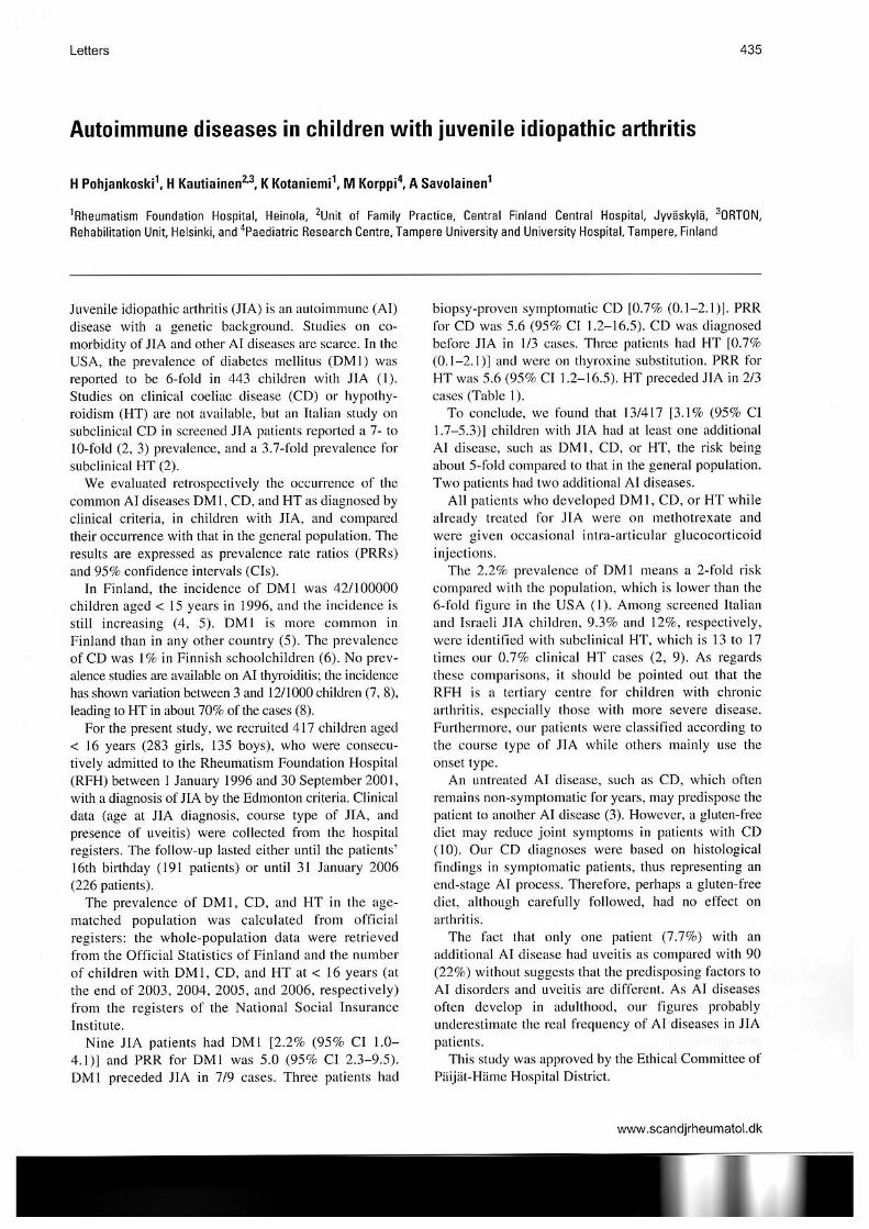
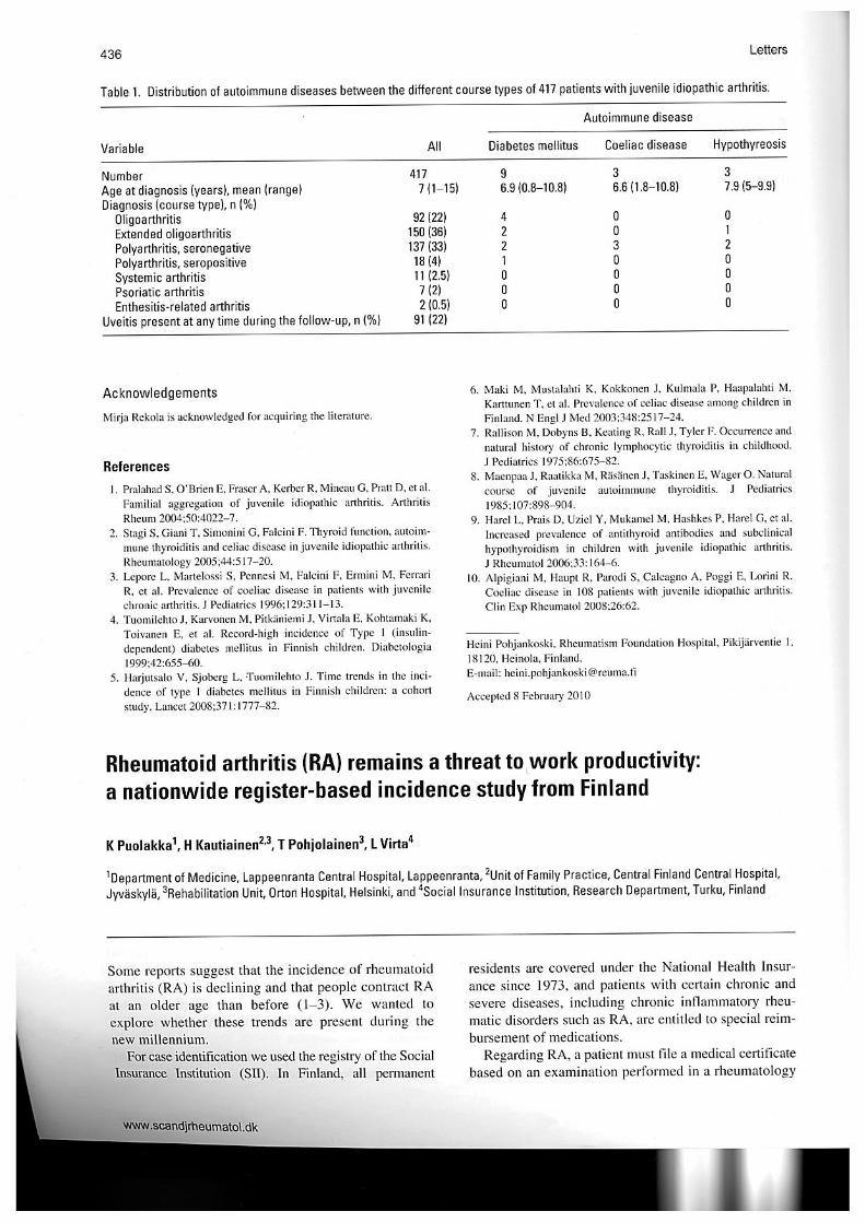
REGULAR ARTICLE
Diabetes, coeliac disease, multiple sclerosis and chronic arthritis in first-degree relatives of patients with juvenile idiopathic arthritisHeini Pohjankoski ([email protected])1, Hannu Kautiainen2,3, Kaisu Kotaniemi4, Matti Korppi5, Anneli Savolainen5
1.Department of Paediatrics, Paijat-Hame District Central Hospital, Lahti, Finland2.Unit of Family Practice, Central Finland Central Hospital, Jyvaskyla, Finland3.Unit of Primary Health Care, Kuopio University Hospital, Kuopio, Finland4.Orton, Children’s Rehabilitation Unit, ORTON Foundation, Helsinki, Finland5.Paediatric Research Centre, Tampere University and University Hospital, Tampere, Finland
KeywordsAutoimmune diseases, Coeliac disease, Chronicarthritis, Diabetes mellitus type 1, Juvenile idiopathicarthritis, Multiple sclerosis
CorrespondenceHeini Pohjankoski, M.D., Department of Pediatrics,Paijat-Hame District Hospital, 15400 Lahti, Finland.Tel: +358-44-4406176 |Fax: +358-3-8192824 |Email: [email protected],[email protected]
Received5 November 2011; revised 26 February 2012;accepted 27 February 2012.
DOI:10.1111/j.1651-2227.2012.02658.x
ABSTRACTAim: To evaluate the occurrence of autoimmune diseases in first-degree relatives of
children with juvenile idiopathic arthritis (JIA) and to compare the figures with published
population data.Materials and methods: Families of the 362 children with recently diagnosed JIA
admitted to Rheumatism Foundation Hospital, Finland, from 1996 to 2001 were contacted
by questionnaires regarding autoimmune diseases in family members. Data were collected
on type 1 diabetes, coeliac disease, multiple sclerosis and chronic arthritis, consisting
mainly of JIA, rheumatoid arthritis, spondyloarthropathy or psoriatic arthritis.Results: In all, 21.4% of the 355 families with a patient with JIA had members with
type 1 diabetes, coeliac disease, multiple sclerosis or chronic arthritis. Thirty-three mothers
and 23 fathers had type 1 diabetes, coeliac disease, multiple sclerosis or chronic arthritis in
15.2% (95% CI 11.6–19.4) of the families, and 23 mothers and 15 fathers had chronic
arthritis in 10.7% (95% CI 7.7–14.5) of the families. When compared with available
research data, the prevalences of rheumatoid arthritis, spondyloarthropathy, psoriatic
arthritis, paediatric type 1 diabetes and JIA (in siblings) were increased in JIA families.
Coeliac disease was as prevalent as in the population.Conclusion: Autoimmune diseases cluster in families with a child with JIA.
INTRODUCTIONJuvenile idiopathic arthritis (JIA) is considered as an auto-immune disease, with the exception of systemic subtype,which is currently considered as an autoinflammatory dis-ease. Autoimmune diseases seem to cluster in children withJIA (1,2), and shared susceptibility genes have been identi-fied during the recent years (3). In fact, observations thatchronic arthritis clusters in families of patients with JIAhave been published since the 1950s (4–7). In addition, JIAhas a tendency to cluster in families (5–7); in a recent study,familial factors had a 13% attributable risk for JIA (7).
In the Netherlands, the prevalence of spondyloarthropa-thy was 40 ⁄ 1000 in parents of children with JIA, comparedwith the population prevalence of 2.5 ⁄ 1000 (8). In Taiwan,the prevalence of autoimmune diseases was 45 ⁄ 1000 infamilies of patients with JIA, but the population prevalencewas not reported (9). Two previous, controlled studies fromthe United States in 2002 and 2004 documented clusteringof autoimmune diseases in families with JIA in children(2,6). The prevalence of autoimmune diseases was161 ⁄ 1000 in first-degree relatives (2), when the populationprevalence was estimated to be 50 ⁄ 1000 (10). In the case–
control setting, autoimmune diseases were more commonin JIA families than in control families, but the differencewas significant only for chronic arthritis and thyroiditis (2).The relatives of patients with JIA had an increased risk forJIA (5). The prevalence of type 1 diabetes among patientswith JIA was sixfold compared with the population preva-lence (6).
Key notes• In a nationwide study, 21.4% of families with JIA
patients had members with type 1 diabetes, coeliac dis-ease, multiple sclerosis or chronic arthritis.
• The prevalences of rheumatoid arthritis, spondyloarthr-opathy and psoriatic arthritis were, but the prevalenceof coeliac disease was not, higher than in the popula-tion.
• The prevalence of type 1 diabetes in children was 4.2-fold compared with recent child population data fromFinland.
Acta Pædiatrica ISSN 0803–5253
ª2012 The Author(s)/Acta Pædiatrica ª2012 Foundation Acta Pædiatrica 2012 101, pp. 767–771 767
The aim of this article is to evaluate the clustering of auto-immune diseases in families of children with JIA. The occur-rences of type 1 diabetes, coeliac disease, multiple sclerosisand chronic arthritis in first-degree relatives are comparedwith other studies.
MATERIAL AND METHODSRheumatism Foundation Hospital in Heinola, Finland, wasuntil 2010 a tertiary centre for juvenile patients with chronicarthritis, especially for the more serious cases. RheumatismFoundation Hospital provided diagnostic, therapeutic andrehabilitation services in paediatric rheumatology nation-wide. The families of all new patients with JIA admitted in1996–2001 were retrospectively identified from the hospitalregisters. All diagnoses were revised according to the ILAREdmonton 2004 criteria (11), using the available informa-tion registered in patient records.
In winter 2003–2004, a questionnaire was sent by post tothe identified 432 patients. In case they did not reply, a newquestionnaire was sent within a year. Finally, 46 patientsdid not respond, 15 letters were returned owing to anunknown address, and nine patients were excluded becauserequired information was incomplete (e.g. adoption, pater-nal information unavailable). Complete data were obtainedfrom the parents and siblings of 362 (84%) patients withJIA, and they belonged to 355 families. The number of sib-lings was 528, and thus the total number of children was890. There were 710 parents, and the number of familymembers (index JIA cases excluded) was 1238.
The questions concerned the diagnoses of chronic arthri-tis, coeliac disease, type 1 diabetes and multiple sclerosis infamily members (first-degree relatives) of the index JIApatients. The families were asked whether patients or theirfirst-degree relatives (mothers, fathers or full siblings) wereentitled to special reimbursement for diabetes or arthritistreatment from Social Insurance Institute, and if entitled,which was the exact diagnosis. The duration of arthritis wasnot enquired about, but Social Insurance Institute grantsreimbursement only for chronic diseases. The diagnoses(recorded according to the ICD-10 classification) were col-lected and had to be made by a physician.
StatisticsStatistical comparison between groups was made by t-testor chi-square test, when appropriate. The prevalence wereexpressed with 95% confidence intervals (95% CI).
EthicsThe study was approved by the Ethics Committee of Paijat-Hame District Central Hospital.
RESULTSDescription of the families and index casesThe 355 families included 24 (5.8%) multiple-JIA families; 6JIA cases in siblings were diagnosed during the study periodin Rheumatism Foundation Hospital and 18 elsewhere or
outside the study period in Rheumatism Foundation Hospi-tal. The mean age of mothers was 45 years (range 28–66)and that of fathers 47 years (31–74). The mean child countper family was 2.6 (range 1–18), with the index JIA patientsincluded.
Of the 362 index JIA patients, 241 (66.6%) were girls and121 (33.4%) boys, and 331 belonged to single-JIA and 31 tomultiple-JIA (24) families. At the end of the study, the meanage of girls was 15.5 years (range 7–27) and that of boys15.7 years (range 7–26). The course types of JIA in the indexpatients are given in Table 1.
Autoimmune diseases in the families and family membersType 1 diabetes, coeliac disease, multiple sclerosis orchronic arthritis were present in 76 families [21.4% (95% CI17.3–26.0)], with no significant difference between multi-ple-JIA and single-JIA families (Data not shown). Thirty-three mothers [9.3% (95% CI 6.5–12.8)] and 23 fathers[6.5% (95% CI 4.2–9.6)] had type 1 diabetes, coeliac dis-ease, multiple sclerosis or chronic arthritis (Table 2). Asboth parents had autoimmune diseases in no case, therewere 56 families [15.8% (95% CI 12.1–20.0)] with autoim-mune disease in the father or mother (Table 2).
Chronic arthritis formed 62.5% of the autoimmune dis-eases in fathers and 71.9% in mothers (Table 2). Fathersand mothers did not differ for the different types of chronicarthritis, but JIA was the only chronic arthritis diagnosis insiblings (Table 3). JIA in at least one sibling was present in6.8% of the families.
Autoimmune diseases were present in 2.7% (95% CI 1.8–3.7) of the 1238 family members of the children with JIAwhen chronic arthritis was not included, and in 7.0% (95%CI 5.6–8.5) when chronic arthritis was included (Table 2).
Results that are not shownThe numbers of autoimmune diseases of patients were toosmall for statistical tests by the course types of JIA inthe index patients and in relation to the presence of autoim-mune diseases or a specific autoimmune disease in thefamily.
When the data on age, sex and course type of JIA werecompared between the 70 eligible families who did not
Table 1 Course type* of juvenile idiopathic arthritis (JIA) in 362 index patients
Course typesGirls (N = 241)n (%)
Boys (N = 121)n (%)
All (N = 362)n (%)
Oligoarthritis, persistent 47 (19.5) 32 (26.5) 79 (21.8)
Oligoarthritis, extended 84 (34.8) 42 (34.7) 126 (34.8)
Polyarthritis seronegative 88 (36.5) 33 (27.3) 121 (33.4)
Polyarthritis seropositive 15 (6.2) 2 (1.6) 17 (4.7)
Systemic onset JIA 4 (1.7) 4 (3.3) 8 (2.2)
Juvenile psoriatic arthritis 1 (0.4) 4 (3.3) 5 (1.4)
Enthesitis-related arthritis 1 (0.4) 4 (3.3) 5 (1.4)
Undefined arthritis 1 (0.4) 0 (0) 1 (0.3)
*The International League Against Rheumatism (ILAR) Edmonton 2004 crite-
ria.
Autoimmune disease clustering in JIA families Pohjankoski et al.
768 ª2012 The Author(s)/Acta Pædiatrica ª2012 Foundation Acta Pædiatrica 2012 101, pp. 767–771
attend the study and the 355 families who attended, no sta-tistically significant differences were found (Data notshown).
Prevalences of autoimmune diseases – a comparison withliteratureThe prevalence of autoimmune diseases in this study and inthe other studies available is given in Table 4. In this study,the parental prevalence of rheumatoid arthritis was22.5 ⁄ 1000, that of spondyloarthropathy 8.5 ⁄ 1000, that ofpsoriatic arthritis 5.6 ⁄ 1000 and that of multiple sclerosis3.1 ⁄ 1000. In children, the JIA prevalence was 34.1 ⁄ 1000(only siblings included) and the type 1 diabetes prevalencewas 15.7 ⁄ 1000. Except multiple sclerosis, the prevalences ofthis study exceeded the published population figures(Table 4). The prevalence of coeliac disease has been wellstudied both in Finland and elsewhere, and our figures werequite similar to the published prevalence (Table 4).
DISCUSSIONThere are three main results in the present retrospectivestudy on the prevalence of autoimmune diseases in 355
Table 2 Autoimmune diseases in families of index juvenile idiopathic arthritis (JIA) patients
Autoimmune disease diagnosisFathers (N = 355)n (%)
Mothers (N = 355)n (%)
Siblings (N = 528)n (%)
Family members* (N = 1238)n (%)
Index JIA patients (N = 362)n (%)
Type 1 diabetes 3 (0.8) 2 (0.6) 6 (1.1) 11 (0.9) 8 (2.2)
Coeliac disease 4 (1.1) 6 (1.7) 7 (1.3) 17 (1.4) 3 (0.8)
Multiple sclerosis 2 (0.6) 3 (0.8) 0 (0) 5 (0.5) 0 (0)
Total (chronic arthritis excluded) 9 (2.5) 11 (3.1) 13 (2.5) 33 (2.7) 11 (3.0)
Chronic arthritis 15 (4.2) 23 (7.0) 18 (3.0) 56 (4.5) –
Total (chronic arthritis included) 23 (6.5) 32 (9.7) 31 (5.9) 86 (7.0) –
There were two autoimmune diseases in two mothers (coeliac disease + chronic arthritis, type 1 diabetes + multiple sclerosis), in one father (coeliac dis-
ease + chronic arthritis) and in two siblings (type 1 diabetes + coeliac disease in both). There were two index JIA patients in one family.
*Index JIA patients not included.
Table 3 Chronic arthritis by specific diagnoses in family members of index juvenileidiopathic arthritis (JIA) patients
Diagnosis
Fathers(N = 355)n (%)
Mothers(N = 355)n (%)
Siblings(N = 528)n (%)
Family members(N = 1238)n (%)
Rheumatoid arthritis 6 (1.7) 10 (2.8) 0 (0) 16 (1.3)
Spondylarthropathy 4 (1.1) 2 (0.6) 0 (0) 6 (0.5)
Psoriatic arthritis 2 (0.6) 2 (0.6) 0 (0) 4 (0.3)
JIA 2 0.6) 4 (1.1) 18 (3.4) 24 (1.9)
Reactive arthritis 1 (0.3) 4 (1.1) 0 (0) 5 (0.4)
Others* 0 (0) 1 (0.3) 0 (0) 1 (0.1)
*Arthritis with non-specified collagenosis.
Table 4 Prevalence (per 1000) of autoimmune diseases in the present and other available studies
Disease
Present study prevalence(95% confidence interval)in first-degree relatives
Prevalence(year; reference) inthe populationFinland
Prevalence (country, year; reference) in the populationOther countries
Chronic arthritis (parents) 45.2 (34.3–58.3) NA NA
Rheumatoid arthritis (parents) 22.5 (12.9–36.3) 8.0 (1993; ref. 13) 6.6 (Sweden, 2010; ref. 27)
Spondylarthropathy (parents) 8.5 (3.1–18.3) 1.5 (1997; ref. 14) 2.1 (Norway, 2005; ref. 28)
Psoriatic arthritis (parents) 5.6 (1.5–1.,4) NA 1.3 (Norway, 2009; ref. 15)
JIA (parents) 8.5 (3.1–18.3) NA NA
JIA (children*) 34.1(20.3–53.3) 0.8 (1985; ref. 16)† 0.9 (Sweden, 1988; ref. 17)‡ 1.5
(Norway, 1994; ref. 17)‡ 1.2 (USA, 2010; ref. 17)‡
Type 1 diabetes (parents) 7.0 (2.3–16.4) NA NA
Type 1 diabetes (children§) 15.7 (8.6–26.3) 3.7 (2011; ref. 20) 1.9 (USA, 1996; ref. 10)
Coeliac disease (parents) 14.1 (6.8–25.7) 19.9 (2001; ref. 22) 9.5 (USA, 2003; ref. 29) 4.8 (Sweden, 2001; ref. 30)
Coeliac disease (children§) 11.2 (5.4–20.6) 10.0 (2001; ref. 23) 3.1 (USA, 2003; ref. 29) 1.3 (Sweden, 2001; ref. 24)
Multiple sclerosis (parents) 3.1 (1.0–7.3) 1.0 (1993; ref. 25) 1.5 (UK, 2005; ref. 26)
NA, not available.
*528 siblings included.†ARA criteria.‡ European League Against Rheumatism criteria.§528 siblings and 362 index juvenile idiopathic arthritis (JIA) children combined.
Pohjankoski et al. Autoimmune disease clustering in JIA families
ª2012 The Author(s)/Acta Pædiatrica ª2012 Foundation Acta Pædiatrica 2012 101, pp. 767–771 769
families with JIA in a child. First, as many as 21.4% of theJIA families had at least one member with type 1 diabetes,coeliac disease, multiple sclerosis or chronic arthritis. Thisfigure is high when compared with the 4.5% occurrence ofautoimmune diseases in JIA families Taiwan (9) but not farfrom the 16.1% occurrence in JIA families in the UnitedStates (2). We did not collect data on thyroiditis, which hasbeen a major autoimmune disease in many previous JIAstudies with 5.5–11.2% prevalence in family members(1,2,9,12). Second, when compared with previously pub-lished population data, the prevalence of rheumatoid arthri-tis, spondyloarthropathy and psoriatic arthritis in parents,and the prevalence of JIA and of type 1 diabetes in children,was increased. And third, there were no significant differ-ences in the occurrence or distribution of autoimmune dis-eases between the single-JIA and multiple-JIA families,which disagreed with the previous US study (2).
Chronic arthritis formed two-thirds of autoimmune dis-eases in parents. In them, the 9.7% occurrence in single-JIAand 25% occurrence in multiple-JIA families are in line withthe previously found 18% occurrence in Finnish multiple-JIA families (5). In the present study, rheumatoid arthritiswas the most common specific diagnosis, especially inmothers. The 22.5 ⁄ 1000 prevalence of rheumatoid arthritiswas 3.5-fold (13) and the 8.5 ⁄ 1000 prevalence of spond-yloarthropathy was 6.7-fold compared with national popu-lation data (14). Surprisingly, the parents withspondyloarthropathy had no sons with juvenile spond-ylarthropathy, and three of the four index JIA patients witha parent with spondylarthropathy were girls. The preva-lence of psoriatic arthritis in Finnish adult population is notknown; in our study, the 5.6 ⁄ 1000 prevalence was 4.6-foldcompared with the prevalence in Norway (15).
The prevalence of JIA was 0.8 ⁄ 1000 in Northern Finlandby American Rheumatism Association (ARA) criteria in1985 (16). The 34.1 ⁄ 1000 prevalence of JIA in siblings inthe present study was 42.6-fold, but the true difference mustbe lower because ARA criteria excluded spondylarthropa-thy. In addition, the study population was less selected thanthe subjects of the present study collected from the special-ized tertiary hospital. The prevalence in the present studywas 22.7- to 28.4-fold when compared with the figures byEuropean League Against Rheumatism (EULAR) criteriafrom the United States, Sweden and Norway (17). The JIAprevalence in parents was 8.5 ⁄ 1000, but no population dataare available for JIA in adults.
In a recent study from Finland, autoimmune diseasesclustered in children with type 1 diabetes and also in theirfirst-degree relatives (18). In that study, the overall preva-lence of autoimmune diseases was 22.0% in the familieswith a child with type 1 diabetes. Although the incidence oftype 1 diabetes in Finland is highest in the world (19), theexact prevalence figures are not known. In a very recentstudy, the prevalence was 3.7 ⁄ 1000 in Finnish children atHelsinki region (20). An old, more than 10 years ago pub-lished estimate from the United States was 1.9 ⁄ 1000 (10).Compared with these Finnish and American figures, theprevalence of paediatric type 1 diabetes in the present study
was 4.2- to 8.3-fold when both siblings and children withJIA were included. Compared with the Finnish populationdata alone (20), the prevalence was about threefold in sib-lings and about sixfold in the children with JIA.
In an earlier study, the prevalence of JIA was increased inthe first-degree relatives of patients with coeliac disease(21). Contrary to expectations, the prevalence of coeliac dis-ease in the present study did not substantially exceed the20 ⁄ 1000 prevalence in Finnish adults (22) or the 10 ⁄ 1000prevalence in Finnish children (23). Unlike in populationstudies, the present study subjects were not screened; thediagnoses were carried out on clinical grounds only. In Swe-den, the prevalence of coeliac disease was 7 ⁄ 1000 among3004 non-screened children (24), and using this figure as areference, the prevalence of coeliac disease in JIA familieswas 1.9-fold in the present study.
The estimated multiple sclerosis prevalence in Finland is1.0 ⁄ 1000, with pronounced regional variations (25). In theUK, the respective figure was 1.5 (26) and considered to behigher than in most other countries. In the United States,the prevalence of multiple sclerosis was 4 ⁄ 1000 among first-and second-degree relatives of patients with JIA (2). In thepresent study, the prevalence of MS in the first-degree rela-tives was 3.1 ⁄ 1000 when adults and children were com-bined, which seems to be higher than in the Finnishpopulation (25).
In the present study, mothers were affected 1.4-fold moreoften than fathers, as expected owing to the higher preva-lence of autoimmune diseases in women (10). In the UnitedStates, the difference was larger, threefold, obviouslybecause of the high frequency of thyroiditis (12).
The study data were collected by questionnaires only. Thepatients in Finland are well aware of their chronic diseasesand exact diagnoses, because chronic diseases are registeredin nationwide files of the Social Insurance Institute forimbursement of the drugs and other treatment costs for cer-tain diseases like type 1 diabetes, multiple sclerosis andchronic arthritis. The epidemiology of coeliac disease hasbeen under active investigation in Finland, and the preva-lence figures are well known. As the study subjects wereidentified from the files of a tertiary hospital specialized forrheumatic diseases and providing services for the wholecountry, the number of study subjects was high: 362 indexcases with JIA and 1238 family members. On the otherhand, the study was not population-based, and obviously,serious cases were overrepresented, as was seen, for exam-ple, in the high number of extended oligoarthritis cases.Only 2% had systemic onset JIA, which may rather be con-sidered as an autoinflammatory than autoimmune disease.In line, no autoimmune co-diseases were found in the fam-ily members of systemic onset patients with JIA.
In conclusion, autoimmune diseases cluster in familieswith JIA in a child. This conclusion is justified, although thepresent prevalence figures were compared with figures fromother studies with varying ways of data collection and prev-alence estimation. Despite of obvious clustering, autoim-mune diseases are so rare that prospective population-based well-matched studies are extremely laborious,
Autoimmune disease clustering in JIA families Pohjankoski et al.
770 ª2012 The Author(s)/Acta Pædiatrica ª2012 Foundation Acta Pædiatrica 2012 101, pp. 767–771
expensive and time-consuming. When treating patients withJIA, vigilance for further autoimmune diseases is warrantednot only for the patients but also for their family members.
ACKNOWLEDGEMENTMirja Rekola is acknowledged for acquiring the literature.
CONFLICTS OF INTERESTNone of the author has any conflicts of interests regardingthis study.
References
1. Stagi S, Giani T, Simonini G, Falcini F. Thyroid function, auto-immune thyroiditis and celiac disease in juvenile idiopathicarthritis. Rheumatology 2005; 44: 517–20.
2. Prahalad S, Shear E, Thompson S, Giannini EH, Glass DN.Increased prevalence of familial autoimmunity in simplex andmultiplex families with juvenile rheumatoid arthritis. ArthritisRheum 2002; 46: 1851–6.
3. Hinks A, Ke X, Barton A, Eyre S, Bowes J, Worthington J, et al.Association of the IL2RA ⁄ CD25 gene with juvenile idiopathicarthritis. Arthritis Rheum 2009; 60: 251–7.
4. Wittinghill M, Evelyn EH, Gervas ST Jr, Lewis ST. The Distri-bution of rheumatoid arthritis, spondylitis and multiple rheu-matoid arthritis in a single large kindred. Proc X Int CongrGenet, Montreal 1958; 2: 314. Abstracts.
5. Saila H, Savolainen A, Kauppi M, Alakulppi N, Tuomilehto-Wolf E, Tuomilehto J, et al. Occurrence of chronic inflamma-tory rheumatic diseases among parents of multiple offspringaffected by juvenile idiopathic arthritis. Clin Exp Rheumatol2003; 21: 263–5.
6. Prahalad S, O¢Brien E, Fraser A, Kerber R, Mineau G, Pratt D,et al. Familial aggregation of juvenile idiopathic arthritis.Arthritis Rheum 2004; 50: 4022–7.
7. Prahalad S, Zeft AS, Pimentel R, Clifford B, McNally B, MineauGB, et al. Quantification of the familial contribution to juvenileidiopathic arthritis. Ann Rheum Dis 2010; 62: 2525–9.
8. Hertzberger-Ten Cate R, Dijkmans BAC. Increased Prevalenceof Spodylarthropathies in parents of children with pauciarticu-lar juvenile chronic arthritis, type 1. Clin Rheumatol 1993; 12:361–3.
9. Huang CM, Yang YH, Chiang BL. Different familial associationpatterns of autoimmune diseases between juvenile-onset sys-temic lupus erythematosus and juvenile rheumatoid arthritis. JMicrobiol Immunol Infect 2004; 37: 88–94.
10. Jacobson D, Gange S, Rose N, Graham N. Epidemiology andEstimated Population burden of selected autoimmune diseasesin the United States. Clin Immunol Immunopathol 1997; 84:223–43.
11. Petty RE, Southwood TR, Manners P, Braum J, Glass DN,Goldenberg J, et al. International League of Associations forRheumatology classification of juvenile idiopathic arthritis: sec-ond revision, Edmonton, 2001. J Rheumatol 2004; 31: 390–2.
12. Zeft A, Shear ES, Thompson SD, Glass DN, Prahalad S. Famil-ial autoimmunity: maternal parent-of-origin effect in juvenileidiopathic arthritis. Clin Rheumatol 2008; 27: 241–8.
13. Hakala M, Pollanen R, Nieminen P. The ARA 1987 Revised cri-teria select patients with clinical rheumatoid arthritis from a
population based cohort of subjects with chronic rheumatic dis-eases registered from drug reimbursement. J Rheumatol 1993;20: 1674–8.
14. Kaipiainen-Seppanen O, Aho K, Heliovaara M. Incidence andprevalence of ankylosing spondylitis in Finland. J Rheumatol1997; 24: 496–9.
15. Nossent JC, Grant JT. Epidemiological and clinical characteris-tics of psoriatic arthritis in northern Norway. Scand J Rheuma-tol 2009; 38: 251–5.
16. Lantto R, von Wendt L. Juvenile rheumatoid arthritis in north-ern Finland. Prague: Abstracts of the 2nd International Sympo-sium on Inflammatory Connective Tissue Diseases inChildhood and Adolescence, 1985.
17. Andersson Gare B. Juvenile arthritis – who gets it, where andwhen? A review of current data on incidence and prevalence.Clin Exp Rheumatol 1999; 17: 367–74.
18. Alhonen S, Korhonen S, Tapanainen P, Knip M, Veijola R.Extended Family History of diabetes and autoimmune diseasesin children with and without type 1 diabetes. Diabetes Care2011; 34: 115–7.
19. Harjutsalo V, Sjoberg L, Tuomilehto J. Time trends in the inci-dence of type 1 diabetes in Finnish children: a cohort study.Lancet 2008; 371: 1777–82.
20. Oilinki T, Otonkoski T, Ilonen J, Knip M, Miettinen PJ. Preva-lence and characteristics of diabetes among Somali childrenand adolescents living in Helsinki, Finland. Pediatr Diabetes2012; 13: 176–80.
21. Neuhausen SL, Steele L, Ryan S, Mousavi M, Pinto M, OsannKE, et al. Co-occurrence of celiac disease and other autoim-mune diseases in celiac disease and their first-degree relatives.J Autoimmun 2008; 31: 160–5.
22. Lohi S, Mustalahti K, Kaukinen K, Laurila K, Collin P, RissanenH, et al. Increasing prevalence of coeliac disease over time. Ali-ment Pharmacol Ther 2007; 26: 1217–25.
23. Maki M, Mustalahti K, Kokkonen J, Kulmala P, Haapalahti M,Karttunen T, et al. Prevalence of celiac disease among childrenin Finland. N Engl J Med 2003; 348:2517–24.
24. Carlsson AK, Axelsson IE, Borulf SK, Bredberg AC, IvarssonSA. Serological screening for celiac disease in healthy 2.5-year-old children in Sweden. Pediatrics 2001; 107: 42–5.
25. Sumelahti M-L, Tienari PJ, Wikstrom J, Palo J, Hakama M.Increasing prevalence of multiple sclerosis in Finland. ActaNeurol Scand 2001; 103: 153–8.
26. Hirst C, Ingram G, Pickersgill T, Swingler R, Compston DAS,Robertson N. Increasing prevalence and incidence of multiplesclerosis in South-east Wales. J Neurol Neurosurg Psychiatry2009; 80: 386–91.
27. Englund M, Joud A, Geborek B, Felson DT, Jacobsson LT, Pet-erssom IF. Prevalence and Incidence of rheumatoid arthritis insouthern Sweden 2008 and their relation to prescribed biolo-gics. Rheumatology (Oxford) 2010; 49: 1563–9.
28. Backland G, Nossent HC, Grant JT. Incidence and prevalenceof ankylosing spondylitis in northern Norway. Arthritis Rheum2005; 53: 850–5.
29. Fasano A, Berti I, Gerarduzzi T, Not T, Colletti RP, Drago S,et al. Prevalence of celiac disease in at-risk and non-at-riskgroups in the United States: a large multicenter study. ArchIntern Med 2003; 163: 286–92.
30. Lagerqvist C, Ivarsson A, Juto P, Persson LA, Hernell O.Screening for adult celiac disease – which serological marker(s)to use? J Intern Med 2001; 250: 241–8.
Pohjankoski et al. Autoimmune disease clustering in JIA families
ª2012 The Author(s)/Acta Pædiatrica ª2012 Foundation Acta Pædiatrica 2012 101, pp. 767–771 771
1
“This is a pre-copy-editing, author-produced PDF of an article accepted for
publication in The Journal of Rheumatology following peer review".
Simultaneous Juvenile Idiopathic Arthritis and Diabetes Mellitus type 1 -
a Nationwide Study.
Pohjankoski Heini, MD*; Kautiainen Hannu, BA,**; Korppi Matti, MD, Professor***;
Savolainen Anneli, MD, PhD, docent***
*Päijät-Häme District Central Hospital, Department of Pediatrics, Lahti, Finland;
** Unit of Family Practice, Central Finland Central Hospital, Jyväskylä, Finland and
Unit of Primary Health Care, Kuopio University Hospital, Finland;
*** Pediatric Research Centre, Tampere University and University Hospital, Tampere,
Finland
Correspondence: Request for reprints: Heini Pohjankoski
Department of Pediatrics
Keskussairaalankatu 7
15140 Lahti
Finland
Abstract
Objective: To describe the occurrence and main clinical and laboratory findings of
patients having both Juvenile Idiopathic Arthritis (JIA) and Diabetes Mellitus type 1
(DM1) in a period of thirty years.
Materials and methods: Eighty-two patients having simultaneous JIA and DM1 were
identified in the reimbursement registers of National Institute of Insurance during 1976-
2005. Data on their clinical history were collected from patient files.
Results: Occurrence of simultaneous JIA and DM1 increased 4,5fold between the first
2
(1976-85) and the last (1996-2005) decade. Prevalence of uveitis was 7%, of RF-
seropositivity 15%; 22% of patients had a third autoimmune disease (AID), 16% had
serious psychiatric problems.
Conclusion: The occurrence of patients with the two diseases, JIA and DM1, increased
in three decades. The prevalence of uveitis was low, the number of seropositive patients
was high and further AIDs were frequent. Patients had multiple additional problems and
necessitated multiprofessional care.
Key words: Juvenile idiopathic arthritis, diabetes mellitus type 1, autoimmunity, uveitis,
psychiatric problems
Introduction
Juvenile idiopathic arthritis (JIA) is the most common rheumatic disease in children, and
the incidence in Finnish children is rather similar to other western populations (1).
Instead, the incidence of diabetes mellitus type1 (DM1) in Finland is the highest in the
world, and still increasing (2). Different autoimmune (AI) diseases seem to cluster in the
same children (3).
The few reports on patients having both JIA and DM1 are mostly case reports (4,5,6,7).
The first pediatric patient with arthritis and diabetes we were able to find was a seven-
year- old girl reported in 1968 (4). Thereafter, ten patients with JIA and DM1 have been
reported, nine of whom also had thyroiditis (5,6,7,8).
Rudolf et al. in 1986 identified seven patients with JIA among 200 diabetic children. Six
of them had polyarthritis with rheumatoid factor (RF) and/or antinuclear antibodies
(ANA) and evidence on thyroid problems. The seventh one was a boy with HLA B27
positive oligoarthritis and family history of ancylosing spondylitis (8).
When 66 Italian JIA patients were screened for pre-diabetic autoantibodies only 3%
showed any positivity at all, and no clinical evidence for DM1 could be verified (9).
We identified from national registers all patients with both JIA and DM1 covering a
period of 30 years (1976 to 2005), and a population of about five million. In Finland
according to an established practice certain chronic diseases in children, like DM1 and
JIA, are all taken care of at the secondary level (central hospitals).
The aim of the present paper is to describe the occurrence and the main clinical and
laboratory findings in the patients with both DM1 and JIA, during the 30 years.
3
Material and methods
The Social Insurance Institution (SII) in Finland maintains registers on individuals
granted with a special reimbursement for medication for defined chronic diseases. From
1966 SII has provided reimbursement for the prescription of drugs for chronic rheumatic
diseases including JIA, and from 1975 for DM. In 1994, non-steroidal anti-inflammatory
drugs (NSAIDs) were excluded from the reimbursed drugs for chronic arthritis.
Injectable drugs e.g. intra articular glucocorticoids are not reimbursed for and so not
registered, since they are administered in inpatient and outpatient clinics and paid by
them. Biologic agents are applied for with specific certificates, which we did not check.
We analyzed patients identified in the SII register on the basis of reimbursement for
medication both for chronic arthritis and DM granted for the first time between January
1st, 1976 and December 31
st, 2005. The register files of the SII provide basic data like
birth date, gender and residential area of the patients and date of the reimbursement
decision.
During the 30-year surveillance period, in all 240 patients were reimbursed for both
chronic arthritis and DM. We excluded 112 patients who were reimbursed for arthritis at
>21 years and for DM <30 years of age. The age limit of 20 for JIA was chosen because
of the possibility that reimbursement was applied for with delay, which for DM is
unlikely. The files of the remaining 128 patients were further checked to ascertain the
diagnoses and the exact age at the onset of the diseases. Twenty had adult onset RA,
three had DM secondary to glucocorticoids and five had DM type 2. Thirteen patients
had been rediagnosed as something else than JIA. Three potential patients were excluded
because their files could not be located.
The remaining 82 patients fulfilling the study criteria were classified according to the
ILAR criteria (10). Data on laboratory markers, radiological changes, uveitis, and the
use of intra articular and systemic glucocorticoids, were collected by charting the
hospital records of the study subjects.
The study patients were divided into two groups based on which disease, JIA or DM1,
started first. The activity of JIA was assessed on the basis of inflammatory parameters
(sedimentation rate, ESR; C-reactive protein; CRP) at disease onset, the presence of
erosions in native radiology and the use of glucocorticoids. The need of systemic
glucocorticoids or the need of >6 annual intra articular injections were set as criterions
of an active disease.
4
Patients were considered having serious psychiatric problems, if they were forwarded
after regular psychological consultation to psychiatrists and had psychiatric medication
(usually anti-depressants), long-term therapy, and/or psychiatric hospital admission.
Ethics. All data were collected from patient files, without contacting the subjects
concerned. The study was done by the permission of The Ministry of Social Affairs and
Health.
Statistical methods. The data are presented as means with standard deviations (SD),
medians with interquartile range (IQR) or counts with percentages. Statistical
comparison between the groups was made by permutation test (Monte Carlo p-value),
Chi-Square test or Fisher exact test, when appropriate. Estimates of occurrence rate
ratios (ORR) were calculated by using Poisson regression models. Gender- and age-
matched samples of the general population were obtained from data in the Official
Statistics of Finland.
Results
Subclassification of JIA:
All 82 patients having JIA and DM1 (55 girls, 27 boys) were classified according to
ILAR criteria. See Table 1.
Occurrence:
Simultaneous JIA and DM1occurence during the 30 year period is shown in Figure 1A.
Occurrence increased statistically significantly (age and sex adjusted p for linearity
<0.001): 4.49fold (95% CI: 2.32 to 8.69) when comparing the first (76-85) and last (96-
05) period.
JIA or DM1 first:
Forty-nine patients (35 girls, 14 boys) had DM1 prior to JIA and 33 (20 girls, 13 boys)
had JIA prior to DM1 (Figure 1B).
Age of onset:
The mean onset age of JIA of all 82 patients was 8.5 years (SD 5,2) and that of DM1 8,1
years (SD 6,4) (p=0.61). The age at the onset of the diseases stayed rather constant
5
during the three surveillance decades.
Laboratory findings:
There were no significant differences between ESR and CRP at disease onset between
the DM-first and JIA-first groups. ANA were found in 18 (22%) patients, with no
difference between the groups. With one exception, RF was positive only in those who
presented with DM first (Table 2).
Erosions:
Erosions in native radiographs were present in 21(43%) in the DM-first and in 13(39%)
in the JIA-first group (p=0.75).
Corticosteroid use
Nine (27%) of the 33 patients with JIA first had been on systemic glucocorticoids at
some time and similarly, nine (27%) had received six or more intra articular injections in
some year. The respective figures for the 49 children with DM first were 14 (29%)
(systemic glucocorticoids) and 26 (53%) ( six or more intra articular injections/year).
Among DM first- 53% and JIA first-group 27% had six or more intra articular
glucocorticoid injections per year (p=0,021).
Use of biologic agents
According to patient files 17 of our patients received biologic agents (etanercept was
first introduced to 11patients and infliximab to 6 patients); all the 17 had DM1 prior to
biologic agent introduction.
Uveitis
Six patients (7%) had chronic uveitis. Five of them had JIA before DM1, four had
oligoarthritis (one extended arthritis), and one had seronegative polyarthritis. The patient
with seronegative polyarthritis had DM1 before JIA. Five patients with chronic uveitis
were ANA negative, only one being ANA positive. A seventh patient, a boy with
oligoarthritis (ANA -/HLAB27+), had experienced several bouts of acute uveitis.
Additional autoimmune diseases
Eighteen out of 82 patients (22%) had a third AI disease, which in 12 cases was
hypothyreosis; 3/11 were RF-seropositive, all were ANA negative (RF and ANA
unknown of the patient who developed psoriatic arthritis). Six had celiac disease.
Psychiatric diseases
Thirteen (16%) patients, three (11%) male, ten (18%) female, had psychiatric disorders
demanding regular, longstanding therapy or medication and/or psychiatric hospital
6
admittance (five depressions, necessitating drug therapy and psychiatric therapy, one
psychosis and drug abuse, one alcohol abuse, one had severe compliance problems in his
teens, two anorexias and one bulimia with depression, one ADHD and compliance
problems).
Discussion
There are five major findings in this study: (1) an increase in the number of patients in
the three study decades, (2) high prevalence of seropositivity , (3) low proportion of
uveitis, (4) a third AI disease in almost a quarter of patients, (5) serious psychiatric
problems in almost 20% of patients.
The increase in occurrence.
The reason for this, in the first hand, must be the increase in DM1 incidence (11). If also
the incidence of JIA has risen, remains to be proved. The analysis of Finnish DM1
incidence between 1965-96 showed an absolute average increase of 0,67 (3,4%) per year
(12). If there is increase in JIA incidence, its magnitude must be modest, compared to
that of DM1. Various studies point to a slight increase in the incidence of JIA in Finland
during the last decades (1,13,14,15).
JIA subclassification
The distribution of JIA subtypes corresponds to that seen in JIA in general, with two
exceptions: there seems to be more (15%) seropositive and less (1%) systemic cases.
Normally, the proportion of seropositive cases is 2,6%, and of systemic disease 4,6%,
calculated from earlier studies (1,3,14,15). The studies of Rudolf and Agrawal show a
similarly high quantity of patients with seropositive JIA (6,8); the cause remains to be
elucidated. Some patients with systemic JIA might be missing, if treated with NSAIDs
and systemic glucocorticoids only, because reimbursement for such medication is not
applied for (intravenous treatment is paid by institutions and oral glucocorticoid
preparations are cheap).
Uveitis
Chronic uveitis was present in six (7%) patients only; five of them had JIA prior to
DM1. The low incidence is in line with our previous report, in which 8% of JIA patients
with another AI disease had uveitis (3). In previous Finnish studies the percentage has
been 21 (13) and 24 (16). Are predisposing factors to uveitis and AI diseases different?
Five of the six patients with chronic uveitis did not have ANA, which is against
numerous previous studies. In spite of this, a 22% ANA positivity was registered in 80
patients, in line with earlier reports. Mean age stayed constant, in accordance with
previous Finnish studies (1,15,17)
7
Additional autoimmune diseases
A fifth of our study patients had a third AI disease. There are plenty of previous studies
on the accumulation of AI diseases in individuals with JIA and in their families
(3,18,19,20,21). In disaccordance with Rudolf and Agrawal (6,8) no clear association
with ANA or RF-positivity could be shown in our twelve patients with simultaneous
JIA, DM1 and hypothyreosis. In six patients the third disease was celiac disease, five of
them had DM1 prior; no clear connection to ANA- or RF-positivity could be seen
among them either.
Psychiatric problems
As early as in 1974 McAnarney et al. in USA found children with chronic arthritis
having more psychological difficulties than their healthy peers (22).
In the latest studies from Western countries, JIA patients seem to have a quite normal
psychosocial outcome, as in a study from the UK on 60 polyarthritics aged 7 to 18 (23),
and in a Finnish one on 123 21 to 26-olds (24).
In Netherlands 4,7% of 233 DM1-patients, aged 9 to 19, had received psychological care
(25).
In a large Finnish birth cohort 12% of boys and 4% of girls born in 1981 had used child
mental health services by 2005 (26). In a cohort of 5 346 children born that year 5,2%
(6,2% of males and 4,1% of females) had been admitted to psychiatric hospital treatment
between 13 and 24 years of age (27).
Reflecting the above results to our patients, the number of serious psychiatric problems
was almost threefold, 16%. The problems were considered serious because treatment by
a psychologist alone was insufficient and the patients had to be remitted to a psychiatrist.
Depression was the most frequent diagnose and could be seen underlying also some
other cases. The high number of patients with serious psychiatric problems was
unanticipated and emphasizes the follow up of the psychological function of the patients
as well as the importance of multiprofessional care.
Glucocorticoid consumption between the two groups
There were only minor differences in glucocorticoid consumption between the groups.
To our surprise those 49 patients with DM1 preceding JIA had been on systemic
glucocorticoids at some phase of their arthritis as frequently as those 33 who had JIA
before DM1 (28% and 27%, respectively), which is against our common JIA treatment
protocol. Normally, we try to avoid systemic glucocorticoids in diabetic patients. Could
it be that their disease was more serious, even if no further evidence (number of
erosions, laboratory parameters) to support such a supposition was found? Among those
49 patients, 53% had more than six intra articular glucocorticoid injections, compared to
27% of patients from JIA prior DM-group (p= 0.021). The aim might have been to avoid
8
systemic glucocorticoids in diabetics.
A case report in 2000 describes onset of DM1 5 months after introduction of etanercept
therapy in a patient with systemic JIA (28). This could, theoretically, be drug induced. In
our study 17 patients received biologic agents (11 etanercept, 6 infliximab); however, all
had their DM1 prior to the biologic agent therapy.
Due to accumulating reports it is obvious that, AIDs in general, and these two diseases
specifically, must have common genetic features. Becker et al. discovered in 1998 that in
some cases clinically distinct AIDs may be controlled by a common set of susceptibility
genes (29). After that a number of different single genes have been shown susceptible
both for JIA and DM1 (30,31,32,33,34).
This is the first nationwide study on patients having DM1 and JIA simultaneously.
Finland is an appropriate country for epidemiological studies, because of the structure of
the health care system and the homogeneity of the population. It is unlikely that many
patients are missing in the SII register, keeping the costs of the diseases in mind, and
equally unlikely that a patient with these diseases would have been treated outside the
national health care system, although after 1994 we may have missed a few cases of
mild oligoarthritis or systemic disease, treated with NSAIDs and/or glucocorticoids only.
Treatment of JIA has became more aggressive during the last decades. There has been
increasing use of reimbursable medication, and more patients thereby appearing in the
database. However considering the prevailing medication practices in Finland their
number must be low. On the other hand salicylates were the only NSAIDs in the 70s and
because these are cheap it may be that in some cases reimbursement was not applied for.
This could explain the lower numbers in the early years of the register.
Conclusion
The number of patients with simultaneous JIA and DM1 has clearly increased in
Finland. Patients with multiple AI diseases need well co-operating multiprofessional
care, and support is necessary not only for patients but for parents as well (23). Vigilance
for further AI diseases is warranted. Several questions call for further studies: how to
best provide psychosocial support? Why do these patients have so much seropositivity?
Why so little uveitis?
Competing interests: None
9
References:
1. Berntson L, Andersson Gäre B, Fasth A, Herlin T, Kristinsson J, Lahdenne P, et al. Nordic
Study Group. Incidence of juvenile idiopathic arthritis in the Nordic countries. A population
based study with special reference to the validity of the ILAR and EULAR criteria. J Rheumatol
2003; 30:2275-82
2. Harjutsalo V, Sjöberg L, Tuomilehto J. Time trends in the incidence of type 1 diabetes in
Finnish children: a cohort study. Lancet 2008; 371:1777-82
3. Pohjankoski H, Kautiainen H, Kotaniemi K, Korppi M, Savolainen A. Autoimmune diseases in
children with juvenile idiopathic arthritis. Scand J Rheumatol 2010; 39:435-6
4. Castleman B, McNeely BU. Case records of the Massachusetts General Hospital, case 44. N
Engl J Med 1968; 279:987-96
5. Fisher M, Nussbaum M, Abrams CAL, Shenker IR. Diabetes Mellitus, Hashimoto´s thyreoiditis
and juvenile rheumatoid arthritis. Am J Dis Child 1980; 134: 93-94.
6. Agrawal S, Meena PD. Simultaneous Occurrence of Type 1 Diabetes Mellitus and Juvenile
Rheumatoid arthritis. Indian Pediatrics 2003; 40:568-71
7. Nagy KH, Lukacs K, Sipos P, Hermann R, Madacsy L, Soltesz G. Type 1 diabetes associated
with Hashimoto´s thyroiditis and juvenile rheumatoid arthritis: a case report with clinical and
genetic investigations. Pediatr Diabetes 2010; 11:579-82
8. Rudolf MCD, Genel Mn, Tamborlane WVJr, Dwyer JM. Juvenile Rheumatoid Arthritis in
children with diabetes mellitus. J Pediatrics 1981; 99:519-524
9. Alpigiani MG, Cerboni M, Bertini I, d`Annunzio G, Haupt R, Iester A, et al. Endocrine
autoimmunity in young patients with juvenile chronic arthritis. Clin Exp Rheumatol 2002;
20:565-8
10. Petty RE, Southwood TR, Manners P, Braun J, Glass DN, Goldenberg J, et al. International
League of Associations for Rheumatology classification of juvenile idiopathic arthritis: second
revision, Edmonton, 2001. J Rheumatol 2004; 31:390-2
11. Myers M, Zimmet P. Halting the accelerating epidemic of type 1 diabetes. Lancet 2008;
371:1730-1
10
12. Tuomilehto J, Karvonen M, Pitkäniemi J, Virtala E, Kohtamäki K, Toivanen L, et al. Record-
high incidence of Type 1 (insulin-dependent) diabetes mellitus in Finnish children. Diabetologia
1999; 42:655-60
13. Kunnamo I, Kallio P, Pelkonen P. Incidence of arthritis in urban Finnish children. A prospective
study. Arthritis Rheum 1986; 29:1232-8
14. Kaipiainen-Seppänen O, Savolainen A. Incidence of chronic juvenile rheumatic diseases in
Finland during 1980-1990. Clin Exp Rheumatol 1996; 14:441-4
15. Kaipiainen-Seppänen O, Savolainen A. Changes in the incidence of juvenile rheumatoid
arthritis in Finland. Rheumatology (Oxford) 2001; 40:928-32
16. Kotaniemi K, Kautiainen H, Karma A, Aho K. Occurrence of uveitis in recently diagnosed
juvenile chronic arthritis: a prospective study. Ophthalmology 2001; 108:2071-5
17. Pruunsild C, Uibo K, Liivamägi H, Tarraste S, Talvik T, Pelkonen P. Incidence of juvenile
idiopathic arthritis in Estonia: a prospective population-based study. Scand J Rheumatol 2007;
36:7-13
18. Prahalad S, Shear E, Thompson S, Giannini EH, Glass DN. Increased Prevalence of Familial
Autoimmunity in Simplex and Multiplex Families with Juvenile Rheumatoid Arthritis. Arthritis
Rheum 2002; 46:1851-6
19. Prahalad S, O´Brien E, Fraser A, Kerber R, Mineau G, Pratt D, et al. Familial aggregation of
Juvenile Idiopathic Arthritis. Arthritis Rheum 2004; 50:4022-7
20. Prahalad S, Zeft AS, Pimentel R, Clifford B, McNally B, Mineau GB, et al. Quantification of
the familial contribution to juvenile idiopathic arthritis. Ann Rheum Dis 2010; 62:2525-9
21. Andersson-Gäre B, Fasth A. The Natural History of Juvenile Chronic Arthritis: A Population
Based Cohort Study. Onset and Disease Process. J Rheumatol 1995; 22:295-307
22. McAnarney ER, Pless IB, Satterwhite B, Friedman SB. Psychological problems of children
with chronic juvenile arthritis. Pediatrics 1974; 53:523-8
11
23. Ding T, Hall A, Jacobs K, David J. Psychological functioning of children and adolescents with
juvenile idiopathic arthritis is related to physical disability but not to disease status.
Rheumatology 2008; 47:660-4
24. Arkela-Kautiainen M, Haapasaari J, Kautiainen H, Vilkkumaa I, Mälkiä E, Leirisalo-Repo M.
Favourable social functioning and health related quality of life of patients with JIA in early
adulthood. Ann Rheum Dis 2005; 64:875-80
25. de Wit M, Snoek FJ. Depressive symptoms and unmet psychological needs of Dutch youth
with type 1 diabetes: results of a web-survey. Pediatr Diabetes. 2011 May;12(3 Pt 1):172-6 doi:
10.1111/j.1399-5448.2010.00673.x
26. Sourander A, Niemelä S, Santalahti P, Helenius H, Piha J. Changes in psychiatric problems and
service use among 8-year-old children: a 16-year population-based time-trend study. J Am Acad
Child Adolesc Psychiatry 2008; 47:317-27
27. Gyllenberg D, Sourander A, Niemelä S, Helenius H, Sillanmäki L, Piha J, et al. Childhood
predictors of later psychiatric hospital treatment: findings from the Finnish 1981 birth cohort
study. Eur Child Adolesc Psychiatry 2010; 19:823-33
28. Bloom BJ. Development of diabetes mellitus during etanercept therapy in a child with systemic
onset juvenile rheumatoid arthritis. Arthritis Rheum 2000; 43:2606-8
29. Becker KG, Simon RM, Bailey-Wilson JE, Freidlin B, Biddison WE, McFarland HF et al.
Clustering of non-major histocompatibility complex susceptibility candidate loci in human
autoimmune diseases. Proc Natl Acad Sci USA 1998; 95:9979-84
30. Angeles-Han S, Prahalad S. The Genetics of Juvenile Idiopathic Arthritis: What Is New in
2010? Curr Rheumatol Rep 2010; 12:87-93
31. Hinks A, Ke X, Barton A, Eyre S, Bowes J, Worthington J, et al. Association of the
IL2RA/CD25 Gene With Juvenile Idiopathic Arthritis. Arthritis Rheum 2009; 60:251-7
32. Hinks A, Martin P, Flynn E, Eyre S, Packham J, Barton A. Investigation of type 1 diabetes and
12
celiac disease susceptibility loci for association with juvenile idiopathic arthritis. Ann Rheum
Dis 2010; 69:2169-72
33. Eike MC, Nordang GBN, Karlsen TH, Boberg KM, Vatn MH on behalf of the IBSEN study
group, Dahl-Jörgensen K, Rönningen KS, Joner G, et al. The FCRL3-169>C polymorphism is
associated with rheumatoid arthritis and shows suggestive evidence of involvement with
juvenile idiopathic arthritis in a Scandinavian panel of autoimmune diseases. Ann Rheum Dis
2008; 67:1287-91
34. Albers HM, Kurreeman FA, Stoeken-Rijsbergen G, Brinkman DMC, Kamphuis SS, van
Rossum MA et al. Association of the Autoimmunity Locus 4q27 With Juvenile Idiopathic
Arthritis. Arthritis Rheum 2009; 60:901-4
13
Table 1.
Subtypes of juvenile idiopathic arthritis (JIA) in 82 patients with both JIA and diabetes
mellitus.
JIA Onset type
N (%)
Course type
N (%)
Systemic onset arthritis 1 (1.2) 1 (1.2)
Oligoarthritis 45 (55) 33 (40)
Oligoarthritis, extended 11 (13)
Polyarthritis, seropositive 12 (15) 12 (15)
Polyarthritis, seronegative 22 (27) 22 (27)
Enthesitis related arthritis
2 (2) 2 (2)
Psoriatic arthritis 0 (0) 1 (1,2)
Table 2. Laboratory findings at disease onset in two patient groups.
DM first (N=49) JIA first (N=33) p value
ESR, median (IQR) 26 (12, 45) 27 (12, 45) 0.62
CRP, median (IQR) 7 (3, 26) 10 (5, 16) 0.90
ANA+ (%) 11 (22) 7 (21) 0.97
RF+(%) 11(22) 1(3) 0,023
14
Figure 1A. Simultaneous occurrence of JIA and DM1 during 30 years, in ten year
episodes. The year of onset of the second disease as end point. The error bars give 95 per
cent confidence intervals.
Figure 1B. Cumulative distribution of difference between onset age of DM and JIA.
Gray band shows the 95 per cent confidence intervals.
Years
1976-85 1986-95 1996-05
Occure
nce o
f both
JIA
and D
M p
er
1 0
00 0
00
0,0
0,5
1,0
1,5
2,0
2,5
3,0
3,5
4,0
n=11
n=27
n=44
Time difference between DM and JIA, years
-25 -20 -15 -10 -5 0 5 10 15 20 25
Perc
enta
ge
0
10
20
30
40
50
60
70
80
90
100
DM before JIA JIA before DM
A B