Embed Size (px)
Citation preview
AD______________
Award Number: W81XWH-10-1-1061
TITLE: Interhemispheric Information Transfer: A New Diagnostic Method for Mild Traumatic Brain Injury
PRINCIPAL INVESTIGATOR: Peter Bergold, Ph.D.
CONTRACTING ORGANIZATION: Research Foundation of State University Brooklyn, NY 11203
REPORT DATE: October 2011
TYPE OF REPORT: Annual
PREPARED FOR: U.S. Army Medical Research and Materiel Command Fort Detrick, Maryland 21702-5012
DISTRIBUTION STATEMENT: Approved for public release; distribution unlimited
The views, opinions and/or findings contained in this report are those of the author(s) and should not be construed as an official Department of the Army position, policy or decision unless so designated by other documentation.
REPORT DOCUMENTATION PAGE Form Approved
OMB No. 0704-0188 Public reporting burden for this collection of information is estimated to average 1 hour per response, including the time for reviewing instructions, searching existing data sources, gathering and maintaining the data needed, and completing and reviewing this collection of information. Send comments regarding this burden estimate or any other aspect of this collection of information, including suggestions for reducing this burden to Department of Defense, Washington Headquarters Services, Directorate for Information Operations and Reports (0704-0188), 1215 Jefferson Davis Highway, Suite 1204, Arlington, VA 22202-4302. Respondents should be aware that notwithstanding any other provision of law, no person shall be subject to any penalty for failing to comply with a collection of information if it does not display a currently valid OMB control number. PLEASE DO NOT RETURN YOUR FORM TO THE ABOVE ADDRESS. 1. REPORT DATE (DD-MM-YYYY) 2. REPORT TYPE 3. DATES COVERED (From - To)
4. TITLE AND SUBTITLE 5a. CONTRACT NUMBER
5b. GRANT NUMBER
5c. PROGRAM ELEMENT NUMBER
6. AUTHOR(S) 5d. PROJECT NUMBER
5e. TASK NUMBER
E-Mail: 5f. WORK UNIT NUMBER
7. PERFORMING ORGANIZATION NAME(S) AND ADDRESS(ES) 8. PERFORMING ORGANIZATION REPORT NUMBER
9. SPONSORING / MONITORING AGENCY NAME(S) AND ADDRESS(ES) 10. SPONSOR/MONITOR’S ACRONYM(S)U.S. Army Medical Research and Materiel Command Fort Detrick, Maryland 21702-5012
11. SPONSOR/MONITOR’S REPORTNUMBER(S)
12. DISTRIBUTION / AVAILABILITY STATEMENTApproved for Public Release; Distribution Unlimited 13. SUPPLEMENTARY NOTES
14. ABSTRACT
15. SUBJECT TERMS
16. SECURITY CLASSIFICATION OF: 17. LIMITATIONOF ABSTRACT
18. NUMBER OF PAGES
19a. NAME OF RESPONSIBLE PERSONUSAMRMC
a. REPORTU
b. ABSTRACTU
c. THIS PAGEU UU
19b. TELEPHONE NUMBER (include area code)
Standard Form 298 (Rev. 8-98) Prescribed by ANSI Std. Z39.18
NUMBER(S) NUMBER(S) NUMBER(S)
12. DISTRIBUTION / AVAILABILITY STATEMENT
Approved for public release; distribution unlimited
12. DISTRIBUTION / AVAILABILITY STATEMENT
Approved for public release; distribution unlimited
12. DISTRIBUTION / AVAILABILITY STATEMENT
Approved for public release; distribution unlimited
12. DISTRIBUTION / AVAILABILITY STATEMENT
Approved for public release; distribution unlimited
12. DISTRIBUTION / AVAILABILITY STATEMENT
Approved for public release; distribution unlimited
12. DISTRIBUTION / AVAILABILITY STATEMENT
Approved for public release; distribution unlimited
12. DISTRIBUTION / AVAILABILITY STATEMENT
Approved for public release; distribution unlimited
12. DISTRIBUTION / AVAILABILITY STATEMENT
Approved for public release; distribution unlimited
12. DISTRIBUTION / AVAILABILITY STATEMENT
Approved for public release; distribution unlimited
12. DISTRIBUTION / AVAILABILITY STATEMENT
Approved for public release; distribution unlimited
13. SUPPLEMENTARY NOTES13. SUPPLEMENTARY NOTES13. SUPPLEMENTARY NOTES13. SUPPLEMENTARY NOTES13. SUPPLEMENTARY NOTES13. SUPPLEMENTARY NOTES13. SUPPLEMENTARY NOTES13. SUPPLEMENTARY NOTES13. SUPPLEMENTARY NOTES13. SUPPLEMENTARY NOTES
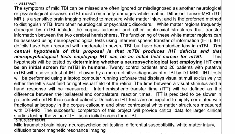
14. ABSTRACTThe symptoms of mild TBI can be missed are often ignored or misdiagnosed as another neurological or psychological disease. mTBI most commonly damages white matter. Diffusion Tensor-MRI (DT-MRI) is a sensitive brain imaging method to measure white matter injury; and is the preferred method to distinguish mTBI from other neurological or psychiatric disorders. White matter regions frequently damaged by mTBI include the corpus callosum and other centroaxial structures that transfer information between the two cerebral hemispheres. The functioning of these white matter regions can be assessed using neuropsychological tests using interhemispheric transfer of information (IHT). IHT deficits have been reported with moderate to severe TBI, but have been studied less in mTBI. The central hypothesis of this proposal is that mTBI produces IHT deficits and that neuropsychological tests employing IHT can be an initial field screen for mTBI. This hypothesis will be tested by determining whether a neuropsychological test employing IHT can be an initial screen for mTBI in humans. Twenty control patients and 20 patients with putative mTBI will receive a test of IHT followed by a more definitive diagnosis of mTBI by DT-MRI. IHT tests will be performed using a laptop computer running software that displays visual stimuli exclusively to either the left visual field or right visual field of the retina. The time between a visual stimulus and a hand response will be measured. Interhemispheric transfer time (ITT) will be defined as the difference between the ipsilateral and contralateral reaction times. ITT is predicted to be slower in patients with mTBI than control patients. Deficits in IHT tests are anticipated to highly correlated with fractional anisotropy in the corpus callosum and other centroaxial white matter structures measured with DT-MRI. The successful completion of this project will provide critical data for larger clinical studies testing the value of IHT as an initial screen for mTBI.
14. ABSTRACTThe symptoms of mild TBI can be missed are often ignored or misdiagnosed as another neurological or psychological disease. mTBI most commonly damages white matter. Diffusion Tensor-MRI (DT-MRI) is a sensitive brain imaging method to measure white matter injury; and is the preferred method to distinguish mTBI from other neurological or psychiatric disorders. White matter regions frequently damaged by mTBI include the corpus callosum and other centroaxial structures that transfer information between the two cerebral hemispheres. The functioning of these white matter regions can be assessed using neuropsychological tests using interhemispheric transfer of information (IHT). IHT deficits have been reported with moderate to severe TBI, but have been studied less in mTBI. The central hypothesis of this proposal is that mTBI produces IHT deficits and that neuropsychological tests employing IHT can be an initial field screen for mTBI. This hypothesis will be tested by determining whether a neuropsychological test employing IHT can be an initial screen for mTBI in humans. Twenty control patients and 20 patients with putative mTBI will receive a test of IHT followed by a more definitive diagnosis of mTBI by DT-MRI. IHT tests will be performed using a laptop computer running software that displays visual stimuli exclusively to either the left visual field or right visual field of the retina. The time between a visual stimulus and a hand response will be measured. Interhemispheric transfer time (ITT) will be defined as the difference between the ipsilateral and contralateral reaction times. ITT is predicted to be slower in patients with mTBI than control patients. Deficits in IHT tests are anticipated to highly correlated with fractional anisotropy in the corpus callosum and other centroaxial white matter structures measured with DT-MRI. The successful completion of this project will provide critical data for larger clinical studies testing the value of IHT as an initial screen for mTBI.
14. ABSTRACTThe symptoms of mild TBI can be missed are often ignored or misdiagnosed as another neurological or psychological disease. mTBI most commonly damages white matter. Diffusion Tensor-MRI (DT-MRI) is a sensitive brain imaging method to measure white matter injury; and is the preferred method to distinguish mTBI from other neurological or psychiatric disorders. White matter regions frequently damaged by mTBI include the corpus callosum and other centroaxial structures that transfer information between the two cerebral hemispheres. The functioning of these white matter regions can be assessed using neuropsychological tests using interhemispheric transfer of information (IHT). IHT deficits have been reported with moderate to severe TBI, but have been studied less in mTBI. The central hypothesis of this proposal is that mTBI produces IHT deficits and that neuropsychological tests employing IHT can be an initial field screen for mTBI. This hypothesis will be tested by determining whether a neuropsychological test employing IHT can be an initial screen for mTBI in humans. Twenty control patients and 20 patients with putative mTBI will receive a test of IHT followed by a more definitive diagnosis of mTBI by DT-MRI. IHT tests will be performed using a laptop computer running software that displays visual stimuli exclusively to either the left visual field or right visual field of the retina. The time between a visual stimulus and a hand response will be measured. Interhemispheric transfer time (ITT) will be defined as the difference between the ipsilateral and contralateral reaction times. ITT is predicted to be slower in patients with mTBI than control patients. Deficits in IHT tests are anticipated to highly correlated with fractional anisotropy in the corpus callosum and other centroaxial white matter structures measured with DT-MRI. The successful completion of this project will provide critical data for larger clinical studies testing the value of IHT as an initial screen for mTBI.
14. ABSTRACTThe symptoms of mild TBI can be missed are often ignored or misdiagnosed as another neurological or psychological disease. mTBI most commonly damages white matter. Diffusion Tensor-MRI (DT-MRI) is a sensitive brain imaging method to measure white matter injury; and is the preferred method to distinguish mTBI from other neurological or psychiatric disorders. White matter regions frequently damaged by mTBI include the corpus callosum and other centroaxial structures that transfer information between the two cerebral hemispheres. The functioning of these white matter regions can be assessed using neuropsychological tests using interhemispheric transfer of information (IHT). IHT deficits have been reported with moderate to severe TBI, but have been studied less in mTBI. The central hypothesis of this proposal is that mTBI produces IHT deficits and that neuropsychological tests employing IHT can be an initial field screen for mTBI. This hypothesis will be tested by determining whether a neuropsychological test employing IHT can be an initial screen for mTBI in humans. Twenty control patients and 20 patients with putative mTBI will receive a test of IHT followed by a more definitive diagnosis of mTBI by DT-MRI. IHT tests will be performed using a laptop computer running software that displays visual stimuli exclusively to either the left visual field or right visual field of the retina. The time between a visual stimulus and a hand response will be measured. Interhemispheric transfer time (ITT) will be defined as the difference between the ipsilateral and contralateral reaction times. ITT is predicted to be slower in patients with mTBI than control patients. Deficits in IHT tests are anticipated to highly correlated with fractional anisotropy in the corpus callosum and other centroaxial white matter structures measured with DT-MRI. The successful completion of this project will provide critical data for larger clinical studies testing the value of IHT as an initial screen for mTBI.
14. ABSTRACTThe symptoms of mild TBI can be missed are often ignored or misdiagnosed as another neurological or psychological disease. mTBI most commonly damages white matter. Diffusion Tensor-MRI (DT-MRI) is a sensitive brain imaging method to measure white matter injury; and is the preferred method to distinguish mTBI from other neurological or psychiatric disorders. White matter regions frequently damaged by mTBI include the corpus callosum and other centroaxial structures that transfer information between the two cerebral hemispheres. The functioning of these white matter regions can be assessed using neuropsychological tests using interhemispheric transfer of information (IHT). IHT deficits have been reported with moderate to severe TBI, but have been studied less in mTBI. The central hypothesis of this proposal is that mTBI produces IHT deficits and that neuropsychological tests employing IHT can be an initial field screen for mTBI. This hypothesis will be tested by determining whether a neuropsychological test employing IHT can be an initial screen for mTBI in humans. Twenty control patients and 20 patients with putative mTBI will receive a test of IHT followed by a more definitive diagnosis of mTBI by DT-MRI. IHT tests will be performed using a laptop computer running software that displays visual stimuli exclusively to either the left visual field or right visual field of the retina. The time between a visual stimulus and a hand response will be measured. Interhemispheric transfer time (ITT) will be defined as the difference between the ipsilateral and contralateral reaction times. ITT is predicted to be slower in patients with mTBI than control patients. Deficits in IHT tests are anticipated to highly correlated with fractional anisotropy in the corpus callosum and other centroaxial white matter structures measured with DT-MRI. The successful completion of this project will provide critical data for larger clinical studies testing the value of IHT as an initial screen for mTBI.
14. ABSTRACTThe symptoms of mild TBI can be missed are often ignored or misdiagnosed as another neurological or psychological disease. mTBI most commonly damages white matter. Diffusion Tensor-MRI (DT-MRI) is a sensitive brain imaging method to measure white matter injury; and is the preferred method to distinguish mTBI from other neurological or psychiatric disorders. White matter regions frequently damaged by mTBI include the corpus callosum and other centroaxial structures that transfer information between the two cerebral hemispheres. The functioning of these white matter regions can be assessed using neuropsychological tests using interhemispheric transfer of information (IHT). IHT deficits have been reported with moderate to severe TBI, but have been studied less in mTBI. The central hypothesis of this proposal is that mTBI produces IHT deficits and that neuropsychological tests employing IHT can be an initial field screen for mTBI. This hypothesis will be tested by determining whether a neuropsychological test employing IHT can be an initial screen for mTBI in humans. Twenty control patients and 20 patients with putative mTBI will receive a test of IHT followed by a more definitive diagnosis of mTBI by DT-MRI. IHT tests will be performed using a laptop computer running software that displays visual stimuli exclusively to either the left visual field or right visual field of the retina. The time between a visual stimulus and a hand response will be measured. Interhemispheric transfer time (ITT) will be defined as the difference between the ipsilateral and contralateral reaction times. ITT is predicted to be slower in patients with mTBI than control patients. Deficits in IHT tests are anticipated to highly correlated with fractional anisotropy in the corpus callosum and other centroaxial white matter structures measured with DT-MRI. The successful completion of this project will provide critical data for larger clinical studies testing the value of IHT as an initial screen for mTBI.
14. ABSTRACTThe symptoms of mild TBI can be missed are often ignored or misdiagnosed as another neurological or psychological disease. mTBI most commonly damages white matter. Diffusion Tensor-MRI (DT-MRI) is a sensitive brain imaging method to measure white matter injury; and is the preferred method to distinguish mTBI from other neurological or psychiatric disorders. White matter regions frequently damaged by mTBI include the corpus callosum and other centroaxial structures that transfer information between the two cerebral hemispheres. The functioning of these white matter regions can be assessed using neuropsychological tests using interhemispheric transfer of information (IHT). IHT deficits have been reported with moderate to severe TBI, but have been studied less in mTBI. The central hypothesis of this proposal is that mTBI produces IHT deficits and that neuropsychological tests employing IHT can be an initial field screen for mTBI. This hypothesis will be tested by determining whether a neuropsychological test employing IHT can be an initial screen for mTBI in humans. Twenty control patients and 20 patients with putative mTBI will receive a test of IHT followed by a more definitive diagnosis of mTBI by DT-MRI. IHT tests will be performed using a laptop computer running software that displays visual stimuli exclusively to either the left visual field or right visual field of the retina. The time between a visual stimulus and a hand response will be measured. Interhemispheric transfer time (ITT) will be defined as the difference between the ipsilateral and contralateral reaction times. ITT is predicted to be slower in patients with mTBI than control patients. Deficits in IHT tests are anticipated to highly correlated with fractional anisotropy in the corpus callosum and other centroaxial white matter structures measured with DT-MRI. The successful completion of this project will provide critical data for larger clinical studies testing the value of IHT as an initial screen for mTBI.
14. ABSTRACTThe symptoms of mild TBI can be missed are often ignored or misdiagnosed as another neurological or psychological disease. mTBI most commonly damages white matter. Diffusion Tensor-MRI (DT-MRI) is a sensitive brain imaging method to measure white matter injury; and is the preferred method to distinguish mTBI from other neurological or psychiatric disorders. White matter regions frequently damaged by mTBI include the corpus callosum and other centroaxial structures that transfer information between the two cerebral hemispheres. The functioning of these white matter regions can be assessed using neuropsychological tests using interhemispheric transfer of information (IHT). IHT deficits have been reported with moderate to severe TBI, but have been studied less in mTBI. The central hypothesis of this proposal is that mTBI produces IHT deficits and that neuropsychological tests employing IHT can be an initial field screen for mTBI. This hypothesis will be tested by determining whether a neuropsychological test employing IHT can be an initial screen for mTBI in humans. Twenty control patients and 20 patients with putative mTBI will receive a test of IHT followed by a more definitive diagnosis of mTBI by DT-MRI. IHT tests will be performed using a laptop computer running software that displays visual stimuli exclusively to either the left visual field or right visual field of the retina. The time between a visual stimulus and a hand response will be measured. Interhemispheric transfer time (ITT) will be defined as the difference between the ipsilateral and contralateral reaction times. ITT is predicted to be slower in patients with mTBI than control patients. Deficits in IHT tests are anticipated to highly correlated with fractional anisotropy in the corpus callosum and other centroaxial white matter structures measured with DT-MRI. The successful completion of this project will provide critical data for larger clinical studies testing the value of IHT as an initial screen for mTBI.
14. ABSTRACTThe symptoms of mild TBI can be missed are often ignored or misdiagnosed as another neurological or psychological disease. mTBI most commonly damages white matter. Diffusion Tensor-MRI (DT-MRI) is a sensitive brain imaging method to measure white matter injury; and is the preferred method to distinguish mTBI from other neurological or psychiatric disorders. White matter regions frequently damaged by mTBI include the corpus callosum and other centroaxial structures that transfer information between the two cerebral hemispheres. The functioning of these white matter regions can be assessed using neuropsychological tests using interhemispheric transfer of information (IHT). IHT deficits have been reported with moderate to severe TBI, but have been studied less in mTBI. The central hypothesis of this proposal is that mTBI produces IHT deficits and that neuropsychological tests employing IHT can be an initial field screen for mTBI. This hypothesis will be tested by determining whether a neuropsychological test employing IHT can be an initial screen for mTBI in humans. Twenty control patients and 20 patients with putative mTBI will receive a test of IHT followed by a more definitive diagnosis of mTBI by DT-MRI. IHT tests will be performed using a laptop computer running software that displays visual stimuli exclusively to either the left visual field or right visual field of the retina. The time between a visual stimulus and a hand response will be measured. Interhemispheric transfer time (ITT) will be defined as the difference between the ipsilateral and contralateral reaction times. ITT is predicted to be slower in patients with mTBI than control patients. Deficits in IHT tests are anticipated to highly correlated with fractional anisotropy in the corpus callosum and other centroaxial white matter structures measured with DT-MRI. The successful completion of this project will provide critical data for larger clinical studies testing the value of IHT as an initial screen for mTBI.
14. ABSTRACTThe symptoms of mild TBI can be missed are often ignored or misdiagnosed as another neurological or psychological disease. mTBI most commonly damages white matter. Diffusion Tensor-MRI (DT-MRI) is a sensitive brain imaging method to measure white matter injury; and is the preferred method to distinguish mTBI from other neurological or psychiatric disorders. White matter regions frequently damaged by mTBI include the corpus callosum and other centroaxial structures that transfer information between the two cerebral hemispheres. The functioning of these white matter regions can be assessed using neuropsychological tests using interhemispheric transfer of information (IHT). IHT deficits have been reported with moderate to severe TBI, but have been studied less in mTBI. The central hypothesis of this proposal is that mTBI produces IHT deficits and that neuropsychological tests employing IHT can be an initial field screen for mTBI. This hypothesis will be tested by determining whether a neuropsychological test employing IHT can be an initial screen for mTBI in humans. Twenty control patients and 20 patients with putative mTBI will receive a test of IHT followed by a more definitive diagnosis of mTBI by DT-MRI. IHT tests will be performed using a laptop computer running software that displays visual stimuli exclusively to either the left visual field or right visual field of the retina. The time between a visual stimulus and a hand response will be measured. Interhemispheric transfer time (ITT) will be defined as the difference between the ipsilateral and contralateral reaction times. ITT is predicted to be slower in patients with mTBI than control patients. Deficits in IHT tests are anticipated to highly correlated with fractional anisotropy in the corpus callosum and other centroaxial white matter structures measured with DT-MRI. The successful completion of this project will provide critical data for larger clinical studies testing the value of IHT as an initial screen for mTBI. 15. SUBJECT TERMSMild traumatic brain injury, neuropsychological testing, differential susceptibility, white matter injury, diffusion tensor magnetic resonance imaging
15. SUBJECT TERMSMild traumatic brain injury, neuropsychological testing, differential susceptibility, white matter injury, diffusion tensor magnetic resonance imaging
15. SUBJECT TERMSMild traumatic brain injury, neuropsychological testing, differential susceptibility, white matter injury, diffusion tensor magnetic resonance imaging
15. SUBJECT TERMSMild traumatic brain injury, neuropsychological testing, differential susceptibility, white matter injury, diffusion tensor magnetic resonance imaging
15. SUBJECT TERMSMild traumatic brain injury, neuropsychological testing, differential susceptibility, white matter injury, diffusion tensor magnetic resonance imaging
15. SUBJECT TERMSMild traumatic brain injury, neuropsychological testing, differential susceptibility, white matter injury, diffusion tensor magnetic resonance imaging
15. SUBJECT TERMSMild traumatic brain injury, neuropsychological testing, differential susceptibility, white matter injury, diffusion tensor magnetic resonance imaging
15. SUBJECT TERMSMild traumatic brain injury, neuropsychological testing, differential susceptibility, white matter injury, diffusion tensor magnetic resonance imaging
15. SUBJECT TERMSMild traumatic brain injury, neuropsychological testing, differential susceptibility, white matter injury, diffusion tensor magnetic resonance imaging
15. SUBJECT TERMSMild traumatic brain injury, neuropsychological testing, differential susceptibility, white matter injury, diffusion tensor magnetic resonance imaging16. SECURITY CLASSIFICATION OF:16. SECURITY CLASSIFICATION OF:16. SECURITY CLASSIFICATION OF:16. SECURITY CLASSIFICATION OF:16. SECURITY CLASSIFICATION OF: 17. LIMITATION
OF ABSTRACT18. NUMBER OF PAGES18. NUMBER OF PAGES
19a. NAME OF RESPONSIBLE PERSONUSAMRMC19a. NAME OF RESPONSIBLE PERSONUSAMRMC
a. REPORT U
b. ABSTRACT Ub. ABSTRACT U
c. THIS PAGE Uc. THIS PAGE U
UU 1212 19b. TELEPHONE NUMBER (include area code)19b. TELEPHONE NUMBER (include area code)
Standard Form 298 (Rev. 8-98)Prescribed by ANSI Std. Z39.18
Table of Contents
Page
Introduction…………………………………………………………….………..….. 4
Body………………………………………………………………………………….. 4
Key Research Accomplishments………………………………………….…….. 11
Reportable Outcomes……………………………………………………………… 11
Conclusion…………………………………………………………………………… 11
References……………………………………………………………………………. 11
Appendices…………………………………………………………………………… -
4
Introduction
Mild TBI (Glasgow Coma Score >13) can be easily missed since its symptoms can be either ignored or misdiagnosed as another neurological or psychological disease [1]. mTBI most commonly damages white matter. Diffusion Tensor-MRI (DT-MRI) is a highly sensitive brain imaging method to measure white matter injury; therefore it is the preferred method to distinguish mTBI from other neurological or psychiatric disorders [2]. MRI scanners needed for DT-MRI can be unavailable to deployed troops; signifying a need for a simple, rapid mTBI screen that decides which soldiers need subsequent DT-MRI. White matter regions that are frequently damaged by mTBI include the corpus callosum and other centroaxial structures that transfer information between the two cerebral hemispheres [2]. The normal functioning of the corpus callosum and other white matter regions can be assessed easily and rapidly using neuropsychological tests that rely upon interhemispheric transfer of information (IHT) [3]. IHT deficits have been reported with moderate to severe TBI, but have been studied less in mTBI [3-7]. The central hypothesis of this proposal is that mTBI produces IHT deficits and that neuropsychological tests employing IHT can be an initial field screen for mTBI. A demonstration that IHT provides an initial screen for mTBI potentially has a large and immediate impact on military health. This hypothesis will be tested by determining whether a neuropsychological test employing IHT can be an initial screen for mTBI in humans. Twenty control patients and 20 patients with putative mTBI will receive a test of IHT followed by a more definitive diagnosis of mTBI by DT-MRI. IHT tests will be performed using a laptop computer running software that displays visual stimuli exclusively to either the left visual field or right visual field of the retina. The time between a visual stimulus and a hand response will be measured. Interhemispheric transfer time (ITT) will be defined as the difference between the ipsilateral and contralateral reaction times. ITT is predicted to be slower in patients with mTBI than control patients. Deficits in IHT tests are anticipated to highly correlated with FA in the corpus callosum following DT-MRI. The successful completion of this project will provide critical data for larger clinical studies testing the value of IHT as an initial screen for mTBI.
Body
Work done at State University of New York-Downstate Medical Center, Brooklyn, NY
PersonnelPeter Bergold, Ph.D. Principal investigatorWilliam Lytton, M.D. Software DeveloperYosef Skolnick, B.S. Software Developer
IRB Approval State University of New York-Downstate Medical Center IRB approval on August 10, 2010.
Summary Software development for the IHT test
5
Interhemispheric information transfer will be measured using custom-written software and a laptop computer. The computer display an arrow to either the left or right hemiretinal field. Upon registering the arrow, the patient presses the space bar with his/her dominant hand. Fifty trials of crossed and uncrossed reaction times will be measured. Interhemispheric transfer will be defined as the difference between the averages of the crossed and uncrossed reactions times. The interhemipsheric information transfer test utilizes the anatomy of the visual system. Each hemiretinal field projects directly to one of the two visual cortices. With intact commissural pathways, the information about the arrow is rapidly transferred to the contralateral hemisphere resulting in virtually identical crossed and uncrossed reaction times. With impaired commissural pathways, transfer across the midline is slower. This is hypothesized to produce a larger difference between the crossed and uncrossed reaction times than if commissural pathways are intact.
Python and the Psychopy API were used to develop the Interhemispheric transfer test. Psychopy API uses OpenGL libraries were used to develop the software to provide superior measurement of reaction times across multiple platforms. The measurement of reaction time was not overly affected by latency differences in the bus and in OS schedulers.
The test begins with a dialog box that allows keying in the identification number and dominant hand of the patient. The nose of the subject is placed 55cm from the center of the screen. The subject fixates on a + sign in the center of the screen while placing his/her dominant hand on the space bar. The subject hits the space bar upon registering the appearance of an arrow either 18cm to the left or right from the + sign. The time that arrow appears randomly varies between 4 and 6 second. This ensures that the subject can not anticipate when the arrow appears. Fifty trials are administered that are randomized to side and to intertrial interval. Interhemispheric transfer time (ITT) will be defined as the difference between the uncrossed and crossed reaction times. To ensure privacy, the identification number of the patient will be the name of the data file of the reaction times.
Work done at University of Texas- Southwestern Medical Center, Dallas, TX
University of Texas- Southwestern Medical Center IRB approval on May 24, 2010.
PersonnelKyle Womack, M.D., Ph.D. Principal investigator, Dallas siteCarlos Marquez de la Plata, Ph.D. Co-principal investigatorChristopher Paliotta, B.S. Research CoordinatorSteve Cannon, M.D. Chair of NeurologyDuke Samson, M.D. Chair of Neurosurgery
6
Other study personnelChristopher Madden, M.D.Lifang Peng, M.S.Nasreen Sayed, M.S.Caryn Harper, M.S., CRCC
IRB approval
IRB approval has been obtained at both the SUNY-Downstate and UT-Southwestern sites.
Study Design
Twenty civilian patients with mild traumatic brain injury are being recruited from Parkland Memorial Hospital, Dallas, TX. The patient’s chart is reviewed for information about the nature of the injury and the first post-resuscitation Glasgow coma Scale. Upon determining that the patient is eligible for the trial, consent will be obtained from the patient or from a legally authorized representative according to the local IRB regulations for informed consent. Eligible TBI subjects will be under the care of the Department of Neurosurgery, the Department of Physical Medicine and Rehabilitation, recruited from Parkland Health and Hospital System. They will be consented face-to face by investigators, study manager, or research assistant. Healthy controls will be age and gender-matched family members of the TBI patients or other healthy volunteers who agree to participate in the study. They will be provided detailed information about the study, their commitment to the study, as well as a copy of the consent form that they signed.
Consent will be obtained from the participant once they become competent to grant consent. Participation in other observational or treatment trials of TBI will not be an exclusion criterion for enrollment into the current study. While this has a small potential to introduce some ambiguity into our study, we believe that this risk is small compared to the expected practical and scientific benefits.
Subjects will be paid $100.00 for each magnetic resonance imaging (MRI) session with neuropsychological testing they undergo. Study participants will have an MRI within 7 days of their injury and, either on the same day or a different day, have neuropsychological testing done by a neuropsychologist on our staff. Participants are paid $100 (check or gift card) if they complete both the MRI and testing. If they do not complete an MRI procedure (e.g. due to claustrophobia while in the scanner), or they want to stop the neuropsychological interview due to fatigue or emotional upset, they will be paid a prorated amount, depending on the task attempted or completed. They will still be paid $50.00 for attempting the scan. if the participant attempts to do the MRI, becomes claustrophobic and has to come out of the scanner, If they do not complete the imaging, they will not have neuropsychological testing. If they complete the MRI but not the neuropsychological testing, they will be paid only for the MRI ($50.00). If they do the MRI and even attempt the testing, they will receive the full $100.00.
7
Both mTBI patients and controls will be scanned using a 3 tesla magnet MRI at the Advanced Imaging Research Center at University of Texas Southwestern Medical Center. Total time to complete each MRI will be about an hour or less. At no stage of the study will IV’s, shots or injections, restraints be required. All research-related MRI studies will be performed at no cost to the subject. Research-related MRI’s will not be made available to the patient’s treating physician unless requested by the patient, if the sole reason for doing the MRI is for research. If the patient requests a copy of the MRI, a digital disc will be provided to the patient or his physician. All of the participants will be patients of the Department of Neurosurgery.
The same day or on another day that is convenient to the study participant, a neuropsychologist will administer the IHT neuropsychological battery, using a laptop computer running tachistoscopic software that displays a plus sign exclusively to either the left visual field or right visual field of the retina.
In the IHT test, the subject places his/her dominant hand on the space bar The computer records the time needed to between projecting the plus sign and the activation of the space bar. In uncrossed determinations, the time elapsed between the projection of the plus sign to the hemiretinal field ipsilateral to the dominant hand and the activation of the space bar. In uncrossed determinations, the time elapsed between the projection of the plus sign to the hemiretinal field contralateral to the dominant hand and the activation of the space bar. Interhemispheric transfer time (ITT) will be defined as the difference between the uncrossed and crossed reaction times. ITT is predicted to be slower in patients with mTBI as compared to control patients.
Deficits in IHT tests are anticipated to highly correlated with the increase in mean diffusion and decrease in FA as measured by DT-MRI. Deficits in interhemispheric transfer time are predicted to positively correlate with decreased FA in multiple white matter areas assayed with DT-MRI. This will suggest that IHT tests are an initial screen for mTBI.
In addition to the IHT, the “battery” of neuropsychological tests will includes the following questionnaires:
Glasgow Outcome Scale- Extended (GOSE)Movement Disorder QuestionnaireEppworth Sleepiness ScaleSatisfaction with LifeBeck Depression InventoryDisability Rating ScaleFunctional Status Examination.
8
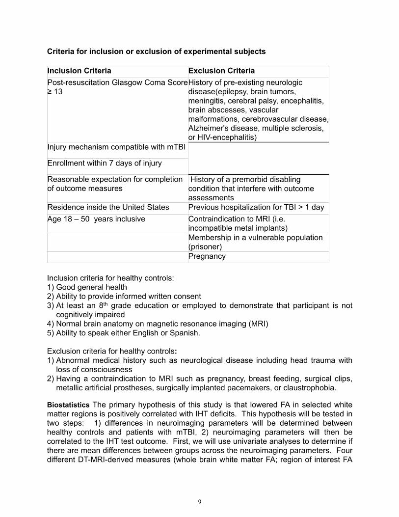
Criteria for inclusion or exclusion of experimental subjects Inclusion Criteria Exclusion CriteriaPost-resuscitation Glasgow Coma Score ≥ 13
History of pre-existing neurologic disease(epilepsy, brain tumors, meningitis, cerebral palsy, encephalitis, brain abscesses, vascular malformations, cerebrovascular disease, Alzheimer's disease, multiple sclerosis, or HIV-encephalitis)
Injury mechanism compatible with mTBI
Enrollment within 7 days of injury
Reasonable expectation for completion of outcome measures
History of a premorbid disabling condition that interfere with outcome assessments
Residence inside the United States Previous hospitalization for TBI > 1 dayAge 18 – 50 years inclusive Contraindication to MRI (i.e.
incompatible metal implants)Membership in a vulnerable population (prisoner)Pregnancy
Inclusion criteria for healthy controls: 1) Good general health2) Ability to provide informed written consent3) At least an 8th grade education or employed to demonstrate that participant is not
cognitively impaired4) Normal brain anatomy on magnetic resonance imaging (MRI)5) Ability to speak either English or Spanish.
Exclusion criteria for healthy controls:1) Abnormal medical history such as neurological disease including head trauma with
loss of consciousness2) Having a contraindication to MRI such as pregnancy, breast feeding, surgical clips,
metallic artificial prostheses, surgically implanted pacemakers, or claustrophobia.
Biostatistics The primary hypothesis of this study is that lowered FA in selected white matter regions is positively correlated with IHT deficits. This hypothesis will be tested in two steps: 1) differences in neuroimaging parameters will be determined between healthy controls and patients with mTBI, 2) neuroimaging parameters will then be correlated to the IHT test outcome. First, we will use univariate analyses to determine if there are mean differences between groups across the neuroimaging parameters. Four different DT-MRI-derived measures (whole brain white matter FA; region of interest FA
9
measures (for 12 different ROIs) and tractography FA measures (for a total of 28 different tracts). In this univariate analysis, group averages of each measure will be compared using the Mann-Whitney rank-sum test between the patients and the normal controls. After differences are determined between groups, we will utilize Receiver Operating Characteristics curves to determine the utility of these neuroimaging parameters to distinguish healthy volunteers from patients with mTBI. Receiver Operating Characteristics curves will be constructed to comparing each neuroimaging measure that showed significant differences between controls and patients to determine the discriminant validity of the potential imaging, as the neuroimaging data of healthy volunteers are considered optimal (i.e., gold standard from which patients’ neuroimaging parameters will deviate). The area under the curves will not be considered to guide the determination of an optimal biomarker, as correlation to outcome will determine this. The Receiver Operating Characteristics will only be used to determine the ability of each neuroimaging parameter to distinguish healthy volunteers from patients with TBI. Second we will utilize correlations within the patient group to determine the best biomarker of mTBI. Each of the neuroimaging measures will be correlated with the IHT outcome using Spearman’s rank correlation coefficient. Given the large number of comparisons, we will use a strict alpha = 0.005 for both group comparisons and correlations. Although this is less strict than a Bonferroni correction, we believe the latter is unduly conservative, as we anticipate that the DT-MRI measures will be highly correlated with each other.
In the first year of the study, we have enrolled 12 patients and 6 controls. The majority of patients have followed procedures. For a small minority of consented patients, we have encountered difficulty in achieving an acute scan. This has largely been due to balancing the patientʼs discharge date, scanning within 7 days of their injury, at an available time for the return patient to scan, and during an available time slot for the imaging center. Fortunately, no major problems have been encountered otherwise. The only protocol deviations has been one patient that was enrolled 36 hours after time of injury. Though the patient had a GCS of 13-15, the patient was drowsy and, by family request, was told to wait until the following morning after injury to be tested and scanned.
In contrast to our relative ease in enrolling patients for the initial scan, we have encountered problems in getting patients to return for their 6-month follow-up. The reasons have been various difficulty to getting transportation to the hospital, forgetfulness and deciding to leave the study. In response, we have refined our approach by making reminder phone calls and placing a substantially higher emphasis on their first scan during consent. There has been no change in the risk/benefit relationship of the research based upon the results.
10
There were personnel changes at the Dallas site during the last reporting period. Dr. Ramon Diaz-Arrastia, the principal investigator, and Ms Carol Moore, the research coordinator left U of Texas Southwestern to take positions at the Uniformed Services University of the Health Sciences. Dr. Diaz-Arrastia was replaced with Dr. Kyle Womack and Ms Moore was replaced with Mr. Paliotta. The personnel changes have not altered any other aspect of the study.
Key Research Accomplishments1) IRB approval at both SUNY-Downstate and UT-Southwestern sites.2) Software Development of IHT software has been completed and is working smoothly at
the Dallas site.3) Finalization of study design4) Ongoing recruitment of both mTBI patients and controls.5) Beginning of 6-month follow-up study.
Reportable OutcomesNone
Conclusion This study has had substantial success enrolling, testing and scanning patients.
References
1." Nelson, N.W., et al., Neuropsychological evaluation of blast-related concussion: Illustrating the challenges and complexities through OEF/OIF case studies. Brain Injury, 2011. 25(5): p. 511-525.
2." Mac Donald, C.L., et al., Detection of Blast-Related Traumatic Brain Injury in U.S. Military Personnel. New England Journal of Medicine, 2011. 364(22): p. 2091-2100.
3." Van der Knaap, L.J., and Van der Ham, J.M., How does the corpus callosum mediate interhemispheric transfer? A review. Behav. Brain Res., 2011. 223: p. 211-221
4." Weber, B., Treyer, V., Oberholzer, N., Jaermann, T., Boesiger, P., Brugger, P., Regard, M., Buck, A., Savazzi, S., Marzi, C.A. , Attention and Interhemispheric Transfer: A Behavioral and fMRI Study. . J. Cogn. Neurosci., 2005. 17: p. 113-123.
5." Peru, A., Beltramello, A., Moro, V., Sattibaldi, L., Berlucchi, G., Temporary and permanent signs of interhemispheric disconnection after traumatic brain injury. . Neuropsychologia., 2003. 41: p. 634-643.
11
6." Benavidez, D.A., Fletcher, J.M., Hannay, H.J., Bland, S.T., Caudle, S.E., Mendelsohn, D.B., Yeakley, J., Brunder, D.G., Harward, H., Song, J., Perachio, N.A., Bruce, D., Scheibel, R.S., Lilly, M.A., Verger-Maestre, K., Levin, H.S., Corpus callosum damage and interhemispheric transfer of information following closed head injury in children. Cortex, 1999. 35, : p. 315-336.
7." Mathias, J.L., Bigler, E.D., Jones, N.R., Bowden, S.C., Barrett-Woodbridge, M., Brown, G.C., Taylor, D.J., Neuropsychological and information processing performance and its relationship to white matter changes following moderate and severe traumatic brain injury: a preliminary study. Appl. Neuropsychol., 2004. 11: p. 134-152.
12