Embed Size (px)
Citation preview

AUS DEM LEHRSTUHL FÜR MUND-KIEFER-UND GESICHTSCHIRURGIE DIREKTOR: PROF. DR. DR. TORSTEN E. REICHERT
DER FAKULTÄT FÜR MEDIZIN DER UNIVERISTÄT REGENSBURG
PRIMARY AND SECONDARY LEIOMYOSARCOMA OF THE ORAL AND PERIORAL REGION- CLINICOPATHOLOGICAL AND IMMUNOHISTOCHEMICAL ANALYSIS
OF A RARE ENTITY WITH A REVIEW OF THE LITERATURE
Inaugural-Dissertation zur Erlangung des Doktorgrades
der Zahnmedizin
der Fakultät für Medizin der Universität Regensburg
vorgelegt von Anna Schütz
2013

!

AUS DEM LEHRSTUHL FÜR MUND-KIEFER-UND GESICHTSCHIRURGIE
DIREKTOR: PROF. DR. DR. TORSTEN E. REICHERT DER FAKULTÄT FÜR MEDIZIN
DER UNIVERISTÄT REGENSBURG
PRIMARY AND SECONDARY LEIOMYOSARCOMA OF THE ORAL AND PERIORAL REGION- CLINICOPATHOLOGICAL AND IMMUNOHISTOCHEMICAL ANALYSIS
OF A RARE ENTITY WITH A REVIEW OF THE LITERATURE
Inaugural-Dissertation zur Erlangung des Doktorgrades
der Zahnmedizin
der Fakultät für Medizin
der Universität Regensburg
vorgelegt von Anna Schütz
2013

Dekan: Prof. Dr. Dr. Torsten E. Reichert
1. Berichterstatter: Prof. Dr. Dr. Torsten E. Reichert
2. Berichterstatter: Prof. Dr. Dr. Peter Proff
Tag der mündlichen Prüfung: 27. Januar 2014
Primary and Secondary Leiomyosarcomaof the Oral and Perioral
Region—Clinicopathological andImmunohistochemical Analysis of a RareEntity With a Review of the LiteratureAnja Sch€utz, DDS,* Ralf Smeets, MD, DDS, PhD,y Oliver Driemel, MD, DDS, PhD,z
Samer George Hakim, MD, DDS, PhD,x Hartwig Kosmehl, MD, PhD,kHenning Hanken, MD, DDS,{ and Andreas Kolk, MD, DDS, PhD#
Purpose: Leiomyosarcoma (LMS) rarely occurs in the head and neck region. These tumors present witha wide range of clinical features, so the diagnosis is predicated on conventional microscopic findingscoupled with immunohistochemical analysis.
Patients and Methods: Clinical and histologic data of 7 patients with LMS of the head and neck wererecorded retrospectively. In addition to routine immunohistochemistry, staining for cell cycle regulatorproteins p16 and p21 was performed.
Results: Five LMSs (4 intraoral, 1 dermal cheek) occurred primarily in the oral and perioral region. TwoLMSs (parietal and sinonasal) were diagnosed as metastases originating from the uterus and pelvis. Treat-ment of the primary LMSs consisted of radical tumor resection with clear margins. Distant metastases fromLMSs were irradiated or excised as palliative treatment. Three of 5 patients (60%) with primarily excisedLMS developed recurrence after an average of 7 months, with lung metastases occurring after 17 months.In 1 patient, cervical lymph node metastases were detected after 10 months. Of all patients, 5 died after anaverage survival period of 2.4 years. The mean survival period of the 5 patients with primary LMS of thehead and neck was 3.3 years. All tumors were positive for vimentin and a-smooth muscle actin, with 57%of tumors showing positive nuclear expression of p16 and 71% of p21. Lack of p16 nuclear expression wasassociated with a shorter mean survival time (1.3 vs 4.3 yr for p16 positivity).
Conclusion: Lung and cervical lymph node metastases often occur in LMS of the head and neck. Presur-gical staging, including gynecologic examination, whole-body computed tomography, and sometimespositron-emission or computed tomography, to rule out LMS metastasis is mandatory. Surgical resectionof the tumor should be given top priority. Lack of p16 reactivity may have a prognostic value for LMSbecause it was related to a trend toward poorer survival.! 2013 American Association of Oral and Maxillofacial SurgeonsJ Oral Maxillofac Surg 71:1132-1142, 2013
Received from the Departments of Oral and Maxillofacial Surgery,
University of Regensburg, Regensburg, Germany and from the
Department of Oral and Maxillofacial Surgery, Technische
Universit€at M€unchen, Klinikum rechts der Isar, Munich, Germany.
*Postgraduate.
yProfessor for Oral and Maxillofacial Surgery, Department of Oral
and Maxillofacial Surgery, University Medical Center Hamburg-
Eppendorf, Hamburg, Germany.
zConsultant Surgeon.xConsultant Surgeon, Department of Maxillofacial Surgery,
University of L€ubeck, L€ubeck, Germany.
kHead, Institute of Pathology, HELIOS-Medical Centre, Erfurt,
Germany.
{Resident, Department of Oral and Maxillofacial Surgery,
University Medical Center Hamburg-Eppendorf, Hamburg, Germany.
#Consultant Surgeon, Department of Oral and Maxillofacial
Surgery, Technische Universit€at M€unchen, Klinikum rechts der
Isar, Munich, Germany.
Address correspondence and reprint requests to Dr Kolk: Depart-
ment of Oral and Maxillofacial Surgery, Technische Universit€at
M€unchen, Ismaninger Str 22, 81675 Munich, Germany; e-mail:
! 2013 American Association of Oral and Maxillofacial Surgeons
0278-2391/12/01729-6$36.00/0
http://dx.doi.org/10.1016/j.joms.2012.12.011
1132
Leiomyosarcomas (LMSs) are rare tumors that arisefrom smooth muscle cells of the myometrium, gastro-intestinal tract, or retroperitoneum.1 They account for7% of soft tissue sarcomas.2 LMSs are found predomi-nantly in the uterus, gastrointestinal tract, or retroper-itoneal space,3 whereas LMSs in the head and neckregion account for only 3%, most likely because ofthe paucity of smooth muscle tissue.4-6 Fewer than0.1% develop in the oral cavity.7 LMSs of the oral andperioral region are divided further into 3 main sub-groups: LMS of the oral soft tissue, LMS of the facialskin, and LMS of the jawbone. Separate from theseare primary LMSs of the head and neck and metastasesoriginating from the female genitalia, predominantlythe uterus.8 LMSs of the head and neck are raretumors with various clinical and histopathologic ap-pearances. Origins of the LMS of the head and neckare probably in the arterial tunica media, ductus lin-gualis, circumvallata papillae, and pluripotent mesen-chymal cells.6 The most frequent sites are thesinonasal tract (19%), the skin and soft tissue (16%),and the esophagus (12%).6
The prognostic significance of the localization ofLMS in the head and neck region is unclear. Becauseof the rare occurrence and poor prognosis of LMS,existing systematic data are insufficient and evidenceof effective therapy is scarce. The new World HealthOrganization classification indicates a poor prognosisfor primary LMS of the sinonasal tract and a variableoutcome of primary LMS of the larynx.1 The progno-sis of LMS in the oral and maxillofacial region is alsopoor, with a large percentage of recurrence and me-tastasis.1,5 The estimated 5-year disease-specific sur-vival rate (DSS) for primary oral LMS is 55%.1,2 Therespective 5-year DSS data of primary LMS of theoral and perioral soft and hard tissues indicate a sur-vival rate from 32% to 62%2,7,9 (Table 1),2,4,7,9-13
because LMS of the oral soft tissue has a betterprognosis than LMS infiltrating the jaws, which hasa significantly higher recurrence rate.1,2 The reasonfor this difference may be easier follow-up treatmentof the soft tissue region, with the possibility of earlierdetection of an initial tumor recurrence than that ofthe jawbone, which can be followed only by imagingmethods.Diagnosis of these tumors is often challenging. Im-
munohistochemistry for vimentin, desmin, or a-smooth muscle actin (ASMA) provides features ofsmooth muscle cell differentiation, which is criticalfor the diagnosis of LMS.14
Proto-oncogenes and suppressor oncogenes thathave contrary functions in cellular growth normallyregulate cellular proliferation. Apart from multipleother changes, neoplastic development is character-ized by a loss of cell cycle control. Proteins p16 andp21 are cell cycle regulators that have been studied
in different human neoplasms, including uterinesmooth muscle tumors.15,16
By inactivating the cyclin-dependent kinase (CDK)that phosphorylates the retinoblastoma protein, pro-tein p16 acts as a CDK inhibitor that slows down theprogression of the cell cycle. Protein p21 is also a po-tent CDK inhibitor and binds to and inhibits the activ-ity of cyclin-CDK2 or -CDK1 complexes and thusfunctions as a regulator of cell cycle progression atthe G1 phase.
The cell cycle regulators p16 and p21 are tightlycontrolled by tumor suppressor protein p53,17 whichis often mutated and overexpressed in LMS.18,19 Thelatter findings suggest a connection between tumorprogression and immunohistochemical expressionlevels of p16 and p21. The overexpression of p16appears to distinguish malignant LMSs from benignleiomyomas.20 The aims of this retrospective studywere to document the clinical presentation, clinicalcourse, and treatment of the rare entity of primaryLMS of the oral cavity and to analyze the impact ofcell cycle proteins p16 and p21 as an adjunct to con-ventional immunohistochemical criteria of LMS ofthe head and neck region.
Patients and Methods
Clinical treatment, follow-up, and histologic data of7 patients with primary LMS of the head and neckwere recorded after therapy. The out- and inpatientmedical records of the university clinics of oral andmaxillofacial surgery in Regensburg and L€ubeck, Ger-many from 1996 through 2008were reviewed. Clinicaldata were correlated with tumor grade, which was as-sessed using the National Cancer Institute system.21
Owing to the retrospective nature of the study, itwas granted a written exemption of the institutionalreview board standards of individual institutions bythe universities of Regensburg and L€ubeck. In addi-tion, all patients signed an informed written consentagreement allowing the use of follow-up data and his-tologic specimens for research purposes.
The diagnosis of a distant metastasis of an occult pri-mary tumor was excluded in 5 cases of primary LMS ofthe head and neck region by a full gynecologic exam-ination and whole-body computed tomography (CT)or magnetic resonance imaging (MRI) at the time of ini-tial diagnosis. An additional positron-emission tomo-graphic or CT scan was performed in the primaryLMS cases when the CT or MRI findings were unclearas to the differential diagnosis of a distant metastasisfrom other origins.
IMMUNOHISTOCHEMISTRY
Formaldehyde-fixated paraffin-embedded tissueblocks from every patient were stained with
SCH€UTZ ET AL 1133

Table 1. LITERATURE SURVEY OF EPIDEMIOLOGY, TREATMENT, AND CLINICAL COURSE OF PRIMARY LEIOMYOSARCOMA OF THE HEAD AND NECK REGION
Source Patients (n) M:F Age (yr) Primary Localization (n) Therapy (n)Relapse(%)
DistantMetastasis
(%)
Follow-UpAfter
Treatment
Mietttinen et al11
(1984)6 (5 case reports +1 own case)
5:1 18-62 mandible (6) OP (6), RT (1),CT (2)
67 50 50% after 24-60 mo,y 33% !Tafter 24 mo,* 1 patient !Tafter 24 mo*
Carter et al12
(1999)11 1.2:1 40 jaw bone (11) OP (10), RT (3),
CT (2)NES 36 36% after 36 mo,y
36% !T after 12 mo,*18 +T after 24 mo*
Dry et al10
(2000)10 1:1.5 34 jaw bone (5), oral soft tissue (5) OP (9), RT (1), CT
(2), unknown (1)20 33 50% after 20 mo,y
40% !T after 49 mo*Ethunandan et al2
(2007)64 1.3:1 43 jaw bone (38), oral soft
tissue (20), facial skin (6)OP (60), RT (14),CT (11)
34 35 5-yr DSS 55% total, 43%with bone infiltration,19% with metastasis
Izumi et al9 (1995) 60 1.4:1 42 jaw bone (27), maxillary sinus(14), oral soft tissue (18),fascial skin (1)
OP (55), RT (4), CT(39), unknown (2)
44 35 2-yr DSS 66%, 5-yr DSS32% total
Kratochvil et al13
(1982)20 4:1 65-70 jaw bone (8), skeleton bones (12) OP (18), RT (6),
CT (2)NES 37 35% after 24 mo,y 45%
after 21 mo,* 20% NESMontgomery et al4
(2002)13 1.2:1 47 jaw bone (5), oral soft tissue (3)
fascial skin (2), neck muscles (2),pharynx (1)
OP (9), unknown (4) 27 55 23% after 67 mo,y
38% !T after 50 mo,*8% +T after 24 mo,*31% NES
Vilos et al7 (2005) 50 1:1.3 44 jaw bone (34), oral soft tissue (15),maxillary sinus (1)
OP (46), RT (14), CT(13), unknown (4)
NES 32 5-yr DSS 62%
Present study 7 1:1.3 60 oral soft tissue (4), fascial skin (1),distant metastasis (2)
OP (6), RT (2) 43 71 71% after 29 mo,y
29% !T after 67 mo*
Abbreviations: CT, chemotherapy; DSS, disease-specific survival rate in years; F, female; M, male; NES, not elsewhere specified; OP, operation with radical resection; RT, radiationtherapy; +T, with tumor; !T, without tumor.* Alive.y Died.
Sch€utz et al. Leimyosarcoma of the oral and perioral region. J Oral Maxillofac Surg 2013.
1134
LEIM
YOSA
RCOMAOFTHEORALANDPERIO
RALREGIO
N

hematoxylin and eosin. For immunohistochemicalmarkers vimentin, desmin, ASMA, and Ki-67, resultsof routine immunohistochemistry were obtained.Staining for cell cycle regulator proteins p16 and p21also was performed with commercially available anti-bodies against p16 (Mtm Laboratories, Germany) andp21 (ScyTek, Zytomed Systems, Berlin, Germany).The sources and dilutions of all primary antibodiesare presented in Table 2. For the negative control, rep-resentative sections were incubated with normal rab-bit serum (1:100; DAKO, Glostrup, Denmark) andprocessed simultaneously as described earlier. Onlynuclear staining was considered a positive reactionfor p16 and p21. The evaluation of immunohistochem-ical staining was performed semiquantitatively andqualitatively by analyzing the percentage of positivelystained tumor cells in 5 representative high-powerfields, as explained later.22
Briefly, the percentage of tumor cells with positivestaining was estimated by counting 1,000 tumor cells.The number of positively stained tumor cells wasscored on a scale of 0 to 4 (0, no tumor cells; 1, 10%tumor cells; 2, 10% to 25% tumor cells; 3, 26% to50% tumor cells; 4, 50% tumor cells). Nuclear stainingintensity was evaluated according to the intensity ofpositive immunostaining as negative (!), weak (+),moderate (++), or strong (+++).Detection of the bound antibody was performed
with the ChemMate detection system (Dako Chem-Mate detection system, Hamburg, Germany) (alkalinephosphatase) and an immunostaining automatic ma-chine (Dako Autostainer, Hamburg, Germany) accord-ing to the manufacturers’ protocols (Table 2).
Results
CLINICAL AND FOLLOW-UP DATA
Seven patients (3 male and 4 female) with primaryLMS of the head and neck region were evaluated.The average age was 60 years (25 to 93 yr). Five LMStumors (4 intraoral, 1 in dermal cheek) occurredprimarily in the oral and perioral region (Table 3,
Fig 1A-D). Two LMS tumors (parietal and sinonasal)were diagnosed as distant metastases originatingfrom the uterus and pelvis. In these 2 cases, therapyof the primary LMS consisted of surgical tumor resec-tion with clear margins. In 3 cases, a selective neck dis-section was performed, and 1 patient underwentirradiation after surgery. Distant LMS metastases weretreated with palliative radiation or excised (Table 3).Independent of the selected therapy, 3 of 5 patients(60%) with primary LMS developed local recurrenceafter an average of 7 months (3 to 10 mo); in addition,lung metastases occurred after 17 months in allpatients. In 1 patient, cervical lymph node metas-tases were detected 10 months after solitary tu-mor resection.
Distant metastases in the head and neck region ofthe LMS of the uterus and pelvis developed parietallyand ethmoidally and in the cavernous sinus and retro-auricularly and in the median skullcap, respectively.Furthermore, metastases occurred in the lung and sub-mandibular gland (Table 3).
Five patients with metastases died after an averagesurvival time of 2.4 years (1.0 to 5.3 yr). Two patientswith primary LMS of the dermal cheek and the floor ofthe mouth were still alive after the end of the maxi-mum 6.5-year follow-up period of this study withoutmetastases or recurrence (4.7 and 6.5 yr, respectively;Table 3).
The mean survival period of all patients was 3.3years (1.0 to 6.5 yr). Because of the small sample,the average survival period of the 5 patients with pri-mary LMS of the head and neck was the same at 3.3years (1.0 to 6.5 yr).
HISTOLOGY
All LMS cases in this research series were assessedfor tumor depth, presence of circumscribed versusinfiltrative tumor borders, mitotic counts per 10high-power fields (5 sets counted), necrosis, nuclearpleomorphism, vascular invasion, and the occur-rence and environment of inflammatory compo-nents. Routine hematoxylin and eosin–stained LMS
Table 2. PRIMARY ANTIBODIES USED IN THIS STUDY
Antibody Clone Producer Dilution Pretreatment
p16 E6H4 Mtm Laboratories — Peroxidase blocking system, pH 9p21 DSC-60.2 Zytomed Systems 1:50 Target Retrieval Solution 1:10, pH 6.1Vimentin V9 Dako 1:4,500 Target Retrieval Solution 1:10, pH 9Desmin D33 Dako 1:200 Target Retrieval Solution 1:10, pH 9ASMA 1A4.(1) Dako 1:300 Target Retrieval Solution 1:10, pH 9Ki-67 MIB1 Dako 1:1,000 Target Retrieval Solution 1:10, pH 6.1
Abbreviation: ASMA, a-smooth muscle actin.
Sch€utz et al. Leimyosarcoma of the oral and perioral region. J Oral Maxillofac Surg 2013.
SCH€UTZ ET AL 1135
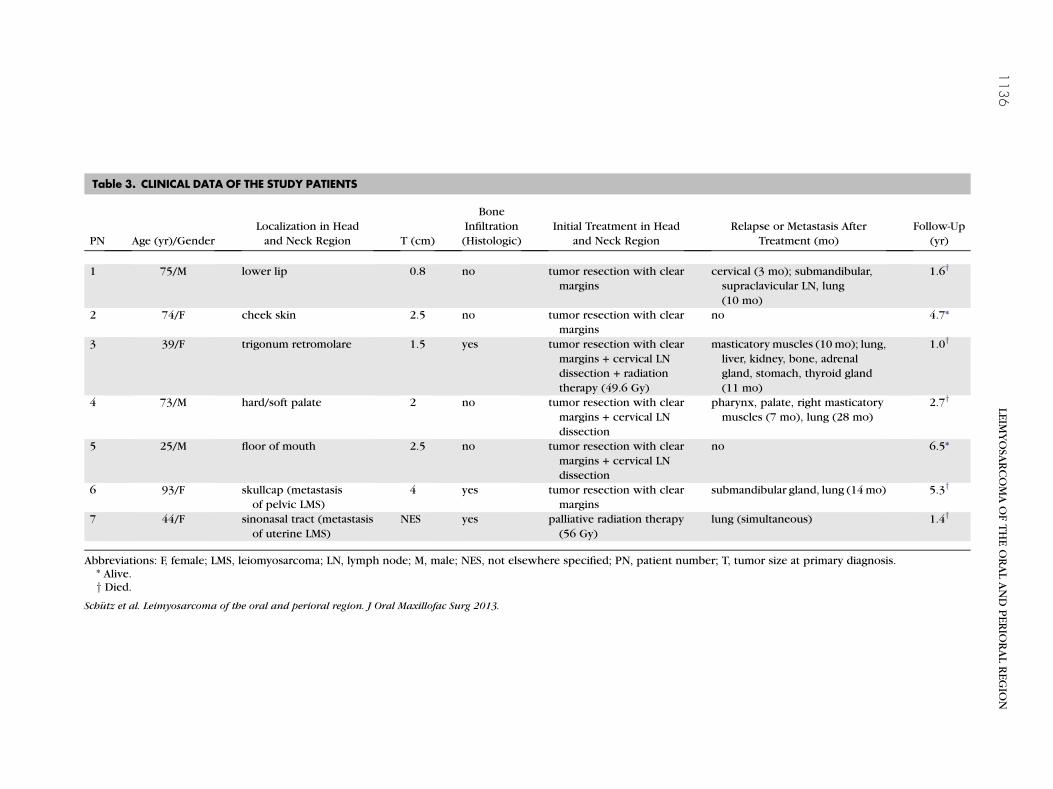
Table 3. CLINICAL DATA OF THE STUDY PATIENTS
PN Age (yr)/GenderLocalization in Headand Neck Region T (cm)
BoneInfiltration(Histologic)
Initial Treatment in Headand Neck Region
Relapse or Metastasis AfterTreatment (mo)
Follow-Up(yr)
1 75/M lower lip 0.8 no tumor resection with clearmargins
cervical (3 mo); submandibular,supraclavicular LN, lung(10 mo)
1.6y
2 74/F cheek skin 2.5 no tumor resection with clearmargins
no 4.7*
3 39/F trigonum retromolare 1.5 yes tumor resection with clearmargins + cervical LNdissection + radiationtherapy (49.6 Gy)
masticatory muscles (10 mo); lung,liver, kidney, bone, adrenalgland, stomach, thyroid gland(11 mo)
1.0y
4 73/M hard/soft palate 2 no tumor resection with clearmargins + cervical LNdissection
pharynx, palate, right masticatorymuscles (7 mo), lung (28 mo)
2.7y
5 25/M floor of mouth 2.5 no tumor resection with clearmargins + cervical LNdissection
no 6.5*
6 93/F skullcap (metastasisof pelvic LMS)
4 yes tumor resection with clearmargins
submandibular gland, lung (14mo) 5.3y
7 44/F sinonasal tract (metastasisof uterine LMS)
NES yes palliative radiation therapy(56 Gy)
lung (simultaneous) 1.4y
Abbreviations: F, female; LMS, leiomyosarcoma; LN, lymph node; M, male; NES, not elsewhere specified; PN, patient number; T, tumor size at primary diagnosis.* Alive.y Died.
Sch€utz et al. Leimyosarcoma of the oral and perioral region. J Oral Maxillofac Surg 2013.
1136
LEIM
YOSA
RCOMAOFTHEORALANDPERIO
RALREGIO
N

slides displayed a rough, tubercular, and infiltratingprocess of growth with fusiform or polygonal cells(Fig 2A). Both cell forms could be seen regularly.A cytoplasm seam with a fine fibrillary eosinophilictexture was always traceable, often with physiologicmitotic figures. At higher magnification, the tumorsexhibited perpendicularly arranged fascicles ofsharply marginated groups of spindle cells with eo-sinophilic cytoplasm and characteristic cigar-shapednuclei, hyperchromatic blunt-ended nuclei, and scat-tered paranuclear vacuoles, which constitute the typ-ical focal histologic features of LMS. Mitotic activitywas found in all tumors (4 to 40 mitoses per 10high-power fields), and necrosis was present inall cases. Vascular invasion was only minimally
visible (Fig 2A, B). All lesions had minimal inflamma-tion that, when present, consisted of scattered lym-phocytes or lymphoid aggregates. The amount ofcollagen fibers between tumor cells was low. Accord-ing to the National Cancer Institute grading system,21
2 of the 5 primary LMSs of the head and neck regionwere assigned to grade 1, 2 to grade 2, and 1to grade 3.
Immunohistochemically, all tumorswere positive forvimentin (5 of 5) and ASMA (5 of 5). Two of 3 examinedtumors expressed desmin. In 2 tumors, the prolifera-tion marker Ki-67 showed positive immunoreactivity.High magnification disclosed blunt-ended nuclei anddelicate cytoplasmic fibrils, with cells displaying strik-ing nuclear pleomorphism (Figs 2B, 3A, B). The
FIGURE1. A, En face view of a leiomyosarcoma of the left cheek. B, Radically resected leiomyosarcoma of the left cheek.C, Leiomyosarcomaat the right side of the floor of the mouth (intraoral tumor mass with necrotic ulcer measuring roughly 3.0 ! 3.0 cm). D, Intraoral view of a leio-myosarcoma in the right retromolar triangle.
Sch€utz et al. Leimyosarcoma of the oral and perioral region. J Oral Maxillofac Surg 2013.
SCH€UTZ ET AL 1137
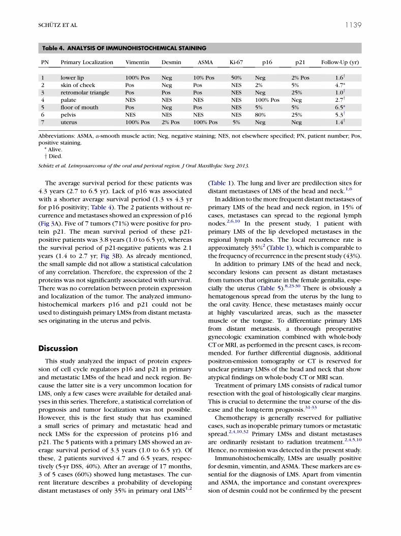
proliferation rate of the primary LMS of the lip was50%, whereas that of the distant metastasis of theLMS of the uterus was only 5%. Four of 7 tumors
(57%) showed positive immunoreactivity for p16(Fig 3A), whereas protein p21 expression was signifi-cantly increased in only 2 cases. The overall expressionof all immunohistochemical markers according to theLMS location is presented in Table 4.
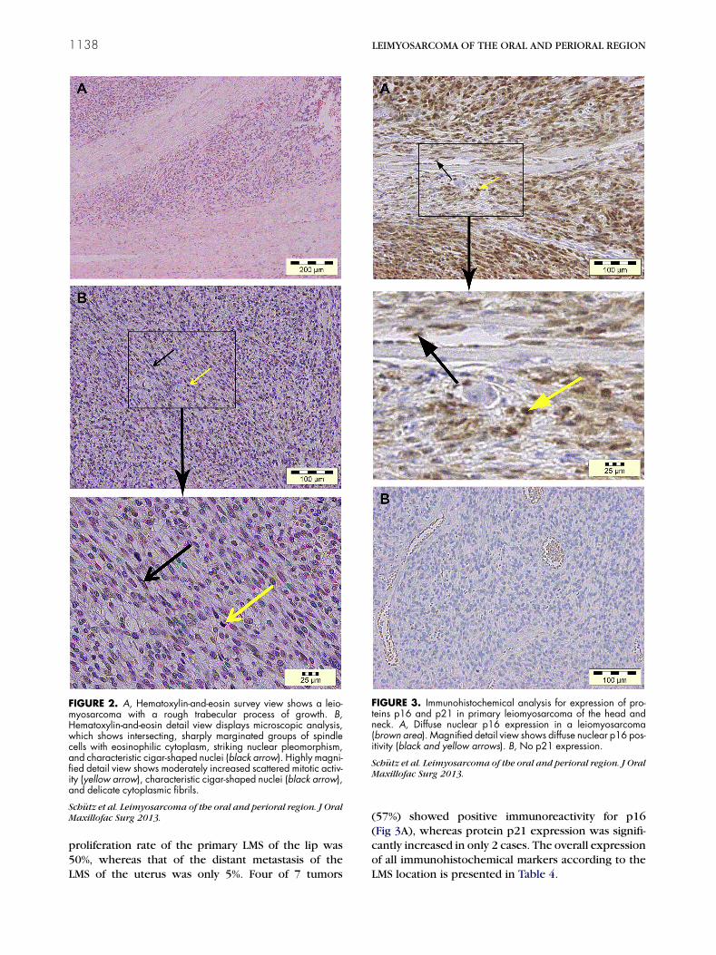
FIGURE 2. A, Hematoxylin-and-eosin survey view shows a leio-myosarcoma with a rough trabecular process of growth. B,Hematoxylin-and-eosin detail view displays microscopic analysis,which shows intersecting, sharply marginated groups of spindlecells with eosinophilic cytoplasm, striking nuclear pleomorphism,and characteristic cigar-shaped nuclei (black arrow). Highly magni-fied detail view shows moderately increased scattered mitotic activ-ity (yellow arrow), characteristic cigar-shaped nuclei (black arrow),and delicate cytoplasmic fibrils.
Sch€utz et al. Leimyosarcoma of the oral and perioral region. J OralMaxillofac Surg 2013.
FIGURE 3. Immunohistochemical analysis for expression of pro-teins p16 and p21 in primary leiomyosarcoma of the head andneck. A, Diffuse nuclear p16 expression in a leiomyosarcoma(brown area). Magnified detail view shows diffuse nuclear p16 pos-itivity (black and yellow arrows). B, No p21 expression.
Sch€utz et al. Leimyosarcoma of the oral and perioral region. J OralMaxillofac Surg 2013.
1138 LEIMYOSARCOMA OF THE ORAL AND PERIORAL REGION
The average survival period for these patients was4.3 years (2.7 to 6.5 yr). Lack of p16 was associatedwith a shorter average survival period (1.3 vs 4.3 yrfor p16 positivity; Table 4). The 2 patients without re-currence and metastases showed an expression of p16(Fig 3A). Five of 7 tumors (71%) were positive for pro-tein p21. The mean survival period of these p21-positive patients was 3.8 years (1.0 to 6.5 yr), whereasthe survival period of p21-negative patients was 2.1years (1.4 to 2.7 yr; Fig 3B). As already mentioned,the small sample did not allow a statistical calculationof any correlation. Therefore, the expression of the 2proteins was not significantly associated with survival.There was no correlation between protein expressionand localization of the tumor. The analyzed immuno-histochemical markers p16 and p21 could not beused to distinguish primary LMSs from distant metasta-ses originating in the uterus and pelvis.
Discussion
This study analyzed the impact of protein expres-sion of cell cycle regulators p16 and p21 in primaryand metastatic LMSs of the head and neck region. Be-cause the latter site is a very uncommon location forLMS, only a few cases were available for detailed anal-yses in this series. Therefore, a statistical correlation ofprognosis and tumor localization was not possible.However, this is the first study that has examineda small series of primary and metastatic head andneck LMSs for the expression of proteins p16 andp21. The 5 patients with a primary LMS showed an av-erage survival period of 3.3 years (1.0 to 6.5 yr). Ofthese, 2 patients survived 4.7 and 6.5 years, respec-tively (5-yr DSS, 40%). After an average of 17 months,3 of 5 cases (60%) showed lung metastases. The cur-rent literature describes a probability of developingdistant metastases of only 35% in primary oral LMS1,2
(Table 1). The lung and liver are predilection sites fordistant metastases of LMS of the head and neck.1,6
In addition to themore frequent distantmetastases ofprimary LMS of the head and neck region, in 15% ofcases, metastases can spread to the regional lymphnodes.2,6,10 In the present study, 1 patient withprimary LMS of the lip developed metastases in theregional lymph nodes. The local recurrence rate isapproximately 35%2 (Table 1), which is comparable tothe frequencyof recurrence in the present study (43%).
In addition to primary LMS of the head and neck,secondary lesions can present as distant metastasesfrom tumors that originate in the female genitalia, espe-cially the uterus (Table 5).8,23-30 There is obviously ahematogenous spread from the uterus by the lung tothe oral cavity. Hence, these metastases mainly occurat highly vascularized areas, such as the massetermuscle or the tongue. To differentiate primary LMSfrom distant metastasis, a thorough preoperativegynecologic examination combined with whole-bodyCTor MRI, as performed in the present cases, is recom-mended. For further differential diagnosis, additionalpositron-emission tomography or CT is reserved forunclear primary LMSs of the head and neck that showatypical findings on whole-body CT or MRI scan.
Treatment of primary LMS consists of radical tumorresection with the goal of histologically clear margins.This is crucial to determine the true course of the dis-ease and the long-term prognosis.31-33
Chemotherapy is generally reserved for palliativecases, such as inoperable primary tumors or metastaticspread.2,4,10,32 Primary LMSs and distant metastasesare ordinarily resistant to radiation treatment.2,4,5,10
Hence, no remissionwas detected in the present study.Immunohistochemically, LMSs are usually positive
for desmin, vimentin, and ASMA. These markers are es-sential for the diagnosis of LMS. Apart from vimentinand ASMA, the importance and constant overexpres-sion of desmin could not be confirmed by the present
Table 4. ANALYSIS OF IMMUNOHISTOCHEMICAL STAINING
PN Primary Localization Vimentin Desmin ASMA Ki-67 p16 p21 Follow-Up (yr)
1 lower lip 100% Pos Neg 10% Pos 50% Neg 2% Pos 1.6y
2 skin of cheek Pos Neg Pos NES 2% 5% 4.7*3 retromolar triangle Pos Pos Pos NES Neg 25% 1.0y
4 palate NES NES NES NES 100% Pos Neg 2.7y
5 floor of mouth Pos Neg Pos NES 5% 5% 6.5*6 pelvis NES NES NES NES 80% 25% 5.3y
7 uterus 100% Pos 2% Pos 100% Pos 5% Neg Neg 1.4y
Abbreviations: ASMA, a-smooth muscle actin; Neg, negative staining; NES, not elsewhere specified; PN, patient number; Pos,positive staining.* Alive.y Died.
Sch€utz et al. Leimyosarcoma of the oral and perioral region. J Oral Maxillofac Surg 2013.
SCH€UTZ ET AL 1139

immunohistochemical data compared with a study byMontgomery et al4 that found an expression of desminin 10 of 12 cases. Although desmin was positive in only40% of cases in this study, the Ki-67 proliferation ratewas analyzed in 2 cases and showed positive nuclearstaining of 5% and 50%, which is in linewith the resultsof other studies of head and neck LMSs.4 Other inves-tigators have reported a Ki-67 proliferation rateof 15%.34
Tumor-suppressor protein p16 regulates the cell cy-cle in the G1 phase by inhibiting the cell proliferationthrough the inhibition of cyclin D-dependent kinasecomplex 4/6.35 A decreased p16 expression hasbeen detected in 5% to 33% of LMSs.36,37 A lack ofnuclear p16 expression in neoplastic cells seems tobe associated with progressive tumor size anddecreased overall survival.38 However, there are onlylimited data regarding the function and impact ofcell cycle regulator proteins p16 and p21 in mesenchy-mal neoplasms. Also, the data on p16 and p21 expres-sions in larger studies of uterine smooth muscletumors are incomplete,39-41 so that there are nocomparable results. In the present study, 4 of 7 casesshowed an increased nuclear expression (>25%) ofp16. Although the p16-negative cases had an averagesurvival period of 1.3 years, the p16-positive cases sur-vived 4.3 years on average. Although this difference isnot statistically significant because of the small num-ber of cases, it indicates a worse prognosis when thereis a lack of p16 activity.Protein p21 is a cyclin kinase inhibitor that is regu-
lated by the tumor suppressor p53. Protein p21 leadsto cell cycle arrest and plays a crucial role in repairing
DNA damage.42,43 Commonly, LMSs express p21,whereas lack of p21 seems to be associated with anincreased risk of recurrence.44 In the present study,themean DSS in the p21-negative cases (2.1 yr) was de-creased compared with the p21-positive cases (3.8 yr).As in the case of p16, a statement regarding the impactof p21 expression on DSS and the chance of recur-rence based on a statistical calculation could notbe made.
Neither p16 nor p21 reactivity could be used to dif-ferentiate primary LMS from metastasis. Kim et al8 de-scribed an increased expression of oncogenes forcellular proliferation and angiogenesis in metastasizedLMS, specifically angiogenin, vascular endothelialgrowth factor, CD31, and von Willebrand factor. Al-though Unver et al45 found that neither p16 nor p21correlated with disease-free or overall survival in gyne-cologic LMS, it is generally accepted that p16 is ex-pressed more frequently and more strongly in LMSscompared with leiomyomas and is a useful antibodyin discriminating LMSs from leiomyomas.39
Interestingly, the present study showed the influ-ence of p16 and p21 expression on the long-term prog-nosis of this disease. To date, there are no othercurrently published studies regarding this issue owingto the rarity of LMS in the head and neck region. Fur-ther multicenter studies are needed to improve thetreatment and prognosis of this sporadic disease andto develop more targeted treatments against meta-static uterine sarcomas.
In conclusion, LMS is an exceedingly rare tumor inthe oral and maxillofacial region and has a poor prog-nosis because of a high local recurrence rate. There
Table 5. LITERATURE SURVEYOF CLINICAL COURSEOF LEIOMYOSARCOMAWITH DISTANTMETASTASIS IN THE HEADAND NECK REGION
Source Age (yr) GenderPrimary
LocalizationLocalization ofMetastasis Treatment Follow-Up (yr)
Allen et al23 (1993) 66 M leg hard palate OP + RT + CT 2y
Allen et al23 (1993) 61 M upper leg mandible OP + RT + CT 3*Allen et al23 (1993) 65 F uterus lower lip OP + CT 1.4*Aslan et al24 (2008) 76 F uterus temporal muscle OP ! RT 3*Bogart et al25 (1990) 58 F lung palate CT + RT 0.4y
Kaziro et al26 (1981) 59 F uterus tongue none unknownNusrath et al27 (2006) 65 F uterus masseter muscle OP + CT 2.3y
Sandruck et al28 (2004) 39 F uterus sphenoid OP + CT + RT 1.1y
Uchino et al29 (1996) 54 F uterus skull OP + CT 2y
Kim et al8 (2009) 56 F uterus right maxilla none 0.3y
Vora and Levin30 (2003) 62 F uterus tongue none NESy
Present study 44 F uterus sinonasal tract OP + RT 1.4y
Present study 93 F pelvis skullcap OP + RT 5.3y
Abbreviations: CT, chemotherapy; F, female; M, male; NES, not elsewhere specified; OP, operation; RT, radiation therapy.* Alive.y Died.
Sch€utz et al. Leimyosarcoma of the oral and perioral region. J Oral Maxillofac Surg 2013.
1140 LEIMYOSARCOMA OF THE ORAL AND PERIORAL REGION

may be a predilection for occurrence in the jawbones,with bone involvement possibly associated with aneven poorer prognosis because of a higherrecurrence rate. In primary LMS, one third of patientsdevelop distant metastases, with lymph node metasta-ses being less frequent. Distant metastases in the headand neck region originating from the female genitaliamust be taken into account in the initial tumor staging;therefore, whole-body imaging and a gynecologicexamination are necessary.Aggressive surgical treatment is necessary for a total
cure. Treatment of primary LMS is radical tumor re-section with histologically clear margins. Whenlymph node metastases are suspected, an additionaluni- or bilateral neck dissection is indicated, depend-ing on the tumor location. Adjuvant radiation andchemotherapy also may have a beneficial effect in de-creasing or delaying the recurrence rate, improvingsurvival time, and sometimes allowing the possibilityof less radical resection.Immunohistochemical markers such as ASMA and
vimentin are important for the diagnosis of LMS.They frequently express cyclin kinase inhibitors p16and p21. Lack of nuclear p16 seems to be associatedwith a trend toward a poorer prognosis.
References
1. Mucke T, Mitchell DA, Tannapfel A, et al: Outcome in adult pa-tients with head and neck sarcomas—A 10-year analysis. J SurgOncol 102:170, 2010
2. Ethunandan M, Stokes C, Higgins B, et al: Primary oral leiomyo-sarcoma: A clinico-pathologic study and analysis of prognosticfactors. Int J Oral Maxillofac Surg 36:409, 2007
3. Mesquita RA, Migliari DA, de Sousa SO, et al: Leiomyosarcoma ofthe buccal mucosa: A case report. J Oral Maxillofac Surg 56:504,1998
4. Montgomery E, Goldblum JR, Fisher C: Leiomyosarcoma of thehead and neck: A clinicopathological study. Histopathology40:518, 2002
5. Nikitakis NG, Lopes MA, Bailey JS, et al: Oral leiomyosarcoma:Review of the literature and report of two cases with assessmentof the prognostic and diagnostic significance of immunohisto-chemical and molecular markers. Oral Oncol 38:201, 2002
6. Yadav R, Bharathan S: Leiomyosarcoma of the buccal mucosa:A case report with immunohistochemistry findings. J Oral Sci50:215, 2008
7. Vilos GA, Rapidis AD, Lagogiannis GD, et al: Leiomyosarcomas ofthe oral tissues: Clinicopathologic analysis of 50 cases. J OralMaxillofac Surg 63:1461, 2005
8. Kim SM, Myoung H, Choung PH, et al: Metastatic leiomyosar-coma in the oral cavity: Case report with protein expression pro-files. J Craniomaxillofac Surg 37:454, 2009
9. Izumi K, Maeda T, Cheng J, et al: Primary leiomyosarcoma of themaxilla with regional lymph node metastasis. Report of a caseand review of the literature. Oral Surg Oral Med Oral PatholOral Radiol Endod 80:310, 1995
10. Dry SM, Jorgensen JL, Fletcher CD: Leiomyosarcomas of theoral cavity: An unusual topographic subset easily mistakenfor nonmesenchymal tumours. Histopathology 36:210,2000
11. Miettinen M, Lehto VP, Ekblom P, et al: Leiomyosarcoma ofthe mandible: Diagnosis as aided by immunohistochemicaldemonstration of desmin and laminin. J Oral Pathol 13:373,1984
12. Carter LC, Aguirre A, Boyd B, et al: Primary leiomyosarcoma ofthe mandible in a 7-year-old girl: Report of a case and reviewof the literature. Oral Surg Oral Med Oral Pathol Oral Radiol En-dod 87:477, 1999
13. Kratochvil FJ III, MacGregor SD, Budnick SD, et al: Leiomyosar-coma of the maxilla. Report of a case and review of the litera-ture. Oral Surg Oral Med Oral Pathol Oral Radiol Endod 54:647, 1982
14. Denk H, Krepler R, Artlieb U, et al: Proteins of intermediate fila-ments. An immunohistochemical and biochemical approach tothe classification of soft tissue tumors. Am J Pathol 110:193, 1983
15. Palazzo JP, Mercer WE, Kovatich AJ, et al: Immunohistochemicallocalization of p21(WAF1/CIP1) in normal, hyperplastic, andneoplastic uterine tissues. Hum Pathol 28:60, 1997
16. Leiser AL, Anderson SE, Nonaka D, et al: Apoptotic and cell cycleregulatory markers in uterine leiomyosarcoma. Gynecol Oncol101:86, 2006
17. Hewedi IH, Radwan NA, Shash LS: Diagnostic value of progester-one receptor and p53 expression in uterine smooth muscletumors. Diagn Pathol 7:1, 2012
18. Mittal K, Demopoulos RI: MIB-1 (Ki-67), p53, estrogen receptor,and progesterone receptor expression in uterine smoothmuscletumors. Hum Pathol 32:984, 2001
19. O’Neill CJ, McBride HA, Connolly LE, et al: Uterine leiomyosar-comas are characterized by high p16, p53 and MIB1 expressionin comparison with usual leiomyomas, leiomyoma variants andsmooth muscle tumours of uncertain malignant potential. Histo-pathology 50:851, 2007
20. Gannon BR, Manduch M, Childs TJ: Differential immunoreactiv-ity of p16 in leiomyosarcomas and leiomyoma variants. Int JGynecol Pathol 27:68, 2008
21. Guillou L, Coindre JM, Bonichon F, et al: Comparative study ofthe National Cancer Institute and French Federation of CancerCenters Sarcoma Group grading systems in a population of410 adult patients with soft tissue sarcoma. J Clin Oncol 15:350, 1997
22. Kolk A, Jubitz N, Mengele K, et al: Expression of Y-box-bindingprotein YB-1 allows stratification into long- and short-term survi-vors ofhead andneckcancerpatients. Br JCancer 105:1864, 2011
23. Allen CM, Neville B, Damm DD, et al: Leiomyosarcoma meta-static to the oral region. Report of three cases. Oral Surg OralMed Oral Pathol Oral Radiol Endod 76:752, 1993
24. Aslan E, Kuzeyli K, Cakir E, et al: Temporalis muscle metastasisof the uterine leiomyosarcoma: A case report. Turk Neurosurg18:215, 2008
25. Bogart SF, Sacks HG, DeMarco LC: Metastatic leiomyosarcoma ofthe palate. J Oral Maxillofac Surg 48:1338, 1990
26. Kaziro GS: Metastatic uterine leiomyosarcoma to the tongue: Re-port of case. J Oral Surg 39:128, 1981
27. Nusrath MA, Kendall CH, Avery CM: Metastatic uterine leiomyo-sarcoma masquerading as a primary lesion of the masseter mus-cle. Int J Oral Maxillofac Surg 35:466, 2006
28. Sandruck J, Escobar P, Lurain J, et al: Uterine leiomyosarcomametastatic to the sphenoid sinus: A case report and review ofthe literature. Gynecol Oncol 92:701, 2004
29. Uchino M, Endo G, Shibata I, et al: Uterine leiomyosarcoma me-tastasis to the skull—Case report. Neurol Med Chir (Tokyo) 36:469, 1996
30. Vora NM, Levin RJ: Metastatic leiomyosarcoma to the tongue.Otolaryngol Head Neck Surg 128:601, 2003
31. Abdin HA, Prabhu SR: Leiomyosarcoma of mandible in a Suda-nese female. Int J Oral Surg 14:85, 1985
32. Lo Muzio L, Favia G, Mignogna MD, et al: Primary intraoral leio-myosarcoma of the tongue: An immunohistochemical study andreview of the literature. Oral Oncol 36:519, 2000
33. Strasser-Fuchs S, Enzinger C, Ropele S, et al: Clinically benignmultiple sclerosis despite large T2 lesion load: Can we explainthis paradox? Mult Scler 14:205, 2008
34. Yavuz E, Gulluoglu MG, Akbas N, et al: The values of intratu-moral mast cell count and Ki-67 immunoreactivity index in dif-ferential diagnosis of uterine smooth muscle neoplasms.Pathol Int 51:938, 2001
35. Liggett WH Jr, Sidransky D: Role of the p16 tumor suppressorgene in cancer. J Clin Oncol 16:1197, 1998
SCH€UTZ ET AL 1141

36. Dei Tos AP, Maestro R, Doglioni C, et al: Tumor suppressor genesand related molecules in leiomyosarcoma. Am J Pathol 148:1037, 1996
37. HashimotoH,DaimaruY, TsuneyoshiM, et al: Leiomyosarcomaofthe external soft tissues. A clinicopathologic, immunohistochem-ical, and electron microscopic study. Cancer 57:2077, 1986
38. Kawaguchi K, Oda Y, Saito T, et al: Mechanisms of inactivation ofthe p16INK4a gene in leiomyosarcoma of soft tissue: Decreasedp16 expression correlates with promoter methylation and poorprognosis. J Pathol 201:487, 2003
39. Bodner-Adler B, Bodner K, Czerwenka K, et al: Expression ofp16 protein in patients with uterine smooth muscle tumors:An immunohistochemical analysis. Gynecol Oncol 96:62, 2005
40. Lee CH, Turbin DA, Sung YC, et al: A panel of antibodies to de-termine site of origin and malignancy in smooth muscle tumors.Mod Pathol 22:1519, 2009
41. D’Angelo E, Spagnoli LG, Prat J: Comparative clinicopathologicand immunohistochemical analysis of uterine sarcomas diag-nosed using the World Health Organization classification sys-tem. Hum Pathol 40:1571, 2009
42. Abdel-Fatah TM, Powe DG, Agboola J, et al: The biological, clin-ical and prognostic implications of p53 transcriptional pathwaysin breast cancers. J Pathol 220:419, 2010
43. Yang W, Qi Q, Zhang H, et al: P21 Waf1/Cip1 polymorphismsand risk of esophageal cancer. Ann Surg Oncol 17:1453, 2010
44. Dobashi Y, Noguchi T, Nasuno S, et al: CDK-inhibitor-associatedkinase activity: A possible determinant of malignant potential insmooth muscle tumors of the external soft tissue. Int J Cancer94:353, 2001
45. Unver NU, Acikalin MF, Oner U, et al: Differential expression ofP16 and P21 in benign and malignant uterine smooth muscletumors. Arch Gynecol Obstet 284:483, 2011
1142 LEIMYOSARCOMA OF THE ORAL AND PERIORAL REGION

1
Deutschsprachige-Zusammenfassung-
1. Einleitung-
Leiomyosarkome (LMS) sind sehr seltene Tumoren der glatten Muskulatur, welche am
häufigsten im Myometrium des Uterus, im Gastrointestinaltrakt und im Retroperitoneum
auftreten [14]. Insgesamt besitzen sie einen Anteil von etwa 7% an allen
Weichgewebssarkomen [13]. Weniger als 5% aller Leiomyosarkome manifestieren sich
im Kopf-Hals-Bereich, weniger als 0,1% treten in der Mundhöhle auf [36]. Nach dem
Ursprungsgewebe können in der oralen und perioralen Region LMS der oralen
Weichgewebe, LMS der Gesichtshaut und LMS der Kieferknochen unterschieden
werden.
Abzugrenzen von den primären LMS des Kopf-Hals-Bereichs sind sekundäre,
metastasierte LMS, am häufigsten ausgehend von den weiblichen Genitalien,
insbesondere vom Uterus [21].
Aufgrund des seltenen Auftretens der Tumoren im Kopf-Hals-Bereich ist die
histopathologische Diagnose mitunter schwierig. Immunhistochemische
Zusatzuntersuchungen, wie etwa die Positivität der Tumoren für Vimentin, Desmin oder
SMA (Smooth Muscle Actin) sind von entscheidender Bedeutung [34]. Die Proteine p16
und p21 sind wichtige Regulatoren des Zellzyklus und werden von p53 kontrolliert [8].
P53 ist in Leiomyosarkomen häufig mutiert und immunhistochemisch exprimiert [31],
weshalb ein Zusammenhang der Tumorprogression und dem immunhistochemischen
Expressionscore von p16 und p21 bestehen könnte. Die Überexpression von p16 scheint
Leiomyosarkome von Leiomyomen und seinen benignen Varianten zu unterscheiden
[15].

2
Die vorliegende retrospektive klinisch-pathologische Studie soll
1. das charakteristische klinische Erscheinungsbild der LMS des Kopf-Hals-
Bereiches und den Verlauf unter Therapie dokumentieren,
2. eine Analyse der Zellzyklus- assoziierten Proteine p16 und p21 in LMS der
Kopf-Hals-Region vorstellen.
-
3
2. Patienten-und-Methoden--
Die vorliegende klinisch-pathologische Studie umfasst sieben Patienten mit einem
histopathologisch gesicherten Leiomyosarkom des Kopf-Hals-Bereiches, welche in den
Jahren 1996 bis 2008 in den Kliniken für Mund-, Kiefer- und Gesichtschirurgie der
Universität Regensburg und der Universität Lübeck behandelt wurden. Epidemiologische
Daten, Symptome, Therapie und klinischer Verlauf wurden anhand der ambulanten und
stationären Patientenakten retrospektiv erhoben.
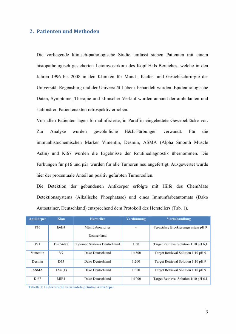
Von allen Patienten lagen formalinfixierte, in Paraffin eingebettete Gewebeblöcke vor.
Zur Analyse wurden gewöhnliche H&E-Färbungen verwandt. Für die
immunhistochemischen Marker Vimentin, Desmin, ASMA (Alpha Smooth Muscle
Actin) und Ki67 wurden die Ergebnisse der Routinediagnostik übernommen. Die
Färbungen für p16 und p21 wurden für alle Tumoren neu angefertigt. Ausgewertet wurde
hier der prozentuale Anteil an positiv gefärbten Tumorzellen.
Die Detektion der gebundenen Antikörper erfolgte mit Hilfe des ChemMate
Detektionssystems (Alkalische Phosphatase) und eines Immunfärbeautomats (Dako
Autostainer, Deutschland) entsprechend dem Protokoll des Herstellers (Tab. 1).
Antikörper Klon Hersteller Verdünnung Vorbehandlung
P16 E6H4 Mtm Laboratories
Deutschland
- Peroxidase Blockierungssystem pH 9
P21 DSC-60.2 Zytomed Systems Deutschland 1:50 Target Retrieval Solution 1:10 pH 6,1
Vimentin V9 Dako Deutschland 1:4500 Target Retrieval Solution 1:10 pH 9
Desmin D33 Dako Deutschland 1:200 Target Retrieval Solution 1:10 pH 9
ASMA 1A4.(1) Dako Deutschland 1:300 Target Retrieval Solution 1:10 pH 9
Ki67 MIB1 Dako Deutschland 1:1000 Target Retrieval Solution 1:10 pH 6,1
Tabelle 1: In der Studie verwendete primäre Antikörper!
4
3. Ergebnisse-
3.1. Klinik-
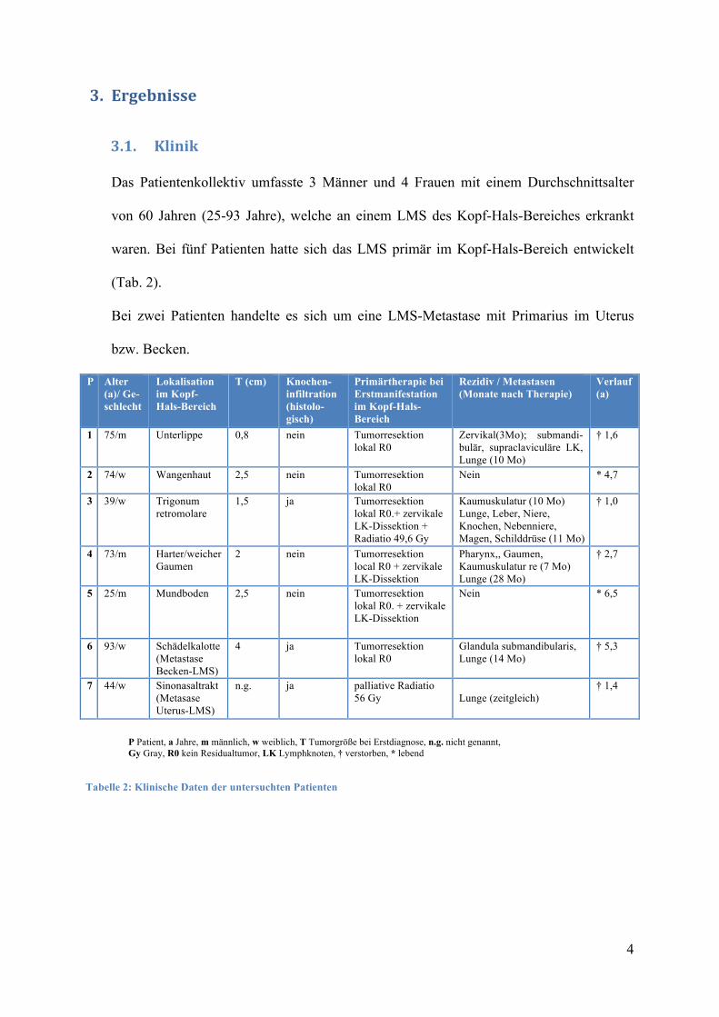
Das Patientenkollektiv umfasste 3 Männer und 4 Frauen mit einem Durchschnittsalter
von 60 Jahren (25-93 Jahre), welche an einem LMS des Kopf-Hals-Bereiches erkrankt
waren. Bei fünf Patienten hatte sich das LMS primär im Kopf-Hals-Bereich entwickelt
(Tab. 2).
Bei zwei Patienten handelte es sich um eine LMS-Metastase mit Primarius im Uterus
bzw. Becken.
P Alter (a)/ Ge-schlecht
Lokalisation im Kopf-Hals-Bereich
T (cm) Knochen-infiltration (histolo-gisch)
Primärtherapie bei Erstmanifestation im Kopf-Hals-Bereich
Rezidiv / Metastasen (Monate nach Therapie)
Verlauf (a)
1
75/m Unterlippe 0,8 nein Tumorresektion lokal R0
Zervikal(3Mo); submandi-bulär, supraclaviculäre LK, Lunge (10 Mo)
† 1,6
2 74/w Wangenhaut 2,5 nein Tumorresektion lokal R0
Nein
* 4,7
3 39/w Trigonum retromolare
1,5 ja Tumorresektion lokal R0.+ zervikale LK-Dissektion + Radiatio 49,6 Gy
Kaumuskulatur (10 Mo) Lunge, Leber, Niere, Knochen, Nebenniere, Magen, Schilddrüse (11 Mo)
† 1,0
4
73/m Harter/weicher Gaumen
2 nein Tumorresektion local R0 + zervikale LK-Dissektion
Pharynx,, Gaumen, Kaumuskulatur re (7 Mo) Lunge (28 Mo)
† 2,7
5 25/m Mundboden 2,5 nein Tumorresektion lokal R0. + zervikale LK-Dissektion
Nein * 6,5
6 93/w Schädelkalotte (Metastase Becken-LMS)
4
ja Tumorresektion lokal R0
Glandula submandibularis, Lunge (14 Mo)
† 5,3
7 44/w Sinonasaltrakt (Metasase Uterus-LMS)
n.g. ja palliative Radiatio 56 Gy
Lunge (zeitgleich)
† 1,4
P Patient, a Jahre, m männlich, w weiblich, T Tumorgröße bei Erstdiagnose, n.g. nicht genannt, Gy Gray, R0 kein Residualtumor, LK Lymphknoten, † verstorben, * lebend
Tabelle 2: Klinische Daten der untersuchten Patienten

5
Die Therapie der primären LMS umfasste in beiden Kliniken die Tumorresektion mit
histologisch tumorfreien Grenzen. Bei drei Patienten wurde eine prophylaktische
Ausräumung der zervikalen Lymphknoten durchgeführt. In einem Fall erfolgte eine
postoperative Radiatio. Die LMS-Metastasen im Kopf-Hals-Bereich wurden palliativ
bestrahlt bzw. reseziert (Tab. 2).
Drei der fünf Patienten (60%) mit primärem LMS im Kopf-Hals-Bereich entwickelten
unabhängig von der gewählten Therapie nach durchschnittlich 7 Monaten (3-10) ein
Tumorrezidiv, nach durchschnittlich 17 Monaten (10-29) zusätzlich Lungenmetastasen.
Bei einem der beiden Patienten mit solitärer Tumorresektion traten 10 Monate
postoperativ Metastasen in den Halslymphknoten auf.
Die LMS des Uterus bzw. Beckens metastasierten in die Keilbeinhöhle und
Siebbeinzellen bzw. in den Sinus cavernosus, retroaurikulär und in die mediane
Schädelkalotte und bildeten weitere Tochtergeschwülste in der Lunge und der Glandula
submandibularis (Tab. 2).
Von insgesamt 7 Patienten waren zum Ende der Auswertung 5 Patienten nach einem
durchschnittlichen Überleben von 2,4 Jahren (1,0-5,3) mit stattgefundener
Fernmetastasierung verstorben. Zwei Patienten mit einem primären LMS der subkutanen
Wangenhaut bzw. des Mundbodens waren zum Abschluss der Auswertung nach 4,7 bzw.
6,5 Jahren ohne Hinweis auf ein Rezidiv oder eine Fernmetastasierung am Leben. (Tab.
2).
Das durchschnittliche Überleben aller Patienten lag zum Ende der Auswertung bei 3,3
Jahren (1,0 - 6,5). Die mittlere Überlebensdauer der fünf Patienten mit einem primären
LMS des Kopf-Hals-Bereiches betrug ebenfalls 3.3 Jahre (1,0-6,5)

6
3.2. Histologie-
Die LMS der eigenen Untersuchungsserie zeigten übereinstimmend ein grobknotiges und
infiltratives Wuchsbild mit spindeligen oder polygonalen Zellen (Abb. 1a und 1b). Beide
Zellformen waren regelmäßig innerhalb eines Tumors sichtbar. Ein Zytoplasmasaum war
immer nachweisbar, oft mit feinfibrillärer eosinophiler Textur. Physiologische Mitosen
waren immer nachweisbar. Der kollagene Fasergehalt zwischen den Tumorzellen war
gering.
Abbildung 1a: HE-Übersichtsaufnahme: LMS mit grobknotiges Wuchsbild.

7
Abbildung 1b: HE-Detailaufnhahme: LMS mit spindeligen und runden Tumorzellen; Nachweis einer hohen Mitoseaktivität durch Mitosefiguren.
Immunhistochemisch zeigten sich die Tumorzellen positiv für das Intermediärfilament
Vimentin (5/5) und den Muskelmarker ASMA (5/5). 2 von 5 untersuchten Tumoren
waren positiv für Desmin (Tab. 3).
P Primär-lokalisation
Vimentin
Desmin ASMA Ki67 p16 p21 Verlauf (a)
1. Unterlippe 100% pos
Neg 10% pos 50% Neg 2% pos † 1,6
2. Wangenhaut Pos Neg Pos n.d. 2% 5% * 4,7 3. Trigonum
retromolare Pos Pos Pos n.d. Neg 25% † 1,0
4 Gaumen n.d. n.d. n.g. n.d. 100% pos
Neg † 2,7
5. Mundboden Pos Neg Pos n.d. 5% 5% * 6,5 6. Becken n.d. n.d. n.d. n.d. 80% 25% † 5,3
7. Uterus 100% pos
2% pos 100% pos
5% Neg Neg † 1,4
P Patient, a Jahre, Pos positive Färbung, Neg negative Färbung, n.d. nicht durchgeführt,† verstorben, * lebend
Tabelle 3: Auswertung der immunhistochemischen Färbungen
8
Bei zwei Tumoren wurde der Proliferationsmarker Ki-67 bestimmt, wobei das LMS der
Lippe eine hohe Proliferationrate von 50% aufwies, das primär im Uterus entstandene
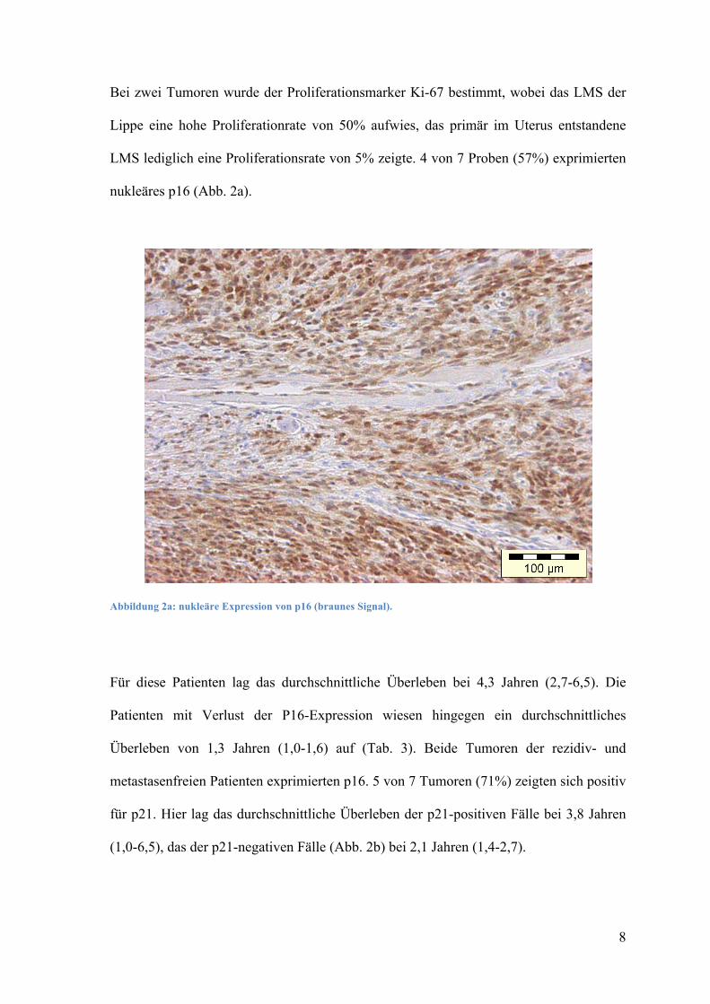
LMS lediglich eine Proliferationsrate von 5% zeigte. 4 von 7 Proben (57%) exprimierten
nukleäres p16 (Abb. 2a).
Abbildung 2a: nukleäre Expression von p16 (braunes Signal).
Für diese Patienten lag das durchschnittliche Überleben bei 4,3 Jahren (2,7-6,5). Die
Patienten mit Verlust der P16-Expression wiesen hingegen ein durchschnittliches
Überleben von 1,3 Jahren (1,0-1,6) auf (Tab. 3). Beide Tumoren der rezidiv- und
metastasenfreien Patienten exprimierten p16. 5 von 7 Tumoren (71%) zeigten sich positiv
für p21. Hier lag das durchschnittliche Überleben der p21-positiven Fälle bei 3,8 Jahren
(1,0-6,5), das der p21-negativen Fälle (Abb. 2b) bei 2,1 Jahren (1,4-2,7).

9
Abbildung 2b: p21 keine Expression
Die Expression der beiden Proteine war nicht miteinander assoziiert. Eine Korrelation mit
der Tumorlokalisation des Primarius konnte nicht beobachtet werden. Ebenso wenig
ermöglichten die immunhistochemischen Marker eine Differenzieung zwischen primären
LMS und LMS-Metastasen.
-

10
4. Diskussion-
Während Leiomyosarkome sich relativ häufig im Uterus und Gastrointestinatrakt
manifestieren [12, 25], entstehen primär im Kopf-Hals-Bereich nur 3% aller LMS [28,
29, 38], da in dieser Region nur wenige glatte Muskelzellen vorliegen. Im Kopf-Hals-
Bereich nehmen LMS vermutlich ihren Ursprung in der Tunika media der Arterien, dem
Ductus lingualis, den Papillae circumvallatae und den pluripotenten Mesenchymalzellen
[4, 38]. Der Sinonasal-Trakt (19%), die Haut und das Weichgewebe (16%) sowie der
Ösophagus (12%) bilden hier die Prädilektionsstellen [38].
Angaben zur prognostischen Bedeutung der Lokalisation der primären LMS innerhalb
des Kopf-Hals-Bereiches sind nicht kongruent. Die aktuelle WHO-Klassifikation der
Tumoren des Weichgewebes und des Knochens beschreibt eine eher schlechte Prognose
der primären LMS des Sinonasal-Traktes und eine variable Prognose bei primärem
Auftreten des LMS im Larynx [16, 33]. Primäre LMS der oralen und perioralen Weich-
und Hartgewebe zeigen in größeren Untersuchungen eine 5-Jahres-Überlebensrate von
32-62% [13, 18, 36] (Tab. 4), wobei LMS der oralen Weichgewebe eine bessere
Prognose aufweisen als LMS der Kieferknochen. Bei den eigenen 5 Patienten mit einem
primären LMS des Kopf-Hals-Bereiches betrug die mittlere Überlebensdauer 3.3 Jahre
(1,0-6,5). Lediglich 2 Patienten (5-JÜR ca. 40%) waren nach 4,7 bzw. 6,5 Jahren noch
am Leben. Eine Zuordnung der Prognose zur Tumorlokalisation konnte aufgrund der
geringen Fallzahl nicht getroffen werden. In 3 von 5 Fällen (60%) kam es nach
durchschnittlich 17 Monaten zu einer Fernmetastasierung in Form von
Lungenmetastasen, was über der in der Literatur angegebenen
Metastasenwahrscheinlichkeit von etwa 35% für primäre orale LMS liegt [13] (Tab. 4).
Die Lunge gilt neben der Leber als Prädilektionsstelle für Fernmetastasen beim LMS des
Kopf-Hals-Bereiches [14, 38].

11
Neben den häufigeren Fernmetastasierungen kann es bei LMS der Kopf-Hals-Region in
etwa 15% der Fälle auch zu regionalen Lymphknotenmetastasierungen kommen [11, 13,
28]. In der eigenen Untersuchungsserie kam es bei einem LMS der Unterlippe zu einer
Tumorabsiedelung in die Halslymphknoten.
Bezüglich der Rezidivhäufigket wird für primäre LMS der oralen Weichgewebe eine
Rezidivrate von etwa 35% angegeben [13] (Tab. 4), was der Rezidivhäufigkeit von 43%
in der eigenen Untersuchung entspricht.

12
Quelle Patien-ten (a)
M: w Alter (a)
Primärlokalisation (n)
Therapie (n)
Rezidiv (%)
Fernmetastasen (%)
Verlauf nach Therapie
Carter et al. (1999) [ ]
11 1,2 :1 40 Kieferknochen (11)
OP (10), RT (3), CT (2)
n.g. 36 † 36% nach 36 Mo, * 36% o T nach 12 Mo, * 18 m T nach 24 Mo
Dry et al. (2000) [ ]
10 1: 1,5 34 Kieferknochen (5), Orale Weichgewebe (5)
OP (9), RT (1), CT (2), unbekannt (1)
20 33 † 50% nach 20 Mo, * 40% o.T. nach 49 Mo
Ethunandan et al. (2007) [ ]
64 1,3: 1 43 Kieferknochen (38), Orale Weichgewebe (20), Gesichtshaut (6)
OP (60), RT (14), CT (11)
34 35 5-JÜR 55% gesamt, 43% bei Knocheninfiltration, 19% bei Metastasen
Izumi et al. (1995) [ ]
60 1,4 : 1 42 Kieferknochen (27), Sinus maxilaris (14), Orale Weichgewebe (18), Gesichtshaut (1)
OP (55), RT (4), CT (39), unbekannt (2)
44 35 2-JÜR 66%, 5 JÜR 32% gesamt
Kratochvil et al. (1982) [ ]
20 4:1 65-70 Kieferknochen (8) Skelettknochen (12)
OP (18), RT (6), CT (2)
n.g. 37 † 35% nach 24 Mo, * 45% nach 21 Mo, 20% n.g.
Montgomery et al. (2002) [ ]
13 1,2 :1 47 Kieferknochen (5), Orale Weichgewebe (3) Gesichtshaut (2), Halsmuskulatur (2), Pharynx (1)
OP (9), unbekannt (4)
27 55 † 23% nach 67 Mo, * 38% o.T. nach 50 Mo, * 8% m.T. nach 24 Mo 31% n.g.
Vilos et al. (2005) [ ]
50 1: 1,3 44 Kieferknochen (34), Orale Weichgewebe (15), Sinus Max. (1)
OP (46), RT (14), CT (13) unbekannt (4)
n.g. 32 5-JÜR 62%
Eigene Studie
7 1 : 1,3 60 Orale Weichgewebe (4), Gesichtshaut (1), Metastasen (2)
OP (6), RT (2) 43 71 † 71% nach 29 Mo * 29% o. T. nach 67 Mo
a Jahr, m männlich, w weiblich, OP Operation, RT Radiatio, CT Chemotherapie, * lebend, † gestorben, o.T. ohne Tumor, m.T. mit Tumor, Mo Monate, JÜR Jahresüberlebensrate, n.g. nicht genannt
Tabelle 4: Literaturübersicht zur Epidemiologie, zur Therapie und zum klinischen Verlauf primärer Leiomyosarkome des Kopf-Halsbereiches

13
Wenn auch selten, so ist bei einem LMS im Kopf-Hals-Bereich dennoch die Möglichkeit
eines fernmetastasierten Tumors in Erwägung zu ziehen (Tab. 5). Als Primärlokalisation
zeigt sich hier vor allem der Uterus, was eine präoperative gynäkologische Untersuchung
empfehlen lässt.
Quelle Alter (a)
Geschlecht Primär-lokalisation
Lokalisation Metastase
Therapie Verlauf (a)
Allen et al.
(1993) [ ]
66 M Bein Harter Gaumen OP+RT+CT † 2
Allen et al.
(1993) [ ]
61 M Oberschenkel Unterkiefer OP+RT+CT * 3
Allen et al.
(1993) [ ]
65 W Uterus Unterlippe OP+CT * 1,4
Aslan et al.
(2008) [ ]
76 W Uterus M. temporalis OP-RT * 3
Bogart et al.
(1990) []
58 W Lunge Gaumen CH+RT † 0,4
Kaziro et al.
(1981) []
59 W Uterus Zunge Keine Unbekannt
Nusrath et al.
(2006) [ ]
65 W Uterus M.masseter OP+CT † 2,3
Sandruck et al.
(2004) [ ]
39 W Uterus Sinus
sphenoidalis
OP+CT+RT † 1,1
Uchino et al.
(1996) [ ]
54 W Uterus Schädel OP+CT † 2
Kim et al.
(2009) []
56 W Uterus Maxilla re Keine † 0,3
Vora and Levin
(2003) []
62 W Uterus Zunge Keine † n.g.
Eigene Studie 44 W Uterus Sinonasal Trakt OP+RT † 1,4
Eigene Studie 93 W Becken Schädelkalotte OP+RT † 5,3
a Jahre, w weiblich, m männlich, Op Operation, RT Radiatio, CT Chemotherapie, * lebend, † verstorben, M Metastasen, n.g. nicht genannt
Tabelle 5: Literaturübersicht zum klinischen Verlauf von in den Kopf-Hals-Bereich metastasierten Leiomyosarkomen
14
Als entscheidend für die Prognose gilt bei allen primären LMS die weit im Gesunden
erfolgte chirurgische Resektion mit histologisch gesicherten, tumorfreien Randschnitten
[2, 24, 26, 27]. Eine Chemotherapie bleibt üblicherweise palliativen Situationen - wie
inoperablen Primärtumoren oder bereits erfolgter Metastasierng - vorbehalten [11, 13, 24,
28]. Gegenüber der Radiatio erweisen sich sowohl primäre LMS als auch LMS-
Metastasen in der Regel als resistent [11, 13, 28, 29]. Auch in der eigenen Studie konnte
die Radiatio weder beim Primärtumor noch bei den Tumormetastasen eine Remission
erzielen.
Die immunhistochemischen Marker ASMA und Vimentin stellen ein wichtiges
Hilfsmittel zur Diagnose eines Leiomyosarkoms dar, was in der eigenen Untersuchung
bestätigt werden konnte. Hingegen zeigte sich Desmin nur in 40% der Fälle als positiv.
Die Ki67-Proliferationsrate wurde nur in 2 Fällen untersucht (5% und 50%). In der Regel
liegt die Zellproliferationsrate von LMS bei über 15% [34].
Das Tumor-Suppressor-Protein p16 reguliert den Zellzyklus in der G1-Phase, indem es
den Cyclin D-abhängigen Kinasekomplex 4/6 und somit die Zellproliferation hemmt
[23]. Eine verringerte p16-Expression wurde in 5-33% von LMS beobachtet [9, 17].
Verlust an nukleärer P16-Expression in neoplastischen Zellen scheint beim LMS der
Weichgewebe mit fortgeschrittener Tumorgröße und schlechterem Gesamtüberleben
assoziiert [19].
In der eigenen Untersuchung zeigte sich in 4 von 7 Fällen eine nukleäre Expression von
p16. Während die p16-negativen Fälle ein durchschnittliches Überleben von 1,3 Jahren
aufwiesen, lag das durchschnittliche Überleben der p16-positiven Fälle bei 4,3 Jahren,
was auf eine Prognoseverschlechterung durch p16-Verlust hindeutet.
Bei p21 handelt es sich ebenfalls um ein Cyclin-Kinase-Inhibitor-Protein. Reguliert durch
den Tumorsuppressor p53 führt p21 zum Zellzyklusarrest und besitzt eine wichtige
15
Funktion bei der Reparatur von DNA-Schäden [1, 39]. Leiomyosarkome der
Weichgewebe exprimieren üblicherweise p21, wobei der Verlust der p21-Expression mit
einem erhöhten Rezidivrisiko einherzugehen scheint [10]. Zwar lag auch in der
vorliegenden Studie das durchschnittliche Gesamtüberleben der p21-negativen Fälle (2,1
Jahre) im Vergleich zu den p21-positiven Fällen (3,8 Jahre) niedriger, eine prognostische
Aussage bezüglich Rezidiv- oder Gesamtüberlebenswahrscheinlichkeit lässt hier jedoch
nicht treffen. Eine Differenzierung zwischen primären LMS und LMS-Metastasen war
weder durch p16 noch durch p21 erkennbar. Kim et al. beschreiben in den metastasierten
LMS eine erhöhte Expression an Onkogenen für die zelluläre Proliferation und die
Angiogenese, etwa Angiogenin, VEGF, CD 31 und vWF [21].
5. Schlussfolgerungen-
1. Primäre Leiomyosarkome des Kopf-Hals-Bereiches lassen Fernmetastasen, v.a. in die
Lunge, bei jedem dritten Patienten erwarten. Regionäre Lymphknotenmetastasen sind
seltener, können aber ebenfalls auftreten. In Betracht zu ziehen ist immer die
Metastasierung eines LMS in den Kopf-Hals-Bereich. Als Primarius findet sich hier
häufig der Uterus. Ein umfassendes präoperatives Ganzkörper-Staging inklusive
gynäkologischer Abklärung ist daher zu empfehlen.
2. Die Therapie von LMS im Kopf-Hals-Bereich besteht in der radikalen chirurgischen
Entfernung des Tumors. Bei Verdacht auf Lymphknotenmetastasen sollten diese mit
exstirpiert werden. Die Strahlentherapie scheint hingegen keinen großen therapeutischen
Wert zu besitzen.
3. Die immunhistochemischen Marker Vimentin und ASMA unterstützen die Diagnostik

16
eins Leiomyosarkoms. LMS exprimieren häufig die Cyclinkinase-Inhibitoren p16 und
p21. Der Verlust von nukleärem p16 scheint mit einer schlechteren Prognose assoziiert.
-
17
Literaturverzeichnis-
1. Abdel-Fatah TM, Powe DG, Agboola J, Adamowicz-Brice M, Blamey RW, Lopez-
Garcia MA, Green AR, Reis-Filho JS, Ellis IO (2010) The biological and prognostic
implications pf p53 transcriptional pathways in breast cancer. J Pathol 220: 419-434
2. Abdin HA, Prabhu SR (1985) Leiomyosarcoma of the mandible in a Sudanese female. Int
J Oral Surg 14: 85-88
3. Allen CM, Neville B, Douglas D, Damm D, Marsh W (1993) Leiomyosarcoma
metastatic to the oral region. Oral Surg Oral Med Oral Pathol 76: 752-756
4. Amarapala H, Tilakaratne WM (2006) Leiomyosarcoma of the oral cavity. Report of
seven cases and review of literature. Oral Oncol Extra 42: 14-17
5. Aslan E, Kuzeyl K, Cakir E, Reis A (2008) Temporalis Muscle Metastasis of the Uterine
Leiomyosarcoma: A Case Report. Turk Neurosurg 18: 215-218
6. Bogart SF, Sacks HG, DeMarco LC (1990) Metastastic leiomyosarcoma of the palate. J
Oral Maxillofac Surg 48: 1338-1340
7. Carter LC, Aguirre A, Boyd B, DeLacure MD (1999) Primary leiomyosarcoma of the
mandible in a 7-year-old girl. Oral Surg Oral Med Oral Pathol Oral Radiol Endod 87:
477-484

18
8. Chen L, Yang B (2008) Immunohistochemical analysis of p16, p53 and Ki-67 expression
in uterine smooth muscle tumors. Int J Gynecol Pathol 27: 326-32
9. Dei Tos AP, Maestro R, Doglioni C, Piccinin S, Libera DD, Boiocchi M, Fletcher CD
(1996) Tumor suppressor genes and related molecules in leiomyosarcoma. Am J Pathol
148: 1037-1045
10. Dobashi Y, Noguchi T, Nasuno S, Katayama K, Kameya T (2001) CDK-inhibitors-
associated kinase activity: a possible determinant of malignant potential in smooth
muscle tumors of the external soft tissue. Int J Cancer 94: 353-62.
11. Dry SM, Jorgensen JL, Fletcher CDM (2000) Leiomyosarcomas of the oral cavity: an
unusual topographic subset easily mistaken for nonmesenchymal tumors. Histopathology
36: 210-220
12. Enzinger FM, Weiss SW (2007) Soft tissue tumors. 5th ed Mosby, St. Louis, pp 545-564
13. Ethunandan M, Stokes C, Higgins B, Spedding A, Way C, Brennan P ( 2007) Primary
oral leiomyosarcoma: a clinico- pathologic study and analysis of prognostic factors. Int J
Oral Maxillofac Surg 36: 409-16.
14. Evans HL, Shipley J (2002) Kapitel In: Fletcher C, Unni KK, Mertens F (eds) World
Health Organisation Classification of Tumours, Pathology &Genetics of Tumours of the
Soft Tissue and Bones. IARC Press, Lyon, pp 131-134

19
15. Gannon BR, Manduch M, Childs TJ (2008) Differential Immunoreactivity of p16 in
leiomyosarcomas and leiomyoma variants. Int J Gynecol Pathol 27: 68-73
16. Gude P, Tisch M, Kraft K, Danz B, Maier H (2006) Leiomyosarkom des Kehlkopfs.
HNO 54: 207-214
17. Hashimoto H, Daimaru Y, Tsuneyoshi M, Enjoji M (1986) Leiomyosarcoma of the
external soft tissues. A clinicopathologic, immunohistochemical, and electron
microscopic study. Cancer 57: 2077-2088
18. Izumi K, Maeda T, Cheng J, Saku T (1995) Primary leiomyosarcomas of the maxilla with
regional lymph node metastasis. Oral Surg Oral Med Oral Pathol 80: 310-319
19. Kawaguchi K, Oda Y, Saito T, Yamamoto H, Tamiya S, Takahira T, Miyajima K,
Iwamoto Y, Tsuneyoshi M (2003) Mechanisms of inactivation of the p16INK4a gene in
leiomyosarcoma of soft tissue: decreased p16 expression correlates with promoter
methylation and poor prognosis. J Pathol 201: 487-495.
20. Kaziro GSN (1981) Metastatic uterine leiomyosarcoma to the tongue: report of a case. J
Oral Surg 39: 128-129
21. Kim SM, Myoung H, Choung PH, Kim MJ, Lee SK, Lee JH (2009) Metastatic
leiomyosarcoma in the oral cavity: Case report with protein expression profiles. J Cranio
Maxillofac Surg 37: 454-460

20
22. Kratochvil FJ, L Commander, MacGregor SD, Budnick SD, Hewan-Lowe K Allsup HW
( 1982) Leiomyosarcoma of the maxilla. Oral Surg 54: 647-655
23. Ligget WH, Sidransky D (1998) Role of p16 tumor suppressor gene in cancer. J Clin
Oncol 16: 1197-1206
24. Lo Muzio L, Favia G, Mignogna MD, Piattelli A, Maiorano E (2000) Primary intraoral
leiomyosarcoma of the tongue: an immunohistochemical study and review of the
literature. Oral Oncol 36: 519-524
25. Mesquita RA, Migliari DA, de Sousa SOM, Alves MR (1998) Leiomyosarcoma of the
Buccal Mucosa : A Case Report. J Oral Maxillofac Surg 56: 504-507
26. Mills SM, Gaffey MJ, Frierson HF (2000) Atlas of Tumor Pathology /Tumors of the
Upper Aerodigestive Tract, American Registry of Pathology. P 342-343
27. Mitsudo K, Tohnai I, Fujimoto Y, Sawaki Y, Sugimura T, Nishiguchi H, Fukui T,
Yamoto N, Ueda M (2006) Leiomyosarcoma of the maxilla: Effective chemotherapy with
docetaxel (DOC) and cisplatin (CDDP) using superselective intra-arterial infusion via
superficial temporal artery. Oral Oncol EXTRA 42: 258-262
28. Montgomery E, Goldblum JR, Fischer C (2002) Leiomyosarcoma of the head and neck: a
clinicopathological study. Histopathology 40: 518-523
21
29. Nikitakis NG, Lopes MA, Bailey JS, Blanchaert RH, Ord RA, Sauk JJ (2002) Oral
leiomyosarcoma: review of the literature and report of two cases with assessment of the
prognostic and diagnostic significance of immunohistochemical and molecular markers.
Oral Oncol 38: 201-208
30. Nusrath MA, Kendall CH, Avery CM (2006) Metastatic uterine leiomyosarcoma
masquerading as a primary lesion of the masseter muscle. Int J Oral Maxillofac Surg 35:
466- 468
31. O’Neill CJ, McBride HA, Connolly LE, McCluggage WG (2007) Uterine
leiomyosarcomas are characterized by high p16, p53 and MIB1 expression in comparison
with usual leiomyomas, leiomyoma variants and smooth muscle tumours of uncertain
malignant potential. Histopathology 50: 851–858.
32. Sandruck J, Escobar P, Lurain J, Fishman D (2004) Uterine leiomyosarcoma metastasic
to the sphenoid sinus: a case report and review of the literature. Gynecol Oncol 92: 701-
704
33. Thompson LDR, Fanburg Smith JC (2005) Hypopharynx, larynx and trachea In: Barnes
L, Eveson JW, Reichart P, Sidransky D (eds) World Health Organisation Classification of
Tumours, Pathology &Genetics, Head and Neck Tumours. IARC Press, Lyon, p 148.
34. Thompson LDR, Fanburg Smith JC (2005) Nasal cavity and paranasal sinuses. In: Barnes
L, Eveson JW, Reichart P, Sidransky D (eds) World Health Organisation Classification of
Tumours, Pathology &Genetics, Head and Neck Tumours. IARC Press, Lyon, pp 37-38

22
35. Uchino M, Endo G, Shibata I, Terao H, Kuramitsu T, Kushida Y, Nakamura N (1996)
Uterine Leiomyosarcoma Metastasis to the Skull. Neurol Med Chir (Tokyo) 36: 469-471
36. Vilos GA, Rapidis AD, Lagogiannis GD, Apostolidis C (2005) Leiomyosarcomas of the
oral tissues: Clinicopathologic Analysis of 50 Cases. J Oral Maxillofac Surg 63: 1461-
1477
37. Vora NM, Levin RJ (2003) Metastatic leiomyosarcoma to the tongue. Otolaryngol Head
Neck Surg 128: 601-602
38. Yadav R, Bharathan S (2008) Leiomyosarcoma of the buccal mucosa: a case report with
immunohistochemistry findings. J Oral Sci 50: 215-218
39. Yang W, Qi Q, Zhang H, Xu W, Chen Z, Wang L, Wang Y, Dong X, Jiao H, Huo Z
(2010) p21 ( Waf1/Cip1 ) Polymorphisms and Risk of Esophageal Cancer. Ann Surg
Oncol [Epub ahead of print].