Embed Size (px)
Citation preview
Neuron, Vol. 12, 571-582, March, 1994, Copyright 0 1994 by Cell Press
Association of Poly(A) mRNA with Microtubules in Cultured Neurons
Gary J. BasselI,* Robert H. Singer,+ and Kenneth S. Kosik* *Center for Neurologic Disease Brigham and Women’s Hospital and Harvard Medical School Boston, Massachusetts 02115 +Department of Cell Biology University of Massachusetts Medical Center Worcester, Massachusetts 01655
Summary
The structural basis for the synthesis of specific proteins within distinct intraneuronal compartments is unknown. We studied the distribution of poly(A) mRNA within cul- tured cerebrocortical neurons using high resolution in situ hybridization to identify cytoskeletal components that may anchor mRNA. After 1 day in culture, poly(A) mRNA was distributed throughout all of the initial neu- rites, including the axon-like process. At 4 days in cul- ture, poly(A) mRNA was distributed throughout the cell body and dendritic processes, but confined to the proxi- mal segment of the axon. Poly(A) mRNA was bound to the cytoskeleton as demonstrated by resistance to deter- gent extraction. Perturbation of microtubules with col- chicine resulted in a major reduction of dendritic poly(A) mRNA; however, this distribution was unaffected by cy- tochalasin. Ultrastructural in situ hybridization revealed that poly(A) mRNA and associated ribosomes were ex- cluded from tightly bundled microtubules.
Introduction
Neurons are highly polarized cells with two morpho- logically and functionally distinct types of processes: axons and dendrites. Protein synthesis is restricted to the somatodendritic compartment in most neu- rons; in contrast, most axons are devoid of polysomes (for review see Steward and Banker, 1992). Therefore, axons rely upon the transport of newly synthesized proteins over considerable distances to reach their correct cytological destinations. Dendrites can syn- thesize proteins where they are required by position- ing specific mRNAs at proximal sites. The localization of microtubule-associated protein 2 (MAP2) mRNA in situ and in culture provides one such example (Garner et al., 1988; Bruckenstein et al., 1990; Kleiman et al., 1990). Transport of RNA into dendrites of hippocam- pal neurons in culture has been demonstrated using sH[uridine] labeling (Davis et al., 1987). Ultrastructural studies have identified polysomes associated with in- ternal membranes beneath the postsynaptic mem- brane of dendritic spines (Steward and Levy, 1982; Steward, 1983). Biochemical analysis of these synapto- dendrosome fractions have demonstrated that the
proteins synthesized are incorporated into synaptic structures(RaoandSteward,1991).Thesedataprovide evidence for the targeting of dendritic mRNA as a mechanism for positioning proteins to their sites of function.
The majority (>85%) of translatable mRNA is bound to the cytoskeleton, whereas untranslated mRNA and monomeric ribosomes are soluble (for reviews see Hesketh and Pryme, 1991; Singer, 1992). The interac- tion of mRNAs with specific cytoskeletal filament sys- tems could provide a mechanism to localize mRNAs to distinct intraneuronal compartments. Analysis of mRNA-cytoskeletal interactions in nonneuronal cells has identified a major role for the actin cytoskeleton in fibroblasts (Sundell and Singer, 1991; Taneja et al., 1992),muscle(Horneand Hesketh,199O),epithelia(Ra- maekers et al., 1983), and HeLa cells (Lenk et al., 1977; Ornelles et al., 1986). Our objective was to identify cytoskeletal components in neurons that anchor mRNA. To derive general mechanisms for the cy- toskeletal localization of mRNA, we focused on the poly(A) sequence, a marker for the 3’ end of most mRNAs (Sheiness and Darnell, 1973). Using fluores- cence in situ hybridization, the effects of cytoskeletal perturbation on the localization of poly(A) mRNA were evaluated. To visualize directly the attachment of mRNA to a cytoskeletal filament, each was detected using double-label immunogold methods and ana- lyzed by electron microscopy.
Results
Compartmentalization of Poly(A) mRNA in Developing Processes The use of primary neuronal cultures to study mRNA localization offers distinct advantages to tissue sec- tions, as they permit direct visualization of individual cells and their complete sets of processes. Compara- blewith the staging of cultured hippocampal neurons (Dotti et al., 1988), most cortical neurons undergo the following developmental sequence: shortly after the neurons have attached to the substrate, they extend actin-rich lamellae (stage 1). These lamellipodia con- solidate to form a relatively symmetric array of minor neurites (stage 2). Within the first 24 hr, one of the minor neurites becomes significantly longer than the others and begins to assume the morphology of an axon (stage 3). Within the next few days, the other neurites develop the tapering and branching charac- teristics of dendrites (stage 4). The compartmentaliza- tion of MAP2, which has been well documented in situ and in culture (Matus et al., 1981,1986; Caceres et al., 1984; Kosik and Finch, 1987), is shown in Figures IA, IC, and IE. At 1 day in culture, MAP2 was distrib- uted throughout the cell body and all of the neurites (Figure IA). In cortical neurons cultured for 4 days,
Neuron 572
Cytoskeletal Association of Neuronal PO&(A) mRNA 573
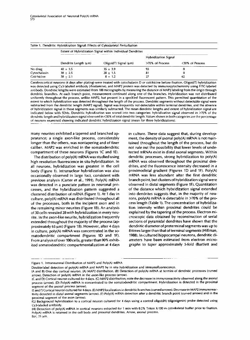
Table 1. Dendritic Hybridization Signal: Effects of Cytoskeletal Perturbation
Extent of Hybridization Signal within Individual Dendrites
Hybridization Signal
Dendrite Length (urn) Oligo(dT) Signal (urn) >70% of Process <30% of Process
No drug 40 f 3.5 35 * 3.9 93 0 Cytochalasin 38 f 2.5 28 * 1.5 81 0 Colchicine 38 f 2.1 8 f 1.2 27 62
Cerebrocortical neurons (4 days after plating) were treated with cytochalasin D or colchicine before fixation. Oligo(dT) hybridization was detected using CyZlabeled antibody (rhodamine), and MAP2 protein was detected by immunocytochemistry using FITC-labeled antibody. Dendritic lengths were estimated from 100 micrographs by measuring the distance of MAP2 labeling from the origin through dendritic branches. At each branch point, measurement continued along one of the branches. Hybridization was not distributed uniformly throughout the process, unlike MAPL, but present in a speckled fluorescent pattern. This permitted quantitation of the extent to which hybridization was detected throughout the length of the process. Dendritic segments without detectable signal were subtracted from the dendritic length (MAP2 signal). Signal was frequently not detectable within terminal dendrites, and the absence of hybridization signal in these segments was similarly subtracted. The mean dendritic lengths and extent of hybridization signal are indicated below with SEMs. Dendritic hybridization was scored into two categories: hybridization signal observed in >70% of the dendritic length and hybridization signal observed in <30% of total dendritic length. Values shown in both categories are the percentage of neurons examined showing indicated dendritic hybridization signal (mean for three hybridizations).
many neurites exhibited a tapered and branched ap- pearance; a single axon-like process, considerably longer than the others, was nontapering and of finer caliber. MAP2 was enriched in the somatodendritic compartment of these neurons (Figures IC and IE).
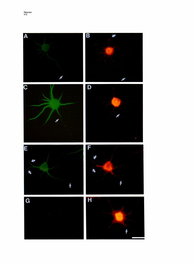
The distribution of poly(A) mRNAwas studied using high resolution fluorescence in situ hybridization. In all neurons, hybridization was greatest in the cell body (Figure 1). lntranuclear hybridization was also occasionally observed in large foci, consistent with previous analysis (Carter et al., 1991). Poly(A) mRNA was detected in a punctate pattern in neuronal pro- cesses, and the hybridization pattern suggested a clustered distribution of mRNA (Figure 1). At 1 day in culture, poly(A) mRNA was distributed throughout all of the processes, both in the incipient axon and in the remaining minor neurites (Figure IB). An analysis of 30cells revealed 28 with hybridization in every neu- rite. In the axon-like neurite, hybridization frequently extended throughout the majority of the process (ap- proximately 65 urn) (Figure IB). However, after 4 days in culture, poly(A) mRNA was concentrated in the so- matodendritic compartment (Figures ID and IF). Fromanalysisofover100celIs,greaterthan90% exhib- ited somatodendritic compartmentalization at 4 days
in culture. These data suggest that, during develop- ment, the density of axonal poly(A) mRNA is not main- tained throughout the length of the process, but do not rule out the possibility that lower levels of unde- tected mRNAs exist in distal axonal segments. Within dendritic processes, strong hybridization to poly(A) mRNA was observed throughout the proximal den- drites, and the fluorescence intensity decreased in a proximodistal gradient (Figures ID and IF). Poly(A) mRNA was less abundant after the first dendritic branch point, but clusters of hybridization signal were observed in distal segments (Figure IF). Quantitation of the distance which hybridization signal extended into dendrites suggests that, in the majority of neu- rons, poly(A) mRNA is detectable in >70% of the pro- cess length (Table 1). The concentration of hybridiza- tion intensity within proximal dendrites could be explained by the tapering of the process. Electron mi- croscopic data obtained by reconstruction of serial sections of pyramidal dendrites have shown that the dendritic diameter of preterminal segments was up to 8 times larger than that of terminal segments (Hillman, 1988). In cultured hippocampal neurons, dendritic di- ameters have been estimated from electron micro- graphs to taper approximately 5-fold (Bartlett and
Figure 1. lntraneuronal Distribution of MAP2 and Poly(A) mRNA
Double-label detection of poly(A) mRNA and MAP2 by in situ hybridization and immunofluorescence. (A and B) One day cortical neuron. (A) MAP2 distribution. (B) Detection of poly(A) mRNA at termini of dendritic processes (curved arrow). Detection of poly(A) mRNA in the axon-like process (arrow). (C and D) Cortical neuron cultured for 4 days. (C) MAP2 distribution; note the decrease in immunoreactivity observed along the axonal process (arrow). (D) Poly(A) mRNA is concentrated to the somatodendritic compartment. Hybridization is detected in the proximal segment of the axonal process (arrow). (E and F) Cortical neuron cultured for4 days.(E) MAP2 localization in dendritic branches (curved arrows). Decrease in MAP2 immunoreac- tivity detected in distal axonal segments (arrow). (F) Poly(A) mRNA detection after a dendritic branch point (curved arrows) and in the proximal segment of the axon (arrow). (G) Background hybridization to a cortical neuron cultured for 4 days using a control oligo(dA) (digoxigenin) probe detected using CyZlabeled antibody. (H) Detection of poly(A) mRNA in cortical neurons extracted for 1 min with 0.2% Triton X-100 in cytoskeletal buffer prior to fixation. Poly(A) mRNA is retained in the cell body and proximal dendrites. Arrow, axonal process. Bar, 25 pm.
Neuron 574
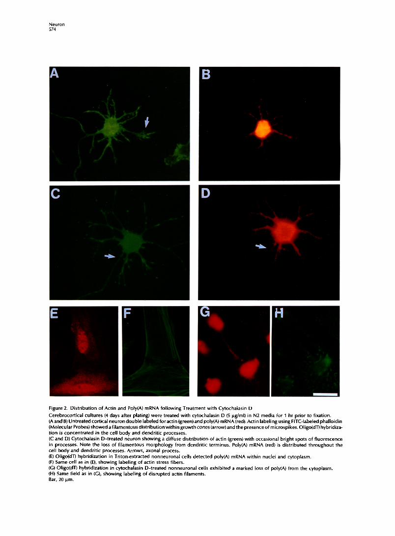
Figure 2. Distribution of Actin and Poly(A) mRNA following Treatment with Cytochalasin D
Cerebrocortical cultures (4 days after plating) were treated with cytochalasin D (5 @ml) in N2 media for 1 hr prior to fixation. (A and B) Untreated cortical neuron double labeled for actin (green) and poly(A) mRNA (red). Actin labeling using FITC-labeled phalloidin (Molecular Probes) showed afilamentous distribution within growth cones (arrow)and the presence of microspikes. Oligo(dT) hybridiza- tion is concentrated in the cell body and dendritic processes. (C and D) Cytochalasin D-treated neuron showing a diffuse distribution of actin (green) with occasional bright spots of fluorescence in processes. Note the loss of filamentous morphology from dendritic terminus. Poly(A) mRNA (red) is distributed throughout the cell body and dendritic processes. Arrows, axonal process. (E) Oligo(dT) hybridization in Triton-extracted nonneuronal cells detected poly(A) mRNA within nuclei and cytoplasm. (F) Same cell as in (E), showing labeling of actin stress fibers. (C) Oligo(dT) hybridization in cytochalasin D-treated nonneuronal cells exhibited a marked loss of poly(A) from the cytoplasm. (H) Same field as in (G), showing labeling of disrupted actin filaments. Bar, 20 pm.

C r 55
oskeletal Association of Neuronal Poly(A) mRNA
Banker, 1984). Therefore, the hybridization signal along the z-axis (perpendicular to the process) would be expected to decrease in a proximodistal gradient.
Although poly(A) mRNA was not detectable in the majority of the axon after 4 days in culture, most neu- rons contained poly(A) within the proximal segment of the axon (Figures ID and IF, arrows). The length of the proximal segment that contained poly(A) mRNA ranged from 2 to IO urn, whereas the entire axonal process was over 150 urn. Hybridization with control oligo(dA) probes (digoxigenin labeled) showed low background throughout the cell and did not reveal fluorescent signal in dendritic processes (Figure IG). Neuronal cultures extracted with 0.2% Triton X-100 in hypertonic buffer exhibited strong hybridization (Figure IH), demonstrating that poly(A) mRNA within the cell body and proximal dendrites was bound to the cytoskeleton.
Effects of Microfilament Perturbation on the Distribution of Poly(A, mRNA Cytochalasins have been previously used to disrupt microfilaments in neurons (Letourneau et al., 1987; Forscher and Smith, 1988) and to release mRNA from the actin cytoskeleton (Ornelles et al., 1986; Sundell and Singer, 1991; Taneja et al., 1992). Cortical neurons (4 days in culture) were treated with 5 ug/ml cytocha- lasin D for 1 hr prior to fixation and hybridization. The cytochalasin dose used here was IO-fold above that used to disrupt mRNA localization in nonneu- ronal cells (Sundell and Singer, 1991; Taneja et al., 1992). Fluorescein isothiocyanate(FITC)-phalloidin la- beling revealed the presence of actin throughout the cell body and processes. Filamentous actin was most evident within flattened growth cones (Figure 2A, arrow). Following cytochalasin treatment, there was a perturbation in the distribution of neuronal actin, most evident as a loss of filamentous actin bundles from dendritic growth cones (Figure 2C). The distribu- tion of actin followingcytochalasin treatment was dif- fuse, although some bright fluorescent spots were observed within processes (possible aggregates of ac- tin) (Figure 2C). The polar morphology of the neuron remained intact, and poly(A) mRNA remained in den- dritic processes, in a proximodistal gradient of de- creasing signal intensity, and remained confined to the proximal segment of the axon (Figure 2D; Table 1). Poly(A) mRNA was also retained in neurites after 1 day in culture following cytochalasin D treatment (data not shown). Therefore, the localization of the majority of poly(A) mRNA within neuronal processes was not dependenton the integrity of microfilaments. There was no evidence of an alteration in the distribu- tion of poly(A) mRNA within the cell body; however, it may be necessary to Triton extract neurons following cy-tochalasin D treatment to eliminate hybridization to soluble mRNA, which was released from microfila- ments but still present in thecell body(region of great- est volume). However, because of the fragility of neu-
ronal cytoskeletons, only the healthiest cultures will resist the Triton extraction. Prior treatment with cy- toskeletal perturbing drugs adds further complica- tions to this procedure, and it cannot be determined as yet whether poly(A) mRNA within the neuronal cell body is bound to actin, as has been reported for other non-process-bearing cells. However, non-process- bearing cells present in the neuronal cultures were found to have actin-associated poly(A) mRNA by com- parison of hybridization in untreated and cytocha- lasin D-treated cells which were Triton extracted (Figures2Eand2G). In thesecells,cytochalasin D treat- ment resulted in a dramatic perturbation of actin stress fibers (Figures IF and IH). The sensitivity of nonneuronal mRNA to cytochalasin D was compara- ble with that observed previously in fibroblasts CTa- neja et al., 1992).
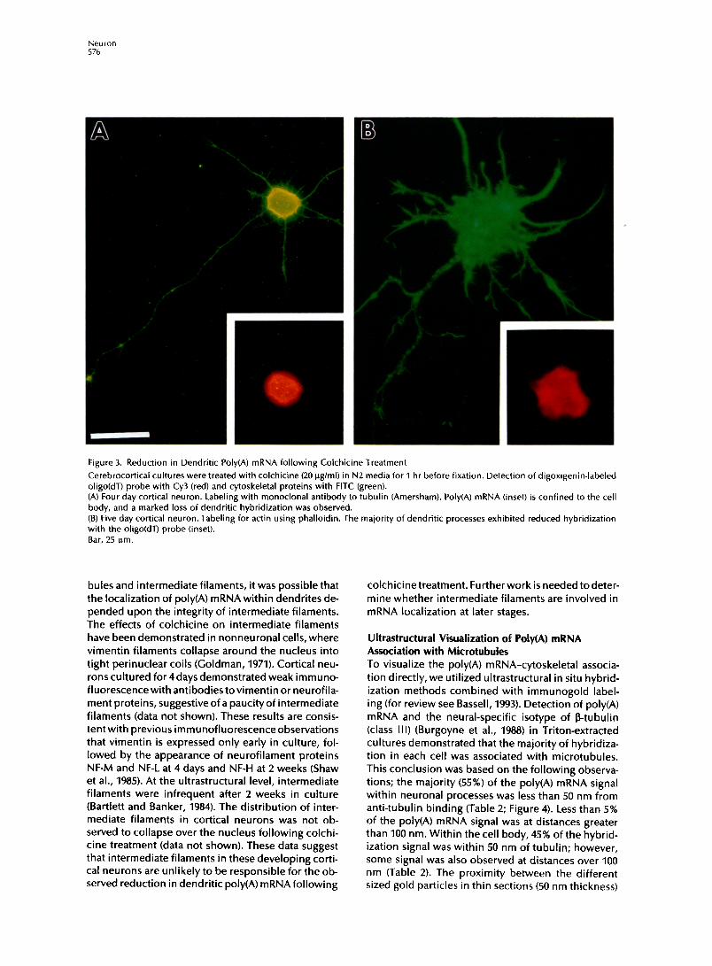
Effects of Microtubule Perturbation on the Distribution of PolyCA) mRNA Since poly(A) mRNA was unaffected by perturbation of microfilaments, we then investigated microtubules for their role in mRNA anchoring. Cortical neurons (4 days in culture) were treated with 10 ug/ml colchi- tine for 1 hr prior to fixation and hybridization. The distribution of tubulin in the cell body of colchicine- treated neurons was less fibrillar, and the caliber of the processes was significantly reduced (Figure 3A). Important to the preservation of neuronal morphol- ogywas the brief time of drug exposure (1 hr), thereby minimizing perturbation to other cytoskeletal compo- nents. For example, the distribution of actin following colchicine treatment was similar to that observed pre- viously without exposure to drug (Figure 38). How- ever, 62% of the neurons showed a major loss of poly(A) mRNA in their dendrites (Table 1); most den- dritic processes had no detectable hybridization (Fig- ures 3A and 3B, inset). In the majority of neurons, poly(A) mRNA was also lost from the proximal seg- ment of the axon. In cultures treated with colchicine 1 day after plating, hybridization was significantly re- duced in all minor neurites (data not shown). The de- polymerization of colchicine labile microtubules was likely to account for the reduction in hybridization intensity within neuronal processes, suggesting a mi- crotubule dependency in the anchoring of poly(A) mRNA. The depolymerization of microtubules may permit poly(A) mRNA to diffuse into the cell body (region of greatest volume). Alternatively, poly(A) mRNA, which was originally associated with dendritic microtubules, may be degraded. A subset of cortical neurons (27%) (Table 1) did not exhibit colchicine- induced alterations in process morphology or den- dritic mRNAdistribution, suggesting drug-stable pop- ulations (Goldschmidt and Steward, 1982). The inability to release dendritic poly(A) mRNA from these neurons (Table 1) might be explained by a population of colchi- tine-resistant microtubules (Ferreira et al., 1989).
Owing to the known interactions between microtu-
Neuron 576
Figure 3. Reduction in Dendritic Poly(A) mRNA following Colchicine Treatment
Cerebrocortical cultures were treated with colchicine (20 @ml) in N2 media for 1 hr before fixation. Detection of digoxigenin-labeled oligo(dT) probe with Cy3 (red) and cytoskeletal proteins with FITC (green). (A) Four day cortical neuron. Labeling with monoclonal antibody to tubulin (Amersham). Poly(A) mRNA (inset) is confined to the cell body, and a marked loss of dendritic hybridization was observed. (B) Five day cortical neuron. Labeling for actin using phalloidin. The majority of dendritic processes exhibited reduced hybridization with the okgo probe (inset). Bar, 25 urn.
bules and intermediate filaments, it was possible that the localization of poly(A) mRNA within dendrites de- pended upon the integrity of intermediate filaments. The effects of colchicine on intermediate filaments have been demonstrated in nonneuronal cells, where vimentin filaments collapse around the nucleus into tight perinuclear coils (Goldman, 1971). Cortical neu- rons cultured for 4 days demonstrated weak immuno- fluorescencewith antibodiestovimentin or neurofila- ment proteins, suggestive of a paucity of intermediate filaments (data not shown). These results are consis- tentwith previous immunofluorescenceobservations that vimentin is expressed only early in culture, fol- lowed by the appearance of neurofilament proteins NF-M and NF-L at 4 days and NF-H at 2 weeks (Shaw et al., 1985). At the ultrastructural level, intermediate filaments were infrequent after 2 weeks in culture (Bartlett and Banker, 1984). The distribution of inter- mediate filaments in cortical neurons was not ob- served to collapse over the nucleus following colchi- tine treatment (data not shown). These data suggest that intermediate filaments in these developing corti- cal neurons are unlikely to be responsible for the ob- served reduction in dendritic poly(A) mRNA following
colchicinetreatment. Furtherwork is needed todeter- mine whether intermediate filaments are involved in mRNA localization at later stages.
Ultrastructural Visualization of Poly(A) mRNA Association with Microtubules To visualize the poly(A) mRNA-cytoskeletal associa- tion directly, we utilized ultrastructural in situ hybrid- ization methods combined with immunogold label- ing (for review see Bassell, 1993). Detection of poly(A) mRNA and the neural-specific isotype of P-tubulin (class III) (Burgoyne et al., 1988) in Triton-extracted cultures demonstrated that the majority of hybridiza- tion in each cell was associated with microtubules. This conclusion was based on the following observa- tions; the majority (55%) of the poly(A) mRNA signal within neuronal processes was less than 50 nm from anti-tubulin binding (Table 2; Figure 4). Less than 5% of the poly(A) mRNA signal was at distances greater than 100 nm. Within the cell body, 45% of the hybrid- ization signal was within 50 nm of tubulin; however, some signal was also observed at distances over 100 nm (Table 2). The proximity between the different sized gold particles in thin sections (50 nm thickness)

Cytoskeletal 577
Association of Neuronal Poly(A) mRNA
Table 2. Ultrastructural Colocalization with Microtubules
Distance between Oligo(dT) (12 nm Particles) and Microtubules (6 nm Particles)
<SO nm 50-100 nm >I00 nm
Neuronal process 55 42 3 Cell body 45 37 18 Nonneuronal 13 20 67
Cortical neurons (4 days in culture) were Triton extracted in cy- toskeletal buffer and fixed as described in the Experimental Pro- cedures. Oligo(dT) (digoxigenin) probes were detected using 12 nm gold-conjugated antibodies, and class III tubulin was de- tected using a monoclonal antibody and a 6 nm gold-labeled secondary antibody. The hybridization signal was evident as clusters of 12 nm particles, i.e., 3 particles per cluster (see Figure 4). Clusters of 12 nm gold were considered as individual hybrid- ization signals to avoid the possibility of scoring a single mRNA molecule more than once. One hundred signals (from 6 cells) were photographed at 50,000x using a JEOL 1200EX transmis- sion electron microscope. The distance between oligo(dT) hy- bridization signals (12 nm particle) and the nearest 6 nm particle (tubulin) was measured in neuronal processes and cell bodies. Similar analysis was performed on non-neuronal cells that did not have processes. These nonneuronal cells were obtained by trypsinization of cortical cells and plating on uncoated cov- erslips; under these conditions, neurons do not attach. Nonneu- ronal tubulin was detected using a monoclonal antibody to a-tubulin, which was not cell-type specific.
of Triton-extracted cells strongly suggests a nonran- dom coincidence between poly(A) mRNA and micro- tubules.
Poly(A) mRNA was not observed to be associated with tightly bundled microtubules, with intermicrotu- bular distances less than 50 nm (Figures 4C-4E). This was not attributed to a paucity of tightly bundled mi- crotubules, as bundles were frequently observed (Fig- ures 4C-4E). It was frequently possible to measure intermicrotubule distances at sites of hybridization if the filaments were positioned within the plane of sectioning and optimally stained with heavy metals. Only 10% of these poly(A) mRNA signals (cluster of 12 nm gold particles) localized to microtubules spaced apart by <SO nm. The majority of poly(A) was associ- ated with the following types of microtubules: parallel microtubules (average intermicrotubule distance of 80 nm; n = 25 hybridization signals), microtubules intersecting at branched angles (nonparallel), and in- dividual microtubules (this association may be attrib- uted to thin sectioning). In dendritic regions that con- tained both tightly bundled and loosely spaced micro- tubules, poly(A) mRNA was preferentially localized to the looser arrangements (Figures 4C-4E). Poly(A) mRNA was also excluded from those microtubules that were tightly bundled in the proximal segment of the axon (data not shown).
The hybridization signal (12 nm gold particles) near microtubules was in the form of clusters of gold parti- cles (2-6 particles per cluster), and multiple clusters were observed in proximity (Figures 4A-4E). These data suggest that multiple poly(A) mRNA molecules
may be aggregated at cytoskeletal attachment sites between microtubules. The ultrastructural approach permitted the morphologic identification of poly- somes, present as electron dense clusters of circular particles (Figure 4A). This identification was confirmed by the disappearance of these clusters (polysomes) when cells were treated with puromycin, which inhib- its ribosome initiation and disengages ribosomes from mRNA (data not shown). Poly(A) mRNA mole- cules were frequently observed in clusters coloca- lized with polysomes (Figure 4A), suggesting that the detected mRNAs were being translated.
To support the above observations, two controls were performed. Nonspecific binding of digoxigenin- labeled oligo(dA) probes showed very low back- ground levels of gold particles and did not colocalize with microtubules (electron microscopy data not shown). Oligo(dT) (digoxigenin) probes hybridized to nonneuronal cells (without processes) did not exhibit frequent interactions with microtubules labeled with antibody to all S-tubulin isotypes (Figure 4F; Table 2), and mRNA was not released from the cytoskeleton following colchicine treatment and Triton extraction (data not shown). These data are consistent with previ- ous reports in fibroblasts, where interactions of mRNA with microtubules were not observed (Singer et al., 1989; Sundell and Singer, 1991; Taneja et al., 1992).
Discussion
In neurons, the mRNA population has not previously been visualized with high resolution, nor have spe- cific cytoskeletal elements that could account for mRNA compartmentalization been identified. Here, we have shown that dendritic localization of poly(A) mRNA requires microtubules. It has been proposed that mRNA localization involves a two-step process (Yisraeli et al., 1990): the transport of newly synthe- sized mRNA from its entrance into the cytoplasm to its region of localization and the subsequent anchor- ing of that mRNA to prevent its diffusion. An alterna- tive hypothesis is that mRNAs diffuse into dendrites and are then anchored to specific dendritic cytoskele- tal components. Recent microinjection experiments of fluorescently labeled myelin basic protein mRNA have permitted kinetic analysis of mRNA localization in oligodendrocytes, which occurs at rates compara- ble with fast transport (Ainger et al., 1993). These data provide evidence that support an active role of the cytoskeleton in both transport and anchoring. The microscopic data obtained here provide information on the steady-state distribution of mRNA and permit visualization of total cytoskeleton-associated mRNA. This study provides the first evidence for an involve- ment of microtubules in the localization of endoge- nous mRNA in a somatic cell. Microtubules have been previously shown to be involved in the localization of untranslated maternal mRNA in oocytes. In Xenopus
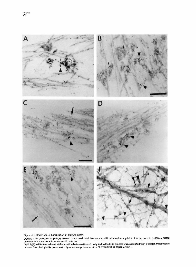
Figure 4. Ultrastructural Localization of Poly(A) mRNA
Double-label detection of poly(A) mRNA (12 nm gold particles) and class III tubulin (6 nm gold) in thin sections of Triton-extracted cerebrocortical neurons from Qday-old cultures. (A) Poly(A) mRNA (arrowhead) at the junction between the cell body and a dendritic process was associated with a Labeled microtubule (arrow). Morphologically preserved polysomes are present at sites of hybridization (open arrow).
Cytoskeletal Association of Neuronal Poly(A) mRNA 579
oocytes, the translocation of veg-7 RNA to the vegetal pole required intact microtubules, whereas the an- choring of veg-7 RNA within the vegetal pole could be perturbed bymicrofilamentdepolymerization (Yis- raeli et al., 1990). In Drosophila nurse cells and oo- cytes, bicoid mRNA transport and anchoring were dependent on microtubules (Pokrywka and Stephen- son, 1991). Digital imaging analysis has suggested that microinjected MBP mRNA in oligodendrocytes formed granules which were near microtubules (Ainger et al., 1993). The peripheral localization of (3-actin mRNA in fibroblasts was observed to be solely dependent on microfilaments (Sundell and Singer, 1991).
The mechanism of dendritic mRNA transport could involve localization of mRNAon microtubules. Axonal microtubules are of uniform polarity with their plus ends oriented distally (Burton and Paige, 1981; Heide- mann et al., 1981), whereas dendrites have a mixed microtubule orientation (Baas et al., 1988). Therefore, the translocation of mRNA along microtubules with minus end distal polarity could explain the concentra- tion of mRNA within dendrites and not axons (Black and Baas, 1989). However, the presence of poly(A) mRNA in minor neurites (Figure IB) and the proximal segment of axonal processes (Figures ID and IF), which have microtubules with plus ends distal to the cell body (Baas et al., 1989), suggest that attachment of mRNA only to microtubules with distal minus ends may not be the only mode of attachment. If microtu- bule polarity is involved in mRNA localization within neuronal processes, it may be that both types of mi- crotubule orientation are utilized to permit localiza- tion of specific dendrite- and axon-directed popula- tions. Although most mRNAs are not detected in mature axons, in situ hybridization analyses have detected the presence of tau mRNA in proximal segments (Litman et al., 1993) and vasopressin and oxytocin mRNAs in axons of the hypothalamo-neuro- hypophysial tract (Jirikowski et al., 1990; Mohr et al., 1991). In this study, poly(A) mRNA was detected in axons, although concentrated in proximal segments during development (Figure 1). The active transport of mRNAs into dendrites and/or axons could involve microtubule-associated motor proteins that move specifically toward plus or minus ends of microtu- bules. Several proteins belonging to either kinesin or dynein families which produce force in preferred directions along microtubules have been identified (Vale, 1992), but none have yet been implicated in mRNA movement.
Anchoring to microtubules may involve the direct binding of mRNAs to tubulin; however, this mecha-
- (B) Dendritic poly(A) mRNA molecules (arrows) observed between parallel series of microtubules. Clusters of poly(A) mRNA (arrowheads) juxtaposed to sites of anti-tubulin binding. (C-D) Dendritic poly(A) mRNA (arrowheads) was not associated with tightly bundled microtubules (arrows). (E) Poly(A) (open arrow) at the edge of bundled microtubules (arrow). Poly(A) and tubulin (arrowhead). (F) Labeling of nonneuronal tubulin (arrows) in a whole-mount preparation. Poly(A) mRNA (arrowheads) was associated with filaments that were unlabeled by anti-tubulin. Bar, 200 nm.
nism would have limited capacity to specify localiza- tion to multiple neuronal domains, since tubulin is distributed throughout the neuron. A more plausible mode of attachment might involve binding to micro- tubule-associated proteins (MAPS), which have been shown to exhibit regional localization within the neu- ron. The ultrastructural distribution of poly(A) mRNA on microtubules was suggestive that mRNA may not be directly bound to the microtubule, but instead bound to associated proteins between adjacent mi- crotubules. Support for this observation is that hy- bridization was not uniformly distributed or directly localized on the surface of microtubules, as was ob- served with an antibody to tubulin. This pattern is similar to previous ultrastructural studies, which have shown that immunogold labeling of MAP2 and MAPlBwasdistributed between microtubules in clus- tered arrangements and not distributed linearly along microtubules (Hirokawa et al., 1988; Sato-Yoshitake et al., 1989). Poly(A) mRNA was frequently present be- tween loosely spaced microtubules and did not make physical contact with the tubule. Supportive of the above hypothesis, in vitro data have shown that iso- lated preparations of MAPS, but not tubulin by itself: bind poly(A) mRNA (Schroder et al., 1982, 1984). It would beworthwhiletodevelopfurther in situ hybrid- ization methodology that permits visualization of ad- ditional cellular structures involved in mRNA localiza- tion. For example, quick-freezing approaches have identified that immunogold labeling of MAPS is observed at cross bridges between microtubules, whereas thin section analysis of Triton-extracted and resin-embedded neurons has only revealed the pres- ence of “fuzzy” structures which colocalize with MAPS (Sato-Yoshitake et al., 1989).
Previous studies have concluded that the majority of mRNAs are not detected in axons, and it is generally believed that most proteins are not synthesized in axons (Steward and Banker, 1992). However, the possi- bility always existed that the detection methods used lacked the sensitivity to detect low levels of mRNA. In 1 day cortical cultures, poly(A) mRNA was detected throughout the majority (>80%) of the axon-like pro- cess; therefore, the in situ hybridization method used was sensitive enough to detect axonal poly(A) mRNA. In contrast, after 4 days in culture, poly(A) mRNA was not detected in the majority (>90%) of the axonal pro- cess and confined to the proximal segment. One ex- planation for the progressive loss of axonal hybridiza- tion is that the majority of newly synthesized poly(A) mRNA is excluded from entering the axon at some point in development. Alternatively, poly(A) mRNA
may enter the axon but get rapidly degraded. In favor of the exclusion hypothesis, poly(A) mRNA within the proximal segment of the axon was associated with microtubules, yet excluded from tightly packed mi- crotubule bundles in neurons cultured for4days (data not shown). As a function of distance from the cell body, poly(A) mRNA declines distally along the axonal process, concommitant with an increase in the den- sity of tightly bundled microtubules. If poly(A) mRNA is bound to certain MAPS, and not to tubulin directly, then the exclusion of most poly(A) mRNA from axons may occur by the absence of those MAPS from tightly bundled axonal microtubules. Alternatively, the bun- dling of microtubules may sterically prevent or limit the ability of large mRNP complexes from entering certain compartments by preventing access to the mi- crotubule except along the edge of the bundle. It would be interesting to know whether the microtu- bules in the incipient axon of neurons cultured for 1 day contain loosely spaced microtubules, but it is difficult to distinguish axonal from dendritic pro- cesses in thin sections at this early stage in culture. Supportiveof this hypothesis,an ultrastructural study on ribosomal distribution in unextracted neurons suggested that bundling of microtubules in the axon may be involved in the confinement of ribosomes within the perikarya (Baas et al., 1987).
Our results demonstrate a major interaction be- tween poly(A) mRNA and the microtubular cytoskele- ton in neurons. Further work is needed to identify whether other filament systems also participate in neuronal mRNA localization. For example, it is possi- ble that mRNAs which are anchored within the cell body are not bound to microtubules. In this model, microtubules within the cell body would serve as tracks for only those mRNAs destined for the pro- cesses. Alternatively, microtubules could be the only filament system involved in mRNA localization within neurons,and specific interactionswith distinct micro- tubule subpopulations and their associated proteins could provide for sorting to specific regions. Identifi- cation of an mRNA-microtubule interaction in neu- rons should focus further efforts on the mechanisms responsible for the transport and anchoring of dis- tinct species of mRNA to their appropriate domains within the neuron and the mechanism that restricts the majority of mRNA from entering the axon during development.
Experimental Procedures
Cell Culture The method of neuronal culture has been described in detail by Banker (for review see Goslin and Banker, 1991) and modified for use with cortical neurons in our laboratory (Kosik and Finch, 1987). Cerebral cortex was dissected from embryonic day 18 rats and digested with 0.25% trypsin in Hanks’balanced salt solution. Tissuewaswashed twice in Hanks’balanced salt solution, placed in modified Eagle’s medium with 10% fetal calf serum, and me- chanically dissociated by pipetting. Neurons were plated at low density (IOOOcells per cm2)on poly-L-lysine-coated coverslips (1.0 mg/ml; overnight). After neurons had attached to the substrate
(2 hr), coverslips were inverted onto a monolayer of astrocytes. The coculture of neurons with glia using this sandwich tech- nique has been previously observed to promote neuronal devel- opment. Astrocytes were prepared from postnatal day 1 rat cor- tex by culturing dissociated cortex in modified Eagle’s medium with 10% fetal calf serum on untreated tissue culture plates. Under these conditions, neurons will not attach to the substrate, and the major cell type cultured is the glial fibrillary acidic pro- tein-positive type-l astrocyte. The coculture was maintained in glutamate-free modified Eagle’s medium with N2 supplements, which include transferrin (100 pg/ml), insulin (5 pg/ml), proges- terone (20 nM), putrescine (100 PM), and selenium dioxide (30 nM). In addition, extra glucose (600 mg/l), sodium pyruvate (1 mM), and ovalbumin (0.1%) were used.
Drug Treatments/Fixation To depolymerize microfilaments, cells were treated with 5 pg/ ml cytochalasin D (Sigma Chemical Co.) in culture media for 1 hr prior to fixation. To depolymerize microtubules, cells were treated with colchicine (IO pglml) (Sigma Chemical Co.) in cul- ture media for 1 hr before fixation. Stock solutions of cytocha- lasin D and colchicine were made up in dimethyl sulfoxide and ethanol, respectively, and the concentration of these solvents was diluted below 0.1% in the culture media, as not to be toxic to neurons. Neurons attached to coverslips were fixed in para- formaldehyde (4% in phosphate-buffered saline with 5 mM MgClJ at 1 and 4 days after plating. Some samples (not exposed to drugs) were extracted with 0.2% Triton X-100 in cytoskeletal buffer (0.1 M NaCI, 0.3 M sucrose, 10 mM PIPES, 3 mM MgC&, 10 WM leupeptin, 2 mM phenylmethylsulfonyl fluoride, 2 mM vanadyl ribonucleoside complex, 2 mM ECTA [pH 6.91) for 1 min at 4OC and fixed in 4% paraformaldehyde (phosphate-buffered saline) for 15 min. This extraction procedure has been widely used to study the association of mRNA with the cytoskeleton (Taneja et al., 1992). Samples that were processed for electron microscopy were fixed in 2% paraformaldehyde, 0.2% glutaral- dehyde (phosphate-buffered saline) following extraction.
Probe Preparation Synthetic oligo(dT) (55 nucleotides) and oligo(dA) (55 nucleo- tides) were 3’end labeled with digoxigenin-IldUTP (Boehringer Mannheim) using terminal transferase (25 pM oligo, 25 mM di- goxigenin dUTP, 140 mM potassium cacodylate, 30 mM Tris- HCI [pH 7.6],1 mM CoCl2,O.l mM dithiothreitol, 100 U of terminal transferase) at 37OC for 1 hr. Probes were purified using a 20 ml C-50 column, and the collected fractions were blotted onto nitrocellulose and detected using an antidigoxigenin-alkaline phosphatase conjugate (Boehringer Mannheim). Positive frac- tions were lyophilized, resuspended, and combined, and the optical density was measured.
Hybridization Cells were washed in phosphate-buffered saline (5 mM MgCI,) and then equilibrated in 15% formamide (Sigma Chemical Co.) 2 x SSC and IO mM sodium phosphate (pH 7.0) at room tempera- ture for IO min. Five nanograms of probe was dried down with Escherichia coli tRNA (IO peg) and sonicated salmon sperm DNA (IO pg) and then suspended in 10 pl of 30% formamide containing 20 mM sodium phosphate (pH 7.0). Probes were mixed with 10 ~1 of hybridization buffer (20% dextran sulfate, 4x SSC, 0.4% bovine serum albumin, 20 mM sodium phosphate [pH 7.01). Cov- erslips were placed cell side down on parafilm containing 20 ~1 of probe mixture and hybridized for 1.5 hr at 37V. After hybrid- ization, coverslips were washed for 20 min in 15% formamide 2x SSC at 37’C and three 10 min washes in 1 x SSC on a rotary shaker at room temperature.
lmmunocytochemistry Probes were detected using affinity purified sheep antibody to digoxigenin conjugated to horseradish peroxidase (Boehringer Mannheim) and affinity purified goat antibody to horseradish peroxidase conjugated to Cy3 (Jackson Immunoresearch). Cy3, which is brighterthan rhodamine, has nearly identical excitation
Cytoskeletal Association of Neuronal PolyfA) mRNA 581
and emission spectraand wasvisualized with a rhodaminefilter. Owing to higher Cy3 emission at lower wavelengths, back- ground levels of Cy3 fluorescence were occasionally observed using the fluorescein filter. Better fluorochrome separation was achieved using an FITC filter equipped with an emission barrier between 520-560 nm. lmmunofluorescence detection of MAP2 was performed simultaneously using monoclonal antibodies to high molecular weight MAP2 (clones AP14-22 obtained from Les- ter Binder) and affinity purified donkey antibody to mouse IgG conjugated to fluorescein (Jackson Immunoresearch). Microtu- bules were detected using a monoclonal antibody to tubulin (Amersham) and fluorochrome-conjugated secondary antibody. Actin was detected using FITC-labeled phalloidin (Molecular Probes). FITC fluorescence was not observed using the rho- damine filter (Figure 1). Antibody incubations were for 1 hr at 37OC in Tris-buffered saline with 1% bovine serum albumin and 0.1% Triton X-100 and were followed by several washes in buffer on a rotary shaker. lmmunofluorescence was viewed using a Zeishxioplan microscope equipped with a 63x Plan-Neofluar objective.
For analysis of dendritic hybridization intensity Uable I), neu- rons were randomly selected under the fluorescein filter that displayed immunostaining for MAPL. After each selection, the cartridge was then shifted to the rhodamine filter to visualize poly(A) mRNA. Cells were photographed under both fluorescein and rhodamine filters. Dendritic length was estimated from mi- crographs by the length of MAP2 labeling (FITC filter). Each pro- cess was divided into 2 pm segments, and the presence or ab- sence of poly(A) mRNA signal (rhodamine filter) was indicated. Processes with hybridization signal in >70% of the segments were scored as having a high dendritic hybridization intensity. Absence of signal in >30% of the segments was scored as having low hybridization intensity.
For electron microscopy, oligo(dT) (digoxigenin) probes were detected using the same antibodies to digoxigenin used for light microscopy, but conjugating 12 nm gold to the secondary anti- body (Jackson Immunoresearch). To label neuronal microtu- bules simultaneously, a monoclonal antibody fJuJ1; obtained from Anthony Frankfurter) to the neural-specific isotype of B-tu- bulin (class III)wasdetected using6 nm gold-conjugated second- ary antibody.
Controls For in situ hybridization, control digoxigenin oligo(dA) probes were hybridized in parallel to oligo(dT) in all experiments. Both oligo(dT) and oligo(dA) hybridizations were evaluated by light and electron microscopy and demonstrated low levels of non- specific hybridization. As an alternative control, hybridization signal could be eliminated when digoxigenin-labeled oligo(dT) probes were hybridized in competition with excess unlabeled oligo(dTr (data not shown).
Electron Microscopy After rinsing in distilled HzO, samples were dehydrated to abso- lute ethanol through a series of graded ethanol. After treatment with propylene oxide poly/bed 812 (1:l) for 30-60 min at room temperature, samples were placed into 108% Epon for 1 hr at room temperature. Beem capsules were filled with fresh 100% poly/bed 812 and quickly inverted onto the surface of the cov- erslips and polymerized for 48 hr at 6o°C. To remove the Beem capsules from the coverslip, samples were immersed in liquid nitrogen for a few seconds and gently tapped to remove the Beem capsule. Thin sections (60-80 nm) were stained with uranyl acetate (1.5% in 50% ethanol) and Reynolds lead citrate for 3 and 5 min, respectively. Samples were thin sectioned parallel to the monolayer, and dendritic and axonal processes were identi- fied at low magnification with a transmission electron micro- scope fJEOL 12OOEX), and various regions were photographed at 50,ooo x
Acknowledgments
We would like to thank Adrianna Ferreira for extensive assis-
tance with neuronal culturesand numerous helpful discussions. We thank Lester Binder for antibodies to MAP2 and Anthony Frankfurter for antibody to class III tubulin. We thank Christine Powers and Peter Paskovitch for electron microscopy assistance. Thiswork was supported by a grant from the Alzheimer’s Disease and Related Disorders Association to G. J. B. and National Insti- tutes of Health grants AGO6601 and NS29031 to K. S. K.
The costs of publication of this article were defrayed in part by the payment of page charges. This article must therefore be hereby marked “advertisement” in accordance with 18 USC Sec- tion 1734 solely to indicate this fact.
Received August 5,1993; revised December 10,1993.
References
Ainger, K., Avossa, D., Morgan, F., Hill, S. J., Barry, C., Barbarese, E., and Carson, 1. H. (1993). Transport and localization of exoge nous MBP mRNA microinjected into oligodendrocytes. J. Cell Biol. 723, 431-441.
Baas, P. W., Sinclair, C. I., and Heidemann, S. R. (19873. Role of microtubules in the cytoplasmic compartmentation of neurons. Brain Res. 420, 73-81.
Baas, P. W., Deitch, J. S., Black, M. M., and Banker, G:A. (1988). Polarity orientation of microtubules in cultured hippocampal neurons: uniformity in the axon and not in the dendrite. Proc. Natl. Acad. Sci. USA 85, 83358338.
Baas, P. W., Black, M. M., and Banker, G. A. (1989). Changes in microtubule polarity orientation during the development of hippocampal neurons in culture. J. Cell Biol. 709, 3085-3094.
Bartlett, W. P., and Banker, G. A. (1984). An electron microscopic study of the development of axons and dendrites by hippocam- pal neurons in culture. J. Neurosci. 4, 19441965.
Bassell, G. J. (1993). High resolution distribution of mRNA within the cytoskeleton. J. Cell. Biochem. 52, 127-133.
Black, M., and Baas, P. W. (1989). The basis of polarity in neurons. Trends Neurosci. 72, 211-214.
Bruckenstein, D. A., Lein, P. J., Higgins, D., and Fremeau, R. T., Jr. (1990). Distinct spatial localization of specific mRNAs in cultured sympathetic neurons. Neuron 5, 809-819.
Burgoyne, R. D., Cambray-Deakin, M., Lewis, S., Sarkar, S., and Cowan, N. (1988). Differential distribution of B-tubulin isotypes in cerebellum. EMBO J. 7, 2311-2319.
Burton, P. R., and Paige, J. L. (1981). Polarity of axonal microtu- bules in the olfactory nerve of the frog. Proc. Natl. Acad. Sci. USA 78, 3269-3273.
Caceres, A., Banker, C. A., Steward, O., Binder, L., and Payne, M. (1984). MAP2 is localized to the dendrites of hippocampal neurons in culture. Dev. Brain Res. 13, 314-318.
Carter, K. C., Taneja, K. L., and Lawrence, J. B. (1991). Discrete nuclear domains of poly-A mRNA and their relationship to the functional organization of the nucleus. J. Cell Biol. 775, 1191- 1202.
Davis, L., Banker, C. A., and Steward, 0. (1987). Selective trans- port of RNA in hippocampal neurons in culture. Nature330,477- 479.
Dotti, C. G., Sullivan, C. A., and Banker, C. A. (1988). The estab- lishment of polarity by hippocampal neurons in culture. 1. Neu- rosci. 8, 1454-1468.
Ferreira, A., Busciglio, J., and Caceres, A. (1989). Microtubule formation and neuritegrowth in cerebellar macroneuronswhich develop in vitro. Dev. Brain Res. 49, 215-228.
Forscher, P., and Smith, S. J. (1988). Actions of cytochalasins on the organization of actin filaments and microtubules in a neu- ronal growth cone. J. Cell Biol. 707, 1505-1516.
Garner, C. C., Tucker, R. P., and Matus, A. (1988). Selective local- ization of mRNA for cytoskeletal protein MAP2 in dendrites. Na- ture 336, 674-679.
Goldman, R. D. (1971). The role of three cytoplasmic fibers in
Neuron 582
BHK-21 cell motility: microtubules and the effects of colchicine. J. Cell Biol. 57, 752-762.
Coldschmidt, R. B., and Steward, 0. (1982). Neurotoxic effects of colchicine: differential susceptibilty of CNS neuronal popula- tions. Neuroscience 7, 695-714.
Goslin, K., and Banker, G. (1991). Culturing Nerve Cells (London, England: MIT Press).
Heidemann, S. R., Landers, j. M., and Hamborg, M. A. (1981). Polarity orientation of axonal microtubules. J. Cell Biol. 97,661- 665.
Hesketh, J. E., and Pryme, 1. F. (1991). Interaction between mRNA, ribosomes and the cytoskeleton. Biochem. J. 277, l-10.
Hillman, D. H. (1988). Dendritic shape and structural determi- nants. In Intrinsic Determinants of Neuronal Form and Function (New York: Alan R. Liss, Inc.).
Hirokawa, N., Hisanaga, S. I., and Shiomura, Y. (1988). MAP2 is a component of crossbridges between microtubules and neuro- filaments in the neuronal cytoskeleton: quick freeze, deep-etch immunoelectron microscopy and reconstitution studies. J. Neu- rosci. 8, 2769-2779.
Horne, S., and Hesketh, J. (1990). Immunological localization of ribosomes in striated rat muscle. Biochem. J. 268, 237-240.
Jirikowski, G. F., Sanna, P. P., and Bloom, F. E. (1990). mRNA coding for oxytocin is present in axons of the hypothalamo- neurohypophysial tract. Proc. Natl. Acad. Sci. USA87,7400-7404.
Kleiman, R., Banker, G., and Steward, 0. (1990). Differential sub- cellular localization of particular mRNAs in hippocampal neu- rons in culture. Neuron 5, 821830.
Kosik, K. S., and Finch, E. A. (1987). MAP2 and tau segregate into dendritic and axonal domains after the elaboration of morpho- logically distinct neurites: an immunocy-tochemical study of cul- tured rat cerebrum. J. Neurosci. 7, 3142-3153.
Lenk, R., Ransom, L., Kaufmann, Y., and Penman, S. (19m. A cytoskeletal structure with associated polyribosomes obtained from HeLa cells. Cell 70, 67-78.
Letourneau, P. C., Shattuck, T. A., and Ressler, A. H. (1987). Pull and push in neurite elongation: observations on the effects of cytochalasin and taxol. Cell Motil. Cytoskel. 8, 193-209.
Litman, P., Barg, J., Rindzoonski, L., and Cinzburg, I. (1993). Sub- cellular localization of tau mRNA in differentiating neuronal cell culture: implications for neuronal polarity. Neuron 70,627-638.
Matus, A., Bernhardt, R.,and Hugh-Jones,T. (1981). High molecu- lar weight MAPS are preferentially associated with dendritic mi- crotubules in brain. Proc. Natl. Acad. Sci. USA 78, 3010-3014.
Matus, A. R., Bernhardt, R., Bodmer, R., and Alaimo, D. (1986). MAP2 and tubulin are differentially distributed in the dendrites of developing neurons. Neuroscience 77, 371-389.
Mohr, E., Fehr, S., and Richter, D. (1991). Axonal transport of neuropeptide encoding mRNAs within the hypothalamo- hypophysial tract of rats. EMBO J. 70, 2419-2424.
Ornelles, D. A., Fey, E. G., and Penman, S. (1986). Cytochalasin releases mRNA from the cytoskeleton and inhibits protein syn- thesis. Mol. Cell. Biol. 6, 1650-1662.
Pokrywka, N.J., and Stephenson, E. C. (1991). Microtubules medi- ate the localization of bicoid RNA during Drosophila oogenesis. Development 773, 55-66.
Ramaekers, F. C., Benedetti, E. L., Dunia, I., Vorstenbosch, P., and Bloemendal, H. (1983). Polyribosomesassociated with micro- filaments in cultured lens cells. Biochim. Biophys. Acta 740,441- 448.
Rao, A., and Steward, 0. (1991). Evidence that protein constit- uents of postsynaptic membrane specializations are locally syn- thesized: analysis of proteins synthesized within synaptosomes. J. Neurosci. 77, 2881-2895.
Sato-Yoshitake, R., Shiomura, Y., Miyasaki, H., and Hirokawa, N. (1989). Microtubule-associated protein IB: molecular structure, localization, and phosphorylation-dependent expression in de veloping neurons. Neuron 3, 229-238.
Schroder, H. C., Bernd, A., Zahn, R. K., and Muller, W. E. G. (1982). Interaction of polyribosomal components and polyribo- nucleotides with microtubule proteins. Mol. Biol. Rep. 8, 233- 237.
Schroder, H. C., Bernd, A., Zahn, R. K., and Muller, W. E. G. (1984). Binding of polyribonucleotides to microtubule proteins. Mech. Ageing Dev. 24, 101-117.
Shaw, G., Banker, C., and Weber, K. (1985). An immunofluores- cence study of neurofilament expression by developing hippo- campal neurons in tissue culture. Eur. J. Cell Biol. 39, 205-216.
Sheiness, D., and Darnell, J. E. (1973). Polyadenylic acid segment in the mRNA becomes shorter with age. Nature 247, 265-268.
Singer, R. H. (1992). The cytoskeleton and mRNA localization. Curr. Opin. Cell Biol. 4, 15-19.
Singer, R. H., Langevin, G. L., and Lawrence, J. B. (1989). Ultra- structural visualization of cytoskeletal mRNAs and their associ- ated proteins using double label in situ hybridization. J. Cell Biol. 708, 2343-2353.
Steward, 0. (1983). Alterations in polysomes associated with den- dritic spines during the reinnervation of the dentate gyrus of the adult rat. J. Neurosci. 3, 177-188.
Steward, 0. S., and Banker, C. A. (1992). Getting the message from the gene to the synapse: sorting and intracellular transport of RNA in neurons. Trends Neurosci. 75, 180-186.
Steward, O., and Levy, W. B. (1982). Preferential localization of polysomes under the base of dendritic spines in granule cells of the dentate gyrus. J. Neurosci. 2, 248-291.
Sundell, C. L., and Singer, R. H. (1991). Requirement of microfila- ments in sorting of actin mRNAs. Science 253, 1275-1277.
Taneja, K. L., Lifshitz, M., Fay, F. S., and Singer, R. H. (1992). PolyfA) RNA codistribution with microfilaments: evaluation by in situ hybridization and quantitativedigital imaging microscopy. J. Cell Biol. 779, 1245-1260.
Vale, R. D. (1992). Microtubule motors: many new models off the assembly line. Trends Biochem. Sci. 77, 300-304.
Yisraeli, J. K., Sokol, S., and Melton, D. A. (1990). A two-step model for the localization of maternal mRNA in Xenopus oo- cytes: involvement of microtubules and microfilaments in the translocation and anchoring of Vgl mRNA. Development 708, 289-298.


![I. INTRODUCTION - arXiv · kinesin walking along microtubules and ribosomes moving along mRNA [2, 3]. These laments act as one-dimensional lanes that allow proteins to move over long](https://img.pdfslide.us/doc/110x75/5fd2cea7d08e451bf247f407/i-introduction-arxiv-kinesin-walking-along-microtubules-and-ribosomes-moving.jpg)