Embed Size (px)
Citation preview

MICROTUBULES AND MAPs
By LINDA A. AMOS AND DANIEL SCHLIEPERMRC Laboratory of Molecular Biology, Hills Road, Cambridge CB2 2QH, UK
I. IntroductionII. Microtubule Structure A. Subunit Lattices B. Atomic Structure of the Protofilament C. Atomic Structure of a Microtubule D. GTP-Binding Sites E. Effect of GTP Hydrolysis on Tubulin Structure F. The Curved Protofilament StructureIII. Dynamic Instability A. Dynamic Behavior and GTP Hydrolysis B. Effects of Assembly-Inhibiting Drugs C. Mechanisms of StabilizationIV. Structural MAPs A. The Tau Family B. MAP1A and MAP1B C. STOPs D. DoublecortinV. Microtubule Destabilizers A. Stathmin (Op18) B. Katanin C. EMAP D. Kinesins as Regulators E. MINUSVI. Proteins That Control Microtubule Location A. γ-Tubulin Ring Complexes B. XMAP215, ch-TOG, Msps Family C. CLIPs/ +TIPs and CLASPs D. The Dynactin Complex E. EB1 and APC F. Hook Proteins G. Centrosomal Coiled-Coil ProteinsVII. Conclusions References
ABSTRACTMicrotubules are very dynamic polymers whose assembly and disassembly is determined by whether their heterodimeric tubulin subunits are in a straight or curved conformation. Curvature is introduced by bending at the interfaces between monomers. Assembly and disassembly are primarily controlled by the hydrolysis of GTP in a site that is completed by the association of two heterodimers. However, a multitude of associat-ed proteins are able to finetune these dynamics so that microtubules are assembled and disassembled where and when they are required by the cell. We review the recent progress that has been made in obtaining a glimpse of the structural interactions involved.
ADVANCES INPROTEIN CHEMISTRY, Vol. 71DOI: 10.1016/S0065-3233(04)71007-4
257

I. INTRODUCTIONWhen microtubules were visualised by electron microscopy, after the improvement of methods of fixation, it was realised that they formed the structural basis of flagellar axonemes and of so-called spindle fibres, as well as occurring as individual filaments in the cytoplasm. Their designation as part of the ʻcytoskeleton ̓suggested that they acted mainly as fixed structural supports. Subsequent research has focussed more and more on their dynamic behaviour and on their role as tracks for motor proteins to transport chromosomes during cell division, for example. Microtubules are found in all eukaryotic cells and are essential for many cellular functions, such as motility, morphogenesis, intracellular transport and cell division. It is that dynam-ic behaviour that allows microtubules to fulfil all these functions in specific places and at appropriate times in the cell cycle.
I1. MICROTUBULE STRUCTUREUnpolymerised tubulin exists as a tight αβ-tubulin heterodimer with binding sites for two molecules of gua-nosine triphosphate (GTP), one exchangeable and the other not. A microtubule is a tube constructed from parallel linear polymers (protofilaments) in which the heterodimers are assembled head to tail in a polar fashion. This polarity is reflected by the distinction between the so-called plus and minus ends of protofila-ments. It can readily be seen by electron microscopy (EM) that each protofilament consists of globular 4nm subunits (Figs 1a & 2); however, α- and β-tubulin are so similar in structure that the two kinds of monomer subunits cannot be distinguished, except at very high resolution. In vivo, microtubules usually have 13 pro-tofilaments, though the number may differ in particular locations. In vitro, it is possible for purified tubulin to assemble with a range of diameters and to contain between 9 and16 protofilaments (Figs 1a & 2). This variation reveals some flexibility in the bonds between adjacent heterodimers, at least in the direction run-ning around the microtubule. When there are 13 protofilaments, they run straight, allowing microtubule-associated motor proteins, such as dynein and kinesin (Hirokawa, 1998; Vale, 2003), to run for long distances along a microtubule without switching lanes; the transport of vesicles and mitochondria along axons, for example, would be difficult if they needed to rotate around microtubules. If microtubules have more or fewer than 13 protofilaments the lattice must rotate slightly, so that the protofilaments wind slowly around the microtubule axis (Wade et al., 1990; Chrétien and Wade, 1991). The amount and direction of rotation is such as to allow the 2D lattice of subunits to close up neatly into tubes with larger or smaller than the standard diameter. In vivo, the occur-rence of microtubules with protofilaments numbers other than 13 appears to be determined by isoforms with specific changes in the amino acid sequences of α- and β-tubulin, as for example in the case of some special-ised 15-protofilament microtubules in neurons of the nematode Caenorhabditis elegans (Savage et al., 1989; Fukushige et al., 1999). These changes presumably allow the formation of a slightly deformed lattice in which a larger number of protofilaments can still be straight.
A. Subunit LatticesMonomers in adjacent protofilaments are slightly staggered (Fig. 2) so they form a set of shallow helices. In a 13-protofilament microtubule 3 shallow helices run in parallel and each makes a complete turn over an axial distance of 12 nm. For a smaller or larger number of protofilaments there may be 2 or 4 shallow helices. It is possible to deduce the number of protofilaments and hence the number of shallow helices in a particular microtubule in an EM image by looking at the Moiré pattern due to the superposition of the front and back of the tube (Fig. 1a). However, the most reliable way of determining the subunit lattice of tubulin monomers is to calculate a Fourier transform (diffraction pattern) of the image (Fig. 1b) and to analyse the positions of the diffraction spots. The data from the Fourier transform can also be used to reconstruct a 3D image of a helically symmetrical specimen.
B. Atomic Structure of the ProtofilamentThe atomic structure of tubulin protofilaments (Figs 3 & 4) is known from 3.5-Å resolution maps, the first of which came from electron crystallography of zinc-induced 2D sheets (Nogales et al., 1998a), subse-quently refined (Löwe et al., 2001). Recent data, including important information about conformational change, have been obtained by X-ray crystallography (Ravelli et al., 2004). Each monomer has a pair of

globular domains set on either side of a central (core) helix H7. The larger globular domain, comprising the N-terminal half of the polypeptide, has the same fold as a family of dinucleotide binding proteins with the so-called Rossmann fold (Nogales et al., 1998b). There is a binding site for the guanosine nucleotide on the plus end surface of this domain, where contact is made with the next subunit in the protofilament. The position of the nucleotide at the centre of the polymerization interface prevents its exchange as long as the subunits remain assembled. The second globular domain of β-tubulin has a binding site for Taxol, which also makes contact with the core helix, on the opposite side from its contact with the nucleotide base. The C-terminal end of each tubulin polypeptide forms two long helices (H11 and H12) connected by a U-turn while the final 13 residues of α-tubulin and 9 residues of β-tubulin are too disordered in the 2D crystals to show up as electron density but are assumed to project out into the solution. These C-terminal residues would be suitable for isoform recognition by tubulin binding proteins, e.g. katanin. They are acidic and negatively charged in physiological conditions.
FIG. 1 (a) Electron microscopy of microtubules assem-bled in vitro. The microtubules shown were assembled from pure pig brain tubulin and rapidly frozen in a very thin layer of ice. Since frozen hydrated specimens are unstained, all the contrast comes from the difference between protein and ice. Microtubules with different numbers of protofilaments (pf) vary in diameter. Also, because their protofilaments run at slightly different an-gles to the microtubule axis, the Moiré patterns created by superposition of the front and back layers of the tubes have different appearances. Protofilaments in a 13-pf segment of a microtubule run straight (white arrow). White arrowheads indicate repeats in the Moiré pattern of a 15-pf microtubule. (Image provided by J. Fan) (b) Diffraction patterns were obtained by calculating the 2D Fourier transforms of individual microtubule im-ages and displaying their amplitudes. For well-ordered specimens, the patterns show a ʻlayerline ̓of peaks at a reciprocal height above the origin of (4nm)-1, arising from the 4nm longitudinal spacing of the tubulin mono-mers. Along the equatorial line, there are peaks arising from the ~5nm lateral separation of the protofilaments (pf). The precise pattern of peaks provides information about how much the tubulin lattice is rotated and, hence, about the number of protofilaments.

15 or 16-pf 13-pf 14-pf12-pf
2-start 3-start 3-start 4-start��
�� ���
���
4nm
5nm seam seam
FIG. 2 Microtubules with varying numbers (12-16) of longitudinal protofilaments (pf). When 13 protofila-ments make up the cylinder, they run straight, but larger or smaller numbers must wind slowly around the axis if the monomer subunits are to line up correctly at the seam. The tilting of the subunit lattice is most obvious in the 15- or 16-pf structure. In this lattice, 4 separate helical lines run through laterally adjacent monomers. For 13- or 14-pf tubes, only 3 shallow helices run in parallel forming the standard ʻ3-start ̓set. Narrower tubes have 2 shallow helices. In this drawing, monomer subunits are represented as darker and lighter spheres to distinguish between α- and β-tubulin. The lattice of subunits, known as the B-lattice, is one in which all or most lateral interactions are α-α or β-β. Perfect helical symmetry (with all lateral interac-tions alike) is possible for B-lattice microtubules with some pf numbers, such as 12, 15 or 16, but a standard 13-pf microtubule can only close with a seam where each α-tubulin monomer makes lateral contact with a β-tubulin subunit. The so-called A-lattice is one in which all lateral contacts would be like this (Amos and Klug, 1974). Most microtubules assembled in vitro from pure tubulin have a B-lattice. Because of their in-stability during isolation, the lattices of truly native microtubules are difficult to investigate.
C. Atomic structure of a microtubuleSeveral long coils loop out from the globular domains. Some are involved in lateral contacts between the protofilaments in a microtubule. A number of groups have docked protofilaments from the zinc-sheet struc-ture solved by electron crystallography to near-atomic resolution (Löwe et al., 2001) into lower-resolution helical microtubule maps imaged by electron cryo-microscopy (Nogales et al., 1999; Amos, 2000; Meurer-Grob et al., 2001; Li et al., 2002). There is general agreement that the ʻM-loops ̓of one protofilament make contact with the GTPase domains of the next one, in the region between helix H3 and the β-sheet (Fig.3). Although the zinc-sheet structure fits into the low resolution envelopes, there are structural differences between tubulin in zinc-sheets and microtubules. It was found that protofilaments in zinc-induced sheets are rotated around their axes by about 20° compared with protofilaments in a normal sheet or opened-out mi-crotubule. As a result, the M-loops make different contacts with the adjacent protofilament, covering part of the surface that corresponds to the outside of a microtubule. Kinesin is, therefore, unable to bind to tubulin in these sheets. The 0.8nm resolution microtubule map of Li et al. (2002) shows that the M-loop also is in a slightly different conformation in a microtubule compared with a zinc-induced sheet.

Taxol
H6
T5
H9
T3
T7
M-loop
GDP
H8
H10
H3
L-loop
N-loop
GTP
GTPase domain
activationdomain
C-term.domain
H7
corehelix
���
���
�
�M-loop
N-loop
FIG.3 Ribbon diagram of an αβ-tubulin heterodimer.The structure solved by electron crystallography using sheets of bovine brain tubulin in complex with Taxol (Nogales et al., 1998a; Löwe et al., 2001) is shown in an orientation corresponding to the inside view of a microtubule. The GTPase domains are coloured pink and the activation domains blue. The core helix that connects the two globular domains in each monomer is coloured yellow and the C-terminal domain on the external surface is green. GTP is sandwiched between the α- and β-tubulin subunits of each heterodimer, being bound to α-tubulin by loops T1-T6 and also makes contact with loop T7 of β-tubulin. The nucleotide bound to β-tubulin has been hydrolysed to GDP through contact with helix H8 and loop T7 of the activation domain of another α-tubulin subunit. Taxol sits in the pocket of β-tubulin on the inside face of microtubules. In α-tubulin, this pocket is occupied by the extended L-loop (prepared using Molscript; Kraulis, 1991).

����
��
��
��
��
��
��
��
�
������
���
��
��
��
���
�
���
��� ���
FIG. 4 Atomic models of tubulin in straight and curved conformations.Ribbon diagrams similar to that in Figure 3, with the same colour scheme. (a,b) Outside and side views of the straight heterodimer (Löwe et al., 2001). (c) Crystal structure of the tubulin-stathmin complex (Ravelli et al., 2004). Stathmin, shown in grey, induces curvature of two tubulin heterodimers. Its N-terminal domain caps one end binding to an α-tubulin subunit. The depolymerising drugs colchicine (CH) and podophyllo-toxin (POD) bind to similar sites on β-tubulin. Pironetin binds to a lysine (K352) on α-tubulin (Usui et al., 2004). Vinblastine (VB), with which pironetin competes, binds to the GTPase domain of β-tubulin, to loop T5 or the loop between H6 and H7 (residues 177-215, Rai and Wolff, 1996). The curvature of the Stathmin helix indicates the degree of the curvature of this complex (~12º/monomer) (prepared using Molscript; Krau-lis, 1991).

When building these docking models, it was already known that the plus end of each protofilament ends with a β-tubulin subunit (Mitchison, 1993; Fan et al., 1996). The orientation of low resolution microtubule maps was also known, from experiments in which sheets growing from the ends of axoneme fragments had been decorated with kinesin motor domains (Hirose et al, 1995a,b). The latter bind stoichiometrically, one motor domain per tubulin heterodimer, and the shape of this complex is sufficiently asymmetric to define which of the two tubulin subunits is α and which is β (Fig. 5a). The orientation of tubulin heterodimers was confirmed by the docking exercises, which also confirmed that the surface consisting of a longitudinal ridge lies on the outside of a microtubule, while the surface exhibiting lateral ridges and containing the Taxol binding site is on the inside (Nogales et al., 1999). The longitudinal ridges seen in 3D EM images correspond to the green helices shown in Figs 3 & 4.
D. GTP-binding sitesGTP is in direct contact with loops T1 to T6 of the GTPase domain. During assembly into protofilaments loop T7 and helix H8, in the activation domain of the next subunit, are brought close to the phosphates of the nucleotide and promote hydrolysis to guanosine diphosphate (GDP) (Nogales et al., 1998b). The idea that the smaller globular domain of tubulin or of FtsZ, the bacterial monomeric homologue of tubulin, acts as a GAP (GTP hydrolysis-activating protein) (Erickson, 1998) to the GTPase domain has been confirmed experimentally by mutagenesis of FtsZ (Mukherjee et al., 2001; Scheffers et al., 2002). The protofilaments of both tubulin and FtsZ thus consist of alternating GTPase domains and activation domains (Figs 3 & 4) (Oliva et al., 2004). After assembly, and hydrolysis, the position of the nucleotide at the centre of the inter-face prevents its exchange from β-tubulin until the subunit disassembles. GTP bound to α-tubulin is non-exchangeable, being permanently trapped between the 2 monomers of the heterodimer, and is never hydro-lysed. β-tubulin has lysine at the lower end of helix H8, in place of glutamic acid (as in α-tubulin E254) or aspartic acid (as in FtsZ), which would be needed for hydrolysis (Nogales et al., 1998b). Because GTP is never hydrolysed in α-tubulin, this interface remains tightly bound when the heterodimer becomes soluble. As first noted by Nogales et al. (1998a, 1999), the other significant difference between α- and β-tubulin is found in the loop L in the activation domain. α-tubulin has a longer L-loop occupying a pocket that provides a binding site in β-tubulin for Taxol and other drugs. This pocket is located on the inner surface of microtu-bules but, in a-tubulin, it is closed by the conserved sequence TVVPGGDL of the extended L-loop. There-fore, each αβ-tubulin heterodimer has one non-exchangeable GTP site and one open Taxol-binding pocket.Loops T1 to T7 surrounding the nucleotide are regions of highest sequence homology between tubulin and FtsZ, the bacterial homologue of tubulin (Löwe and Amos, 1998; Nogales et al., 1998b). The high affinity of this site for nucleotide was demonstrated by a remarkable experiment with FtsZ (Andreu et al., 2002). Unlike tubulin, which is very unstable and loses all activity, the FtsZ of Methanococcus jannaschii could be refolded after being unfolded with guanidinium hydrochloride. Nucleotide was released during unfolding of the protein but as much as 80% rebound when the denaturant was diluted 50-fold in fresh buffer and the FtsZ refolded.
E. Effect of GTP hydrolysis on tubulin structureWhen a microtubule disassembles, after GTP hydrolysis, its protofilaments roll up to form rings or pieces of a ring (Watts et al., 2002; Nogales et al., 2003). This is because GTP hydrolysis promotes bends in protofila-ments. However, while GDP-bound protofilaments are still associated together as a microtubule or 2D sheet, the contacts between neighbouring subunits constrain them to remain in a straight form. The resulting ten-sion is proposed to store conformational energy that is released during depolymerisation. Thus, the structure solved by Nogales and colleagues corresponds to the “strained” straight state. In microtubules, nucleotide hydrolysis produces a small conformational change that shows up as a 2–4% reduction in the length of the tubulin dimer. This was discovered by comparing microtubules assembled with GTP with those assembled with GMPCPP (guanylyl-(α,β)-methylene-diphosphonate) (Hyman et al., 1995). While the microtubules hydrolyse GTP quickly and thus have GDP bound to most of their β-subunits, they hydrolyse GMPCPP very slowly and the microtubules can be seen in a GTP-like state. The difference in longitudinal spacing can be measured in diffraction patterns (Fig. 1b). Microtubules assembled with GMPCPP are relatively stable. The nucleotide-dependent change in spacing is indicative of a conformational change that puts the protofilaments into the straight, but strained, condition responsible for the dynamic instability of microtubules. The change

in subunit length presumably involves movement of some of the loops that are involved in longitudinal bonds. Also, the core helix H7 (connected at one end to loop T7, and at the other end to the loop N, which is involved in both longitudinal and lateral inter-subunit contacts) may also shift, tilt or even shorten. Such a change might explain how hydrolysis of the nucleotide bound to β-tubulin may lead to changes in α-tubulin as well.
F. The Curved Protofilament StructureA surprising observation is that when a protofilament forms a ring, it appears to bend at all the interfaces between monomers, both between and within heterodimers. The bending at all interfaces was first seen by electron microscopy (Watts et al., 2002; Nogales et al., 2003) and has been confirmed in cocrystals of tubu-lin and the tubulin-sequestering protein stathmin (Gigant et al., 2000; Ravelli et al., 2004). It is unexpected because there is still GTP bound in the intra-dimer interface, so there is no nucleotide hydrolysis driving a conformational change here. The bending occurs even in the absence of destabilising agents such as stath-min or drugs such as colchicine. The core helix is a likely means of communication from the top to the bottom of the β-subunit. This cooperative mechanism is presumably advantageous during the rapid disas-sembly phase of microtubule dynamics. It remains to be seen how communication along the protofilaments axis is actually achieved. As well as the observed variation in the number of protofilaments in microtubules, and hence variations in their curvature, whole microtubules can bend and twist without snapping or coming under great elastic strain. This is apparent from images of fluorescently labelled microtubules growing and shrinking in living cells (e.g. Rodionov et al., 1999). When a microtubule bends, individual protofilaments are bent in a variety of directions. It is likely, therefore, that there are multiple ʻbent ̓states for dimers and protofilaments and there is no certainty that the curved conformations induced by different agents of disas-sembly are identical.
III. DYNAMIC INSTABILITYMicrotubules, especially those that make up the mitotic spindle, are in a delicate state of balance between assembly and disassembly. This is because both the formation of the spindle and the movement of chromo-somes to opposite spindle poles depend on carefully coordinated extension or shrinkage at both ends of the microtubules in the spindle. The end of a microtubule that terminates with β-tubulin is more dynamic than the other end, which has an α-tubulin monomer as its final subunit. In cells, microtubules usually grow out from some sort of organising centre from which the more dynamic end, known as the plus end, is able to grow and shrink, while the minus end may not be able to change. If both ends are free, as in vitro, assembly and disassembly can occur from either end, though at different rates. Microtubules can continue to grow as long as the free tubulin concentration is above a critical level. The critical concentration at a minus end is somewhat higher than at a plus end and thus minus ends tend to stop growing first.
A. Dynamic behaviour and GTP hydrolysisEven when the tubulin concentration is above the critical level, however, it is observed that any microtubule end may suddenly stop growing and begin to shrink rapidly. The switch is a stochastic process; individual plus ends shrink rapidly while others are still growing (see Desai and Mitchison, 1997). The change from growth to shrinkage has been termed a ʻcatastropheʼ. After a while, a microtubule end shrinking in vivo may ʻpause ̓in a state intermediate between the growing and shrinking states, though this does not occur in the case of microtubules assembled in vitro from pure tubulin. In either situation, a microtubule that has shrunk may begin to grow again; the latter process is known as a ʻrescueʼ. Microtubules tend to disassemble when cells are cooled below their normal temperature and reassemble when they are rewarmed but they show dynamic instability even under constant warm conditions.The hydrolysis rate of GTP by unpolymerized tubulin dimer is very low (0.054 min-1 at most; David-Pfeuty et al., 1979; Erickson, 1995) but it dramatically increases during the polymerization into microtubules (21 min-1; Melki et al., 1996). The GTP bound to α-tubulin is non-exchangeable, being trapped between the 2 monomers of the heterodimer (the so-called N-site), and is apparently never hydrolysed. However, when heterodimers associate to form protofilaments, GTP on β-tubulin is hydrolysed to GDP as a consequence of the interaction with α-tubulin in the next dimer. It is not clear how soon this exchangeable GTP (on the so-called E-site) is hydrolysed after the addition of a new dimer to the plus end but it may be very quick.

The ʻcap ̓of dimers containing GTP on a microtubule end may be as little as one subunit deep (Walker et al., 1991). Unpolymerised tubulin dimer with GDP bound has a curved conformation. Double rings of 12-16 GDP-bound dimers have an average bend per monomer of 11°-15º. However, the GDP protofilament struc-ture that exists throughout most of the microtubule is constrained to form straight protofilaments by contacts between neighbouring subunits in the lattice, which has been proposed to store conformational energy and to release it during depolymerisation. Assembly of microtubules appears to take place mainly through addition of individual heterodimers to the ends of protofilaments. During rapid assembly at the plus end, there is usually a group of protofilaments in part of the microtubule wall that takes the lead (Chrétien et al., 1995) and may extend as a narrow sheet for some distance beyond the end of the closed tube. Such sheets have not been seen at minus ends, where growth is more even. Disassembly at either end appears to be a cooperative process, since the ends of disas-sembling microtubules have been seen splaying apart and bending outwards into a curved conformation. In some circumstances, long segments of protofilaments are shed as spirals and 30–40-nm diameter rings can form. It has been postulated that catastrophes, pauses and rescues at the plus end are caused by random loss or restoration of the GTP tubulin cap. This would happen by hydrolysis of the nucleotide or attachment of fresh GTP-bound tubulin heterodimers, respectively. Thus, the stochastic events responsible for changes in behaviour may be spontaneous conformational changes that are propagated along either individual protofila-ments or small groups of protofilaments. The bending of protofilaments during disassembly is a manifesta-tion of a cooperative change.
B. Effects of Assembly-Inhibiting DrugsThe role of microtubules in the process of segregating duplicated chromosomes before cell division makes them an important target for antimitotic drugs. A variety of drugs are known to inhibit microtubule assem-bly and thereby stall cells in mitosis, when microtubules are most dynamic and least stable. It seems likely that most of these compounds, if not all of them, favour the curved conformation of tubulin. The crystal structure of tubulin in complex with stathmin and colchicine (Gigant et al., 2000; Ravelli et al., 2004) con-firmed earlier indications that colchicine binds to β-tubulin near to the interface between monomers (Fig.4). Podophyllotoxin was seen to bind to a similar site. This binding location necessarily requires a distortion within the dimer structure that would inhibit its polymerization into straight protofilaments. In contrast, vinblastine and pironetin bind to regions that form contacts between dimers. For vinblastine, which can turn protofilaments into fairly tightly wound helices, cross-linking experiments identified a binding region somewhere on residues 175–213 of β-tubulin (Rai and Wolff, 1996). This peptide includes regions that are involved in longitudinal polymerisation contacts between dimers (Fig. 4). A surprising feature of a recently discovered compound called pironetin is that its binding to a-tubulin inhib-its the binding of vinblastine to β-tubulin (Kondoh et al., 1999; Usui et al., 2004). Such competition suggests that the two drugs occupy overlapping sites in the interface between tubulin heterodimers, where they must each bind without actually disassembling the protofilaments. In contrast, colchicine binds more readily to the intra-dimer site if either vinblastine or pironetin is already bound to the inter-dimer interface. The bind-ing of either drug apparently forces protofilaments to be curved, with the intra-dimer interfaces partially opened-up. Another microtubule disassembler, cryptophycin-1, induces the formation of 24nm diameter rings, each containing eight tubulin dimers (Watts et al., 2002). Digitally processed EM images of these rings showed 13° bends at intra-dimer contacts and 32° bends at inter-dimer contacts. Although the drug binds only to the β-subunit, it protects both α- and β-tubulin against proteolysis by trypsin, indicating that conformational changes were induced in specific regions of both subunits.
C. Mechanisms of StabilizationSince the structure of Taxol has been solved to high resolution repeatedly, by X-ray crystallography and NMR, Snyder et al. (2001) were able to correlate the electron density in the Taxol-binding pocket in the map of tubulin sheets with all the known Taxol conformations and thus identify the one most likely to be present. This is a T-shaped or butterfly-like structure, opened-up to expose a hydrophobic surface that interacts with a hydrophobic patch on the surface of β-tubulin. A recent study of tubulin sheets complexed with epothilone B, a compound that competes with Taxol and has similar microtubule-stabilising effects (Bollag et al., 1995; Giannakakou et al., 2000), found it to bind in the same pocket as Taxol but it interacts with the protein

via quite different groups of atoms in the polypeptide (Nettles et al., 2004). Other stabilising drugs such as discodermolide (ter Haar et al., 1996), eleutherobin (Long et al., 1998; Hamel et al., 1999) and the sarco-dictins (Hamel et al., 1999) are structurally diverse, yet all compete with Taxol for binding to microtubules, apparently because they bind to the same pocket on β-tubulin. It is clear that this common binding site is of immense importance for controlling the assembly of tubulin polymers. This pocket in β-tubulin, where microtubule-stabilising drugs from different organisms can bind, also binds the assembly-promoting repeat motifs of tau protein (and other MAPs). It lies above the β-sheet of the second domain and next to the core helix (Figs 3 & 4), so that anything that fills it is in contact with the core helix and with the M-loop. When assembled in microtubules, the pocket is located on the inside face. In α-tubulin, the corresponding pocket is closed by the extended L-loop. One proposed mechanism for stabilising the assembled state is that structures that hold the M-loop in place stabilise lateral contacts between proto-filaments (Nogales et al., 1999; Li et al., 2002). An alternative possibility is that these structures hold the GTPase and second domains in a relative orientation that favours the straight protofilament conformation, which would otherwise be compromised by GTP hydrolysis (Amos and Löwe, 1999). The two mechanisms may be combined.The effect of the GTP analogue GMPCPP (Hyman et al., 1992), which tubulin can hydrolyse only very slowly, shows that the GTP-bound state of β-tubulin makes microtubules very stable. The conformation of α-tubulin is permanently stable because of the non-exchangeable, non-hydrolysed GTP as well as the pres-ence of the extended loop. It is not really clear why it is advantageous to have a permanently stable α-subu-nit, but it may be because it makes the properties of the two ends of a microtubule more distinct. GTP binds on the other side of the core helix from Taxol and the contact may exert a similar influence on the relative orientations of the GTPase domain, the core helix and the second domain. The presence of GTPʼs γ-phos-phate will also have a direct effect on the interface between α- and β-tubulin, strengthening the bond be-tween heterodimers. It is notable that Taxol stabilized microtubules are relatively flexible and brittle (Dye et al., 1993; Venier et al., 1994), suggesting that the inter-dimer bonds are not strengthened in this case.
IV. STRUCTURAL MAPs In interphase, microtubules are stabilised by several kinds of proteins that are found all along microtubules and are called microtubule associated proteins (MAPs). They tend to have repeating domains, which allow each MAP molecule to associate with more than one tubulin dimer. This produces a doubly effective method of controlling assembly, in that the conformations of several tubulin dimers may be individually stabilised and also the subunits are crosslinked. The binding of these structural MAPs is in turn controlled by kinases and phosphatases (Cassimeris and Spittle, 2001); during mitosis they are phosphorylated and detach from tubulin, whose assembly and disassembly comes under the control of proteins that operate more at the ends of microtubules. Differentiated cells, such as neurons, do not divide; however, as microtubules and MAPs are slowly transported along axons (Baas and Buster, 2004), the MAPs may be phosphorylated in particular places, at times when structural plasticity is required for making synapses or other contacts.
A. The Tau FamilyThe mammalian neuronal proteins MAP2 and tau belong to a widespread family of MAPs. MAP2 is concen-trated in dendrites and the taus, a set of smaller polypeptides, are found in axons. In non-neuronal mam-malian cells there is a high molecular weight protein called MAP4; in Xenopus a similar protein is called XMAP230. Homologous proteins have also been found in invertebrates (Goedert et al., 1996; Heidary and Fortini, 2001). All are extended molecules, lacking in detectable secondary structure and unusually heat-sta-ble. Each consists of a microtubule-binding domain, containing from one to five copies of a semi-conserved motif, preceded by an N-terminal ʻprojection ̓domain that extends from the outer surface of the microtubule. The side-to-side spacing between microtubules containing MAPs depends on the size of the projection do-mains, which seem to repel rather than crosslink neighbouring microtubules. They do, however, contain sites that interact with other proteins, such as the cortical layer, including actin, on the cell membrane (see Cas-simeris and Spittle, 2001, for further references). When overexpressed in vivo or added in excessive amounts to microtubules in vitro, tau accumulates on microtubule surfaces and interferes with the movement of motor proteins along them (Ackmann et al., 2000; Stamer et al., 2002). But at normal levels, tau and related MAPs clearly assist motor transport by creating space around microtubules (Chen et al., 1992).

N
�
�
��
���
��Since Taxol and other microtubule-stabilising drugs are each found only in a specific group of organisms, the highly conserved site on the inside surface of β-tubulin in which they sit presumably evolved to bind some more widespread natural stabilising agents, such as the structural MAPs. There is, indeed, evidence that at least part of the repeating motif in tau binds to a site on β-tubulin that overlaps with the Taxol-binding site (Kar et al., 2003a). This was shown by labelling one of the motifs with a nanogold particle and localis-ing the gold by 3D analysis of electron micrographs. The specimens were microtubules assembled with tubulin and tau and then decorated with kinesin motor domains which, as mentioned above, bind to tubulin dimers with a characteristic polarity and allow the distinction between α- and β-tubulin. Difference maps between images of microtubules containing labelled and unlabelled tau gave a peak on the inside surface of β-tubulin (Fig. 5). Supporting evidence that Taxol and discodermolide both compete with tau for overlap-ping binding sites came from binding assays in which pelleted microtubules contained less tau in the pres-ence of these drugs (Kar et al., 2003a,b). Sequence comparisons suggest that the tau repeat motif binds to β-tubulin in a similar way as the extended L-loop closes (and stabilizes) the pocket in α-tubulin. The con-served sequence (TVVPGGDL) here closely resembles the PGGG signature in the sequence THVPGGGN from tau and in the other conserved repeats found in tau and related MAPs. These repeat motifs could be the natural substrates to bind to the Taxol binding pocket on the inside of the microtubules. It is also possible that successive repeat domains bind along the shallow helices of tubulin monomers and link 3 or 4 adjacent protofilaments together. This would explain rapidly growing triplets of protofilaments in some conditions when tubulin and MAPs are mixed (Mandelkow et al., 1984). Regions of tau outside the repeat region almost certainly bind to sites on the outer surface and support the N-terminal domain that projects out. This projection domain is thought to have important roles in determining the spacing between microtubules in axons and possibly in binding to other structures such as the axonal
FIG. 5 3D EM shows how kinesin and tau bind to microtubules. (a) Reconstruction of a microtubule decorated with kinesin heads (ochre). One kinesin head binds per ab-tubulin heterodimer (grey) and, due to its asym-metric form, can be used to distinguish between the subunits.
(b) Inside view of a microtubule which was coassembled with gold-labeled tau and decorated with kinesin heads. The kinesin heads can be seen on the outside through the holes between the proto-filaments. The labeled repeat motif of tau binds to the inside face of microtubule. The averaged nano-gold density (yellow), which is attached to a repeat motif of tau through a linker, can only be seen near the taxol binding site of β-tubulin, but not on the a subunit (Kar et al., 2003a). The ribbon diagram of the refined zinc-sheet structure is also shown for reference (see Fig. 3).

membrane (Baas and Buster, 2004). The link between the N-terminal projection domain of tau on the out-side of the microtubules and the repeat motifs on the inside surface could be provided by tau s̓ proline-rich flanking region; it would thread through one of the holes between protofilaments. A model of the arrange-ment of a complete tau molecule in a microtubule is shown in Figure 6a. Clearly this sort of arrangement can only be reached by co-assembling tubulin and tau, which would happen naturally in vivo. Other models in which tau binds only to the outer surface (e.g. Al-Bassam et al., 2002), apparently overlapping with the kinesin binding site, are based on experiments in which preassembled microtubules were stabilised with Taxol before the addition of tau. The difference between the two modes of binding has been nicely high-lighted by a stopped-flow kinetic analysis: Makrides et al. (2004) showed that labelled tau bound to preas-sembled microtubules exchanges freely with freshly added unlabelled protein, whereas coassembled tau is nonexchangeably bound to the polymer. Besides filling the pocket on β-tubulin that seems to be a primary control centre for assembly, tau and related MAPs provide two additional forms of stabilisation. Firstly, the loops that occupy the pockets are interconnected in the repeat domain, thus crosslinking three or four dimers, probably in adjacent protofila-ments (Kar et al., 2003a). Secondly, the molecules have other domains that almost certainly bind well to the outer surface of a microtubule and probably run along a protofilament covering several tubulin dimers (Kar et al., 2003b). Fig. 6b shows the charge distribution of tau. The 3 different domains have characteristic net charges. The negatively charged N-terminal projection domain is repelled by the outer microtubule surface, which has the same charge. The positive proline-rich region binds well to this surface, while the less positive repeat region could bind at the inside of the microtubules. MAPs are known to have a stiffening effect on microtubules (Dye et al., 1993; Felgner et al., 1997). Overall, their binding should both favour the straight heterodimer conformation and hold the protofilaments together.
B. MAP1A and MAP1BThe two closely-related neuronal proteins MAP1A and MAP1B are also highly extended and apparently unstructured in solution. Their sequences contain many short repetitive motifs, especially in the N-terminal microtubule-binding domain where there is a predominance of basic residues, and it is likely that they bind to the negatively charged outer surface of the microtubule, though little has been published about the struc-ture of MAP1. The repeats bear no obvious relationship to any part of the microtubule binding domains of tau or MAP2. In vitro, MAP1A has been shown to increase nucleation and stimulate microtubule elonga-tion (Pedrotti and Islam, 1994) but has less power than MAP2 to stabilise the polymer (Vaillant et al., 1998). However, MAP1B can apparently substitute for tau in vivo, since knockout mice are viable without one or other of these proteins but die if both are missing (Takei et al., 2000).
C. STOPsIn vertebrates, resistance of microtubules to cold is largely due to their association with the class of microtu-bule-associated proteins known as STOPs (Stable Tubule Only Polypeptides). These are calmodulin-binding and calmodulin-regulated proteins and their microtubule stabilising activity has been ascribed to micro-tubule-binding motifs that also bind calmodulin. The repeating sequence is different from that in tau and MAP2 but includes an EGGP motif that may fold up as a sharp turn like the extended L-loop of α-tubulin. It is, therefore, conceivable that part of each STOP repeat binds to the pocket on β-tubulin. The proteins seem to have more specific functions in nerve cells than simply to stabilise microtubules, since STOP-null knock-out mice show synaptic defects and abnormal behaviour (Bosc et al., 2003).
D. DoublecortinDoublecortin is so-named because its mutation leads to brain developmental disorders which can include the formation in the brain of a ʻdouble cortex ̓due to an additional band of aberrantly placed neurons. The 30kD N-terminal domain of the protein binds to and stabilises microtubules in vitro. It enhances polymerisation and can also bundle microtubules because the tubulin binding part of the molecule consists of a tandem re-peat of ʻDCX domains ̓that may crosslink two tubulin subunits either in the same or different microtubules (Fig. 6c). Another protein, LIS1, appears to have a similar function in the brain but targets microtubule ends rather than binding all along their length (Tanaka et al., 2004). The structure of the N-terminal DCX do-mains has been solved to high resolution by NMR and X-ray crystallography (Fig. 7a). The domains have

tau P-rich
tau projection
tau repeat regionC
N
tubulinC-termini
RepeatsProline-richProjection C-trmN CMicrotubule-binding
Tau charges
+
-+
-
(a)
(b)
kinesinheads
DCXdomains
(c) S/P-richN N-DC C-DC CDCX plan
FIG. 6 Model of tau molecules and DCX domains interacting with a micro-tubule.(a) Diagram of a microtubule with the inside surface exposed. The acidic C-terminal segment of each tubulin monomer projects from the outer surface (short black stubs). The longer projec-tions represent the N-terminal domains of tau molecules whose C-terminal seg-ments, including the repeat motifs, bind strongly to the inside of the microtubule (Kar et al, 2003a,b). Tauʼs proline-rich region binds to the outer surface along the protofilaments; the connecting piece of polypeptide is sandwiched between two protofilaments during assembly. Ki-nesin motor domains are shown bound to the protofilament on the left (light grey dropshapes). They can probably move past the tau projections without diffi-culty. The binding site for doublecortin DCX domains (Moores et al., 2004) is also indicated (white ovals). It is close to the tau/taxol binding site but on the external, rather than the internal, side of the M-loop. Its position would also not interfere with kinesin binding. (b) Dis-tribution of basic and acidic amino acid residues in human 4-repeat tau (modi-fied from Goedert et al., 1994). The fairly acidic N-terminal segment forms a projection (shown in (a)), that is repelled by the negatively charged tubulin sur-face. The proline-rich region, with a net positive charge, interacts quite strongly with the microtubule surface. The net positive charge of the repeat region is much less than the proline-rich region. Its sequence consists of 3 or 4 semi-con-served repeats with motifs similar to the extended loop in α-tubulin (Kar et al., 2003a). Each motif binds to β-tubulin in the pocket that corresponds to the ex-tended loop in α-tubulin i.e. where Taxol has been seen to bind. (c) Scheme for the doublecortin mol-ecule. The N-terminal DCX domain (N-DC) binds only to microtubules. Its structure is shown in Fig. 7a. The C-terminal DCX domain (C-DC) binds to both microtubules and soluble tubulin dimers.
also been imaged bound to microtubules (Moores et al., 2004), making doublecortin the only MAP, apart from tau and MAP2c (Al-Bassam et al., 2002; Kar et al., 2003a), to have been studied so far by 3D analysis of EM images.When microtubules were decorated with molecules consisting of tandem domains, single globular domains were seen binding in the longitudinal groove between protofilaments, at intervals of 8nm, i. e. the spacing of αβ-heterodimers (see Fig. 6a). The most significant observation from the EM analysis was that the molecules selectively bound to and stabilised 13-protofilament microtubules, presumably because each domain binds in a ster-eospecific way to two protofilaments. This result shows, for the first time, that a basal template is not necessary to specify 13-pro-tofilament microtubules but that their diameter can be controlled by proteins that bind to microtubule sides. It is not clear whether both domains from each DCX molecule were bound identically,

linking adjacent sites, or whether only one was bound and the other was tethered to it but was too mobile to appear in the averaged image of many molecules. The two domains are known to have slightly different properties: the N-terminal domain binds only to microtubules, whereas the C-terminal domain binds to both microtubules and soluble tubulin dimers (Kim et al., 2003). The second domain might, therefore, be active only at a microtubule end.
����������
����������������
��
��
��������������
�����
�����
�����
FIG. 7 Atomic models of MAP domainsThree globular domains all bind to microtubules but do not appear to be homologous.
(a) Human doublecortin N-terminal DCX domain (PDB code 1MJD), solved by NMR, shows a ubiquitin-like fold (Kim et al., 2003). The domain has been docked into a 3D map of the protein bound to microtubules, as recon-structed from EM images (Moores et al., 2004). The regions marked PF are thought to make contact with two adjacent tubulin protofilaments.
(b) N-terminal CAP-Gly domain, as found in CLIPs and the dynactin heavy chain. The structure shown is from C. elegans (PDB code 1LPL), solved by X-ray crystallography (Li et al., 2002), and contains three β-strands.
(c) The N-terminal domain of human EB1 (PDB code 1PA7), solved by X-ray crystallography (Hayashi and Ikura, 2003), contains a single calponin-homology (CH) domain consisting of several α-helices. Tandem CH do-mains are often found in actin-binding proteins; this is the first example of a tubulin-binding CH domain.
V. MICROTUBULE DESTABILISERSA. Stathmin (Op18)The tubulin-sequestering protein called stathmin has been crystallised with tubulin (Gigant et al., 2000; Ravelli et al., 2004). Each molecule runs along-side a pair of tubulin heterodimers and its amino-terminal domain caps one of the a-tubulin subunits, preventing any further interactions (Fig. 4c). In addition to ~12∞ bends between the tubulin monomers, there are changes within each subunit compared with the straight conformation (Fig. 4a,b), including a relative rotation between the GTPase and activation domains. Contacts between subunits are preserved by local movements of helices H6, H7 and H8 and loop T5.
B. KataninKatanin, a member of the AAA (ATPases associated with different cellular activities) superfamily, is a heterodimer of 60- and 80-kDa subunits. In the presence of ATP and microtubules, it forms a transient hexadimer, which can be seen in electron micrographs. It is capable of disrupting contacts
between αβ-tubulin heterodimers in the wall of microtubules. This results in severing the microtubules into short pieces (reviewed by Quarmby, 2000, and Vale, 2000). One important role is in preparing free lengths of microtubule for transport along axons (Baas and Buster, 2004). Davis et al. (2002) proposed that katanin binds preferentially to lattice defects in microtubules, e.g. where a αβ-tubulin building block is missing, rather than to random locations. These defects do occur, at least in microtubules that are assembled in vitro, as shown by scanning force microscopy with single-protein resolution (Schaap et al., 2004). The severing mechanism of microtubule dynamics modulation is intriguing, and is consistent with in vivo results from two members of the katanin family: Human spastin promotes microtubule disassembly (Errico et al., 2002) and the MEI-1/MEI-2 complex is needed for C. elegans meiosis (Srayko et al., 2000). As MEI-1/MEI-2

interacts differently with microtubules containing different β-tubulin isoforms, it could directly regulate meiotic spindle microtubule function. The specific binding site was mapped to the extreme C-terminus of b-tubulin (Lu et al., 2004). Katanin might be regulated by several mechanisms. One way would involve MAPs interfering with kataninʼs access to the microtubule lattice (McNally et al., 2002).
C. EMAPA 77-kDa WD repeat protein, called EMAP from sea urchins reduces the frequency of rescues by 8-fold without producing a change in the frequency of catastrophe (Hamill et al., 1998). The mechanism for this activity is still to be investigated. EMAP-like proteins have also been identified in starfish, sanddollars, and mammals.
D. Kinesins as RegulatorsMany members of the kinesin family are involved in regulating microtubule dynamics. The Kinesin-13 (Kin-I) subfamily do not move along microtubules but instead use energy provided by ATP to depolymerise microtubules at their ends (Walczak et al., 1996; Desai et al., 1999; Homma et al., 2003). Binding of ATP to a Kinesin-13 motor domain attached to a tubulin heterodimer causes the latter to bend and the protofilament to roll up. The activity is catalytic since the motor domain dissociates from the bent tubulin after ATP hy-drolysis. Two crystal structures of Kinesin-13 motor domains show relatively small differences from con-ventional kinesins (Shipley et al., 2004; Ogawa et al., 2004). The two groups agree that an extended loop L2 is of major importance. Whereas most of the loops that interact with tubulin lie on the plus-end half of the motor domain and are thought to interact with β-tubulin, loop L2 extends towards the minus end, in a suit-able position to interact with α-tubulin. When attached to microtubules, motor proteins in the strongly-bind-ing states (with ATP or a nonhydrolysable analogue bound to the motor domain or with an empty nucleotide binding site) both produce a small bend in the tubulin heterodimer (Figure 8; Hirose et al., 1999). If this activity is exaggerated by more strongly binding versions of L2 and other loops, it can presumably cause de-polymerisation. Ogawa et al. (2004) also found the class-specific N-terminal neck to be a long rigid helical structure that would extend into the inter-protofilament groove and thereby inhibit the motor from binding except at the ends of microtubule. Most Kinesin-13 molecules also have an additional N-terminal domain compared with conventional kinesin and this may also have a role in targeting microtubule ends. Even some kinesins that act as normal motor proteins appear to be involved in controlling the length of microtubules in cells (Hunter and Wordeman, 2000). The minus-end-directed motor Ncd has been seen to shorten micro-tubules in vitro (Endow et al., 1994). It is also required for efficient transport of the end-controlling Msps protein (see section VI.B) to spindle poles (Ohkura et al., 2001). Other, non-depolymerising, kinesins may act solely by transporting other molecules to the ends of microtubules.Several kinesin members have microtubule binding sites that are separate from the motor domain and lead to crossbridging of microtubules. MKLP1 is an interesting example. In vitro it can bundle and slide antipar-allel microtubules apart. Microtubule binding involves a highly basic N-terminal extension of the motor, which may interact with the C-terminal acidic tail of tubulin (Mishima et al., 2004). It is thought to help elongate the anaphase spindle by crossbridging microtubules that overlap in the midzone. However, it also has a role in triggering cytokinesis by crossbridging astral microtubules from opposite poles (Inoue et al., 2004)E. MINUSThe small polypeptide MINUS is a novel type of microtubule regulator, having been proposed to act as a nucleation suppressor (Shahani et al., 2001).
VI. PROTEINS THAT CONTROL MICROTUBULE LOCATION Apart from the structural MAPs discussed above, there are many proteins that affect microtubule behaviour. In cells, many factors affect plus end dynamics and also play roles in the assembly of higher order struc-tures. Growing microtubules use dynamic instability to search with their tips for structures that are capable of capturing them. Thus, microtubules are “guided” toward specialized membrane domains, chromosomes and other components of the cell. A wide variety of assembly regulators and adapters that form links with these structures are found at microtubule ends, especially the plus end. Their distributions and activities are reviewed in detail elsewhere (Cassimeris, 1999; Cassimeris and Spittle, 2001; Schroer, 2001; Miyamoto et

Strongly bound Weakly bound
tub tub
tub tub
K K
N N
�
�
�
�
� �
� �
Kinesin
Ncd
al., 2003; Kline-Smith and Walczak, 2004). Plus-end tracking proteins (+TIPs) that actively remain at the tips of growing microtubules are reviewed by Schyler and Pellman (2001), Galjart and Perez (2003) and Vaughan (2004). Here, we try to summarise what is known from a structural point of view. The minus end is less active but is also subject to stabilising and destabilising activity (Dammermann et al., 2003).
A. γ-Tubulin Ring ComplexesThe protein complexes that make up microtubule-organizing centres usually include a third kind of tubulin known as γ-tubulin, which binds but probably does not hydrolyse GTP. There is tentative evidence that it may be capable of assembling into protofilaments (Llanos et al., 1999; Inclán and Nogales, 2001). γ-tubu-lin complexes have been detected in the form of ~25-nm diameter rings in centrosomes (γ-TuRCs) (Moritz and Agard, 2001) but cells also contain smaller γ-tubulin containing complexes (Leguy et al., 2000;Job et al., 2003). Also present in each type of complex are various special MAPs, known as Dgrips in the case of the Drosophila complexes that have been studied in most detail. γ-TuRCs have been proposed to provide a basal template for the 13-protofilament microtubule lattice (Moritz and Agard, 2001). In an alternative model, based on the observation that both interfaces involved in the formation of protofilaments are con-served in γ-tubulin, the ring is proposed to be a stored form of a γ-tubulin protofilament that can straighten to stabilize an αβ-tubulin microtubule lattice by lateral interactions (Erickson, 2000). It is even possible that γ-tubulin merely serves as a seed for one αβ-tubulin protofilament that initiates microtubule assembly (Leguy et al., 2000).
B. XMAP215, ch-TOG, Msps FamilyThis very widespread family of proteins is important in controlling the local interactions of microtubules with many other structures (see e.g. Ohkura et al., 2001). XMAP215 is an assembly promoter that can in-crease microtubule plus end growth by seven- to tenfold but also appears to stabilise minus ends. Its usual effect is to increase the average length of microtubules (Kinoshita, 2002). According to Popov et al. (2001), it is the N-terminal domain (residues 1-560) of XMAP215 that protects plus ends from catastrophe promot-ers such as the Kin I kinesin XKCM1. These workers found that the C-terminal domain (residues 1168-2065) also bound to tubulin, even as a separate fragment, but did not stabilise assembly. However, there seems to be no general agreement about which parts of the molecules are important for microtubule stabili-
FIG. 8 3D EM shows the effect of motor binding on tubulin structure: Sections through 3D EM maps of microtubules decorated with conventional kinesin (K) or ncd (N) motor do-mains: the sections pass through the microtu-bule axis (left-hand edge of each box), through a tubulin protofilament (tub) and then through the middle of the attached heads. The weak in-teraction (with ADP in the nucleotide-binding site) appears similar for both kinesin and ncd, though ncd may make an additional bond to α-tubulin via the L2 loop, which is very short in kinesin. Strong binding of either motor (without nucleotide or with the non-hydrolys-able analogue AMPPNP (Adenosine-5ʼ-[(β,γ)-imido]triphosphate) bound) appears to induce the same change in the tubulin dimer, as indicated by dashed lines through the tubulin monomers: compared with the ADP state (or with an undecorated microtubule – not shown), in which both monomers appear equal, the outer tip of the β-tubulin subunit tilts closer to that of the α-tubulin subunit (Hirose et al., 1999).

sation (Ohkura et al., 2001). Two hypotheses have been considered for the mechanism by which XMAP215 promotes polymerization of microtubule plus ends (Spittle et al., 2000). By oligomerising soluble tubulin dimers, it might catalyse the addition of several dimers per association event; alternatively, it might promote a defined structure at the growing end that either adds dimers more rapidly or is less likely to go into the paused state.Cassimeris et al. (2001) showed that the XMAP215 molecule from Xenopus is long, thin and flexible and can bind to protofilaments along their long axis; its length, 60±18nm, is sufficient to span seven or eight tubulin dimers. Incubation of XMAP215 and tubulin at 4°C resulted in the assembly of curved protofila-ments. A major part of the polypeptide (Fig. 9a) is made up of HEAT motifs (Huntingtin/Elongation factor 3/protein phosphatase 2A/TOR1 repeats, which are related to armadillo repeats and typically form protein-protein interaction surfaces). The atomic structure of a series of HEAT motifs (Fig. 9b) shows a curved row of α-helical hairpins. Proteins in the XMAP215 family may, therefore, consist of concertinas of short helices with more extended connecting regions.The smaller proteins Stu2p from Saccharomyces cerevisiae and Alp14 and Dis1 from Schizosaccharomyces pombe, that are homologous to XMAP215, have fewer repeats in the N-terminal domain (Fig. 9a) and there is no obvious sequence homology in the C-terminal domain. Another difference is that Stu2p slows polym-erisation rates in vitro rather than increasing them (van Breugel et al., 2003). Stu2p depolymerised microtu-bules in vitro by binding directly to plus ends, hindering tubulin dimer addition and increasing catastrophe rates. A stu2 mutant had longer microtubules than normal in interphase, which may correspond to the in vitro conditions, whereas during mitosis microtubules were shorter than normal. XMAP215 can also have a destabilising effect under certain conditions (Shirasu-Hiza et al., 2003). EM images of microtubules as-sembled in vitro with GMPCPP and incubated with XMAP215 revealed a rolled-up structure characteristic of shrinkage. This observation suggests that XMAP215 can operate a protofilament peeling mechanism, similar to that of KinI kinesins. Shirasu-Hiza et al. proposed that the plus ends of microtubules stabilized by GMPCPP, previously thought to resemble the GTP caps on growing microtubules, may instead mimic the paused state. Since Chrétien et al. (1995) found that loss of the sheet-like protofilament extensions of grow-ing microtubule plus ends correlated with slower growth, it is possible that the paused state corresponds to a blunt-ended, closed tube structure. It seems likely that XMAP215 and Stu2p both disrupt the paused state and either increase polymerisation or cause depolymerisation, depending on conditions such as free tubulin concentration. In vivo, their activity would vary with the cell cycle or the location within the cell.
�������������������������������������������������
��������������
��
���������������
���
���
���
FIG. 9 XMAP215/Stu2p polypeptides(a) A plan showing the arrangement of domains. Xenopus XMAP215 has 5 TOG domains, each containing up to 5 HEAT repeats. Similar proteins are found in hu-mans (ch-TOG), Drosophila (Msps) and C. elegans (Zyg-9). Homologous proteins from S. cerevisiae (Stu2p) and S. pombe (Alp14 and Dis1/Mtc1) are less than half the length of those from higher organisms.
(b) The structure of part of a typical HEAT-motif protein (Groves et al., 1999), showing what one of the TOG repeats may look like. If the whole molecule is long enough to cover 7-8 tubulin dimers (Cassimeris et al., 2001), the inter-repeats must have a more stretched-out structure; each TOG repeat plus an inter-repeat may bind to an 8nm dimer.

At the start of mitosis, this family of proteins is important in organising spindle poles. In Drosophila, for ex-ample, Msps (mini-spindles) interacts via its C-terminal with D-TACC (the Drosophila ʻtransforming acidic coiled-coil-containingʼ) protein (Lee et al., 2001). This and other members of the family interact, directly or indirectly, with a wide variety of other proteins (Ohkura et al., 2001)
C. CLIPs/+TIPs and CLASPsProteins that bind specifically to microtubule plus ends have been named +TIPs (+end-tracking proteins; Carvalho et al., 2003). They are a subset of CLIPs (cytoplasmic linker proteins), some of which appear to be capable of binding along the length of microtubules but associate preferentially with the ends. Green fluores-cent protein-labelled CLIP170 association with growing tips has been observed directly (Perez et al., 1999). Its behaviour has been explained either as surfing (sliding along with the growing end) or as treadmilling. Specific binding to growing plus ends may occur because these proteins bind preferentially to narrow sheets rather than closed microtubules; possibly part of the binding site lies on the side of a protofilament or even on the inside surface. Alternatively, they may be able to distinguish between tubulin containing GTP rather than GDP in the exchangeable site. A final possibility, that could include aspects of the previous two, is that they bind to soluble tubulin, making it more likely to add to a microtubule, and then dissociate from the polymer after the conformational change in tubulin due to GTP hydrolysis. In fission yeast, growing micro-tubules are stable until they reach the cell end, provided Tip1p is bound. In its absence, catastrophes are no longer restricted to times when the microtubule ends contact the membrane at the end of the cell (Brunner and Nurse, 2000).The CAP-Gly motif is a conserved glycine-rich sequence of about 42 residues found in the microtubule-binding domains of the various CLIPs, in the heavy chain (P150) of the dynactin complex and in the Golgi protein GMAP210 (Infante et al., 1999; Pernet-Gallay et al., 2002). The structure of a globular domain (Fig. 7.7b) containing this motif has been solved by X-ray crystallography (Li et al., 2002). CLIP170 (also called restin) has two such domains near its N-terminus and binds specifically to the plus ends of growing microtu-bules (Perez et al.,1999). CLIP115, a neuronal CLIP, has a single CAP-Gly domain as does the P150 chain of dynactin. The CAP-Gly domain(s) are typically followed by a stretch of sequence that is able to dimerise in the form of a coiled-coil. Finally, a smaller globular C-terminal region of each protein is thought to be involved in linking to membranous structures. Orbit/MAST proteins, also known as CLASPs (CLIP-associated proteins), are involved in the regulation of microtubule dynamics and bind to microtubule plus ends via CLIP115 or CLIP170. Active CLASP sup-presses microtubule assembly and axon outgrowth (Lee et al., 2004), whereas activated APC (see below) promotes microtubule assembly and axon outgrowth.
D. The Dynactin ComplexDynactin is a 1200 kD protein complex that is usually found in association with the motor protein, cyto-plasmic dynein, an equally large complex. Together, they are active in a wide variety of processes such as axonal transport, organelle movement, nuclear localisation and chromosome separation (reviewed by Allan, 1996; Holleran et al., 1998). Although dynein is able to bind to membranes and membranous organelles by itself, it apparently cannot move them for long distances along microtubules without help from dynactin (King et al., 2003). It remains to be seen how the two complexes interact to produce movement. Dynactin itself can bind all along microtubules but shows a preference for the ends of microtubules in cells as a result of association with CLIP170 (Valetti et al., 1999; Vaughan et al., 2002). The dynactin complex contains at least 10 distinct subunits (Schroer, 1996). The dominant structure seen by EM (Schafer et al., 1994; Eckley et al., 1999) is a 37nm actin-like filament (consisting mainly of monomers of the actin-related protein, Arp1), from which a sidearm projects (Fig. 9). A dimer of dynactin heavy chains (P150glued) is the main component of this sidearm. Each chain has a globular CAP-Gly domain at its N-terminus, followed by a long coiled-coil segment that effects the dimerisation. Some other components of the complex have been identified by antibody labelling (Schafer et al., 1994). A central region of P150 is linked to the dynein intermediate chain by a protein called dynamitin (Vaughan and Vallee, 1995; Valetti et al., 1999), which forms the stem of the dynein complex (Fig. 10a,c).

������
���������
����������������
���������������������
�������������
���
��������������
������������������
�����������������
� �
����������������
������
���
���
���
���������
������
FIG. 10 The dynactin complex and cytoplasmic dynein(a) Model of the 3D structure of dynactin based on EM images of shadowed molecules (redrawn from Schroer, 1996). The cargo-binding domain is a short filament assembled from Arp1 (centractin) plus some standard actin, with capping proteins at each end. The P50 (dyna-mitin) component helps to link the filament to the heavy chain P150glued. Globular CAP-Gly domains (see Fig. 7b) at the end of the thin sidearm bind to tubulin.
(b) The binding regions along the dynactin heavy chain.
(c) Hypothetical model of the interaction between dyn-actin and cytoplasmic dynein. Both large complexes can bind independently with both microtubules and cargo but also associate with each other. Each of two dynein heavy chains (dyn HC) forms a multidomain globular head from which extends a long thin ʻstalkʼ. The stalks of freshly isolated active dimers are closely associated with each other (Fan and Amos, 2001). A small globular domain at the end of each stalk has a microtubule (MT)-binding site. Dynein intermediate chains (dyn IC) form part of the ʻstem ̓of the molecule, used for associating with dynactin and with cargo. What is known about the atomic structures of dynein subunits has been reviewed recently by Sakato and King (2004).
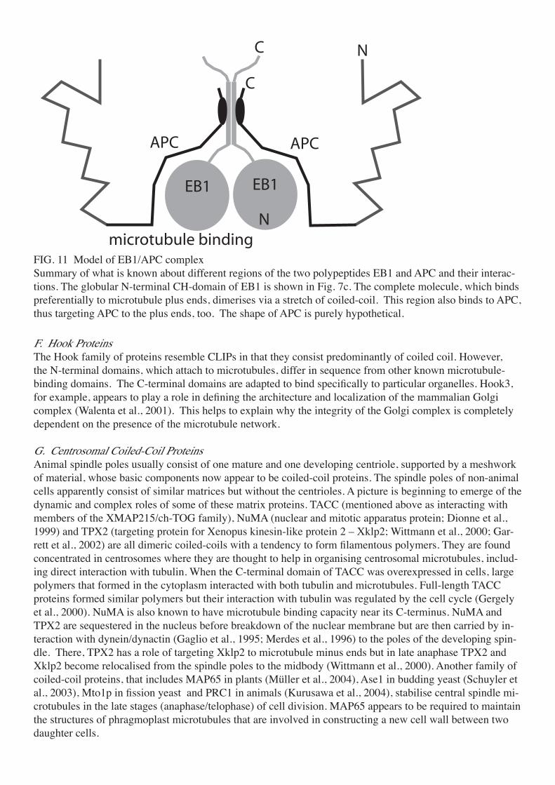
E. EB1 and APCEB1 (end-binding protein 1) is involved in a variety of macromolecular complexes at microtubule plus ends including those that make contact with the cell membrane (Bu and Su, 2003). It cooperates with another plus end protein, the tumour suppressor, adenomatous polyposis coli (APC – not to be confused with APC/C, the anaphase-promoting complex/cyclosome, which is a ubiquitinase). Loss of this interaction, due to mutations in APC, may lead to colon cancer. EB1 and APC both localise to kinetochores (containing plus ends) during mitosis (Fodde et al., 2001). In centrosomes (where minus ends are located), they are assumed to be associ-ated with the plus ends of very short, newly initiated microtubules (Rehberg and Graf, 2002).The globular N-terminal CH domain of EB1, for which a crystal structure is known and which shows no obvious homology to the globular CAP-Gly domain of the CLIPs (Fig. 7c), binds preferentially to the plus ends of microtubules. The extended C-terminal domain dimerises via a stretch of coiled-coil and also binds to APC. Thus, APC is targeted to plus ends too, where it promotes microtubule polymerisation. Neither pro-tein alone has this effect, which is regulated by phosphorylation of APC by Cdc2, disrupting EB1 binding to the N-terminal region of APC. This N-terminal domain forms a weakly-bonded coiled-coil dimer (Day and Alber, 2000), while the middle of the polypeptide includes a cysteine-rich region, with armadillo repeats that interact with β-catenin. It is not yet known how APC promotes tubulin assembly. However, the C-terminal domain contains a microtubule-binding site with a high proportion of positively charged amino acids and shows some sequence homology to the proline-rich region of tau (Deka et al., 1998). The EB1 binding do-main is at the C-terminus of APC and includes a binding site for a PDZ protein-protein interaction domain. PDZ domain proteins are frequently associated with the plasma membrane.
�
���
�
�
�
��������������������
���
���������
���
���
FIG. 11 Model of EB1/APC complexSummary of what is known about different regions of the two polypeptides EB1 and APC and their interac-tions. The globular N-terminal CH-domain of EB1 is shown in Fig. 7c. The complete molecule, which binds preferentially to microtubule plus ends, dimerises via a stretch of coiled-coil. This region also binds to APC, thus targeting APC to the plus ends, too. The shape of APC is purely hypothetical.
F. Hook ProteinsThe Hook family of proteins resemble CLIPs in that they consist predominantly of coiled coil. However, the N-terminal domains, which attach to microtubules, differ in sequence from other known microtubule-binding domains. The C-terminal domains are adapted to bind specifically to particular organelles. Hook3, for example, appears to play a role in defining the architecture and localization of the mammalian Golgi complex (Walenta et al., 2001). This helps to explain why the integrity of the Golgi complex is completely dependent on the presence of the microtubule network.
G. Centrosomal Coiled-Coil ProteinsAnimal spindle poles usually consist of one mature and one developing centriole, supported by a meshwork of material, whose basic components now appear to be coiled-coil proteins. The spindle poles of non-animal cells apparently consist of similar matrices but without the centrioles. A picture is beginning to emerge of the dynamic and complex roles of some of these matrix proteins. TACC (mentioned above as interacting with members of the XMAP215/ch-TOG family), NuMA (nuclear and mitotic apparatus protein; Dionne et al., 1999) and TPX2 (targeting protein for Xenopus kinesin-like protein 2 – Xklp2; Wittmann et al., 2000; Gar-rett et al., 2002) are all dimeric coiled-coils with a tendency to form filamentous polymers. They are found concentrated in centrosomes where they are thought to help in organising centrosomal microtubules, includ-ing direct interaction with tubulin. When the C-terminal domain of TACC was overexpressed in cells, large polymers that formed in the cytoplasm interacted with both tubulin and microtubules. Full-length TACC proteins formed similar polymers but their interaction with tubulin was regulated by the cell cycle (Gergely et al., 2000). NuMA is also known to have microtubule binding capacity near its C-terminus. NuMA and TPX2 are sequestered in the nucleus before breakdown of the nuclear membrane but are then carried by in-teraction with dynein/dynactin (Gaglio et al., 1995; Merdes et al., 1996) to the poles of the developing spin-dle. There, TPX2 has a role of targeting Xklp2 to microtubule minus ends but in late anaphase TPX2 and Xklp2 become relocalised from the spindle poles to the midbody (Wittmann et al., 2000). Another family of coiled-coil proteins, that includes MAP65 in plants (Müller et al., 2004), Ase1 in budding yeast (Schuyler et al., 2003), Mto1p in fission yeast and PRC1 in animals (Kurusawa et al., 2004), stabilise central spindle mi-crotubules in the late stages (anaphase/telophase) of cell division. MAP65 appears to be required to maintain the structures of phragmoplast microtubules that are involved in constructing a new cell wall between two daughter cells.

VII. CONCLUSIONSProtein domains that bind to tubulin and microtubules come in a variety of forms: single long a-helices (stathmin), helical coiled-coils (APC), helical hairpins (XMAP215/ch-TOG), extended random coil (tau) and 3 unrelated globular folds (EB1, CAP-Gly and doublecortin). Most of these have not yet been observed by EM or X-ray crystallography when bound to tubulin but it seems likely that each type of structure binds differently, possibly without competing with the others or with the motor proteins that move along microtu-bules. Some proteins, such as tau, MAP1 and MAP2 bind equally well at any position along a microtubule, stabilising the tubulin lattice directly. This is done by a combination of crosslinking different tubulin dimers and binding to specific sites that control either the conformation of the whole subunit or that of loops in-volved in binding to other subunits. The binding of DCX domains to sites in the grooves between protofila-ments strongly favours the formation of 13-protofilament microtubules, without requiring a basal template. The doublecortins seem to occur only in mammalian nerve cells but it seems likely that molecules which control microtubule curvature in this way are widespread. Other proteins, such as XMAP215, CLIP170 or EB1, control microtubule dynamics by binding to the growing or shrinking ends. At present we have a very limited understanding of how the ends are targeted or how their dynamics are influenced but the current research in this area is likely to produce more information soon. Growth may be stimulated by stabilising tubulin in its straight (GTP-bound) conformation, shrinkage by promoting the curved (GDP-bound) confor-mation. End-binding proteins often work together to provide sophisticated control of the behaviour of the ends.
ACKNOWLEDGMENTS D. S. is supported by an award from the Leverhulme Trust to Dr Jan Löwe, whom we thank for inspiring discussions.
REFERENCESAckmann, M., Wiech, H., and Mandelkow, E. (2000). Nonsaturable binding indicates clustering of tau on the microtubule surface in a paired helical filament-like conformation. J. Biol. Chem. 275, 30335-30343.Al-Bassam, J., Ozer, R. S., Safer, D., Halpain, S., and Milligan, R. A. (2002). MAP2 and tau bind longitudi-nally along the outer ridges of microtubule protofilaments. J. Cell Biol. 157, 1187-1196.Allan, V. (2000). Dynactin. Curr. Biol. 10, R432.Amos, L., and Klug, A. (1974). Arrangement of subunits in flagellar microtubules. J. Cell Sci. 14, 523-549.Amos, L. A. (2000). Focusing-in on Microtubules. Curr. Op. Struct. Biol. 10, 236-241.Amos, L. A., and Löwe, J. (1999). How taxol stabilises microtubule structure. Chem. Biol. 6, R65-R69.Andreu, J. M., Oliva, M. A., and Monasterio, O. (2002). Reversible unfolding of FtsZ cell division proteins from archaea and bacteria: comparison with eukaryotic tubulin folding and assembly. J. Biol. Chem. 277, 43262-43270.Baas, P. W., and Buster, D. W. (2004). Slow axonal transport and the genesis of neuronal morphology. J. Neurobiol. 58, 3-17.Bollag, D. M., McQueney, P. A., Zhu J., Hensens O., Koupal L., Liesch, J., Goetz M., Lazarides E., and Woods, C. M. (1995). Epothilones, a new class of microtubule-stabilizing agents with a taxol-like mecha-nism of action. Cancer Res. 55, 2325–2333.Bosc, C., Oenarier, E., Andrieux A., and Job, D. (1999). STOP proteins. Cell Struct. Funct. 24, 393-399.Brunner, D., and Nurse, P. (2000). CLIP170-like Tip1p spatially organizes microtubular dynamics in fission yeast. Cell 102, 695-704.Bu, W., and Su, L. K. (2003). Characterization of functional domains of human EB1 family proteins. J. Biol. Chem. 278, 49721-49731.Carvalho, P., Tirnauer J. S., and Pellman D. (2003). Surfing on microtubule ends. Trends Cell Biol. 13, 229-237.Cassimeris, L. (1999). Accessory protein regulation of microtubule dynamics throughout the cell cycle. Curr. Opin. Cell Biol. 11, 134-141.Cassimeris, L., and Spittle, C. (2001). Regulation of microtubule-associated proteins. Int. Rev. Cytol. 210, 163-226.Cassimeris, A., Gard, D., Tran, P. T., and Erickson, H. P. (2001). XMAP215 is a long thin molecule that does not increase microtubule stiffness. J. Cell Sci. 114, 3025-3033.

Chen, J., Kanai, Y., Cowan N. J., and Hirokawa, N. (1992). Projection domains of MAP2 and tau determine spacings between microtubules in dendrites and axons. Nature 360, 674-676.Chrétien, D. and Wade, R. H. (1991). New data on the microtubule surface lattice. Biol. Cell 71, 161-174.Chrétien, D., Fuller S. D., and Karsenti, E. (1995). Structure of growing microtubule ends: two-dimensional sheets close into tubes at variable rates. J. Cell Biol. 129, 1311-1328.Dammermann, A., Desai, A., and Oegema, K. (2003). The minus end in sight. Curr. Biol. 13, R614-R624.David-Pfeuty, T., Simon, C., and Pantaloni, D. (1979). Effect of antimitotic drugs on tubulin GTPase activity and self-assembly. J. Biol. Chem. 254, 11696-11702.Davis, L. J., Odde, D. J., Block, S. M., and Gross, S. P. (2002). The importance of lattice defects in katanin-mediated microtubule severing in vitro. Biophys. J. 82, 2916-2927.Day, C. L., and Alber, T. (2000). Crystal structure of the amino-terminal coiled-coil domain of the APC tu-mor suppressor. J. Mol. Biol. 301, 147-156.Deka, J., Kuhlmann, J., and Müller, O. (1998). A domain within the tumor suppressor protein APC shows very similar biochemical properties as the microtubule-associated protein tau. Eur. J. Biochem. 253, 591-597.Desai, A., and Mitchison, T. J. (1997). Microtubule polymerization dynamics. Ann. Rev. Cell Dev. Biol. 13, 83-117.Desai, A., Verma S., Mitchison T. J., and Walczak C. E. (1999). Kin I kinesins are microtubule-destabilizing enzymes. Cell 96, 69-78.Dionne, M. A., Howard L. and Compton D. A. (1999). NuMA is a component of an insoluble matrix at mi-totic spindle poles. Cell Motil Cytoskel. 42, 189-203.Dye, R. B., Fink, S. P., and Williams, R. C. J. (1993). Taxol-induced flexibility of microtubules and its re-versal by MAP2 and Tau. J. Biol. Chem. 268, 6847-6850.Eckley, D. M., Gill, S. R., Melkonian, K. A., Bingham, J. B., Goodson, H. V., Heuser, J. E., and Schroer, T. A. (1999). Analysis of dynactin subcomplexes reveals a novel actin-related protein associated with the Arp1 minifilament pointed end. J Cell Biol. 147, 307-320.Endow, S. A., Kang, S. J., Satterwhite, L. L., Rose, M. D., Skeen, V. P., and Salmon, E. D. (1994). Yeast Kar3 is a minus-end microtubule motor protein that destabilizes microtubules preferentially at the minus ends. EMBO J. 13, 2708-2713.Erickson, H. P. (1995). FtsZ, a prokaryotic homolog of tubulin? Cell 80, 367-370.Erickson, H. P. (1998). Atomic structures of tubulin and FtsZ. Trends Cell Biol. 8, 133-137.Erickson, H. P. (2000). γ-tubulin nucleation: template or protofilament? Nat. Cell Biol. 2, E93-E96.Errico, A., Ballabio, A., and Rugarli, E. I. (2002). Spastin, the protein mutated in autosomal dominant he-reditary spastic paraplegia, is involved in microtubule dynamics. Hum. Mol. Genet. 11, 153-163.Fan, J., and Amos L. A. (2001). Antibodies to cytoplasmic dynein heavy chain map the surface and inhibit motility. J. Mol. Biol. 307, 1317-1327.Fan, J., Griffiths, A., Lockhart, D. A., Cross, R. A., and Amos, L. A. (1996). Microtubule minus ends can be labelled with a phage display antibody specific to alpha-tubulin. J. Mol. Biol. 259, 325-330.Felgner, H., Frank R., Biernat J., Mandelkow E.-M., Mandelkow E., Ludin B., Matus A., and Schliwa M. (1997). Domains of neuronal microtubule-associated proteins and flexural rigidity of microtubules. J. Cell Biol. 138, 1067-1075.Fodde, R., Kuipers, J., Rosenberg, C., Smits, R., Kielman, M., Gaspar, C., van Es, J. H., Breukel, C., Wi-egant, J., Giles, R. H., and Clevers, H. (2001). Mutations in the APC tumour suppressor gene cause chromo-somal instability. Nat. Cell Biol. 3, 433-438.Fukushige, T., Siddiqui, Z. K., Chou, M., Culotti, J. G., Gogonea, C. B., Siddiqui, S. S., and Hamelin, M. (1999). MEC-12, an alpha-tubulin required for touch sensitivity in C. elegans. J. Cell Sci. 112, 395–403.Gaglio, T., Saredi, A., and Compton, D. A. (1995). NuMA is required for the organization of microtubules into aster-like mitotic arrays. J. Cell Biol. 131, 693-708.Galjart, N., and Perez, F. (2003). A plus-end raft to control microtubule dynamics and function. Curr. Opin. Cell Biol. 15, 48-53.Garrett, S., Auer, K., Compton, D. A., and Kapoor, T. M. (2002). hTPX2 is required for normal spindle mor-phology and centrosome integrity during vertebrate cell division. Curr. Biol. 12, 2055-2059.Gergely, F., Karlsson, C., Still, I., Cowell, J., Kilmartin, J., and Raff, J. W. (2000). The TACC domain identi

fies a family of centrosomal proteins that can interact with microtubules. Proc. Natl. Acad. Sci. U. S. A. 97, 14352-14357.Giannakakou, P., Gussio, R., Nogales, E., Downing, K. H., Zaharevitz, D., Bollbuck, B., Poy, G., Sackett, D., Nicolaou, K.C., and Fojo, T. (2000). A common pharmacophore for epothilone and taxanes: molecular basis for drug resistance conferred by tubulin mutations in human cancer cells. Proc. Natl. Acad. Sci. U. S. A. 97, 2904-2909.Gigant, B., Curmi, P. A., Martin-Barbey, C., Charbaut, E., Lachkar, S., Lebeau, L., Siavoshian, S., Sobel, A., and M. Knossow (2000). The 4Å X-ray structure of a tubulin:stathmin-like domain complex. Cell 102, 809-816.Goedert, M., Jakes, R., Spillantini, M. G., and Crowther, R. A. (1994). Tau protein and Alzheimerʼs disease. In Microtubules (Hyams, J. and Lloyd, C., Eds.), Wiley, New York.Goedert, M., Baur, C. P., Ahringer, J., Jakes, R., Hasegawa, M., Spillantini, M. G., Smith, M. J., and Hill, F. (1996). PTL-1, a microtubule-associated protein with tau-like repeats from the nematode Caenorhabditis elegans. J. Cell Sci. 109, 2661-2572.Groves, M. R., Hanlon, N., Turowski, P., Hemmings, B. A., and Barford, D. (1999). The structure of the protein phosphatase 2A PR65/A subunit reveals the conformation of its 15 tandemly repeated HEAT motifs. Cell 8, 99-110.Hamel, E., Sackett, D. L., Vourloumis, D. and Nicolaou, K. C. (1999). The coral-derived natural products eleutherobin and sarcodictyins A and B: effects on the assembly of purified tubulin with and without micro-tubule-associated proteins and binding at the polymer taxoid site. Biochemistry 38, 3490–3498.Hamill, D. R., Howell, B., Cassimeris, L., and Suprenant, K. A. (1998). Purification of a WD repeat protein, EMAP, that promotes microtubule dynamics through an inhibition of rescue. J. Biol. Chem. 273, 9285-9291.Hayashi, I., and Ikura, M. (2003). Crystal structure of the amino-terminal microtubule-binding domain of end-binding protein 1 (EB1). J. Biol. Chem. 278, 36430-36434.Heidary, G., and Fortini, M. E. (2001). Identification and characterization of the Drosophila tau homolog. Mech. Dev. 108, 171-178.Hirokawa, N. (1998). Kinesin and dynein superfamily proteins and the mechanism of organelle transport. Science 279, 519-526.Hirose, K., Fan, J., and Amos, L. A. (1995a). Re-examination of the polarity of microtubules and sheets decorated with kinesin motor domain. J. Mol. Biol. 251, 329-333.Hirose, K., Lockhart, A., Cross, R. A., and Amos, L. A. (1995b). Nucleotide-dependent angular change in kinesin motor domain bound to tubulin. Nature 376, 277-279.Hirose, K., Löwe, J., Alonso, M., Cross, R. A., and Amos, L. A. (1999). 3D electron microscopy of the inter-action of kinesin with tubulin. Cell Struct. Func. 24, 277-284.Holleran, E. A., Karki, S., and Holzbaur, E. L. F. (1998). The role of the dynactin complex in intracellular motility. Int. Rev. Cytol. 182, 69-109.Homma, N., Takei Y., Tanaka Y., Nakata T., Terada S., Kikkawa M., Noda Y., and Hirokawa N. (2003). Kinesin superfamily protein 2A (KIF2A) functions in suppression of collateral branch extension. Cell 114, 229-239.Hunter, A. W., and Wordeman L. (2000). How motor proteins influence microtubule polymerization dynam-ics. J. Cell Sci. 113, 4379-4389.Hyman, A. A., Salser, S., Drechsel, D. N., Unwin, N., and Mitchison, T. J. (1992). Role of GTP hydrolysis in microtubule dynamics: information from a slowly hydrolyzable analogue, GMPCPP. Mol. Biol. Cell 3, 1155-1167.Hyman, A. A., Chrétien, D., Arnal, I., and Wade, R. H. (1995). Structural changes accompanying GTP hydrolysis in microtubules: information from a slowly hydrolyzable analogue guanylyl-(α,β)-methylene-di-phosphonate. J. Cell Biol. 128, 117-125.Inclán, Y. F., and Nogales, E. (2001). Structural models for the self-assembly and microtubule interactions of γ-, δ- and ε-tubulin. J. Cell Sci. 114, 413-422.Infante, C., Ramos-Morales, F., Fedriani, C., Bornens, M., and Rios, R. M. (1999). GMAP-210, a cis-Golgi network-associated protein, is a minus end microtubule-binding protein. J. Cell Biol. 145, 83-98.Inoue, Y. H., Savoian, R. M., Suzuki, T., Mathe, E., Yamamoto, M. T., and D. M. Glover (2004). Mutations in orbit/mast reveal that the central spindle is comprised of two microtubule populations, those that initiate

cleavage and those that propagate furrow ingression. J. Cell Biol. 166, 49-60.Job, D., Valiron O., and Oakley, B. (2003). Microtubule nucleation. Curr. Opin. Cell Biol. 15, 111-117.Kar, S., Fan, J., Smith, M. J., Goedert, M., and Amos, L. A. (2003a). Repeat motifs of tau bind to the insides of microtubules in the absence of taxol. EMBO J. 22, 1-8.Kar, S., Florence, G.J., Paterson, I., and Amos, L. A. (2003b). Discodermolide interferes with the binding of tau protein to microtubules. FEBS Letters 539, 34-36.Kim, M. H., Cierpicki, T., Derewenda, U., Krowarsch, D., Feng, Y., Devedjiev, Y., Dauter, Z., Walsh, C. A., Otlewski, J., Bushweller, J. H., and Derewenda, Z. S. (2003). The DCX-domain tandems of doublecortin and doublecortin-like kinase. Nat. Struct. Biol. 10, 324-333.Kinoshita, K., Habermann, B., and Hyman, A. A. (2002). XMAP215: a key component of the dynamic mi-crotubule cytoskeleton. Trends Cell Biol. 12, 267-273.Kline-Smith, S. L., and Walczak, C. E. (2004). Mitotic spindle assembly and chromosome segregation: refo-cusing on microtubule dynamics. Mol. Cell. 15, 317-327.Kondoh, M., Usui, T., Nishikiori, T., Mayumi, T., and Osada H. (1999). Apoptosis induction via microtubule disassembly by an antitumour compound, pironetin. Biochem. J. 340, 411-416.Kraulis, P. J. (1991). Molscript - a program to produce both detailed and schematic plots of protein struc-tures. J. Appl. Crystallogr. 24, 946-950.Kurasawa, Y., Earnshaw, W. C., Mochizuki, Y. , Dohmae, N., and Todokoro, K. (2004). Essential roles of KIF4 and its binding partner PRC1 in organized central spindle midzone formation. EMBO J. 23, 3237–3248.Lee, H., Engel, U., Rusch, J., Scherrer, S., Sheard, K., Van Vactor, D. (2004). The microtubule plus end tracking protein Orbit/MAST/CLASP acts downstream of the tyrosine kinase Abl in mediating axon guid-ance. Neuron (2004) 42, 913-926.Lee, M. J., Gergely, F., Jeffers, K., Peak-Chew S. Y., Raff J.W. (2001). Msps/XMAP215 interacts with the centrosomal protein D-TACC to regulate microtubule behaviour. Nat. Cell Biol. 3, 643-649.Leguy, R., Melki, R., Pantaloni, D., and Carlier, M. F., (2000). Monomeric γ-tubulin nucleates microtubules. J. Biol. Chem. 275, 21975-21980.Li, H., DeRosier, D., Nicholson, W., Nogales, E., and Downing, K. (2002). Microtubule structure at 8Å reso-lution. Structure 10, 1317-1328.Li, S., Finley, J., Liu, Z. J., Qiu, S. H., Chen, H., Luan, C. H., Carson, M., Tsao, J., Johnson, D., Lin, G., Zhao, J., Thomas, W., Nagy, L. A., Sha, B., DeLucas, L. J., Wang, B. C., and Luo, M. (2002) Crystal struc-ture of the cytoskeleton-associated protein glycine-rich (CAP-Gly) domain. J. Biol. Chem. 277, 48596-48601.Llanos, R., Chevrier, V., Ronjat, M., Meurer-Grob, P., Martinez, P., Frank, R., Bornens, M., Wade, R. H., Wehland, J., and Job, D. (1999). Tubulin binding sites on γ-tubulin: identification and molecular characteri-zation. Biochemistry 38, 15712-15720.Long, B. H., Carboni, J. M., Wasserman, A. J., Cornell, L. A., Casazza, A. M., Jensen, P. R., Lindel, T., Feni-cal, W., and Fairchild, C. R. (1998). Eleutherobin, a novel cytotoxic agent that induces tubulin polymeriza-tion, is similar to paclitaxel (Taxol). Cancer Res. 58, 1111-1115.Löwe, J., and Amos, L. A. (1998). Crystal structure of the bacterial cell-division protein FtsZ. Nature 391, 203-206.Löwe, J., Li, H., Downing, K. H., and Nogales, E. (2001). Refined structure of αβ_tubulin at 3.5Å resolu-tion. J. Mol. Biol. 313, 1045-1057.Lu, C., Srayko, M., and Mains, P. E. (2004). The Caenorhabditis elegans microtubule-severing complex MEI-1/MEI-2 katanin interacts differently with two superficially redundant β-tubulin isotypes. Mol. Biol. Cell 15, 142-150.Makrides, V., Massie, M. R., Feinstein, S. C., and Lew, J. (2004). Evidence for two distinct binding sites for tau on microtubules. Proc. Natl. Acad. Sci. U. S. A. 101, 6746-6751.Mandelkow, E., Schultheiss, R., and Mandelkow, E.-M. (1984). Assembly and 3-dimensional image recon-struction of tubulin hoops. J. Mol. Biol. 177, 507-529.McNally, K. P., Buster, D. W., and McNally, F. J. (2002). Katanin-mediated microtubule severing can be regulated by multiple mechanisms. Cell Motil. Cytoskel. 53, 337-349.Melki, R., Fievez S., and Carlier M.-F. (1996). Continuous monitoring of Pi release following nucleotide

hydrolysis in actin or tubulin assembly using 2-amino-6-mercapto-7-methylpurine ribonucleoside and pu-rine-nucleoside phosphorylase as an enzyme-linked assay. Biochemistry 35, 12038-12045.Merdes, A., Heald, R. Samejima, K., Earnshaw, W.C. and Cleveland, D.W. (2000). Formation of spindle poles by dynein/dynactin-dependent transport of NuMA. J. Cell Biol. 149, 851-861.Meurer-Grob, P., Kasparian, J., and Wade, R. H. (2001). Microtubule structure at improved resolution. Bio-chemistry 40, 8000-8008.Mishima, M., Pavicic, V., Gruneberg, U., Nigg, E. A., and Glotzer, M. (2004). Cell cycle regulation of cen-tral spindle assembly. Nature 430, 908-913.Mitchison, T. J. (1993). Localization of an exchangeable GTP-binding site at the plus end of microtubules. Science 261, 1044–1047.Miyamoto, D. T., Perlman, Z. E., Mitchison, T. J., Shirasu-Hiza, M. (2003). Dynamics of the mitotic spindle - potential therapeutic targets. Prog. Cell Cycle Res. 5, 349-360.Moores, C. A., Perderiset M., Francis, F., Chelly, J., Houdusse, A., and Milligan, R. A. (2004). Mechanism of microtubule stabilization by doublecortin. Mol. Cell 14, 833-839.Moritz, M., and Agard, D. A. (2001). γ-Tubulin complexes and microtubule nucleation. Curr. Op. Struct. Biol. 11, 174-181.Mukherjee, A., Saez, C., and Lutkenhaus, J. (2001). Assembly of an FtsZ mutant deficient in GTPase activ-ity has implications for FtsZ assembly and the role of the Z ring in cell division. J. Bacteriol. 183, 190-197.Müller, S., Smertenko, A., Wagner, V., Heinrich, M., Hussey, P. J., and Hauser, M. T. (2004). The plant mi-crotubule-associated protein AtMAP65-3/PLE is essential for cytokinetic phragmoplast function. Curr. Biol. 14, 412-417.Nettles, J. H., Li, H., Cornett, B., Krahn, J. M., Snyder, J. P., and Downing, K. H. (2004). The binding mode of epothilone A on β-tubulin by electron crystallography. Science 305, 866-869.Nogales, E., Wolf, S., and Downing, K. H. (1998a). Structure of the αβ-tubulin dimer by electron crystallog-raphy. Nature 391, 199-203.Nogales, E., Downing, K. H., Amos, L. A., and Löwe, J. (1998b). Tubulin and FtsZ form a distinct family of GTPases. Nat. Struct. Biol. 5, 451-458.Nogales, E., Whittaker, M., Milligan, R. A., and Downing, K. H. (1999). High resolution model of the mi-crotubule. Cell 96, 79-88.Nogales, E., Wang H.-W., and Niederstrasser, H. (2003). Tubulin rings: which way do they curve? Curr. Op. Struct. Biol. 13, 256-261.Ogawa, T., Nitta, R., Okada, Y., and Hirokawa, N. (2004) A common mechanism for microtubule destabiliz-ers-M type kinesins stabilize curling of the protofilament using the class-specific neck and loops. Cell, 116, 591-602.Ohkura, H., Garcia, M. A., and Toda, T. (2001). Dis1/TOG universal microtubule adaptors - one MAP for all? J. Cell Sci. 114, 3805-3812.Oliva, M. A., Cordell, S. C., and Löwe, J. (2004). Structural insights into FtsZ protofilament formation. Nat. Struct. Mol. Biol. 11, 1243-1250.Pedrotti, B., and Islam, K. (1996). Dephosphorylated but not phosphorylated microtubule associated protein MAP1B binds to microfilaments. FEBS Lett. 388, 131-133.Perez, F., Diamantopoulos, G. S., Stalder, R., and Kreis, T. E. (1999). CLIP-170 highlights growing microtu-bule ends in vivo. Cell 96, 517-527.Pernet-Gallay, K., Antony, C., Johannes, L., Bornens, M., Goud, B., and Rios, R. M. (2002). The overex-pression of GMAP-210 blocks anterograde and retrograde transport between the ER and the Golgi appara-tus. Traffic 3, 822-832.Popov, A. V., Severin, F., and Karsenti, E. (2002). XMAP215 is required for the microtubule-nucleating activity of centrosomes. Curr. Biol. 12, 1326-1330.Quarmby, L. (2000). Cellular samurai: katanin and the severing of microtubules. J. Cell Sci. 113, 2821-2827.Rai, S. S., and Wolff, J. (1996). Localization of the vinblastine-binding site on β-tubulin. J. Biol. Chem. 271, 14707-14711.Ravelli, R. B. G., Gigant, B., Curmi, P. A., Jourdain, I., Lachkar, S., Sobel, A., and Knossow, M. (2004). Insight into tubulin regulation from a complex with colchicine and a stathmin-like domain. Nature 428, 198-202.

Rehberg, M., and Graf, R. (2002). Dictyostelium EB1 is a genuine centrosomal component required for proper spindle formation. Mol. Biol. Cell 13, 2301-2310.Rodionov, V., Nadezhdina, E., and Borisy, G. (1999). Centrosomal control of microtubule dynamics. Proc. Natl. Acad. Sci. U. S. A. 96, 115-120.Sakato, M., and King, S. M. (2004). Design and regulation of the AAA+ microtubule motor dynein. J. Struct. Biol. 146, 58-71.Savage, C., Hamelin, M., Culotti, J. G., Coulson, A., Albertson, D. G., and Chalfie, M. (1989). Mec-7 is a β-tubulin gene required for the production of 15-protofilament microtubules in Caenorhabditis elegans. Genes Dev. 3, 870–881.Schaap, I. A. T., de Pablo, P. J., and Schmidt, C. F. (2004). Resolving the molecular structure of microtubules under physiological conditions with scanning force microscopy. Eur. Biophys. J. 33, 462-467.Schafer, D. A., Gill, S. R., Cooper, J. A., Heuser, J. E., and Schroer, T. A. (1994). Ultrastructural analysis of the dynactin complex: an actin-related protein is a component of a filament that resembles F-actin. J. Cell Biol. 126, 403-412.Scheffers, D. J., de Wit, J. G., den Blaauwen, T., and Driessen, A. J. (2002). GTP hydrolysis of cell division protein FtsZ: evidence that the active site is formed by the association of monomers. Biochemistry 41, 521-529.Schroer, T. A. (1996). Structure and function of dynactin. Semin. Cell Dev. Biol. 7, 321-328.Schroer, T. A. (2001). Microtubules don and doff their caps: dynamic attachments at plus and minus ends. Curr. Opin. Cell Biol. 13, 92-96.Schuyler, S. C., and Pellman, D. (2001). Microtubule “plus-end-tracking proteins”: the end is just the begin-ning. Cell 105, 421-424.Schuyler, S. C., Liu, J. Y., and Pellman, D. (2003). The molecular function of Ase1p: evidence for a MAP-dependent midzone-specific spindle matrix. J. Cell Biol. 160, 517-528.Shahani, N., Fath, T., and Brandt, R. (2001). MINUS: A negative regulator of microtubule nucleation. Mol. Biol. Cell 12, 937.Shipley, K., Hekmat-Nejad, M., Turner, J., Moores, C., Anderson, R., Milligan, R., Sakowicz, R. and Fletter-ick, R. (2004). Structure of a kinesin microtubule depolymerization machine. EMBO Journal 23, 1422-1432.Shirasu-Hiza, M., Coughlin, P., and Mitchison, T. (2003). Identification of XMAP215 as a microtubule-destabilizing factor in Xenopus egg extract by biochemical purification. J. Cell Biol. 161, 349-358.Snyder, J. P., Nettles, J. H., Cornett, B., Downing, K. H., and Nogales, E. (2001). The binding conformation of Taxol in β-tubulin: a model based on electron crystallographic density. Proc. Natl. Acad. Sci. U. S. A. 98, 5312-5316.Spittle, C., Charrasse, S., Larroque, C., and Cassimeris, L. (2000). The interaction of TOGp with microtu-bules and tubulin. J. Biol. Chem. 275, 20748-20753.Srayko, M., Buster, D. W., Bazirgan, O. A., McNally, F. J., and Mains, P. E. (2000). MEI-1/MEI-2 katanin-like microtubule severing activity is required for Caenorhabditis elegans meiosis. Gens. Dev. 14, 1072-1084.Stamer, K., Vogel, R., Thies, E., Mandelkow, E., and Mandelkow, E.-M. (2002). Tau blocks traffic of or-ganelles, neurofilaments and APP vesicles in neurons and enhances oxidative stress. J. Cell Biol. 156, 1051-1063.Takei, Y., Teng, J., Harada, A., and Hirokawa, N. (2000). Defects in axonal elongation and neuronal migra-tion in mice with disrupted tau and map1b genes. J. Cell Biol. 150, 989-1000.Tanaka, T., Serneo, F. F., Higgins, C., Gambello, M. J., Wynshaw-Boris, A., and Gleeson, J. G. (2004). Lis1 and doublecortin function with dynein to mediate coupling of the nucleus to the centrosome in neuronal migration. J. Cell Biol. 165, 709-721.ter Haar, E., Kowalski, R. J., Hamel, E., Lin, C. M., Longley, R. E., Gunasekera, S. P., Rosenkranz, H. S., and Day, B. W. (1996). Discodermolide, a cytotoxic marine agent that stabilizes microtubules more potently than Taxol. Biochemistry 35, 243-250.Usui, T., Watanabe, H., Nakayama, H., Tada, Y., Kanoh, N., Kondoh, M., Asao, T., Takio, K., Watanabe, H., Nishikawa, K., Kitahara, T., and Osada, H. (2004). The anti-cancer natural product pironetin selectively targets Lys352 of α-tubulin. Chem. Biol. 11, 799-806Vaillant, A. R., Müller, R., Langkopf, A., and Brown, D. L. (1998). Characterization of the microtubule-binding domain of microtubule-associated protein 1A and its effects on microtubule dynamics. J. Biol.

Chem. 273, 13973-13981.Vale, R. D. (2000). AAA proteins: lords of the ring. J. Cell Biol. 150, F13-F19.Vale, R. D. (2003). The molecular motor toolbox for intracellular transport. Cell 112, 467-480.Valetti, C., Wetzel, D. M., Schrader, M., Hasbani, M. J., Gill, S. R., Kreis, T. E., and Schroer, T. A. (1999). Role of dynactin in endocytic traffic: effects of dynamitin overexpression and colocalization with CLIP-170. Mol. Biol. Cell 10, 4107-4120.van Breugel, M., Drechsel, D., and Hyman, A. (2003). Stu2p, the budding yeast member of the conserved Dis1/XMAP215 family of microtubule-associated proteins is a plus end-binding microtubule destabilizer. J. Cell Biol. 161, 359-369.Vaughan, K. T. (2004). Surfing, regulating and capturing: are all microtubule-tip-tracking proteins created equal? Trends Cell Biol. 14, 491-496.Vaughan, K. T., and Vallee, R. B. (1995). Cytoplasmic dynein binds dynactin through a direct interaction between the intermediate chains and p150Glued. J. Cell Biol. 131, 1507-1516.Vaughan, P. S., Miura, P., Henderson, M., Byrne, B. and Vaughan, K.T. (2002). A role for regulated binding of p150Glued to microtubule plus ends in organelle transport. J. Cell Biol. 158, 305-319.Venier, P., Maggs, A. C. , Carlier, M. F., and Pantaloni, D. (1994). Analysis of microtubule rigidity using hydrodynamic flow and thermal fluctuations. J. Biol. Chem. 269, 13353-13360.Wade, R. H., Chrétien, D., and Job, D. (1990). Characterization of microtubule protofilament numbers. How does the surface lattice accommodate? J. Mol. Biol. 212, 775-786.Walczak, C. E., Mitchison, T. J., and Desai A. (1996). XKCM1: a Xenopus kinesin-related protein that regu-lates microtubule dynamics during mitotic spindle assembly. Cell 84, 37-47.Walenta, J. H., Didier, A. J., Liu, X., and Kramer, H. (2001). The Golgi-associated hook3 protein is a mem-ber of a novel family of microtubule-binding proteins. J. Cell Biol. 152, 923-934.Walker, R., Pryer, N., and Salmon, E. (1991). Dilution of individual microtubules observed in real time in vitro: evidence that cap size is small and independent of elongation rate. J. Cell Biol. 114, 73-81.Watts, N. R., Cheng, N., West, W., Steven, A. C., and Sackett, D. L. (2002). The cryptophycin-tubulin ring structure indicates two points of curvature in the tubulin dimer. Biochemistry 41, 12662-12669.Wittmann, T., Wilm, M. , Karsenti, E., and Vernos, I. (2000). TPX2, a novel Xenopus MAP involved in spin-dle pole organization. J. Cell Biol. 149, 1405-1418.