Embed Size (px)
Citation preview
Jeejeebhoy et al | 1 Etiology of Maternal Cardiac Arrest and Mortality Appendix
Appendix: Etiology of Maternal Cardiac Arrest and Mortality
The most frequent causes of maternal cardiac arrest include bleeding, heart failure,
amniotic fluid embolism (AFE), and sepsis,1 and the most common causes of maternal
mortality that may also lead to cardiac arrest include cardiac disease, sepsis,
preeclampsia/eclampsia, hemorrhage, cerebrovascular events, AFE, complications from
anesthesia, and thrombosis/thromboembolism.2,3 However, data from the British Centre
for Maternal and Child Enquiries (CMACE) report illustrate that the approach to the
etiology of a maternal cardiac arrest must be more inclusive,2 and a broader approach to
the possible etiology of a maternal cardiac arrest is necessary in order to have the best
chance to identify and correctly treat causative factors and thereby give the patients,
mother and baby, the best chance of survival. It should also be recognized that women
are entering into pregnancy with more comorbidities and risk factors than seen
historically.2,4 During pregnancy, the chance exists for the additional development of
maternal and fetal complications, which may result in acute deterioration. Therefore, the
cause of the maternal cardiac arrest may often be multifactorial. Diagnostic testing and
treatment of disease is almost always more complicated in pregnancy, given that there are
2 patients to consider, the mother and the baby. Therefore, rescuers should have a basic
understanding of diagnostic approaches and treatment considerations. This appendix
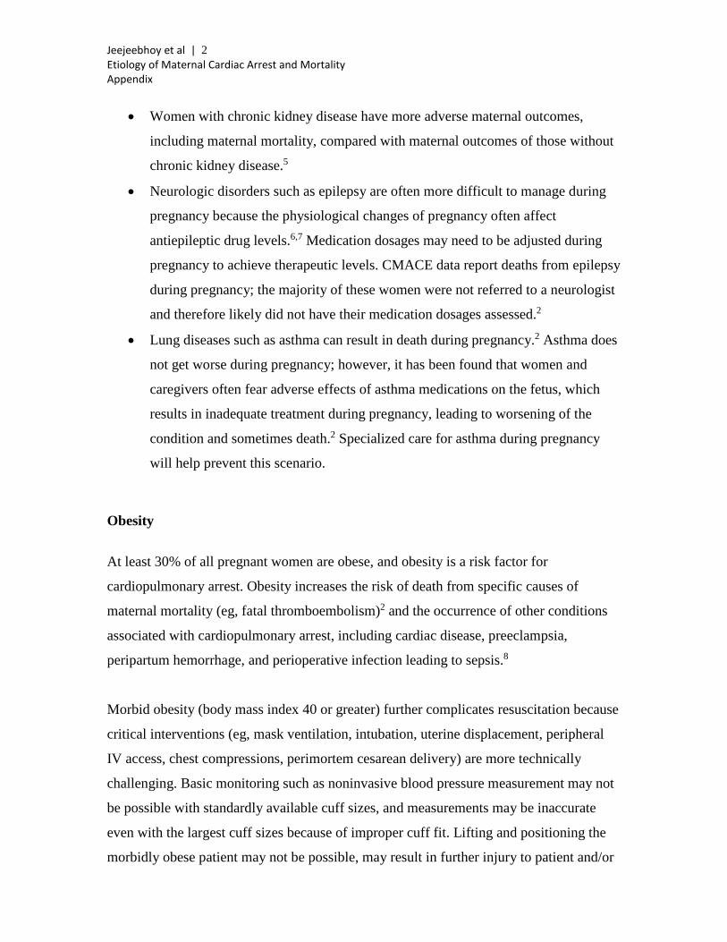
includes a discussion of the important etiologies of maternal mortality (listed in table 1),
and, where appropriate, diagnostic testing and treatment recommendations are discussed.
Chronic Health Problems That Exist Before Pregnancy Chronic health problems that exist before pregnancy can put the pregnant woman at risk
for cardiac arrest and mortality. A list of all possible health problems and concerns with
pregnancy is outside the scope of this report. However, important examples are discussed
below. It is important that pregnant women who have preexisting chronic health
problems receive specialized care during their pregnancy and into the postpartum period.2
Jeejeebhoy et al | 2 Etiology of Maternal Cardiac Arrest and Mortality Appendix
• Women with chronic kidney disease have more adverse maternal outcomes,
including maternal mortality, compared with maternal outcomes of those without
chronic kidney disease.5
• Neurologic disorders such as epilepsy are often more difficult to manage during
pregnancy because the physiological changes of pregnancy often affect
antiepileptic drug levels.6,7 Medication dosages may need to be adjusted during
pregnancy to achieve therapeutic levels. CMACE data report deaths from epilepsy
during pregnancy; the majority of these women were not referred to a neurologist
and therefore likely did not have their medication dosages assessed.2
• Lung diseases such as asthma can result in death during pregnancy.2 Asthma does
not get worse during pregnancy; however, it has been found that women and
caregivers often fear adverse effects of asthma medications on the fetus, which
results in inadequate treatment during pregnancy, leading to worsening of the
condition and sometimes death.2 Specialized care for asthma during pregnancy
will help prevent this scenario.
Obesity
At least 30% of all pregnant women are obese, and obesity is a risk factor for
cardiopulmonary arrest. Obesity increases the risk of death from specific causes of
maternal mortality (eg, fatal thromboembolism)2 and the occurrence of other conditions
associated with cardiopulmonary arrest, including cardiac disease, preeclampsia,
peripartum hemorrhage, and perioperative infection leading to sepsis.8
Morbid obesity (body mass index 40 or greater) further complicates resuscitation because
critical interventions (eg, mask ventilation, intubation, uterine displacement, peripheral
IV access, chest compressions, perimortem cesarean delivery) are more technically
challenging. Basic monitoring such as noninvasive blood pressure measurement may not
be possible with standardly available cuff sizes, and measurements may be inaccurate
even with the largest cuff sizes because of improper cuff fit. Lifting and positioning the
morbidly obese patient may not be possible, may result in further injury to patient and/or

Jeejeebhoy et al | 3 Etiology of Maternal Cardiac Arrest and Mortality Appendix
staff, or may require a specialized lift team that may not be immediately available. These
factors may negatively affect the ability of responding teams to render optimal care
during maternal cardiac arrest.
Jeejeebhoy et al | 4 Etiology of Maternal Cardiac Arrest and Mortality Appendix
Table 1. Most Common Etiologies of Maternal Arrest and Mortality Letter Cause Etiology A Anesthetic complications High neuraxial block
Hypotension Loss of airway Aspiration Respiratory depression Local anesthetic systemic toxicity
Accidents/trauma Trauma Suicide
B Bleeding Coagulopathy Uterine atony Placenta accreta Placental abruption Placenta previa Retained products of conception Uterine rupture Surgical Transfusion reaction
C Cardiovascular causes Myocardial infarction Aortic dissection Cardiomyopathy Arrhythmias Valve disease Congenital heart disease
D Drugs Oxytocin Magnesium Drug error Illicit drugs Opioids Insulin Anaphylaxis
E Embolic causes Amniotic fluid embolus Pulmonary embolus Cerebrovascular event Venous air embolism
F Fever Sepsis Infection
G General H’s and T’s H Hypertension Preeclampsia
Eclampsia HELLP syndrome, intracranial bleed
HELLP indicates hemolysis, elevated liver enzymes, and low platelet count.
Jeejeebhoy et al | 5 Etiology of Maternal Cardiac Arrest and Mortality Appendix
AAnesthetic Complications Anesthesia-related maternal mortality has decreased nearly 60% over the past few
decades9; however, a significant proportion (1% to 4%) of maternal deaths are still
directly related to complications from anesthesia.1,10-12 Although case-fatality rates for
general anesthesia are decreasing, rates for regional anesthesia are increasing.9 Many of
these complications are avoidable and potentially reversible. Although complications
from anesthesia accounted for 8% of maternal cardiac arrests in the 1998-2011 US
Nationwide Inpatient Sample, 82% of these women survived to hospital discharge.1
High Neuraxial Block and Hypotension
Epidemiology: With increasing use of neuraxial anesthetic techniques in the obstetric
setting,9 neuraxial anesthesia−related complications such as high spinal blocks have
increased. In an analysis of closed claims related to regional anesthesia and analgesia in
England, approximately half were related to obstetric cases.13
Pathophysiology: The innervation to the diaphragm is C3, C4, and C5; therefore, high
spinals will paralyze the diaphragm if the cephalad extent of the block reaches or exceeds
this level. Paralysis will result in a respiratory arrest with the potential for full
cardiopulmonary arrest if ventilation is not immediately instituted. In addition, high
spinal blocks cause significant sympathectomy. As a result, decreased systemic vascular
resistance may precipitate marked hypotension. Persistent severe hypotension may result
in cardiac arrest if left untreated. Severe sinus bradycardia or arrest during spinal or
epidural anesthesia is also described, presumably due to a Bezold-Jarisch reflex and/or
blockage of cardiac accelerator fibers at T1-4 with unopposed vagal tone.14,15
Diagnosis: Early recognition and immediate institution of resuscitative measures is
essential to optimize outcomes.14,15 Spinal-induced cardiopulmonary arrest should be
suspected if it occurs soon after administration of spinal anesthesia or after epidural bolus
(because of the potential for intrathecal migration of the catheter and/or high block from
Jeejeebhoy et al | 6 Etiology of Maternal Cardiac Arrest and Mortality Appendix
the epidural itself). Underlying and contributing causes for cardiac arrest complicating
spinal anesthesia should always be investigated; one case report after an investigation of
cesarean sections revealed dilated cardiomyopathy related to pregnancy.16
Treatment: The liberal use of phenylephrine, ephedrine (progressing to epinephrine and
atropine if necessary), and vasopressin; repeated, frequent blood pressure measurement;
aggressive intravascular fluid repletion; inferior vena caval decompression; and external
or transvenous pacing may help prevent the hypotension associated with high or total
spinal but will not prevent high or total spinal from occurring and causing paralysis of the
diaphragm. If high or total spinal occurs, management should focus on oxygenation,
intubation, and ventilation if apnea, loss of consciousness, or both occur; administration
of vasopressors to increase systemic vascular resistance (early and aggressive
administration of epinephrine, vasopressin, ephedrine, and phenylephrine); fluid
resuscitation with colloid or crystalloids; adequate left uterine displacement to restore the
effective circulating blood volume; inotropes (epinephrine and ephedrine) and
chronotropes (atropine) if bradycardia presents; and chest compressions as necessary.
Loss of Airway and Aspiration
Epidemiology: The risk of loss of maternal airway and gastric aspiration has decreased
with increasing use of regional anesthetic techniques.9 However, airway-related problems
still cause maternal death.9-12 Aspiration pneumonitis accounted for 7% of maternal
cardiac arrests in a 1998-2011 US Nationwide Inpatient Sample.1 The reported survival
to hospital discharge during maternal cardiac arrests associated with aspiration
pneumonitis is 83%.1
Pathophysiology: The physiology of pregnancy results in increased edema and friability
of the oropharyngeal mucosa as well as increased acidity of gastric contents and
decreased lower esophageal sphincter tone. These physiological changes may synergize
and result in more technically challenging airway control. If aspiration occurs in an
unprotected airway, highly acidic gastric contents may enter the pulmonary tree and
injure the lung parenchyma, causing pneumonitis.
Jeejeebhoy et al | 7 Etiology of Maternal Cardiac Arrest and Mortality Appendix
Diagnosis: Hypoventilation, airway obstruction, or both have been reported during
induction, emergence, extubation, or recovery and often in the setting of inadequate
anesthesia supervision or expertise. Aspiration may be diagnosed by the presence of
gastric contents in the oropharynx and may be associated with hypoxia.10,11
Treatment: Common strategies used to prevent aspiration include the use of
nonparticulate antacids to temporarily (about 15 minutes) neutralize acidic pH before
induction of anesthesia and agents (such as metoclopramide) that increase lower
esophageal sphincter tone. The quality of the evidence was poor, but the findings suggest
that the combination of antacids plus H2 antagonists was more effective than no
intervention and superior to antacids alone in preventing low gastric pH.17 Meticulous
airway examination, documentation, and communication, in conjunction with proper
equipment (videolaryngoscopic capability, supraglottic devices with orogastric ports,
bougies, etc), and the presence of a difficult-airway cart and experts in airway
management all confer added safety to control of the airway in the parturient. If
pulmonary aspiration of gastric contents is suspected, management strategies include use
of an endotracheal tube and mechanical ventilation with 100% oxygen and positive end-
expiratory pressure 5 cm H2O; suction airway; bronchoscopy if necessary; and
bronchodilators, fluids, and inotropes as needed.18 Steroids and antibiotics should not be
used in the acute phase, because there is no evidence of an improved outcome.18
Respiratory Depression
Epidemiology: Although very rare, maternal respiratory depression and arrest is a
potentially serious risk after neuraxial (epidural and intrathecal) or intravenous opioid
administration.19 In 1 report, 73% of these patients died or experienced permanent brain
damage after respiratory depression involving neuraxial opioids.20 Early recognition and
effective management often result in good outcomes.21
Pathophysiology: Opioids may cause hypoventilation and/or apnea, particularly when
administered by multiple routes (eg, oral, intravenous, neuraxial) to high-risk patients
Jeejeebhoy et al | 8 Etiology of Maternal Cardiac Arrest and Mortality Appendix
(eg, obstructive sleep apnea), although opioid-induced respiratory arrest may occur in any
patient.
Diagnosis: Opioid-induced respiratory arrest in the setting of labor should be suspected if
respiratory arrest occurs within recent (within 30 minutes) administration of intravenous
epidural or intrathecal neuraxial opioids.19,21,22 Delayed (6 to 18 hours postdose)
respiratory depression after use of neuraxial morphine (due to rostral spread in
cerebrospinal fluid and brainstem penetration) has also been described.19,23 Continuous
infusion of short-acting opiates has also been associated with maternal apnea.24
Therefore, monitoring for this potential anesthetic complication should be of adequate
duration, depending on the opioid used.25
Treatment: Prevention of respiratory depression and arrest from opioids depends on
appropriate monitoring of the respiratory rate and capnography, widespread education of
staff, readily available reversal agents (eg, naloxone), and ventilation equipment. In the
setting of suspected opioid-induced respiratory depression, treatment protocols should
focus on rapid provision of supplemental oxygen and immediate administration of the
opioid reversal agent naloxone. If naloxone fails to reverse respiratory depression or
arrest, prompt bag-mask ventilation and/or endotracheal intubation should be performed.
Treatment and intensive monitoring should be maintained until signs and symptoms of
respiratory depression have resolved.26
Local Anesthetic Systemic Toxicity
Epidemiology: With the increasing use of neuraxial anesthesia in obstetrics, one possible
anesthetic-related etiology for cardiac arrest is local anesthetic systemic toxicity
(LAST).9
Pathophysiology: Pregnancy may enhance sensitivity to local anesthetics, and LAST-
induced cardiac toxicity may be particularly resistant to conventional resuscitative
interventions.
Jeejeebhoy et al | 9 Etiology of Maternal Cardiac Arrest and Mortality Appendix
Diagnosis: LAST-induced cardiac arrest should be suspected if an arrest occurs soon
after an epidural local anesthetic “top-up.”27 If LAST-induced cardiac arrest is suspected,
lipid emulsion therapy should be given in addition to standard resuscitative measures.28,29
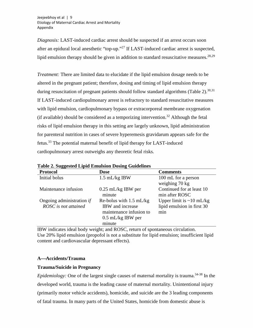
Treatment: There are limited data to elucidate if the lipid emulsion dosage needs to be
altered in the pregnant patient; therefore, dosing and timing of lipid emulsion therapy
during resuscitation of pregnant patients should follow standard algorithms (Table 2).30,31
If LAST-induced cardiopulmonary arrest is refractory to standard resuscitative measures
with lipid emulsion, cardiopulmonary bypass or extracorporeal membrane oxygenation
(if available) should be considered as a temporizing intervention.32 Although the fetal
risks of lipid emulsion therapy in this setting are largely unknown, lipid administration
for parenteral nutrition in cases of severe hyperemesis gravidarum appears safe for the
fetus.33 The potential maternal benefit of lipid therapy for LAST-induced
cardiopulmonary arrest outweighs any theoretic fetal risks.
Table 2. Suggested Lipid Emulsion Dosing Guidelines Protocol Dose Comments Initial bolus 1.5 mL/kg IBW 100 mL for a person
weighing 70 kg Maintenance infusion 0.25 mL/kg IBW per
minute Continued for at least 10 min after ROSC
Ongoing administration if ROSC is not attained
Re-bolus with 1.5 mL/kg IBW and increase maintenance infusion to 0.5 mL/kg IBW per minute
Upper limit is ~10 mL/kg lipid emulsion in first 30 min
IBW indicates ideal body weight; and ROSC, return of spontaneous circulation. Use 20% lipid emulsion (propofol is not a substitute for lipid emulsion; insufficient lipid content and cardiovascular depressant effects). AAccidents/Trauma
Trauma/Suicide in Pregnancy
Epidemiology: One of the largest single causes of maternal mortality is trauma.34-38 In the
developed world, trauma is the leading cause of maternal mortality. Unintentional injury
(primarily motor vehicle accidents), homicide, and suicide are the 3 leading components
of fatal trauma. In many parts of the United States, homicide from domestic abuse is
Jeejeebhoy et al | 10 Etiology of Maternal Cardiac Arrest and Mortality Appendix
more common than fatal motor vehicle accidents.37,39,40 Suicide represents up to 10% of
maternal mortality but may be decreased in pregnancy compared with nonpregnant
controls.39,41 Approximately 1% to 4% of pregnant women are admitted for treatment of
traumatic injury, yet there are few educational strategies targeted toward
prevention/management of maternal trauma. Pregnancy should be tested for in all trauma
cases involving women of childbearing age. Use of illicit drugs and alcohol, domestic
abuse, and depression are all important contributors to maternal trauma; thus, a high
index of suspicion should be maintained when treating young injured women.
Pathophysiology: Maternal death is associated with penetrating injury, high injury
severity score, and head injury. Pregnancy-related morbidity occurs in about 25% of
cases and may include placental abruption, uterine rupture, preterm delivery, and need for
cesarean delivery.
Diagnosis: Diagnosis is performed by physical examination, monitoring of uterine
activity and fetal heart rate, and ultrasound evaluation (depending on gestational age).
Fetal-maternal hemorrhage (detected with a Kleihauer-Betke test) complicates 10% to
30% of cases.
Treatment: The response of the resuscitation team to traumatic injury includes an
expedited use of perimortem cesarean delivery. If the mother has an obviously lethal
injury, such as a fatal gunshot wound to the head, then continued maternal resuscitation is
not indicated after the mother arrives in the emergency department. Emergency cesarean
delivery should be performed. Considerations for immediate cesarean delivery include
the extent of the injury and the time from presumed cardiac arrest until arrival in the
emergency department. In general, >30 minutes of absent uterine blood flow is not
associated with fetal survival. However, in many situations, this information may not be
immediately obvious. Other considerations for immediate cesarean delivery include fetal
age and the extent of maternal injury. Each situation is different. In maternal suicide, the
means of the suicide may direct resuscitative efforts. A study of the California birth
database found drug overdose to be the most common means of maternal suicide
Jeejeebhoy et al | 11 Etiology of Maternal Cardiac Arrest and Mortality Appendix
attempts.42 Rh-negative patients should receive Rh-D immunoglobulin within 72 hours of
injury. Anesthetic management should be directed toward maternal oxygenation and
perfusion. Airway management includes prevention of aspiration, expert help, and
insertion of a smaller-than-usual endotracheal tube. Drugs (choice and dose) should be
adapted primarily to suit maternal injuries and, when possible, pregnancy. If pregnancy is
confirmed, gestational age should be assessed while providers adhere to advanced trauma
life support guidelines. Treating the mother appropriately is beneficial for both mother
and fetus. The only guideline modifications include provision of supplemental oxygen,
preference for intravenous access above the diaphragm, and left lateral positioning
whenever possible.
BBleeding
Epidemiology: Hemorrhage complicates approximately 3% to 4% of all births, but fewer
than 1 in 1000 women with hemorrhage also experience cardiac arrest.1 Regardless,
antepartum or postpartum hemorrhage accounts for 38% of all cardiac arrests during
hospitalization for delivery.1
Pathophysiology: Maternal mortality reviews have suggested that hemorrhagic cardiac
arrest often follows delays in recognition, treatment, or escalation of care for women with
obstetric hemorrhage.2,43,44 In some cases, hemorrhage may be concealed (eg, abruption,
retroperitoneal hemorrhage). The bleeding pregnant patient can lose 1500 mL before any
clinical manifestations of compromise.45 Occasionally, catastrophic hemorrhage can
present abruptly or overwhelm standard therapies (eg, amniotic fluid embolism).
Although 80% of obstetric hemorrhage may be caused by uterine atony, retained
placenta, including the spectrum of morbidly adherent placentation (ie, placenta accreta,
increta, and percreta), is an important etiology of massive hemorrhage and peripartum
hysterectomy and has been linked to the escalation of cesarean birth rates.46,47 Uterine
infection (eg, chorioamnionitis) has also been associated with unremitting uterine atony
that requires peripartum hysterectomy.48 Additional etiologies of massive maternal
hemorrhage include abruption, uterine rupture, uterine inversion, placenta previa, and
coagulation defects.49 Other risk factors for peripartum obstetric hemorrhage include
Jeejeebhoy et al | 12 Etiology of Maternal Cardiac Arrest and Mortality Appendix
prolonged labor, episiotomy, preeclampsia, operative delivery, and uterine overdistention
(ie, multiple gestations and polyhydramnios).50 Finally, vascular catastrophe is a rare but
highly lethal cause of hemorrhagic arrest (eg, aortic rupture, splanchnic arterial rupture)
that appears to be more common in pregnancy (see aortic dissection in “Cardiovascular
Causes”).51
Hemorrhage typically causes hypovolemic arrest. Alternatively or additionally, blood
product transfusion may generate hypocalcemic or hyperkalemic arrest, anaphylactic
reaction to blood products, or respiratory arrest as a result of resuscitation-induced
pulmonary or airway edema.
Diagnosis: Rescuers need to perform concurrent resuscitation and evaluation to diagnose
the source of bleeding, which may guide specific intervention and treatment. History and
physical examination may also help reveal the etiology. Transabdominal ultrasound at the
bedside may be beneficial in localizing the diagnosis. In the arrest patient with a closed
abdomen, transabdominal ultrasound may be used to diagnose concealed hemorrhage
(retroperitoneal hemorrhage, hepatic rupture) and localize the source of bleeding. The
Focused Assessment with Sonography in Trauma protocol should be combined with
comprehensive uterine evaluation (eg, endometrial stripe, signs of partial uterine
inversion). Serial arterial blood gas analysis may help identify acid base and electrolyte
abnormalities as well as abnormalities of gas exchange. Hematologic measurements,
including the hematocrit, platelet count, and fibrinogen level may not correlate acutely
with the degree of blood loss, so transfusion should be started when hemorrhage is
suspected clinically rather than waiting for laboratory results. Finally, hemostatic
monitoring with thromboelastography or thromboelastometry may facilitate diagnosis of
coagulopathy.
Treatment: To prevent cardiac arrest, include obstetric hemorrhage protocols that
facilitate a multidisciplinary approach to ensure timely recognition and treatment to stop
the bleeding and ensure end-organ perfusion. Uterotonics, uterine evacuation, and uterine
tamponade constitute early intervention. Hemodynamic and hemostatic resuscitation
Jeejeebhoy et al | 13 Etiology of Maternal Cardiac Arrest and Mortality Appendix
become increasingly important as the quantity and velocity of blood loss increase.
Nevertheless, patient survival ultimately depends on the timely escalation of surgical
interventions to control the source of bleeding.
Use of massive transfusion with appropriate proportions of packed red blood cells and
fresh frozen plasma and cryoprecipitate requires early activation.52 Aggressive hemostatic
and volume resuscitation requires a large multiprofessional team of clinicians and careful
coordination with the blood bank, clinical lab, and clinical pharmacy. Institutional
massive transfusion protocols can accelerate and enhance access to large volumes of
blood products and timely clinical lab turnaround. Coagulopathy is best treated by
product replacement and maintenance of normothermia. Early antifibrinolytic therapy
(eg, tranexamic acid) has been shown to increase survival in patients with traumatic
hemorrhage, and although currently there is insufficient evidence to confirm this benefit
in postpartum hemorrhage,53 there is no evidence of harm to the maternal patient. In
contrast, no trials have been attempted to evaluate the benefit of recombinant factor VIIa,
a drug that has been associated with serious thrombotic complications in survivors.
Exploratory laparotomy with uterine compression sutures, pelvic vascular ligation, and/or
vascular embolization may provide successful treatment. Hysterectomy may be required
when other measures have been unsuccessful. Exploratory laparotomy may allow ligation
of vascular bleeding pedicles and gain access to the uterus. Compression sutures of the
uterus and serial pelvic vessel ligation can decrease blood loss. If unsuccessful,
hysterectomy should be performed. Pledgeted sutures may help achieve hemostasis when
suturing highly friable vascular beds.54
Alternatively or additionally, abdominal packing may be necessary to temporarily control
bleeding during resuscitation. Vascular radiological embolization may be a viable
adjunct, but this would not be the modality of choice during cardiac arrest. It may be
helpful as a postresuscitation maneuver in a more stabilized but critically ill patient in
whom repetitive surgical procedures should be avoided. In the event of cardiac arrest, a
vertical abdominal skin incision will facilitate rapid surgical maneuvers. Examination of
the liver and retroperitoneal space for hemorrhage may be important if the cause of
Jeejeebhoy et al | 14 Etiology of Maternal Cardiac Arrest and Mortality Appendix
cardiac arrest is unknown. The vertical incision also opens the abdomen for manual
compression of the aorta, which has been described as a lifesaving temporizing maneuver
for women with catastrophic pelvic hemorrhage.55
Both hyperkalemic and hypocalcemic arrest are best treated with calcium chloride and
chest compressions. In addition, hyperkalemia should be treated with drugs that shift
potassium into cells (eg, sodium bicarbonate, insulin and dextrose, and a β2-agonist),
followed by therapies that accelerate potassium clearance (eg, diuretics, dialysis).
Insufficient and difficult peripheral venous access may be rescued with central line
insertion or intraosseous needle insertion; any fluid, blood product, or medication can be
administered by the intraosseous route while additional venous access is secured.56
Postresuscitation hypothermia instituted for neuroprotection may exacerbate
coagulopathy.
CCardiovascular Causes
Cardiac Risk Stratification
In the presence of maternal cardiac disease, the hemodynamic changes of pregnancy may
result in decompensation or death of the mother or fetus. In western civilizations, most
maternal cardiac disease is congenital in origin, due in large part to the success of cardiac
surgery in the past few decades. This may cause significant maternal morbidity but not
necessarily mortality. Predictors of maternal cardiac events have been studied largely
retrospectively, and predictors of maternal cardiac complications have been identified
(Table 3). There are significant limitations in the use of risk scores to predict maternal
cardiac events because many studies are retrospective, may have different definitions of a
specific lesion (heart failure, for example), and are highly dependent on the populations
studied. The study by Siu et al57 reviewed patients with both congenital and acquired
heart disease, whereas the study of Drenthen et al58 studied only patients with congenital
heart disease. Known important risk factors such as pulmonary arterial hypertension and
aortic dilatation were not identified in either of these studies and thus are
underrepresented in these cohorts. Therefore, the use of predictive risk scores should be
only a part of a prepregnancy assessment. Patients with Eisenmenger syndrome, for
Jeejeebhoy et al | 15 Etiology of Maternal Cardiac Arrest and Mortality Appendix
example, have a maternal mortality approaching 30% and as such are usually counseled
to avoid pregnancy.
The World Health Organization categorizes maternal risk as grades I to IV. Patients in
class III are at significant risk of maternal mortality or severe morbidity. Patients in class
IV are at extremely high risk of severe morbidity or mortality; for these patients,
pregnancy is contraindicated.59,60 Other pathologies included in class III (not mentioned
in Table 1) are a systemic right ventricle, Fontan circulation, and an aorta of 40 to 45 mm
in Marfan syndrome and 45 to 50 mm with a bicuspid aortic valve. Patients in class IV
include those with pulmonary arterial hypertension of any cause, prior peripartum
cardiomyopathy (PPCM) with any residual impairment of ventricular function, native
severe coarctation, an aorta >45 mm in Marfan syndrome, and an aorta >50 mm with a
bicuspid aortic valve.
Cardiac Disease in Pregnancy
Cardiac disease remains the number 1 cause of maternal mortality in developed countries,
albeit a relatively rare event.2 Leading causes include sudden adult death syndrome,
aortic dissection, myocardial infarction, ischemic heart disease, and PPCM. In this
context, current trends in lifestyle, including the increasing prevalence of metabolic
syndrome, diabetes, smoking, hypertension, and advanced maternal age, must be taken
into consideration because they undoubtedly contribute to the growing incidence of
ischemic heart disease in pregnancy. In the CMACE study,2 from 2006 to 2008, 30 of 50
women (60%) who died from cardiac disease were overweight or obese, with half having
a body mass index ≥30.
Myocardial Infarction
Epidemiology: Myocardial infarction and ischemic heart disease are important causes of
maternal death, with most infarcts occurring in the period 6 weeks postpartum.61
Pregnancy is a risk factor for acute myocardial infarction (AMI), increasing risk 3- to 4-
fold in comparison with the nonpregnant state. Maternal age adds to this risk, so that risk
of pregnancy-related AMI is 30 times higher in women >40 years of age versus those <20
Jeejeebhoy et al | 16 Etiology of Maternal Cardiac Arrest and Mortality Appendix
years of age.59 AMI is more common in multigravidas (66%),62 and approximately three-
quarters of these involve the anterior wall. Almost half of acute infarcts are related to
coronary dissection; others are related to coronary atheroma, reflecting the impact of
lifestyle factors such as increasing maternal age, obesity, and smoking. Indeed, in the
CMACE study,2 all women who died from ischemic heart disease had identifiable risk
factors. A US population−based study reported that age >35 years and black race were
significant risk factors for pregnancy-related myocardial infarction, with odds >5 times
higher for black women 35 years of age and older. Multivariable analysis, however,
eliminated race as a risk factor, suggesting that black women have an increased
prevalence of other risk factors.63 Independent risk factors include chronic hypertension,
diabetes, advanced maternal age, and eclampsia.61 Smoking may increase risk 8-fold.
Thrombophilia is also a risk factor, which is not surprising in view of the
hypercoagulability of pregnancy, which is further increased in the presence of
thrombophilia. Women diagnosed during the prenatal period may have a higher mortality
and more pregnancy complications.61 The risk of death from AMI in pregnancy has been
reported to be as high as 37%, but recent data suggest the risk is about 5%.63 This may be
related, in part, to the rapidity with which patients with acute coronary syndromes are
transported to the catheterization laboratory in the contemporary era and the widespread
use of percutaneous coronary intervention (PCI). Mortality appears twice as high in
women diagnosed with AMI in the peripartum period compared with the antepartum or
postpartum period.62
Pathophysiology: Delay in diagnosis and treatment is common because chest pain in
pregnancy is common and may reflect reflux disease, with a failure to perform an
electrocardiogram to facilitate diagnosis and, where PCI is unavailable, a failure to
administer thrombolytic therapy because of the concern for hemorrhage. Failure to
proceed rapidly to open the occluded coronary artery either by PCI or thrombolysis is the
most common cause of cardiac arrest with ventricular tachycardia or fibrillation.
Cardiogenic shock may also occur due to acute ventricular failure with low cardiac
output.
Jeejeebhoy et al | 17 Etiology of Maternal Cardiac Arrest and Mortality Appendix
Diagnosis: The criteria for diagnosis of AMI in pregnant women are generally the same
as in nonpregnant patients and consist of symptoms, electrocardiographic changes, and
cardiac markers, although patients may initially have normal troponin levels.
Nonetheless, elevated troponin levels should heighten the consideration of AMI as the
etiology. Echocardiography is safe and can be used in a timely way to assess regional
wall motion abnormalities. The differential diagnoses in this context are aortic dissection,
acute pulmonary embolism, and preeclampsia; thus, appropriate investigations for a
woman presenting with chest pain should include an electrocardiogram, chest x-ray,
troponin levels, an echocardiogram (which may include a transesophageal
echocardiogram, depending on available expertise), computed tomography, and/or
magnetic resonance imaging.
Treatment: When the diagnosis of AMI is suspected, an invasive approach with prompt
transfer to a cardiac catheterization laboratory for consideration of PCI is preferable to
thrombolysis because coronary angiography will also diagnose coronary dissection.
Morphine may be used for pain control and has no teratogenic effects. β-Adrenergic
blocking agents generally can be used safely, as can antiplatelet therapy with low-dose
aspirin (81 mg/d). Thienopyridine derivatives such as clopidogrel and ticlopidine may be
used when necessary, but data about the safety of these drugs in the fetus are sparse.
Ideally, clopidogrel should be discontinued for 1 week before regional anesthesia or
cesarean section. Inotropic support may be necessary if cardiac output is impaired. In the
context of profound hemodynamic instability, inotropic support may be administered and
an intra-aortic balloon pump may be implanted concomitantly with the catheterization
procedure. Because drug-eluting stents require prolonged dual antiplatelet treatment, bare
metal stents should be used when necessary. Thrombolytic therapy should be reserved for
life-threatening AMI when there is no access to a catheterization laboratory, because its
use is associated with an increased risk of placental hemorrhage.64 It may also contribute
to further progression of a coronary dissection and for these reasons should be considered
a second choice to primary PCI. If possible, the patient should be treated in a coronary
care unit that can also provide maternal monitoring as well as a comprehensive maternal-
fetal medicine service. In cases of hemodynamic instability and cardiac deterioration, a
Jeejeebhoy et al | 18 Etiology of Maternal Cardiac Arrest and Mortality Appendix
plan should be established for urgent delivery of a potentially viable fetus. Surgical
myocardial revascularization may be considered, usually when there is extensive
coronary dissection and failure of PCI to reopen the vessel. Limited data are available
about the safety and outcomes of these procedures, however.
Aortic Dissection
Epidemiology: Aortic dissection is another major cause of cardiac death, particularly in
the context of connective tissue abnormalities such as Marfan syndrome, Ehlers-Danlos
syndrome type IV, bicuspid aortic valve, familial aortic dissection, and Turner syndrome.
In addition to the hemodynamic changes of pregnancy, hormonal changes render the
aortic wall more vulnerable to dissection, with a fragmentation of the reticulin fibers,
reduced mucopolysaccharides, and loss of the normal corrugation of elastic fibers. Type
A and type B dissections occur, particularly in those with Marfan syndrome. Most
dissections occur in the third trimester (50%) or the early postpartum period60,65 during
periods of maximum hemodynamic stress. Women with dilated aortas or prior aortic
dissection are at particularly high risk during pregnancy. The diagnosis of dissection
should always be considered in pregnant women who present with chest pain. Early
mortality is high.
Dissections typically propagate in an antegrade direction but may extend retrogradely.
Acute coronary compromise and severe aortic regurgitation may occur along with cardiac
tamponade, which is the most common cause of cardiac arrest.
Pathophysiology: Cardiac arrest may occur from hemorrhage, acute aortic regurgitation
(typically with a type A dissection), dissection of a coronary artery, and cardiac
tamponade.
Diagnosis: Emergent echocardiographic imaging, transthoracic or transesophageal, is
typically the fastest modality to delineate the etiology.
Jeejeebhoy et al | 19 Etiology of Maternal Cardiac Arrest and Mortality Appendix
Treatment: Women with one of these connective tissue abnormalities and an aortic root
diameter >4.5 cm should be counseled against pregnancy because of the risk of
dissection.60,65 This is particularly true when there is a family history of dissection or the
aorta has undergone a period of rapid dilatation. For those with Marfan syndrome, no safe
aortic diameter exists, and there is a 1% risk of dissection even with a normal aortic root
diameter.66 For those with Turner syndrome, who often are of short stature, an aortic
diameter index >27 mm/m2 is associated with a high risk of dissection, which carries a
maternal mortality of approximately 11%.60 Associated risk factors in this group include
concomitant bicuspid aortic valve, coarctation of the aorta, and hypertension.
Patients with aortic pathology (ascending aorta >4 cm or less in those with a low body
surface area) should be monitored throughout pregnancy by a multidisciplinary team that
includes cardiologists with expertise and training in the management of high-risk patients
with cardiovascular disease.60 Monitoring should include imaging with echocardiography
at regular intervals, the frequency of which is determined by the clinical context and size
of the aorta, but generally at 6- to 8-week intervals and for 6 months postpartum.59 β-
Blocking agents are generally used to keep blood pressure under strict control and to
reduce the shear stresses on the aortic wall, although there are scant data to support their
use except in the context of Marfan syndrome. Hospitalization of high-risk patients
should be considered between 28 and 32 weeks of gestation, with elective fetal lung
maturation at 26 weeks of gestational age. Hospitalization should be undertaken in a
center where cardiothoracic surgery is available. Prophylactic surgery should be
considered during pregnancy if the aortic diameter is >50 mm and increasing rapidly.60
Aortic dissection in pregnancy is a surgical emergency, and the diagnosis must be made
promptly, typically with transesophageal echocardiography, computed tomography, or
magnetic resonance imaging, depending on local facilities and expertise. Senior
cardiothoracic surgeons along with the team of cardiology, obstetric, and anesthesia staff
must proceed rapidly to the cardiac operating room to repair the dissection. After 30
weeks of gestation, immediate cesarean section with delivery of the fetus in the cardiac
operating room followed immediately by cardiac surgery seems to be the most promising
Jeejeebhoy et al | 20 Etiology of Maternal Cardiac Arrest and Mortality Appendix
option to save the life of the mother and the unborn child. Cardiopulmonary bypass can
be instituted simultaneously during cesarean section (femoral or axillary cannulation).65
Cardiac surgery should be performed where neonatal intensive care facilities are
available. If the fetus is not yet viable, high-pressure, high-flow (>2.4 L/min per square
meter) normothermic perfusion is preferable for cardiopulmonary bypass, because
hypothermia decreases placental flow and causes fetal bradycardia with an increased
likelihood of intrauterine death or hypoxic-ischemic fetal insult.65,67 If possible,
maintaining uterine displacement by placing the patient in the left lateral recumbent
position during cardiopulmonary bypass will help avoid aortocaval compression and
minimize fetal risk.67 Continuous fetal cardiac monitoring may decrease the risk to the
fetus, as will close monitoring of serum potassium concentration (goal <5 mmol/L),
because prolonged cardioplegia may increase potassium levels.
For type B dissection, medical therapy to strictly control blood pressure is the preferred
strategy in the absence of rupture or malperfusion.65 In this situation, there is a high
incidence of fetal death, probably due to compromise of the internal iliac or ovarian
arteries impairing placental blood flow.
Indications for surgical repair include leakage or rupture, progressive aortic dilatation,
extension of the dissection, and recurrent pain.
Cardiomyopathy
Epidemiology: Cardiomyopathies are rare but remain an important cause of death.68 This
includes PPCM; patients with a prior history of PPCM should be counseled about the risk
of recurrence. The incidence varies from around 1 in 4000 pregnancies in the United
States to 1 in 300 pregnancies in Haiti, suggesting an environmental influence or
common genetic mutation.69 The etiology and pathophysiology are poorly understood,
but inflammation and autoimmune processes may play a role. Risk factors include
multiparity, race (being black), older maternal age, smoking, hypertension, and
preeclampsia. Most cases present in the puerperium when the increased hemodynamic
burden of pregnancy has diminished. Subsequent normalization of ventricular function
Jeejeebhoy et al | 21 Etiology of Maternal Cardiac Arrest and Mortality Appendix
occurs in up to half of patients with PPCM and is more likely if the ejection fraction is
>30% at the time of diagnosis. A subsequent pregnancy carries a 30% to 50% risk of
recurrence of PPCM, which may result in further clinical deterioration and even death,
particularly when ventricular function failed to normalize before the subsequent
pregnancy.68,70 Appropriate counseling before pregnancy is therefore imperative to
prevent a cardiac catastrophe.
Other causes of cardiomyopathy include idiopathic dilated cardiomyopathy, familial
cardiomyopathy, noncompaction, and ischemic cardiomyopathy. With all of these
cardiomyopathies, women are at high risk of heart failure if the ejection fraction is <40%,
and close monitoring with a multidisciplinary team in a tertiary center should be advised.
When the ejection fraction is <30%, maternal mortality is increased, and termination of
the pregnancy should be considered.60 In accordance with the guidelines, patients with
congestive heart failure and an ejection fraction <35% should have had implantation of
an automated implantable cardioverter-defibrillator (ICD) because of the increased risk of
ventricular arrhythmia and sudden cardiac death.
Hypertrophic cardiomyopathy: Pregnancy in women with hypertrophic cardiomyopathy
(HCM) is usually well tolerated because the volume load of pregnancy reduces left
ventricular outflow obstruction. Those at increased risk of cardiac arrest are very
symptomatic before pregnancy with dyspnea, angina, syncope, and arrhythmias.
Typically, these patients have a high left ventricular outflow tract gradient or clinically
important diastolic dysfunction.71,72 For patients with HCM, risk factors for sudden death
include a history of out-of-hospital arrest, family history of sudden death, history of
ventricular tachycardia, massive left ventricular hypertrophy on echocardiography (septal
wall thickness >3 cm), presence of scar by delayed enhancement on magnetic resonance
imaging, history of syncope, and either ventricular tachycardia or a decrease in blood
pressure on treadmill exercise testing. When the 2 largest studies of pregnancy and HCM
were combined in recent years,71,72 only 2 deaths occurred in 470 pregnancies in 227
patients. Both had high-risk features before pregnancy (massive left ventricular
hypertrophy with severe outflow obstruction, symptoms of heart failure, and a family
Jeejeebhoy et al | 22 Etiology of Maternal Cardiac Arrest and Mortality Appendix
history of malignancy). Typically, such patients would have received an automated ICD
before pregnancy. In other patients, pregnancy is usually well tolerated, and the risk of
cardiac arrest is therefore very low. The decision to place an ICD for prevention of
sudden death should be individualized, but in general, patients at the highest risk are
considered candidates.73
Pathophysiology: Severe impairment of ventricular function with dilated
cardiomyopathies may lead to intractable heart failure and ventricular tachycardia or
fibrillation. Malignant ventricular arrhythmias may occur with HCM and high-risk
features as outlined above.
Diagnosis: Diagnosis can be made with a transthoracic echocardiogram.
Treatment: Most severe heart failure in pregnancy is related to PPCM, and guidelines for
the management of acute heart failure apply, except that angiotensin-converting enzyme
inhibitors and angiotensin receptor blockers are contraindicated in pregnancy. Bed rest
and sodium and fluid restriction are important. Digoxin, hydralazine, oral nitrates used in
combination with hydralazine, β-blockers, and diuretics can all be used with relative
safety, although the latter may decrease placental perfusion and should be used
judiciously. Aldosterone antagonists should be avoided because of antiandrogenic effects
on the fetus. Anticoagulation with heparin should be considered in those with a very low
ejection fraction, particularly those with PPCM who have a high incidence of ventricular
thrombi and cerebral embolism.68 Complex ventricular arrhythmias may precipitate
cardiac arrest in this context.
In severe cases, nitrates and inotropes may be used, and in such circumstances,
consideration should be given to transferring the patient to a center where intra-aortic
balloon pump counterpulsation, ventricular assist devices, and transplant teams are
available. Extracorporeal membrane oxygenation may be necessary for those with
cardiogenic shock. Urgent cardiac transplantation may need to be considered.68 Urgent
Jeejeebhoy et al | 23 Etiology of Maternal Cardiac Arrest and Mortality Appendix
delivery irrespective of gestation should be considered in women with hemodynamic
instability despite treatment.
For women with HCM, pregnancy should be avoided in those with high-risk features.
Implantation of an automated ICD can prevent cardiac arrest in high-risk patients with
HCM. Maintenance of β-blocker therapy (the mainstay of treatment) should be continued
or even increased during pregnancy and through delivery to alleviate left ventricular
outflow obstruction. Dehydration and anemia should be avoided, as should tocolytic
therapy. Gentle incremental diuresis can be used for symptoms of heart failure. Delivery
should be accomplished in high-risk centers; spinal block should be avoided in case of
hypotension, and blood loss should be promptly replaced.
Mechanical Valve Prostheses
Epidemiology: Death may also occur from thrombosis of a mechanical prosthetic valve,
although this is a rare cause of cardiac death. Optimum anticoagulation strategies are
challenging in this situation, and there are clear data that anticoagulation with warfarin is
safer for the mother then either low-molecular-weight or unfractionated heparin.74 The
use of heparin for even a relatively short time (typically in the first trimester to avoid the
risk of fetal embryopathy) more than doubles the risk of mechanical valve thrombosis.75
Because pregnancy is a markedly prothrombotic state, valve thrombosis and death can
occur even in the context of therapeutic anti-Xa levels with the use of low-molecular-
weight heparin.76 The mechanical mitral valve, which is the most vulnerable to thrombus
formation, is particularly problematic.60 Optimum anticoagulation with the selected
anticoagulant is essential throughout pregnancy, and patients should be managed in a
tertiary care center with a multidisciplinary approach. Anticoagulant targets may need to
be measured weekly.
Although cardiac arrest is uncommon, it may occur in the setting of a thrombosed
mechanical prosthesis, particularly with left-sided valves (either mitral or aortic), and
results from low cardiac output and/or ventricular arrhythmia.
Jeejeebhoy et al | 24 Etiology of Maternal Cardiac Arrest and Mortality Appendix
Pathophysiology: Pregnancy is a hypercoagulable state with an increased concentration
of clotting factors, decreased fibrinolysis, and increased platelet adhesiveness. Most valve
thromboses occur in the setting of a tilting disc mitral prosthesis with heparin therapy,
which is a less optimum anticoagulant than warfarin during pregnancy. Even with careful
and meticulous monitoring in a tertiary care center, valve thrombosis may occur. Because
anticoagulation is interrupted during both vaginal and cesarean delivery, prosthetic valve
thrombosis may occur in the peripartum period.
Diagnosis: Emergent transthoracic echocardiography can facilitate the diagnosis to
identify the thrombosed valve.
Treatment: The only interventional therapy of benefit in this situation is emergent valve
replacement with concomitant delivery of the fetus if viable.
Congenital Heart Disease
Epidemiology: Patients with severely reduced function of the systemic ventricle and
those with cyanotic heart disease, particularly those with pulmonary hypertension, are at
most risk of adverse cardiac events. In those with severe pulmonary vascular disease and
a right-to-left shunt (Eisenmenger syndrome), maternal mortality was 30% to 50% in
early series, with death usually occurring in the last trimester or peripartum.77
Patients with severe symptomatic aortic stenosis, usually secondary to a bicuspid aortic
valve, should be counseled against a pregnancy, because the increase in cardiac output
and afterload reduction that accompanies pregnancy exaggerates the aortic gradient.
Patients may present with syncope, angina, or heart failure, and malignant arrhythmias
and death may occur.60 Such patients also have an associated aortopathy and an increased
risk of aortic dissection and rupture.
Patients with d-transposition of the great arteries after an atrial switch procedure
(Mustard or Senning operation) are vulnerable to heart failure because the systemic
ventricle is a morphological right ventricle. Patients with l-transposition of the great
Jeejeebhoy et al | 25 Etiology of Maternal Cardiac Arrest and Mortality Appendix
arteries are also at risk because the systemic ventricle is a right ventricle and vulnerable
to failure, particularly in the context of systemic atrioventricular valve regurgitation,
which may progress as pregnancy advances. Patients with single-ventricle physiology
after the Fontan procedure are also vulnerable to heart failure, and both atrial and
ventricular arrhythmias may intervene, causing profound hemodynamic instability and
cardiac arrest.
In Eisenmenger syndrome, cardiac arrest usually occurs with a progressive decrease in
peripheral resistance with hypotension causing more right-to-left shunting and cyanosis,
or abruptly with pulmonary thrombosis. Patients are at special risk of cardiac arrest
particularly in the third trimester and in the early postpartum weeks. Progressive right-
sided heart failure may also occur. Even with contemporary pulmonary vasomodulator
drugs (nitric oxide and sildenafil), maternal mortality may occur in 33% of patients and
may occur in those with minimal disability before pregnancy.78
Pathophysiology: Eisenmenger syndromeright-sided heart failure, in situ pulmonary
thrombosis, and progressive cyanosis with decrease in peripheral resistance; aortic
stenosisangina, syncope, heart failure, and ventricular arrhythmias.
Diagnosis: If the diagnosis is unknown, emergent echocardiography is the only modality
that might facilitate diagnosis in an emergent situation.
Treatment: Aortic stenosisseverely symptomatic patients refractory to medical therapy
may be considered for percutaneous valvuloplasty if the valve is noncalcified and free of
regurgitation. If the fetus is viable, early delivery by cesarean section followed by
emergency valve replacement may be lifesaving.67 In women whose pregnancy is <30
weeks in duration, emergency aortic valve replacement with the precautions previously
outlined for cardiopulmonary bypass should be undertaken.
Eisenmenger syndrome: Hospital admission to a tertiary care center well in anticipation
of delivery is warranted, with management by a multidisciplinary care team. Treatment of
Jeejeebhoy et al | 26 Etiology of Maternal Cardiac Arrest and Mortality Appendix
right-sided heart failure is often warranted, and concomitant therapy with sildenafil may
help reduce pulmonary artery pressure. Intravenous prostacyclin analogues can also be
considered. Inhaled nitric oxide may be helpful peripartum in those with unstable
hemodynamics. The use of anticoagulants is controversial, because cyanotic patients are
at increased risk of bleeding, but death may occur from pulmonary thrombosis.
High-Risk Cardiac Arrhythmia Substrate
Epidemiology: In addition to the cardiac conditions mentioned above, genetic conditions
associated with a high risk of sudden arrhythmic death include those related to
disturbances of the cardiac ion channels (channelopathies), which include the long QT
syndrome, catecholaminergic polymorphic ventricular tachycardia, Brugada syndrome,
and short QT syndrome. Of the channelopathies, the long QT syndrome is the most
studied. Observational reports from registries have demonstrated an increased incidence
of adverse cardiac events (syncope or ventricular arrhythmias) during the postpartum
period, but not during the pregnancy, which actually was associated with decreased risk.
Women with the LQT2 genotype are especially at high risk.79,80 Reports on other
channelopathies are scant and mainly limited to single case reports or very small series.81
Pathophysiology: There is a diversity of syndromes, but in general, these genetic
mutations affect the proper function of ion channels within the heart, predisposing the
patient to life-threatening arrhythmias, mainly ventricular fibrillation and torsades de
pointes.
Diagnosis: The diagnosis is made based on clinical presentation and electrocardiographic
and genetic testing.
Treatment: The most studied channelopathy during pregnancy is the long QT syndrome,
for which β-blockers have been shown to be effective in preventing events.79,80
ICD Implantation in High-Risk Patients
Jeejeebhoy et al | 27 Etiology of Maternal Cardiac Arrest and Mortality Appendix
ICD therapy has been shown to be well tolerated during pregnancy, although the number
of patients reported in the literature is small. Natale et al reported no significant increase
in complications due to ICD therapy or ICD shocks in 44 women who received or already
had ICDs during pregnancy.82 When ICD shocks are required, no adverse effects to the
fetus have been reported. ICD therapy should therefore be considered for pregnant
patients who are at high risk for sudden cardiac death or patients who plan to conceive in
the future.82,83 Related to implantation of an ICD during pregnancy, one common concern
is the risk of radiation exposure to the fetus. This risk is likely overstated, as today’s
ICDs can be implanted safely with very small doses of fluoroscopy or even none at all. In
fact, common cardiovascular interventions can be performed with relatively low radiation
exposure to the fetus, well below the 50 mGy threshold generally considered associated
with increased risk.84 After serious consideration of risk versus benefit, if implantation of
an ICD is deemed necessary, careful planning and additional precautions should be taken
to minimize radiation exposure.85 Similarly, in rare cases, catheter ablation of
arrhythmias might be necessary. This can now be done using nonfluoroscopic 3-
dimensional mapping technology, which significantly decreases the amount of radiation
required during the study and in some cases can even be done with no radiation at all.86
D—Drugs
Oxytocin
Epidemiology: Oxytocin is commonly used for induction and augmentation of labor and
prevention of uterine atony and postpartum hemorrhage after vaginal or cesarean
delivery.
Pathophysiology: Oxytocin is a potent systemic vasodilator with negative inotropic
effects, and its use may result in cardiovascular side effects, including hypotension,
tachycardia, and myocardial ischemia.87 In patients who develop hemodynamic
instability before or immediately after cardiac arrest, administration of oxytocin may
precipitate rearrest because of sudden decreases in pre- or afterload that decrease stroke
volume and systemic vascular resistance, respectively.
Jeejeebhoy et al | 28 Etiology of Maternal Cardiac Arrest and Mortality Appendix
Diagnosis: Although uterine atony is a common etiology of obstetric hemorrhage and
may precipitate cardiac arrest, administration of oxytocin during chest compressions must
be balanced against other medical and/or surgical interventions available to the
emergency response team. Oxytocin may precipitate or contribute to cardiovascular
collapse if administered in large (>5 international units) bolus doses.88
Treatment: To prevent oxytocin-mediated hypotension, the smallest effective dose should
be used when oxytocin is administered as an uterotonic agent.89,90 Slow infusion rather
than bolus administration is preferable,91 and a concomitant phenylephrine infusion can
minimize the hemodynamic effect of oxytocin on systemic vascular resistance.92
Magnesium
Epidemiology: Magnesium sulfate (intravenous or intramuscular) is commonly used for
seizure prophylaxis in preeclampsia, tocolysis in threatened preterm labor, and fetal
neuroprotection of extreme preterm (<32 weeks of gestation) fetuses.93
Pathophysiology: Magnesium is a mild generalized vasodilator, a tocolytic (depresses
smooth muscle contraction), and a central nervous system depressant. Both maternal
(flushing, lethargy, nausea, pulmonary edema, respiratory depression, cardiac arrest,
uterine atony) and neonatal symptoms of toxicity (respiratory distress, decreased cerebral
perfusion, hypotonia, feeding difficulties) are potentially associated with its
administration.93,94 Although magnesium-induced uterine atony is theoretically possible,
evidence that magnesium administration is associated with hemorrhage is equivocal.95
Decreased renal excretion of magnesium is a major cause of toxicity, but maternal and
fetal magnesium toxicity can present regardless of impaired renal function.
Diagnosis: Toxicity should be immediately suspected in any patient receiving
magnesium in respiratory and/or cardiac arrest.
Jeejeebhoy et al | 29 Etiology of Maternal Cardiac Arrest and Mortality Appendix
Treatment: Magnesium should be stopped immediately and empiric calcium treatment
administered. Calcium gluconate IV/IO 30 mL 10% solution or calcium chloride IV/IO
10 mL 10% solution should be administered immediately.96-98 Cardiopulmonary arrest
resulting from magnesium sulfate overdose has been successfully treated with aggressive
resuscitation and calcium administration.97
E—Embolic Causes
Amniotic Fluid Embolism
Epidemiology: AFE complicates between 2 and 8 of 100 000 deliveries.99-101 High-
quality supportive care can result in good outcomes, with survival >70% in most
contemporary surveillance reports.99-101 One in 4 women who experience AFE also
experience cardiopulmonary arrest; more than half of these women survive.1
Nevertheless, some cases of AFE may be fatal regardless of medical care.
Pathophysiology: Cardiovascular pathophysiology after AFE appears to progress from
pulmonary vasospasm and hypertension to right-sided heart failure and later left-sided
heart failure.102,103 Any of these cardiovascular derangements may be sufficiently severe
to cause cardiac arrest; alternatively or additionally, disseminated intravascular
coagulopathy may produce massive hemorrhage resulting in hypovolemic cardiac arrest.
Diagnosis: Although cardiac arrest may be the first sign of AFE, most women present
first with changes in mental status, dyspnea, hypotension, fetal bradycardia, maternal
seizure, or rarely, hemorrhage with coagulopathy.104,105 AFE most frequently presents
during labor but can also manifest at the time of delivery104 or in the immediate
postpartum period.32,106
Treatment: Treatment for AFE-related maternal cardiopulmonary arrest follows the
principles of maternal basic life support and advanced cardiovascular life support plus
specific treatment for coagulopathy and hemorrhage and aggressive interventions to
support the cardiovascular system. Although bleeding may be anticipated after AFE,107
for women who experience cardiopulmonary arrest outside the operating room,
Jeejeebhoy et al | 30 Etiology of Maternal Cardiac Arrest and Mortality Appendix
perimortem cesarean delivery should be completed at the bedside before consideration of
whether or not to transport the patient to an operating room. After delivery, uterine
packing and manual compression of the aorta may be effective strategies to limit any
hemorrhage that may develop in the immediate postpartum period.
In anticipation of massive hemorrhage and coagulopathy after return of spontaneous
circulation, providers should secure large-bore venous access, administer uterotonics, and
activate the institutional massive transfusion protocol to provide both hemodynamic and
hemostatic support. Point-of-care viscoelastic monitoring may reveal a profound
fibrinolysis that indicates antifibrinolytic therapy (eg, tranexamic acid or α-aminocaproic
acid).108 Prothrombotic agents (eg, recombinant factor VIIa) are available but may be
associated with thrombotic complications and should be considered a therapy of last
resort.109,110
Case reports have described the successful use of cardiopulmonary bypass and
extracorporeal membrane oxygenation to treat AFE.29,100,102,108-110 These technologies
may exert their greatest effect by filtering noxious substances from the blood; even
continuous hemofiltration has been described as therapeutic.111,112 Institutions should
evaluate whether any of these technologies are available on an emergent basis for
obstetric patients and are the most effective means of activation.
Inhaled nitric oxide or prostacyclin may be indicated to treat pulmonary hypertension.113
Case reports also describe right ventricular support devices for right-sided heart failure114
and intra-aortic balloon counterpulsation for management of left-sided heart failure.115
Finally, although hypothermia improves neurologic outcomes for survivors of cardiac
arrest, its use may exacerbate coagulopathy116,117 and should be carefully considered in
women with evidence of coagulopathy induced by AFE.
Jeejeebhoy et al | 31 Etiology of Maternal Cardiac Arrest and Mortality Appendix
Thromboembolism
Epidemiology: Thromboembolism is an important cause of maternal mortality. According
to the CMACE data, thromboembolism is the fourth leading cause of maternal deaths.2
The major cause of maternal mortality from thromboembolism is pulmonary embolism
and central vein thrombosis.2 The most recent CMACE data showed a dramatic decrease
in deaths due to thromboembolism, which is attributed to more widespread
thromboprophylaxis and enhanced recognition of women at risk. There are many risk
factors for thromboembolism in pregnancy118; however, obesity is the most important of
these.2 Important strategies for preventing maternal mortality from thromboembolism
include identifying at-risk women, using weight-specific guidelines for specific
thromboprophylaxis when necessary, and investigating chest symptoms when necessary
based on the patient’s risk of thromboembolism.2
Pulmonary Embolism
Epidemiology: Pulmonary embolism is an important cause of cardiac arrest in women at
all stages of pregnancy and into the postpartum period.
Pathophysiology: Preexisting risk factors include age >35 years, obesity with body mass
index >30 kg/m2, parity >3, previous thromboembolism, thrombophilia, gross varicose
veins, paraplegia, sickle cell disease, and chronic medical conditions. Transient risk
factors include hyperemesis, ovarian hyperstimulation syndrome, dehydration, long-haul
travel, surgical procedures (cesarean section), infection, and immobility.
Diagnosis: If pulmonary embolism is suspected during cardiac arrest, investigation can
include a transesophageal echocardiogram. But chest compressions should not be
interrupted for investigation; therefore, other tests such as V/Q scan and computed
tomography pulmonary angiography will not be possible while the patient is in cardiac
arrest. If there is a high index of suspicion of pulmonary embolism, treatment can be
considered empirically. If there is evidence of pulmonary embolism, then treatment
should be rapidly initiated.
Jeejeebhoy et al | 32 Etiology of Maternal Cardiac Arrest and Mortality Appendix
Treatment: Recommendations for the treatment of life-threatening pulmonary embolism
include thrombolysis.119,120 Although thrombolysis can result in both maternal and fetal
complications, case reports of successful treatment with embolectomy with and without
thrombolysis have been reported.121,122 Risk-benefit ratio assessment should govern its
use.
Cerebrovascular Events
Epidemiology: Cerebral venous thrombosis (CVT) accounts for 2% of all strokes during
pregnancy.123 Cerebral thrombosis is a cause of indirect mortality during pregnancy.2 The
incidence of CVT is estimated to be 12 per 100 000 during the peripartum period, which
is almost 12 times the incidence of CVT in the general population.124,125 The risk is
highest in the third trimester and the initial 4 weeks postpartum.123
Pathophysiology: CVT is a cause of stroke and is defined as thrombosis of the dural sinus
and/or cerebral veins.123 The increased risk of CVT during pregnancy is due to the
hypercoagulability. Risk factors for CVT during pregnancy include increasing maternal
age, increasing hospital size, cesarean delivery, hypertension, infections, and excessive
vomiting.123 Volume depletion is believed to be the reason for increased risk in the
postpartum period. The large hemorrhagic lesion or multiple lesions and diffuse brain
edema can cause transtentorial herniation, resulting in death.123 Other causes of death in
the setting of CVT are status epilepticus, medical complications, and pulmonary
embolism.123
Diagnosis: Diagnosis is made on clinical suspicion with confirmation by imaging. The
patient usually has a headache from the increased intracranial pressure. This may be the
only clinical clue to the diagnosis of CVT.123 Other findings include focal neurological
findings and papilledema.123 Magnetic resonance imaging T2-weighted imaging plus
magnetic resonance venography is the imaging modality of choice to confirm the
diagnosis of CVT.
Jeejeebhoy et al | 33 Etiology of Maternal Cardiac Arrest and Mortality Appendix
Treatment: The treatment of choice for CVT during pregnancy is low-molecular-weight
heparin in full anticoagulant doses, which should be continued until at least 6 weeks
postpartum.123
Venous Air Embolism
Epidemiology: Air embolism can cause cardiac arrest in pregnancy. Historically, venous
air embolism is responsible for about 1% of all maternal deaths.126
Pathophysiology: Venous air embolism is caused by air that is released from ruptured
veins and then travels into the central venous system and embolizes into the pulmonary
artery.127 Risk factors for venous air embolism include cesarean delivery, Trendelenburg
position, placental abruption, placenta previa, exteriorization of the uterus, manual
extraction of the placenta, severe preeclampsia, hemorrhage, and hypovolemia.127
Diagnosis: Symptoms of venous air embolism include chest tightness or shortness of
breath. Findings can include hypotension, decreased oxygen saturation, decrease in
PETCO2, and increased end-tidal nitrogen prearrest. Air embolism can be diagnosed by
echocardiogram (transthoracic and transesophageal); once the condition is diagnosed,
treatment should be rapidly initiated.127
Treatment127: Treatment consists of a multidisciplinary approach that involves prevention
of further gas entry, gas removal, and maintenance of hemodynamic stability. Hydration
is an important first step in management, and in a patient undergoing an operation, the
surgical field should be flooded with fluid. The team could insert a central venous
catheter to extract the air. During resuscitation, inotropes may be required. Hyperbaric
oxygen therapy can be considered.
F—Fever
Sepsis
Epidemiology: Sepsis in the pregnant woman is relatively infrequent but remains one of
the 5 leading causes of pregnancy-related death around the world. Bacteremia occurs in
Jeejeebhoy et al | 34 Etiology of Maternal Cardiac Arrest and Mortality Appendix
<1% of obstetric patients,128 and only a small proportion of these develop septic shock,
which has been estimated to occur in 0.002% to 0.01% of all deliveries.129 Sepsis was the
only direct cause of maternal death to increase in frequency in the most recent
Confidential Enquiries report, with 26 deaths from 2006 to 2008 compared with 13 to 18
in the previous triennia,2 largely due to community-acquired group A streptococcal
genital tract infection.
Pathophysiology: Septic shock is characterized by refractory hypotension and inadequate
delivery of oxygen, producing end-organ hypoperfusion and lactic acidosis. Cardiac
arrest may result from myocardial ischemia (as a result of the sepsis process or inotropic
drug therapy) or severe acidosis or as a result of marked hypotension.
Diagnosis: The CMACE report lists “red flag” signs and symptoms of sepsis in
pregnancy. In addition to the common characteristics, such as fever, tachycardia, and
dyspnea, important other concerns include renal angle tenderness or the woman who is
“generally unwell, unduly anxious, distressed.”2 Sepsis may occur as a result of
nonobstetric infections (commonly pyelonephritis or pneumonia) or obstetric infection
(chorioamnionitis, postpartum endometritis, and wound infections).
Treatment: Deaths due to sepsis may be avoided by prompt recognition and treatment.
Early administration of antibiotics has a dramatic effect on outcome, with survival (in a
nonpregnant cohort) decreasing by about 7% for each hour of delay in administration of
antibiotic after onset of hypotension.130 Management of sepsis in the pregnant patient is
similar to that in the nonpregnant patient. Because the fetus is at risk in the presence of
maternal hypotension, this must be rapidly managed initially with volume resuscitation
and left lateral positioning. Vasopressor drugs may reduce uterine and therefore placental
perfusion, but the benefits of correcting maternal hemodynamics usually outweigh this
concern. The usual intensive care vasopressors are typically used, despite the fact that
norepinephrine, epinephrine, and dopamine may adversely affect uterine blood flow.131
Short-term management of maternal hypotension secondary to neuraxial anesthesia with
ephedrine or phenylephrine is often used without a problem in small bolus doses or
Jeejeebhoy et al | 35 Etiology of Maternal Cardiac Arrest and Mortality Appendix
infusion.132,133 Cardiac arrest in the patient with sepsis may be a result of inadequate fluid
resuscitation, and marked hypotension may be interpreted as pulseless electrical activity.
Consideration should be given to aggressive fluid resuscitation.
Influenza/Acute Respiratory Distress Syndrome
Epidemiology: Data from influenza pandemics have demonstrated an increased mortality
rate in pregnant women compared with a matched nonpregnant cohort. Reports after the
2009 H1N1 influenza A pandemic described a high incidence of severe disease and
respiratory failure in pregnant women, with hypoxemic respiratory failure and a
significant mortality rate.134 Influenza vaccination is an important prophylactic
intervention, but a low uptake of vaccination was identified in pregnant patients, some of
whom went on to develop severe respiratory failure.135
Pathophysiology: The pregnant woman’s immune system changes to allow tolerance to
paternally derived fetal antigens, with a downregulation of cell-mediated immunity
balanced by an intact or upregulated humoral immune response.136 These changes may
predispose the pregnant woman to more severe manifestations of certain infections,
including some viral and fungal infections. In addition, the pregnant woman may be more
susceptible to the development of acute respiratory distress syndrome, related to factors
such as increased circulating blood volume and hypoalbuminemia, but an immunological
effect may also play a role. The pregnant state or the process of labor and delivery may
produce an inflammatory change in the lungs, priming the lungs for the development of
acute respiratory distress syndrome.137 Cardiac arrest may result from the marked
hypoxemia that may occur in these patients.
Diagnosis: During an influenza outbreak, a pregnant woman with pulmonary infiltrates
should be considered to have influenza pneumonitis. Diagnosis can be confirmed by
immunoassay or polymerase chain reaction of a nasopharyngeal swab specimen.
Jeejeebhoy et al | 36 Etiology of Maternal Cardiac Arrest and Mortality Appendix
Treatment: Treatment with antiviral therapy within 48 hours of onset of symptoms
improves outcome. Management of cardiac arrest in these patients should emphasize the
importance of rapidly correcting hypoxemia.
G—General
The pregnant patient would also be at risk for causes of cardiac arrest in the nonpregnant
patient, and these etiologies should be considered. Some of these etiologies listed below
overlap with others highlighted in this appendix. These collective etiologies are often
referred to as the “H’s and T’s”: hypoxia, hypovolemia, hydrogen ion (acidosis), hypo-
/hyperkalemia, hypothermia, toxins, tamponade (cardiac), tension pneumothorax,
thrombosis (pulmonary), and thrombosis (coronary). More extensive discussion is outside
the scope of this appendix.
H—Hypertension
Epidemiology: Preeclampsia and its variants complicate about 7% of all pregnancies,
leading to morbidity and mortality in both mother and fetus.138 In a British surveillance
report from 2006, eclampsia occurred in 27.5 cases per 100 000 maternities.139 US data
reveal a higher rate of 82 per 100 000 deliveries between 1995 and 2004.140 Overall,
14.5% of all cardiac arrests during hospitalization for delivery may be related to
preeclampsia and its variants.1
Pathophysiology: Preeclampsia and its variants can lead to cardiac arrest through a
variety of pathophysiological processes, including cerebral hemorrhage; eclampsia
leading to hypoxia or stroke; pulmonary edema leading to hypoxia; and hepatic failure or
rupture leading to profound hemorrhage. Preeclampsia itself can lead to
thrombocytopenia and disseminated intravascular coagulation, resulting in massive
hemorrhage.49 Due to the complex pathophysiological presentation, multiple organ
systems must be evaluated during resuscitation.
Diagnosis: The hallmark of preeclampsia and its variant is hypertension: systolic blood
pressure ≥140 mm Hg more than 4 to 6 hours apart and/or diastolic blood pressure ≥90
Jeejeebhoy et al | 37 Etiology of Maternal Cardiac Arrest and Mortality Appendix
mm Hg more than 4 to 6 hours apart in the sitting position. Because this is a multisystem
disease, evaluation of liver function, renal function, and hematologic system, including
coagulation parameters, is required. Proteinuria is no longer required for the diagnosis.141
Catastrophic complications such as stroke may alter the presentation. A toxicology screen
should also be included. If labetalol is given, the toxicology screen will be positive for
barbiturates.
Treatment: Timely recognition and treatment of severe preeclampsia/eclampsia is the
best preventive strategy to avoid arrest. Cerebral autoregulation is compromised, and
women are at risk of intracerebral hemorrhage when blood pressure reaches a level of
160/110 mm Hg.142 Treatment of systolic blood pressures ≥160 mm Hg or diastolic blood
pressures ≥110 mm Hg with antihypertensive agents is essential. Two systems solutions
have been proposed to ensure timely treatment for severe hypertension: (1) prompt
bedside evaluation by a senior clinician and/or (2) standing orders that empower nurses to
initiate antihypertensive therapy.143 The use of magnesium sulfate to prevent or treat
eclampsia can be lifesaving.2,141 Currently, delivery of the fetus and placenta is the only
intervention known to treat the underlying pathophysiology of preeclampsia. Attempting
to balance perinatal prematurity against severity of maternal disease can lead to insidious
disease progression, which can result in significant maternal morbidity leading to cardiac
arrest.
Airway management in the setting of preeclampsia and eclampsia is fraught with danger.
Airway edema associated with preeclampsia increases the risk of difficult laryngoscopy
and failed intubation. In addition, if laryngoscopy and/or endotracheal intubation are
undertaken without meticulous hemodynamic management, the resulting abrupt increase
in blood pressure can overwhelm already injured cerebral endothelium and lead to
intracerebral hemorrhage. For these reasons, endotracheal intubation in the setting of
preeclampsia/eclampsia should only be attempted (1) by airway management experts, (2)
after other airway management strategies fail (eg, bag-mask ventilation, supraglottic
airway ventilation), or (3) after the onset of cardiopulmonary arrest. The diagnosis of
Jeejeebhoy et al | 38 Etiology of Maternal Cardiac Arrest and Mortality Appendix
preeclampsia/eclampsia is critical information for providers called to manage the
obstetric airway.
The patient with cardiac arrest and preeclampsia should be delivered within 5 minutes;
uterine evacuation will facilitate resuscitation and also begin to reverse the underlying
pathophysiology. Magnesium sulfate should be instituted in a 4 to 6 g intravenous
loading dose over 15 to 20 minutes, with a maintenance dose of 2 g/h unless
hypermagnesemia is thought to be the cause of arrest (see section on drugs). Oxygenation
and ventilation are crucial to management, especially if intracranial hemorrhage is
suspected; however, before return of spontaneous circulation, standard ventilation with a
goal of maintaining normocarbia will help preserve cardiac output and cerebral blood
flow.144,145 After return of spontaneous circulation, neuroradiologic evaluation should be
performed to identify intracranial processes, including hemorrhage and mass effect
leading to intracranial hypertension. Intracranial surgical intervention may be required.
Hyperventilation may be considered after return of spontaneous circulation to avoid
brainstem herniation in the patient with intracranial hypertension. However, this
maneuver reduces cerebral blood flow and may exacerbate anoxic brain injury.144,145
Blood product replacement may be necessary to reverse coagulopathy.
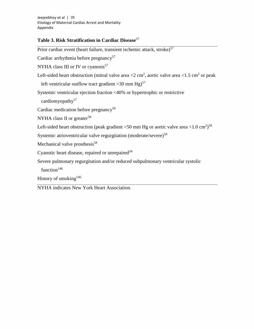
Jeejeebhoy et al | 39 Etiology of Maternal Cardiac Arrest and Mortality Appendix
Table 3. Risk Stratification in Cardiac Disease57
Prior cardiac event (heart failure, transient ischemic attack, stroke)57
Cardiac arrhythmia before pregnancy57
NYHA class III or IV or cyanosis57
Left-sided heart obstruction (mitral valve area <2 cm2, aortic valve area <1.5 cm2 or peak
left ventricular outflow tract gradient >30 mm Hg)57
Systemic ventricular ejection fraction <40% or hypertrophic or restrictive
cardiomyopathy57
Cardiac medication before pregnancy58
NYHA class II or greater58
Left-sided heart obstruction (peak gradient >50 mm Hg or aortic valve area <1.0 cm2)58
Systemic atrioventricular valve regurgitation (moderate/severe)58
Mechanical valve prosthesis58
Cyanotic heart disease, repaired or unrepaired58
Severe pulmonary regurgitation and/or reduced subpulmonary ventricular systolic
function146
History of smoking146
NYHA indicates New York Heart Association.
Jeejeebhoy et al | 40 Etiology of Maternal Cardiac Arrest and Mortality Appendix
REFERENCES
1. Mhyre JM, Tsen LC, Einav S, Kuklina EV, Leffert LR, Bateman BT. Cardiac arrest during hospitalization for delivery in the United States, 1998-2011. Anesthesiology. 2014;120:810-818.
2. Cantwell R, Clutton-Brock T, Cooper G, Dawson A, Drife J, Garrod D, Harper A, Hulbert D, Lucas S, McClure J, Millward-Sadler H, Neilson J, Nelson-Piercy C, Norman J, O'Herlihy C, Oates M, Shakespeare J, de Swiet M, Williamson C, Beale V, Knight M, Lennox C, Miller A, Parmar D, Rogers J, Springett A. Saving Mothers' Lives: Reviewing maternal deaths to make motherhood safer: 2006-2008. The Eighth Report of the Confidential Enquiries into Maternal Deaths in the United Kingdom. BJOG. 2011;118(suppl 1):1-203.
3. Centers for Disease Control and Prevention. Pregnancy mortality surveillance system. 2014. http://www.cdc.gov/reproductivehealth/maternalinfanthealth/pmss.html. Accessed May 12, 2014.
4. Kuklina E, Callaghan W. Chronic heart disease and severe obstetric morbidity among hospitalisations for pregnancy in the USA: 1995-2006. BJOG. 2011;118:345-352.
5. Nevis IF, Reitsma A, Dominic A, McDonald S, Thabane L, Akl EA, Hladunewich M, Akbari A, Joseph G, Sia W, Iansavichus AV, Garg AX. Pregnancy outcomes in women with chronic kidney disease: a systematic review. Clin J Am Soc Nephrol. 2011;6:2587-2598.
6. Kamyar M, Varner M. Epilepsy in pregnancy. Clin Obstet Gynecol. 2013;56:330-341.
7. Haider B, von Oertzen J. Neurological disorders. Best Pract Res Clin Obstet Gynaecol. 2013;27:867-875.
8. Catalano PM. Management of obesity in pregnancy. Obstet Gynecol. 2007;109:419-433.
9. Hawkins JL, Chang J, Palmer SK, Gibbs CP, Callaghan WM. Anesthesia-related maternal mortality in the United States: 1979-2002. Obstet Gynecol. 2011;117:69-74.
10. McClure JH, Cooper GM, Clutton-Brock TH. Saving mothers' lives: reviewing maternal deaths to make motherhood safer: 2006-8: a review. Br J Anaesth. 2011;107:127-132.
11. Mhyre JM, Riesner MN, Polley LS, Naughton NN. A series of anesthesia-related maternal deaths in Michigan, 1985-2003. Anesthesiology. 2007;106:1096-1104.
12. Al-Foudri H, Kevelighan E, Catling S. CEMACH 2003–5 Saving mothers’ lives: lessons for anaesthetists. Continuing Education in Anaesthesia. Crit Care Med. 2010;10:81–87.
13. Szypula K, Ashpole KJ, Bogod D, Yentis SM, Mihai R, Scott S, Cook TM. Litigation related to regional anaesthesia: an analysis of claims against the NHS in England 1995-2007. Anaesthesia. 2010;65:443-452.
14. Caplan RA, Ward RJ, Posner K, Cheney FW. Unexpected cardiac arrest during spinal anesthesia: a closed claims analysis of predisposing factors. Anesthesiology. 1988;68:5-11.
Jeejeebhoy et al | 41 Etiology of Maternal Cardiac Arrest and Mortality Appendix
15. Geffin B, Shapiro L. Sinus bradycardia and asystole during spinal and epidural anesthesia: a report of 13 cases. J Clin Anesth. 1998;10:278-285.
16. Hawthorne L, Lyons G. Cardiac arrest complicating spinal anaesthesia for caesarean section. Int J Obstet Anesth. 1997;6:126-129.
17. Paranjothy S, Griffiths JD, Broughton HK, Gyte GM, Brown HC, Thomas J. Interventions at caesarean section for reducing the risk of aspiration pneumonitis. Cochrane Database Syst Rev. 2014;2:CD004943.
18. Janda M, Scheeren TW, Noldge-Schomburg GF. Management of pulmonary aspiration. Best Pract Res Clin Anaesthesiol. 2006;20:409-427.
19. Carvalho B. Respiratory depression after neuraxial opioids in the obstetric setting. Anesth Analg. 2008;107:956-961.
20. Weinger MB. Dangers of Postoperative Opioids: APSF Workshop and White Paper Address Prevention of Postoperative Respiratory Complications. APSF Newsletter. 2007;21:61, 63-67.
21. Marr R, Hyams J, Bythell V. Cardiac arrest in an obstetric patient using remifentanil patient-controlled analgesia. Anaesthesia. 2013;68:283-287.
22. Kuczkowski KM. Respiratory arrest in a parturient following intrathecal administration of fentanyl and bupivacaine as part of a combined spinal-epidural analgesia for labour. Anaesthesia. 2002;57:939-940.
23. Sultan P, Gutierrez MC, Carvalho B. Neuraxial morphine and respiratory depression: finding the right balance. Drugs. 2011;71:1807-1819.
24. Stocki D, Matot I, Einav S, Eventov-Friedman S, Ginosar Y, Weiniger CF. A randomized controlled trial of the efficacy and respiratory effects of patient-controlled intravenous remifentanil analgesia and patient-controlled epidural analgesia in laboring women. Anesth Analg. 2014;118:589-597.
25. Horlocker TT, Burton AW, Connis RT, Hughes SC, Nickinovich DG, Palmer CM, Pollock JE, Rathmell JP, Rosenquist RW, Swisher JL, Wu CL. Practice guidelines for the prevention, detection, and management of respiratory depression associated with neuraxial opioid administration. Anesthesiology. 2009;110:218-230.
26. Cannesson M, Nargues N, Bryssine B, Debon R, Boselli E, Chassard D. Intrathecal morphine overdose during combined spinal-epidural block for Caesarean delivery. Br J Anaesth. 2002;89:925-927.
27. Camorcia M. Testing the epidural catheter. Curr Opin Anaesthesiol. 2009;22:336-340.
28. Bern S, Weinberg G. Local anesthetic toxicity and lipid resuscitation in pregnancy. Curr Opin Anaesthesiol. 2011;24:262-267.
29. Suresh MS, LaToya Mason C, Munnur U. Cardiopulmonary resuscitation and the parturient. Best Pract Res Clin Obstet Gynaecol. 2010;24:383-400.
30. Neal JM, Mulroy MF, Weinberg GL. American Society of Regional Anesthesia and Pain Medicine checklist for managing local anesthetic systemic toxicity: 2012 version. Reg Anesth Pain Med. 2012;37:16-18.
31. Weinberg GL. Treatment of local anesthetic systemic toxicity (LAST). Reg Anesth Pain Med. 2010;35:188-193.
32. Ecker JL, Solt K, Fitzsimons MG, MacGillivray TE. Case records of the Massachusetts General Hospital. Case 40-2012. A 43-year-old woman with
Jeejeebhoy et al | 42 Etiology of Maternal Cardiac Arrest and Mortality Appendix
cardiorespiratory arrest after a cesarean section. N Engl J Med. 2012;367:2528-2536.
33. Folk JJ, Leslie-Brown HF, Nosovitch JT, Silverman RK, Aubry RH. Hyperemesis gravidarum: outcomes and complications with and without total parenteral nutrition. J Reprod Med. 2004;49:497-502.
34. King JC. Strategies to reduce maternal mortality in developed countries. Curr Opin Obstet Gynecol. 2013;25:117-123.
35. Brown S, Mozurkewich E. Trauma during pregnancy. Obstet Gynecol Clin North Am. 2013;40:47-57.
36. Samandari G, Martin SL, Kupper LL, Schiro S, Norwood T, Avery M. Are pregnant and postpartum women: at increased risk for violent death? Suicide and homicide findings from North Carolina. Matern Child Health J. 2011;15:660-669.
37. Palladino CL, Singh V, Campbell J, Flynn H, Gold KJ. Homicide and suicide during the perinatal period: findings from the National Violent Death Reporting System. Obstet Gynecol. 2011;118:1056-1063.
38. Heron M. Deaths: leading causes for 2008. Natl Vital Stat Rep. 2012;60:1-94. 39. Romero VC, Pearlman M. Maternal mortality due to trauma. Semin Perinatol.
2012;36:60-67. 40. Chang J, Berg CJ, Saltzman LE, Herndon J. Homicide: a leading cause of injury
deaths among pregnant and postpartum women in the United States, 1991-1999. Am J Public Health. 2005;95:471-477.
41. Marzuk PM, Tardiff K, Leon AC, Hirsch CS, Portera L, Hartwell N, Iqbal MI. Lower risk of suicide during pregnancy. Am J Psychiatry. 1997;154:122-123.
42. Gandhi SG, Gilbert WM, McElvy SS, El Kady D, Danielson B, Xing G, Smith LH. Maternal and neonatal outcomes after attempted suicide. Obstet Gynecol. 2006;107:984-990.
43. Della Torre M, Kilpatrick SJ, Hibbard JU, Simonson L, Scott S, Koch A, Schy D, Geller SE. Assessing preventability for obstetric hemorrhage. Am J Perinatol. 2011;28:753-760.
44. Saucedo M, Deneux-Tharaux C, Bouvier-Colle MH. Ten years of confidential inquiries into maternal deaths in France, 1998-2007. Obstet Gynecol. 2013;122:752-760.
45. Campbell TA, Sanson TG. Cardiac arrest and pregnancy. J Emerg Trauma Shock. 2009;2:34-42.
46. Callaghan WM, Kuklina EV, Berg CJ. Trends in postpartum hemorrhage: United States, 1994-2006. Am J Obstet Gynecol. 2010;202:353 e351-e356.
47. Bateman BT, Mhyre JM, Callaghan WM, Kuklina EV. Peripartum hysterectomy in the United States: nationwide 14 year experience. Am J Obstet Gynecol. 2012;206:63 e61-68.
48. Hernandez JS, Nuangchamnong N, Ziadie M, Wendel GD Jr, Sheffield JS. Placental and uterine pathology in women undergoing peripartum hysterectomy. Obstet Gynecol. 2012;119:1137-1142.
49. Mhyre JM, Shilkrut A, Kuklina EV, Callaghan WM, Creanga AA, Kaminsky S, Bateman BT. Massive blood transfusion during hospitalization for delivery in New York State, 1998-2007. Obstet Gynecol. 2013;122:1288-1294.
Jeejeebhoy et al | 43 Etiology of Maternal Cardiac Arrest and Mortality Appendix
50. Zelop CM. Postpartum hemorrhage: becoming more evidence-based. Obstet Gynecol. 2011;117:3-5.
51. la Chapelle CF, Schutte JM, Schuitemaker NW, Steegers EA, van Roosmalen J. Maternal mortality attributable to vascular dissection and rupture in the Netherlands: a nationwide confidential enquiry. BJOG. 2012;119:86-93.
52. Pacheco LD, Saade GR, Costantine MM, Clark SL, Hankins GD. The role of massive transfusion protocols in obstetrics. Am J Perinatol. 2013;30:1-4.
53. Shakur H, Elbourne D, Gulmezoglu M, Alfirevic Z, Ronsmans C, Allen E, Roberts I. The WOMAN Trial (World Maternal Antifibrinolytic Trial): tranexamic acid for the treatment of postpartum haemorrhage: an international randomised, double blind placebo controlled trial. Trials. 2010;11:40.
54. Rich NM, Mattox KL, Hirschberg A. Vascular Trauma. Philadelphia, PA: Elsevier Saunders; 2004.
55. Belfort MA, Zimmerman J, Schemmer G, Oldroyd R, Smilanich R, Pearce M. Aortic compression and cross clamping in a case of placenta percreta and amniotic fluid embolism: a case report. AJP Rep. 2011;1:33-36.
56. Chatterjee DJ, Bukunola B, Samuels TL, Induruwage L, Uncles DR. Resuscitation in massive obstetric haemorrhage using an intraosseous needle. Anaesthesia. 2011;66:306-310.
57. Siu SC, Sermer M, Colman JM, Alvarez AN, Mercier LA, Morton BC, Kells CM, Bergin ML, Kiess MC, Marcotte F, Taylor DA, Gordon EP, Spears JC, Tam JW, Amankwah KS, Smallhorn JF, Farine D, Sorensen S. Prospective multicenter study of pregnancy outcomes in women with heart disease. Circulation. 2001;104:515-521.
58. Drenthen W, Boersma E, Balci A, Moons P, Roos-Hesselink JW, Mulder BJ, Vliegen HW, van Dijk AP, Voors AA, Yap SC, van Veldhuisen DJ, Pieper PG. Predictors of pregnancy complications in women with congenital heart disease. Eur Heart J. 2010;31:2124-2132.
59. Roos-Hesselink JW, Duvekot JJ, Thorne SA. Pregnancy in high risk cardiac conditions. Heart. 2009;95:680-686.
60. Regitz-Zagrosek V, Blomstrom Lundqvist C, Borghi C, Cifkova R, Ferreira R, Foidart JM, Gibbs JS, Gohlke-Baerwolf C, Gorenek B, Iung B, Kirby M, Maas AH, Morais J, Nihoyannopoulos P, Pieper PG, Presbitero P, Roos-Hesselink JW, Schaufelberger M, Seeland U, Torracca L. ESC Guidelines on the management of cardiovascular diseases during pregnancy: the Task Force on the Management of Cardiovascular Diseases during Pregnancy of the European Society of Cardiology (ESC). Eur Heart J. 2011;32:3147-3197.
61. Ladner HE, Danielsen B, Gilbert WM. Acute myocardial infarction in pregnancy and the puerperium: a population-based study. Obstet Gynecol. 2005;105:480-484.
62. Roth A, Elkayam U. Acute myocardial infarction associated with pregnancy. J Am Coll Cardiol. 2008;52:171-180.
63. James AH, Jamison MG, Biswas MS, Brancazio LR, Swamy GK, Myers ER. Acute myocardial infarction in pregnancy: a United States population-based study. Circulation. 2006;113:1564-1571.
Jeejeebhoy et al | 44 Etiology of Maternal Cardiac Arrest and Mortality Appendix
64. Leonhardt G, Gaul C, Nietsch HH, Buerke M, Schleussner E. Thrombolytic therapy in pregnancy. J Thromb Thrombolysis. 2006;21:271-276.
65. Immer FF, Bansi AG, Immer-Bansi AS, McDougall J, Zehr KJ, Schaff HV, Carrel TP. Aortic dissection in pregnancy: analysis of risk factors and outcome. Ann Thorac Surg. 2003;76:309-314.
66. Meijboom LJ, Vos FE, Timmermans J, Boers GH, Zwinderman AH, Mulder BJ. Pregnancy and aortic root growth in the Marfan syndrome: a prospective study. Eur Heart J. 2005;26:914-920.
67. John AS, Gurley F, Schaff HV, Warnes CA, Phillips SD, Arendt KW, Abel MD, Rose CH, Connolly HM. Cardiopulmonary bypass during pregnancy. Ann Thorac Surg. 2011;91:1191-1196.
68. Sliwa K, Hilfiker-Kleiner D, Petrie MC, Mebazaa A, Pieske B, Buchmann E, Regitz-Zagrosek V, Schaufelberger M, Tavazzi L, van Veldhuisen DJ, Watkins H, Shah AJ, Seferovic PM, Elkayam U, Pankuweit S, Papp Z, Mouquet F, McMurray JJ. Current state of knowledge on aetiology, diagnosis, management, and therapy of peripartum cardiomyopathy: a position statement from the Heart Failure Association of the European Society of Cardiology Working Group on peripartum cardiomyopathy. Eur J Heart Fail. 2010;12:767-778.
69. Sliwa K, Fett J, Elkayam U. Peripartum cardiomyopathy. Lancet. 2006;368:687-693.
70. Duran N, Gunes H, Duran I, Biteker M, Ozkan M. Predictors of prognosis in patients with peripartum cardiomyopathy. Int J Gynaecol Obstet. 2008;101:137-140.
71. Thaman R, Varnava A, Hamid MS, Firoozi S, Sachdev B, Condon M, Gimeno JR, Murphy R, Elliott PM, McKenna WJ. Pregnancy related complications in women with hypertrophic cardiomyopathy. Heart. 2003;89:752-756.
72. Autore C, Conte MR, Piccininno M, Bernabo P, Bonfiglio G, Bruzzi P, Spirito P. Risk associated with pregnancy in hypertrophic cardiomyopathy. J Am Coll Cardiol. 2002;40:1864-1869.
73. Maron BJ. Contemporary insights and strategies for risk stratification and prevention of sudden death in hypertrophic cardiomyopathy. Circulation. 2010;121:445-456.
74. McLintock C, McCowan LM, North RA. Maternal complications and pregnancy outcome in women with mechanical prosthetic heart valves treated with enoxaparin. BJOG. 2009;116:1585-1592.
75. Chan WS, Anand S, Ginsberg JS. Anticoagulation of pregnant women with mechanical heart valves: a systematic review of the literature. Arch Intern Med. 2000;160:191-196.
76. Yinon Y, Siu SC, Warshafsky C, Maxwell C, McLeod A, Colman JM, Sermer M, Silversides CK. Use of low molecular weight heparin in pregnant women with mechanical heart valves. Am J Cardiol. 2009;104:1259-1263.
77. Gleicher N, Midwall J, Hochberger D, Jaffin H. Eisenmenger's syndrome and pregnancy. Obstet Gynecol Surv. 1979;34:721-741.
78. Bedard E, Dimopoulos K, Gatzoulis MA. Has there been any progress made on pregnancy outcomes among women with pulmonary arterial hypertension? Eur Heart J. 2009;30:256-265.
Jeejeebhoy et al | 45 Etiology of Maternal Cardiac Arrest and Mortality Appendix
79. Rashba EJ, Zareba W, Moss AJ, Hall WJ, Robinson J, Locati EH, Schwartz PJ, Andrews M. Influence of pregnancy on the risk for cardiac events in patients with hereditary long QT syndrome. LQTS Investigators. Circulation. 1998;97:451-456.
80. Seth R, Moss AJ, McNitt S, Zareba W, Andrews ML, Qi M, Robinson JL, Goldenberg I, Ackerman MJ, Benhorin J, Kaufman ES, Locati EH, Napolitano C, Priori SG, Schwartz PJ, Towbin JA, Vincent GM, Zhang L. Long QT syndrome and pregnancy. J Am Coll Cardiol. 2007;49:1092-1098.
81. Sharif-Kazemi MB, Emkanjoo Z, Tavoosi A, Kafi M, Kheirkhah J, Alizadeh A, Sadr-Ameli MA. Electrical storm in Brugada syndrome during pregnancy. Pacing Clin Electrophysiol. 2011;34:e18-e21.
82. Natale A, Davidson T, Geiger MJ, Newby K. Implantable cardioverter-defibrillators and pregnancy: a safe combination? Circulation. 1997;96:2808-2812.
83. Schuler PK, Herrey A, Wade A, Brooks R, Peebles D, Lambiase P, Walker F. Pregnancy outcome and management of women with an implantable cardioverter defibrillator: a single centre experience. Europace. 2012;14:1740-1745.
84. Colletti PM, Lee KH, Elkayam U. Cardiovascular imaging of the pregnant patient. AJR Am J Roentgenol. 2013;200:515-521.
85. Damilakis J, Theocharopoulos N, Perisinakis K, Manios E, Dimitriou P, Vardas P, Gourtsoyiannis N. Conceptus radiation dose and risk from cardiac catheter ablation procedures. Circulation. 2001;104:893-897.
86. Ferguson JD, Helms A, Mangrum JM, DiMarco JP. Ablation of incessant left atrial tachycardia without fluoroscopy in a pregnant woman. J Cardiovasc Electrophysiol. 2011;22:346-349.
87. Dyer RA, Butwick AJ, Carvalho B. Oxytocin for labour and caesarean delivery: implications for the anaesthesiologist. Curr Opin Anaesthesiol. 2011;24:255-261.
88. Thomas TA, Cooper GM. Maternal deaths from anaesthesia. An extract from Why mothers die 1997-1999, the Confidential Enquiries into Maternal Deaths in the United Kingdom. Br J Anaesth. 2002;89:499-508.
89. Butwick AJ, Coleman L, Cohen SE, Riley ET, Carvalho B. Minimum effective bolus dose of oxytocin during elective Caesarean delivery. Br J Anaesth. 2010;104:338-343.
90. Carvalho JC, Balki M, Kingdom J, Windrim R. Oxytocin requirements at elective cesarean delivery: a dose-finding study. Obstet Gynecol. 2004;104:1005-1010.
91. Thomas JS, Koh SH, Cooper GM. Haemodynamic effects of oxytocin given as i.v. bolus or infusion on women undergoing Caesarean section. Br J Anaesth. 2007;98:116-119.
92. Dyer RA, Reed AR, van Dyk D, Arcache MJ, Hodges O, Lombard CJ, Greenwood J, James MF. Hemodynamic effects of ephedrine, phenylephrine, and the coadministration of phenylephrine with oxytocin during spinal anesthesia for elective cesarean delivery. Anesthesiology. 2009;111:753-765.
93. Pryde PG, Mittendorf R. Contemporary usage of obstetric magnesium sulfate: indication, contraindication, and relevance of dose. Obstet Gynecol. 2009;114:669-673.
Jeejeebhoy et al | 46 Etiology of Maternal Cardiac Arrest and Mortality Appendix
94. Lu JF, Nightingale CH. Magnesium sulfate in eclampsia and pre-eclampsia: pharmacokinetic principles. Clin Pharmacokinet. 2000;38:305-314.
95. Altman D, Carroli G, Duley L, Farrell B, Moodley J, Neilson J, Smith D. Do women with pre-eclampsia, and their babies, benefit from magnesium sulphate? The Magpie Trial: a randomised placebo-controlled trial. Lancet. 2002;359:1877-1890.
96. Vanden Hoek TL, Morrison LJ, Shuster M, Donnino M, Sinz E, Lavonas EJ, Jeejeebhoy FM, Gabrielli A. Part 12: cardiac arrest in special situations: 2010 American Heart Association Guidelines for Cardiopulmonary Resuscitation and Emergency Cardiovascular Care. Circulation. 2010;122:S829-861.
97. Swartjes JM, Schutte MF, Bleker OP. Management of eclampsia: cardiopulmonary arrest resulting from magnesium sulfate overdose. Eur J Obstet Gynecol Reprod Biol. 1992;47:73-75.
98. Kfuri TA, Morlock L, Hicks RW, Shore AD. Medication errors in obstetrics. Clin Perinatol. 2008;35:101-117, viii-ix.
99. Abenhaim HA, Azoulay L, Kramer MS, Leduc L. Incidence and risk factors of amniotic fluid embolisms: a population-based study on 3 million births in the United States. Am J Obstet Gynecol. 2008;199:49 e41-e48.
100. Knight M, Berg C, Brocklehurst P, Kramer M, Lewis G, Oats J, Roberts CL, Spong C, Sullivan E, van Roosmalen J, Zwart J. Amniotic fluid embolism incidence, risk factors and outcomes: a review and recommendations. BMC Pregnancy Childbirth. 2012;12:7.
101. Kramer MS, Rouleau J, Liu S, Bartholomew S, Joseph KS. Amniotic fluid embolism: incidence, risk factors, and impact on perinatal outcome. BJOG. 2012;119:874-879.
102. Shechtman M, Ziser A, Markovits R, Rozenberg B. Amniotic fluid embolism: early findings of transesophageal echocardiography. Anesth Analg. 1999;89:1456-1458.
103. Stanten RD, Iverson LI, Daugharty TM, Lovett SM, Terry C, Blumenstock E. Amniotic fluid embolism causing catastrophic pulmonary vasoconstriction: diagnosis by transesophageal echocardiogram and treatment by cardiopulmonary bypass. Obstet Gynecol. 2003;102:496-498.
104. Clark SL, Hankins GD, Dudley DA, Dildy GA, Porter TF. Amniotic fluid embolism: analysis of the national registry. Am J Obstet Gynecol. 1995;172:1158-1167; discussion 1167-1159.
105. Knight M, Tuffnell D, Brocklehurst P, Spark P, Kurinczuk JJ. Incidence and risk factors for amniotic-fluid embolism. Obstet Gynecol. 2010;115:910-917.
106. Margarson MP. Delayed amniotic fluid embolism following caesarean section under spinal anaesthesia. Anaesthesia. 1995;50:804-806.
107. Tuffnell DJ. United kingdom amniotic fluid embolism register. BJOG. 2005;112:1625-1629.
108. Stroup J, Haraway D, Beal JM. Aprotinin in the management of coagulopathy associated with amniotic fluid embolus. Pharmacotherapy. 2006;26:689-693.
109. Leighton BL, Wall MH, Lockhart EM, Phillips LE, Zatta AJ. Use of recombinant factor VIIa in patients with amniotic fluid embolism: a systematic review of case reports. Anesthesiology. 2011;115:1201-1208.
Jeejeebhoy et al | 47 Etiology of Maternal Cardiac Arrest and Mortality Appendix
110. Simpson E, Lin Y, Stanworth S, Birchall J, Doree C, Hyde C. Recombinant factor VIIa for the prevention and treatment of bleeding in patients without haemophilia. Cochrane Database Syst Rev. 2012;3:CD005011.
111. Kaneko Y, Ogihara T, Tajima H, Mochimaru F. Continuous hemodiafiltration for disseminated intravascular coagulation and shock due to amniotic fluid embolism: report of a dramatic response. Intern Med. 2001;40:945-947.
112. Ogihara T, Morimoto K, Kaneko Y. Continuous hemodiafiltration for potential amniotic fluid embolism: dramatic responses observed during a 10-year period report of three cases. Ther Apher Dial. 2012;16:195-197.
113. McDonnell NJ, Chan BO, Frengley RW. Rapid reversal of critical haemodynamic compromise with nitric oxide in a parturient with amniotic fluid embolism. Int J Obstet Anesth. 2007;16:269-273.
114. Nagarsheth NP, Pinney S, Bassily-Marcus A, Anyanwu A, Friedman L, Beilin Y. Successful placement of a right ventricular assist device for treatment of a presumed amniotic fluid embolism. Anesth Analg. 2008;107:962-964.
115. Hsieh YY, Chang CC, Li PC, Tsai HD, Tsai CH. Successful application of extracorporeal membrane oxygenation and intra-aortic balloon counterpulsation as lifesaving therapy for a patient with amniotic fluid embolism. Am J Obstet Gynecol. 2000;183:496-497.
116. Whelihan MF, Kiankhooy A, Brummel-Ziedins KE. Thrombin generation and fibrin clot formation under hypothermic conditions: an in vitro evaluation of tissue factor initiated whole blood coagulation. J Crit Care. 2014;29:24-30.
117. Wolberg AS, Meng ZH, Monroe DM 3rd, Hoffman M. A systematic evaluation of the effect of temperature on coagulation enzyme activity and platelet function. J Trauma. 2004;56:1221-1228.
118. Calderwood CJ, Thanoon OI. Venous thromboembolism in pregnancy. Obstetrics, Gynaecology and Reproductive Medicine. 2013;23:227-230.
119. Bates SM, Greer IA, Middeldorp S, Veenstra DL, Prabulos AM, Vandvik PO. VTE, thrombophilia, antithrombotic therapy, and pregnancy: Antithrombotic Therapy and Prevention of Thrombosis, 9th ed: American College of Chest Physicians Evidence-Based Clinical Practice Guidelines. Chest. 2012;141:e691S-e736S.
120. McLintock C, Brighton T, Chunilal S, Dekker G, McDonnell N, McRae S, Muller P, Tran H, Walters BN, Young L. Recommendations for the diagnosis and treatment of deep venous thrombosis and pulmonary embolism in pregnancy and the postpartum period. Aust N Z J Obstet Gynaecol. 2012;52:14-22.
121. Ilsaas C, Husby P, Koller ME, Segadal L, Holst-Larsen H. Cardiac arrest due to massive pulmonary embolism following caesarean section. Successful resuscitation and pulmonary embolectomy. Acta Anaesthesiol Scand. 1998;42:264-266.
122. Ayad S, Tetzlaff JE. Massive pulmonary embolism in a patient undergoing Cesarean delivery. J Clin Anesth. 2012;24:582-585.
123. Saposnik G, Barinagarrementeria F, Brown RD Jr, Bushnell CD, Cucchiara B, Cushman M, deVeber G, Ferro JM, Tsai FY. Diagnosis and management of cerebral venous thrombosis: a statement for healthcare professionals from the
Jeejeebhoy et al | 48 Etiology of Maternal Cardiac Arrest and Mortality Appendix
American Heart Association/American Stroke Association. Stroke. 2011;42:1158-1192.
124. Coutinho JM, Zuurbier SM, Aramideh M, Stam J. The incidence of cerebral venous thrombosis: a cross-sectional study. Stroke. 2012;43:3375-3377.
125. James AH. Venous thromboembolism in pregnancy. Arterioscler Thromb Vasc Biol. 2009;29:326-331.
126. Kaunitz AM, Hughes JM, Grimes DA, Smith JC, Rochat RW, Kafrissen ME. Causes of maternal mortality in the United States. Obstet Gynecol. 1985;65:605-612.
127. Kim CS, Liu J, Kwon JY, Shin SK, Kim KJ. Venous air embolism during surgery, especially cesarean delivery. J Korean Med Sci. 2008;23:753-761.
128. Fernandez-Perez ER, Salman S, Pendem S, Farmer JC. Sepsis during pregnancy. Crit Care Med. 2005;33:S286-293.
129. Barton JR, Sibai BM. Severe sepsis and septic shock in pregnancy. Obstet Gynecol. 2012;120:689-706.
130. Kumar A, Roberts D, Wood KE, Light B, Parrillo JE, Sharma S, Suppes R, Feinstein D, Zanotti S, Taiberg L, Gurka D, Cheang M. Duration of hypotension before initiation of effective antimicrobial therapy is the critical determinant of survival in human septic shock. Crit Care Med. 2006;34:1589-1596.
131. James FM 3rd, Greiss FC Jr, Kemp RA. An evaluation of vasopressor therapy for maternal hypotension during spinal anesthesia. Anesthesiology. 1970;33:25-34.
132. Habib AS. A review of the impact of phenylephrine administration on maternal hemodynamics and maternal and neonatal outcomes in women undergoing cesarean delivery under spinal anesthesia. Anesth Analg. 2012;114:377-390.
133. Shnider SM, de Lorimier AA, Holl JW, Chapler FK, Morishima HO. Vasopressors in obstetrics. I. Correction of fetal acidosis with ephedrine during spinal hypotension. Am J Obstet Gynecol. 1968;102:911-919.
134. Jamieson DJ, Honein MA, Rasmussen SA, Williams JL, Swerdlow DL, Biggerstaff MS, Lindstrom S, Louie JK, Christ CM, Bohm SR, Fonseca VP, Ritger KA, Kuhles DJ, Eggers P, Bruce H, Davidson HA, Lutterloh E, Harris ML, Burke C, Cocoros N, Finelli L, MacFarlane KF, Shu B, Olsen SJ, Novel Influenza APWG. H1N1 2009 influenza virus infection during pregnancy in the USA. Lancet. 2009;374:451-458.
135. Pierce M, Kurinczuk JJ, Spark P, Brocklehurst P, Knight M. Perinatal outcomes after maternal 2009/H1N1 infection: national cohort study. BMJ. 2011;342:d3214.
136. Priddy KD. Immunologic adaptations during pregnancy. J Obstet Gynecol Neonatal Nurs. 1997;26:388-394.
137. Lapinsky SE. Pregnancy joins the hit list. Crit Care Med. 2012;40:1679-1680. 138. Rebordosa C, Zelop CM, Kogevinas M, Sorensen HT, Olsen J. Use of
acetaminophen during pregnancy and risk of preeclampsia, hypertensive and vascular disorders: a birth cohort study. J Matern Fetal Neonatal Med. 2010;23:371-378.
139. Knight M. Eclampsia in the United Kingdom 2005. BJOG. 2007;114:1072-1078.
Jeejeebhoy et al | 49 Etiology of Maternal Cardiac Arrest and Mortality Appendix
140. Wallis AB, Saftlas AF, Hsia J, Atrash HK. Secular trends in the rates of preeclampsia, eclampsia, and gestational hypertension, United States, 1987-2004. Am J Hypertens. 2008;21:521-526.
141. American College of Obstetricians and Gynecologists Taskforce on Hypertension in Pregnancy. Hypertension in pregnancy. Washington, DC: American College of Obstetricians and Gynecologists; 2013.
142. Ghulmiyyah L, Sibai B. Maternal mortality from preeclampsia/eclampsia. Semin Perinatol. 2012;36:56-59.
143. Clark SL, Christmas JT, Frye DR, Meyers JA, Perlin JB. Maternal mortality in the United States: predictability and the impact of protocols on fatal postcesarean pulmonary embolism and hypertension-related intracranial hemorrhage. Am J Obstet Gynecol. 2014.
144. Bratton SL, Chestnut RM, Ghajar J, McConnell Hammond FF, Harris OA, Hartl R, Manley GT, Nemecek A, Newell DW, Rosenthal G, Schouten J, Shutter L, Timmons SD, Ullman JS, Videtta W, Wilberger JE, Wright DW. Guidelines for the management of severe traumatic brain injury. XIV. Hyperventilation. J Neurotrauma. 2007;24(suppl 1):S87-S90.
145. Brain Trauma Foundation, American Association of Neurological Surgeons, Congress of Neurological Surgeons. Guidelines for the management of severe traumatic brain injury. J Neurotrauma. 2007;24(suppl 1):S1-S106.
146. Khairy P, Ouyang DW, Fernandes SM, Lee-Parritz A, Economy KE, Landzberg MJ. Pregnancy outcomes in women with congenital heart disease. Circulation. 2006;113:517-524.